1. Introduction
Angioleiomyomas are benign tumors originating from smooth muscle cells of the tunica media of vascular structures, typically presenting as small, painful nodules. They have a predilection for women in the fourth to sixth decade of life [
1,
2] and account for 4.5–7.7% of benign soft tissue tumors [
1,
3] but only 0.2% of those occurring in the foot [
4]. Despite this rarity, reports in foot and ankle literature have steadily increased in recent years. A 2025 systematic review from Switzerland identified 62 studies of foot and ankle angioleiomyomas in recent literature [
5], while another review and case series from Spain analyzed 14 case series comprising 172 angioleiomyomas [
2], underscoring the growing recognition of this entity and its impact on timely diagnosis and efficient patient care.
Clinically, angioleiomyomas present as firm, mobile subcutaneous masses measuring approximately 0.5–2 cm [
6], which may gradually enlarge and cause discomfort [
1,
6,
7], particularly with footwear or activity. Although less common, exophytic presentations have also been reported [
8]. Pain is a common but nonspecific symptom, often leading to a broad differential diagnosis that includes lipoma, schwannoma, ganglion cyst, and other soft tissue masses. Current literature also reflects debate regarding the most frequent anatomic location: one 2025 systematic review reported a majority occurrence in the forefoot (36%) [
1], whereas another found the rearfoot most commonly affected (40%) [
2]. Because these tumors grow slowly and imaging findings may be subtle, preoperative diagnosis remains uncommon [
5].
Histologically, angioleiomyomas consist of mature smooth muscle cells arranged concentrically around vascular channels, a feature used for definitive diagnosis [
9]. They are classified into capillary (solid), venous, and cavernous subtypes, with the solid type being most prevalent [
2,
7]. While generally benign, malignant transformation to angioleiomyosarcoma is reported in rare instances [
10]. Calcification occurs in fewer than 3.3% of cases and may be visualized histologically and radiographically [
2,
5].
Imaging modalities such as plain radiographs, ultrasound (US), and MRI have been utilized, though none are definitive. MRI typically demonstrates a well-circumscribed lesion with high T2 signal intensity and a low-signal fibrous rim and may reveal the “dark reticular sign,” a hypo- or isointense linear or branched structure corresponding to vascular channels [
5]. Ultrasound can occasionally detect internal vascularity [
11], while radiographs may reveal calcifications in a minority of cases [
2,
5].
Surgical excision followed by histopathological examination remains the standard of care [
4], particularly for symptomatic lesions. Recurrence is uncommon, with reported rates ranging from 2.1% to 2.8% [
2,
5].
We present a unique case of angioleiomyoma occurring in the heel of a 54-year-old woman. This report underscores the diagnostic challenges and the importance of including angioleiomyoma in the differential diagnosis of chronic, painful heel masses.
2. Case Report
A 54-year-old female with no significant past medical history initially presented to the clinic in March 2018 with pain localized to the posterior–lateral aspect of the heel. She reported a gradual onset of symptoms that had progressively worsened over a two-year period. Conservative measures aimed at offloading the area—such as footwear modifications and padding—had provided minimal relief. Initial clinical and radiographic evaluation suggested insertional Achilles tendinosis and a mild Haglund’s deformity. No abnormal soft tissue mass was appreciated at that time. Management included anti-inflammatory modalities, changes in shoe gear, orthotic support, and consideration for formal physical therapy.
The patient returned to the clinic about three years later (November 2021) with persistent pain. Repeat radiographs demonstrated a mild Haglund’s deformity without any acute osseous abnormalities. The patient elected to continue nonoperative treatment, including formal physical therapy, with the plan to pursue surgical options only if pain persisted or began to limit daily function.
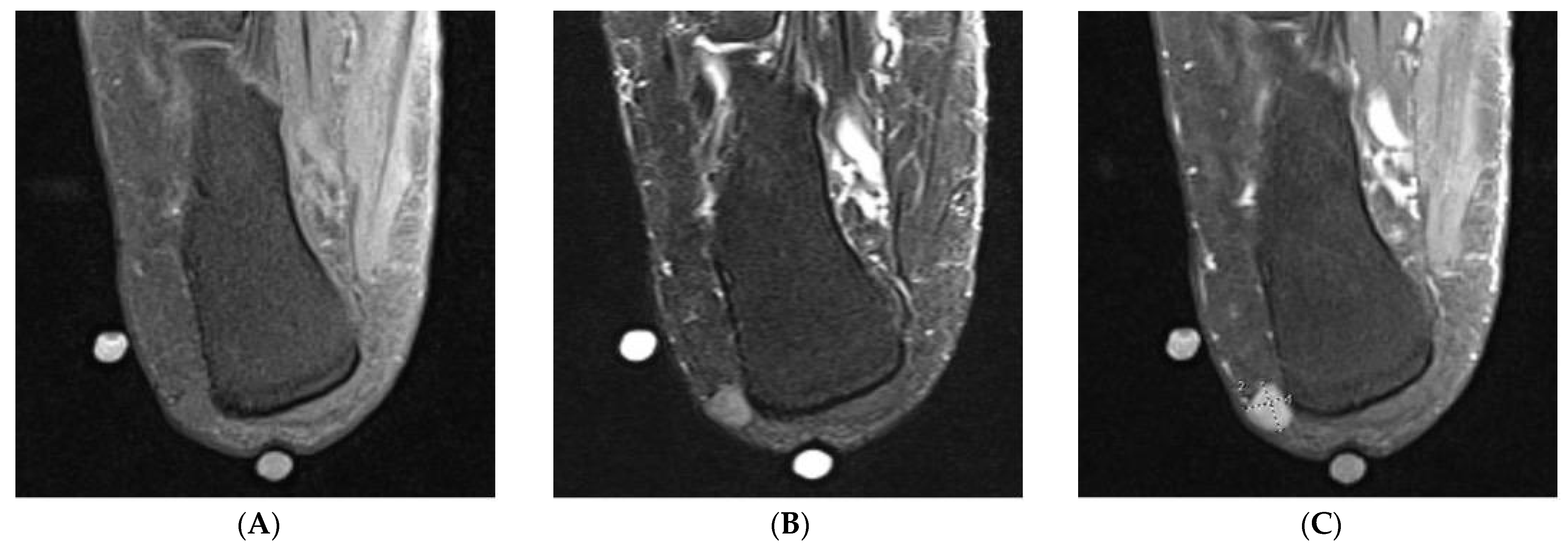
Two years after that visit (August 2023), the patient returned with worsening posterior–lateral heel pain and a newly palpable nodule in the same region. Given the new findings, an MRI was obtained. MRI revealed a well-circumscribed soft tissue mass adjacent to the lateral calcaneal cortex, measuring approximately 11 mm × 8 mm × 10 mm, without evidence of osseous involvement. The lesion was isointense to skeletal muscle on T1-weighted images and homogeneously hyperintense on T2-weighted images with a hypointense fibrous capsule (
Figure 1). The differential diagnosis included both benign and malignant entities, such as vascular malformation, peripheral nerve sheath tumor, metastatic lesion, and soft tissue sarcoma. Orthopedic oncology was consulted and recommended an excisional biopsy to establish a definitive diagnosis.
3. Operative Report and Patient Outcomes
The patient underwent surgical intervention in October 2023. Monitored anesthesia care (MAC) was utilized with a local nerve block. They were positioned laterally on the operating table. Perioperative antibiotic prophylaxis was administered with intravenous cefazolin (Ancef). The operative extremity was prepped and draped in a sterile fashion using standard aseptic technique. The limb was then elevated and exsanguinated with an Esmarch bandage, which was used as the tourniquet.
A linear incision was made over the palpable soft tissue mass. Careful blunt dissection was carried down to the level of the lesion. The mass was identified and excised in its entirety. Intraoperatively, it was noted that the mass was intertwined with a cutaneous nerve. A segment of this nerve was excised along with the mass and submitted for pathological evaluation. Layered closure of the wound was performed, and a multilayer compressive dressing was applied. The patient was placed in a tall CAM boot and allowed to bear weight as tolerated in a protected manner. A short course of oral antibiotics was prescribed immediately postoperatively for prophylactic purposes. Postoperative healing was uneventful.
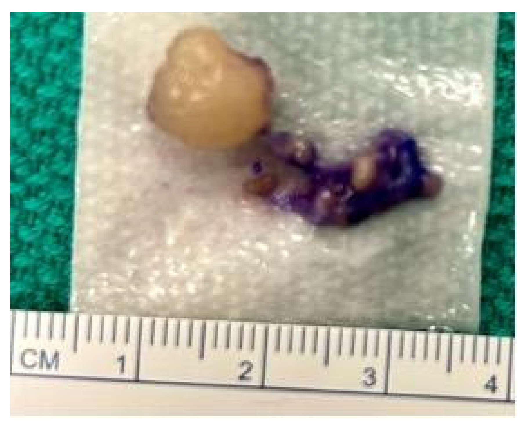
Macroscopically, the excised lesion was indurated, encapsulated, and tan-white in color (
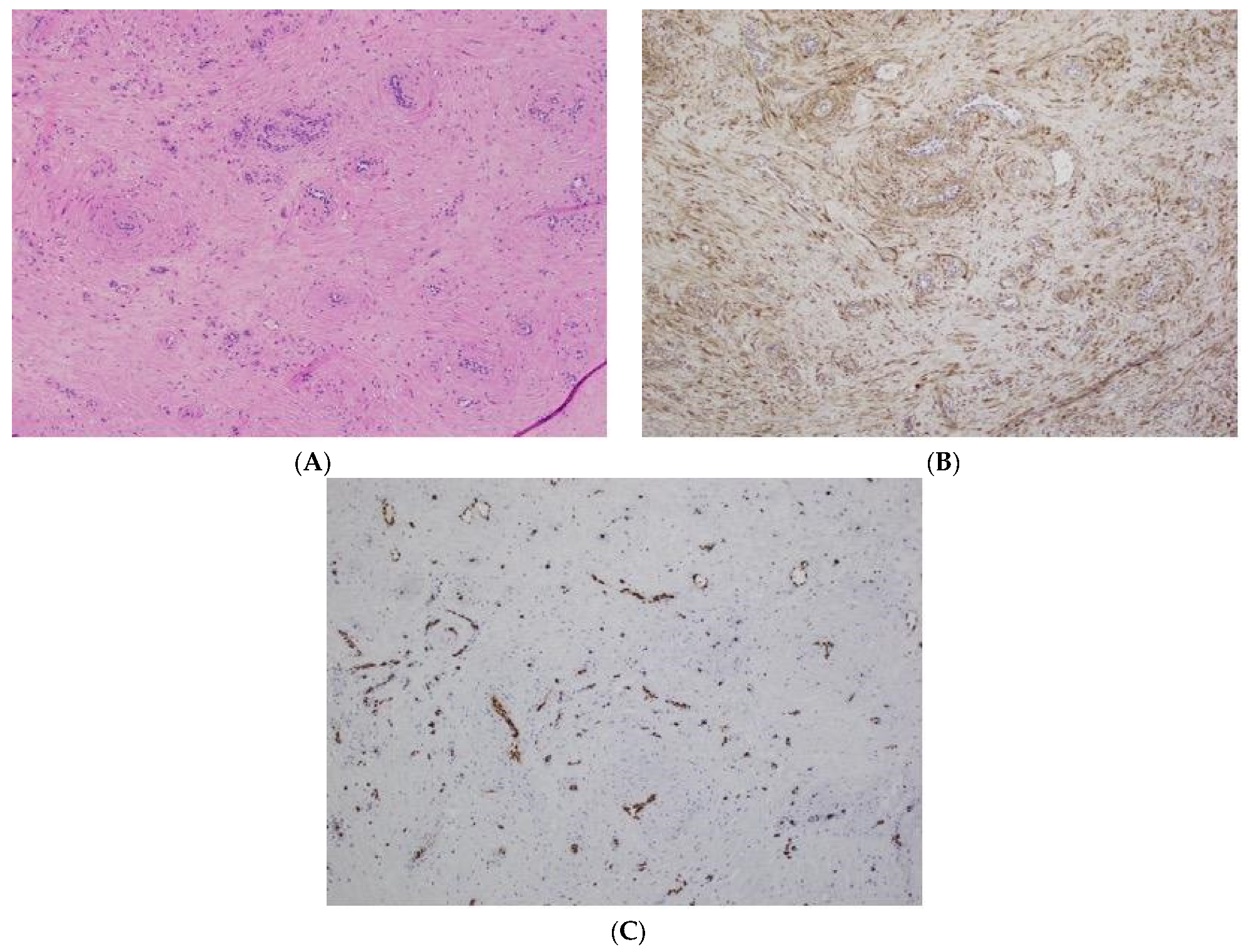
Figure 2). Histopathologic examination (
Figure 3A–C) demonstrated a benign-appearing spindle cell neoplasm with interspersed thick-walled blood vessels. There was no evidence of cytologic atypia, mitotic activity, or necrosis. Immunohistochemistry revealed diffuse positivity for smooth muscle actin (SMA) and patchy, weak positivity for desmin (
Figure 3B). The vascular endothelium stained positive for erythroblast transformation-specific regulated gene (ERG) (
Figure 3C). These findings were consistent with a diagnosis of angioleiomyoma. The case was reviewed with orthopedic oncology, who confirmed the diagnosis of a benign soft tissue tumor. No additional surveillance was deemed necessary. There was no evidence of recurrence at 12 months post-op, and the patient returned to full activity. The patient did note some residual numbness at the surgical site but was otherwise pain-free and without functional restrictions.
4. Discussion
Several reports of angioleiomyomas in the foot describe a slow-growing mass initially mistaken for other benign lesions, including lipoma, fibroma, ganglion cyst, schwannoma [
6,
12,
13], and even posterior tibial artery pseudoaneurysm [
14]. As in our case, misdiagnosis often results in treatment delays. For our patient, the time from symptom onset to surgical excision was seven years, aligning with the reported average of 5.6 ± 1.6 years [
5], underscoring the diagnostic challenge associated with this pathology.
Consistent with most published literature, angioleiomyomas are rarely diagnosed preoperatively and are typically confirmed through surgical excision and histopathologic analysis [
5]. While surgical excision is not always required, it has been shown to significantly reduce pain and improve functional outcomes [
4]. In our case, surgery was pursued due to recurrent pain that limited footwear options and activities of daily living, with notable postoperative improvement.
MRI revealed a well-circumscribed subcutaneous lesion that was isointense to skeletal muscle on T1-weighted imaging, hyperintense on T2, and demonstrated avid post-contrast enhancement. The “dark reticular sign” described in a recent systematic review [
5] was not clearly appreciated in our case. Other differentials were excluded based on MRI features, including absence of T1 hypointensity (hemangioma, glomus tumors), lack of lobulation (giant cell tumor, lipoma), and weak contrast enhancement (neurogenic tumors) [
15]. Nonetheless, other hallmark MRI findings, such as a well-defined, round, or oval shape and a hypointense fibrous capsule with hyperintense T2 signal, were present, further validating the utility of MRI in preoperative evaluation.
Histologically, the lesion displayed thick-walled vascular channels and smooth muscle proliferation without cytologic atypia, most consistent with a venous-type angioleiomyoma, the second most common subtype (23%) [
5,
7]. This subtype has been associated with similar delays to treatment (approximately seven years) [
7] and is painful in roughly 37% of cases [
16], often due to compression of adjacent neural structures, as observed in our patient.
Matos et al. (2025) recently proposed that combining ultrasound with MRI may offer the highest diagnostic accuracy for preoperative identification [
5]. However, even in this most comprehensive review of foot and ankle angioleiomyomas to date, delays in diagnosis and treatment remain prevalent. Our case reinforces the need to maintain a broad differential diagnosis when evaluating chronic, painful soft tissue masses of the foot and ankle to ensure timely and effective management.
5. Conclusions
We report a case of a venous-type angioleiomyoma in the posterolateral heel of a 54-year-old female, presenting as a long-standing, slow-growing, painful mass. Clinical diagnosis of angioleiomyoma remains challenging due to its often indolent and nonspecific presentation. MRI proved useful in characterizing the lesion preoperatively, demonstrating features consistent with those described in recent literature. Surgical excision provided symptom relief and allowed for definitive histopathologic diagnosis. This case highlights the importance of considering angioleiomyoma in the differential diagnosis of chronic, painful, soft tissue masses in the lower extremity, thereby improving awareness and diagnostic accuracy and optimizing patient care.
6. Bias and Limitations
Inherent to case reports, there exists a potential for selection and reporting bias, as outcomes are presented from a single patient experience and may not be generalizable to broader populations. To mitigate potential bias, this report adheres to the CARE (Case Report) guidelines for transparent case reporting and includes objective clinical and MR imaging to substantiate clinical outcomes. Furthermore, all treatment decisions were made based on standard clinical protocols and in consultation with the patient, with outcomes corroborated through imaging and functional assessment. Comparative discussion was limited to similar cases in the existing literature to contextualize findings without overextending conclusions beyond the scope of this case.
Author Contributions
Conceptualization, A.W.; methodology, A.W.; investigation, A.W.; data curation, A.W., H.B. and D.N.; formal analysis, J.W.K.-S.; visualization, J.W.K.-S.; writing—original draft preparation, A.W., H.B. and D.N.; writing—review and editing, J.B.; supervision, J.B. All authors have read and agreed to the published version of the manuscript.
Funding
This research did not receive any specific grant from funding agencies in the public, commercial, or not-for-profit sectors.
Institutional Review Board Statement
According to the policies of the Atrium Health Wake Forest Baptist Medical Center Institutional Review Board, this single-patient case report using fully de-identified clinical information does not constitute human subjects research and therefore did not require IRB review. The report was prepared in accordance with the principles of the Declaration of Helsinki, and all reasonable measures were taken to protect patient privacy and confidentiality.
Informed Consent Statement
Informed consent was obtained from the subject involved in the study.
Data Availability Statement
The data supporting the findings are clearly cited within the article. Requests for further data access may be considered by the corresponding author in accordance with institutional and regulatory requirements.
Conflicts of Interest
The authors declare no conflicts of interest.
References
- Murata, H.; Matsui, T.; Horie, N.; Sakabe, T.; Konishi, E.; Kubo, T. Angioleiomyoma with Calcification of the Heel: Report of Two Cases. Foot Ankle Int. 2007, 28, 1021–1025. [Google Scholar] [CrossRef] [PubMed]
- Córdoba-Fernández, A.; Mir-Gil, J.; Díaz-Baena, C.; Ballesteros-Mora, M.; Córdoba-Jiménez, V.E.; Castro-Méndez, A. Clinical and Demographic Aspects of Foot Angioleiomyomas: Case Reports and Systematic Review. Surgeries 2025, 6, 66. [Google Scholar] [CrossRef]
- Toepfer, A.; Harrasser, N.; Recker, M.; Lenze, U.; Pohlig, F.; Gerdesmeyer, L.; von Eisenhart-Rothe, R. Distribution patterns of foot and ankle tumors: A university tumor institute experience. BMC Cancer 2018, 18, 735. [Google Scholar] [CrossRef] [PubMed]
- Ipponi, E.; Bechini, E.; Ruinato, A.D.; De Franco, S.; D’ARienzo, A.; Andreani, L. Angioleiomyoma of the foot: Clinical and functional outcomes of surgical treatment in a case series. J. Am. Podiatr. Med. Assoc. 2024, 114, 23-025. [Google Scholar] [CrossRef] [PubMed]
- Matos, M.J.; Soares, S.; Schwab, J.M.; Tannast, M.; Seidel, A. Foot and ankle angioleiomyoma: A systematic review. BMC Musculoskelet. Disord. 2025, 26, 246. [Google Scholar] [CrossRef] [PubMed]
- Bodapati, V.S.; Sunderamoorthy, D. Angioleiomyoma—Rare soft tissue tumor of the foot and ankle: Review of two patients and review of the literature. J. Surg. Case Rep. 2021, 2021, rjab535. [Google Scholar] [CrossRef] [PubMed]
- Hachisuga, T.; Hashimoto, H.; Enjoji, M. Angioleiomyoma: A clinicopathologic reappraisal of 562 cases. Cancer 1984, 54, 126–130. [Google Scholar] [CrossRef] [PubMed]
- Dreyer, L.M.; Eastman, D.; Atwood, R.; Johnson, L.J.T. Exophytic Angioleiomyoma on the Plantar Foot: A Case Study. J. Am. Podiatr. Med. Assoc. 2020, 110, 19013. [Google Scholar] [CrossRef] [PubMed]
- Weiss, S.W.; Goldblum, J.R. Soft tissue tumors. In Enzinger and Weiss’s Soft Tissue Tumors, 5th ed.; Elsevier: Philadelphia, PA, USA, 2007; pp. 701–705. [Google Scholar]
- Herren, D.B.; Zimmermann, A.; Büchler, U. Vascular leiomyoma in an index finger undergoing malignant transformation. J. Hand Surg. 1995, 20, 484–487. [Google Scholar] [CrossRef] [PubMed]
- Park, H.J.; Kim, S.S.; Lee, S.Y.; Choi, Y.J.; Chung, E.C.; Rho, M.H. Sonographic appearances of soft tissue angioleiomyomas. J. Ultrasound Med. 2012, 31, 1589–1595. [Google Scholar] [CrossRef] [PubMed]
- Sprinkle, R.L.B., 3rd; Sangueza, O.P.; Wells, A.E. Angioleiomyoma of the Lower Leg. J. Am. Podiatr. Med. Assoc. 2017, 107, 244–247. [Google Scholar] [CrossRef] [PubMed]
- Moriarty, J.; Sottile, J.; Thakurdial, T.; Wrzolek, M.; Liu, Y. Angioleiomyoma of the foot. J. Am. Podiatr. Med. Assoc. 2019, 109, 397–400. [Google Scholar] [CrossRef] [PubMed]
- Ciaramella, M.A.; Positano, R.C.; Schneider, D.B.; Doolan, J.J.; Forlines, M.; Positano, R.G. Angioleiomyoma (vascular leiomyoma) presenting as a pseudoaneurysm of the tibialis posterior artery. J. Am. Podiatr. Med. Assoc. 2019, 109, 253–258. [Google Scholar] [CrossRef] [PubMed]
- Yoo, H.J.; Choi, J.A.; Chung, J.H.; Oh, J.H.; Lee, G.K.; Choi, J.Y.; Hong, S.H.; Kang, H.S. Angioleiomyoma in soft tissue of extremities: MRI findings. Am. J. Roentgenol. 2009, 192, W291–W294. [Google Scholar] [CrossRef] [PubMed]
- Kozlov, M.; Levit, E.; Husain, S.; Mardakhaev, E. Angioleiomyoma of the Ankle: Case Report and Literature Review of a Rare Benign Soft Tissue Tumor. Cureus 2024, 6, 16. [Google Scholar] [CrossRef] [PubMed]
| Disclaimer/Publisher’s Note: The statements, opinions and data contained in all publications are solely those of the individual author(s) and contributor(s) and not of MDPI and/or the editor(s). MDPI and/or the editor(s) disclaim responsibility for any injury to people or property resulting from any ideas, methods, instructions or products referred to in the content. |
© 2026 by the authors. Published by MDPI on behalf of the American Podiatric Medical Association (APMA). Licensee MDPI, Basel, Switzerland. This article is an open access article distributed under the terms and conditions of the Creative Commons Attribution (CC BY) license.