Article

Extracorporeal Magnetotransduction Therapy (EMTT) for Midfoot Osteoarthritis: A Prospective Pilot Study of Pain and Functional Outcomes

Department of Orthopedic Surgery, Mayo Clinic, Rochester, MN 55905, USA
*
Author to whom correspondence should be addressed.
J. Am. Podiatr. Med. Assoc. 2026, 116(4), 51; https://doi.org/10.3390/japma116040051
Submission received: 4 March 2026 / Revised: 4 July 2026 / Accepted: 6 July 2026 / Published: 10 July 2026

Abstract

Extracorporeal magnetotransduction therapy (EMTT) is a high-energy electromagnetic modality that delivers field strengths of 80–150 mT, effective transduction power > 60 kT/s, and dual-frequency architecture with pulse repetition near 8 Hz and oscillations of 100–300 kHz. This prospective pilot study evaluated pain and functional outcomes in patients with symptomatic midfoot osteoarthritis (OA). Eight patients (13 feet; five bilateral and three unilateral cases) with radiographic Kellgren–Lawrence (KL) grade 3 or 4 midfoot OA received eight EMTT sessions over four weeks. Outcomes were measured at baseline, after the eighth treatment at four weeks, and at 3-month and 6-month follow-up using the 0–10 Visual Analog Scale (VAS) and Foot and Ankle Ability Measure—Activities of Daily Living (FAAM-ADL). VAS demonstrated a significant overall effect of time (F(3,36) = 3.93, p = 0.016), with mean scores decreasing from 4.15 ± 1.63 at baseline to 2.62 ± 1.98 at 3 months and 3.00 ± 2.08 at 6 months. Bonferroni-adjusted baseline-to-follow-up contrasts approached significance at 3 months (p = 0.067) and were not significant at 6 months (p = 0.150). FAAM-ADL improved significantly over time (F(3,30) = 13.76, p < 0.001), increasing from 61.7% ± 9.9% at baseline to 81.8% ± 11.1% at 6 months. No treatment-related adverse events were observed; one participant proceeded to surgery by 6 months and was classified as a treatment failure. These preliminary findings suggest that EMTT is safe and may provide clinically meaningful functional improvement in selected patients with midfoot OA; larger sham-controlled trials are needed.

1. Introduction

Midfoot osteoarthritis (OA) is a common and often disabling condition that can profoundly limit a patient’s mobility and quality of life. Population-level studies report that roughly 12% of adults over 50 years old demonstrate symptomatic radiographic evidence of midfoot OA, and up to 80% of those individuals describe their pain as functionally limiting [1]. The degenerative changes can involve the tarsometatarsal (TMT), naviculocuneiform (NC), talonavicular (TN), and calcaneocuboid (CC) joints. Causes of osteoarthritis of the foot are often multifactorial, including mechanical factors such as obesity, prior injury, and repetitive axial loading, along with demographic influences like female sex, older age, and altered lower limb alignment [1,2]. Despite its prevalence, the midfoot has received far less research attention than other weight-bearing joints such as the knee or hip.
Conservative management remains the first-line approach for most patients [3]. Such treatments often include over-the-counter or custom orthoses, activity modification, weight management, and footwear adjustments aimed at reducing mechanical strain [4,5]. Additionally, nonsteroidal anti-inflammatory drugs (NSAIDs) and targeted injections, including corticosteroids or ketorolac, are used for symptomatic control, although these interventions rarely alter disease progression and their benefits are usually short-lived.
When nonoperative options fail, midfoot arthrodesis is regarded as the definitive surgical option. Outcomes, however, are variable. Reported union rates range from 74% to 92%, with reoperation rates between 10% and 35% and hardware removal rates approaching 25% [6,7]. While pain relief after successful fusion can be substantial, loss of motion can place additional stress on adjacent joints, leading to secondary degenerative changes over time [8,9]. These long-term risks, combined with extended recovery demands, highlight the need for more effective noninvasive alternatives before considering surgical fusion.
Extracorporeal Magnetotransduction Therapy (EMTT) has recently emerged as a potential option within this treatment gap. EMTT delivers high-frequency, oscillating electromagnetic fields that penetrate deeply into musculoskeletal tissues. While pulsed electromagnetic field (PEMF) devices have been used for decades, their real-world impact has been inconsistent due, in part, to relatively low field strengths (<8 mT) and time-intensive treatment schedules [10]. EMTT was developed to potentially overcome these shortcomings through a dual-frequency design combining a pulse repetition rate of approximately 8 Hz with high-frequency oscillations of 100–300 kHz. This configuration produces field strengths of 80–150 mT and an effective transduction power > 60 kT/s, potentially allowing deeper tissue engagement [11,12].
The proposed mechanism of EMTT involves Faraday’s law of induction, whereby fluctuating magnetic fields generate electrical currents in tissue. These currents may mimic endogenous cellular repair phenomena, including piezoelectric and electroporative effects that influence ion exchange and cellular metabolism. Laboratory studies have reported increased expression of vascular endothelial growth factor (VEGF) and types I and III collagen, suggesting angiogenic and osteogenic potential [11,12,13,14,15]. EMTT and related electromagnetic modalities may also act on inflammatory pathways by downregulating nuclear factor kappa-light-chain-enhancer of activated B cells (NF-κB) signaling and associated cytokines, including tumor necrosis factor-alpha (TNF-α) and interleukin-6 (IL-6), thereby reducing inflammatory processes implicated in cartilage breakdown [16,17]. Building on this biological rationale, our pilot study was designed to evaluate the effect of EMTT on pain and functional outcomes in patients with symptomatic midfoot OA.

2. Materials and Methods

This prospective, single-center pilot study was conducted at the Mayo Clinic in Rochester, MN. The study was approved by the Mayo Clinic Institutional Review Board (IRB # 24-002661). A total of eight patients (n = 8) with a confirmed clinical and radiographic diagnosis of symptomatic midfoot OA were enrolled. Demographic data were extracted via retrospective chart review. In instances where specific data points were unavailable because of incomplete institutional records or unsuccessful medical record number (MRN) linkage, those cases were excluded from the specific subanalysis in a pairwise fashion. No data imputation was performed given the pilot nature of the study. Treated foot was used as the unit of analysis because pain, function, and KL grade were foot-specific; the potential nonindependence of bilateral feet is addressed as a limitation. Patients were initially contacted through the electronic health portal system if they had expressed interest in trialing alternative nonoperative therapy for symptomatic midfoot OA. Other patients were consulted in the clinic at the time of diagnosis. All patients signed written informed consent before enrollment. Staging of OA was completed according to the Kellgren–Lawrence (KL) classification system.

2.1. Inclusion Criteria

Patients were eligible to participate if they were older than 18 years and had a clinical and radiographic diagnosis of symptomatic midfoot OA. Midfoot OA included the tarsometatarsal (TMT), naviculocuneiform (NC), calcaneocuboid (CC), talonavicular (TN), or a combination of these joints.

2.2. Exclusion Criteria

Patients were ineligible for the study if they had an active infection, pacemaker, active cancer, inflammatory arthritis, fibromyalgia, inability to discontinue anti-inflammatory medications, current steroid use, pregnancy, retained shrapnel, metal in the eye, implantable devices that were not compatible with magnetic resonance imaging, a concurrent foot condition or injury, prior foot surgery for osteoarthritis, injection in the midfoot within 2 months, foot implants directly in the primary treatment area, or foot tattoos directly in the primary treatment area.

2.3. Intervention

Participants received a total of eight EMTT sessions administered twice weekly, every 2–3 days, over a 4-week period. EMTT was performed using the MAGNETOLITH device (Curamedix, Storz Medical, Tägerwilen, Switzerland). During each session, EMTT parameters were set to a pulse repetition frequency of 8 Hz and an intensity level of 8, corresponding to an approximate magnetic field strength of 80 mT, for a total of 10,000 impulses over approximately 20 min.
The MAGNETOLITH device was placed dorsally over the midfoot when one foot was treated. When both feet were treated, the treatment handpiece was positioned between both feet. No local anesthesia was used. Patients were instructed to avoid NSAIDs and other anti-inflammatory medications during the treatment window, beginning 2 weeks before the first treatment and continuing until 8 weeks after the final treatment session.

2.4. Outcome Measures

Patient-reported outcome metrics were assessed at four time points: baseline before treatment initiation, post-treatment after the eighth EMTT session at 4 weeks, 3 months after the first treatment, and 6 months after the first treatment. The primary outcomes were pain and function. Pain was measured using the 0–10 Visual Analog Scale (VAS). Function was assessed using the Foot and Ankle Ability Measure—Activities of Daily Living (FAAM-ADL) subscale. The FAAM-ADL consists of 21 items, each scored on a 0–4 scale, and is converted to a percentage score, with higher scores indicating better function. Items marked not applicable were excluded from the denominator according to standard FAAM scoring.

2.5. Statistical Analysis

Analyses were conducted with a custom Python 3 statistical routine using standard scientific libraries. Continuous variables, including VAS and FAAM-ADL scores, are presented as means ± standard deviations (SDs) with t-based 95% confidence intervals (CIs). Categorical variables are presented as counts and percentages. Descriptive statistics were calculated at baseline, post-treatment, 3 months, and 6 months. A repeated-measures analysis of variance (ANOVA) was used to evaluate the main effect of time for VAS (13 feet with complete data across all four time points) and FAAM-ADL (11 feet with complete data across all four time points). The 6-month FAAM-ADL analysis excluded two grade 4 feet from one participant who proceeded to surgery and marked all FAAM-ADL items as not applicable at 6 months; no imputation was performed. Post hoc pairwise comparisons among the four time points used Bonferroni adjustment for all six possible contrasts, with baseline-to-follow-up contrasts emphasized in the Results. Statistical significance was set at p < 0.05. Clinical significance was interpreted using prespecified, literature-based thresholds: at least a 1.1-point reduction in VAS pain on the 0–10 scale and at least an 8-percentage-point improvement in FAAM-ADL [18,19]. Treatment failure was defined as lack of clinically meaningful improvement or progression to surgical intervention. KL-based subgroup analyses were exploratory. Changes from baseline to 6 months were calculated as ΔVAS = baseline VAS minus 6-month VAS and ΔFAAM-ADL = 6-month FAAM-ADL minus baseline FAAM-ADL; therefore, positive change values indicate improvement. Spearman rank correlations were calculated within each KL grade between ΔVAS and ΔFAAM-ADL and reported with two-sided p values.

3. Results

3.1. Patient Cohort

A total of eight patients were enrolled. Due to bilateral pathology in several participants, 13 treated feet were analyzed: five patients had bilateral OA and three had unilateral OA. The cohort consisted of 8 patients, all female, with ages ranging from 58 to 81 years (mean age, 68.1 ± 7.6 years) and mean body mass index (BMI) of 34.69 ± 7.43 kg/m2. Initial KL classification was used to characterize midfoot OA severity. All 13 treated feet had VAS scores at baseline, post-treatment, 3 months, and 6 months. FAAM-ADL data were available for 13 feet at baseline, post-treatment, and 3 months and for 11 feet at 6 months. One patient opted for surgical intervention by the 6-month time point and was classified as a treatment failure (Table 1).

3.2. Pain Outcomes (VAS)

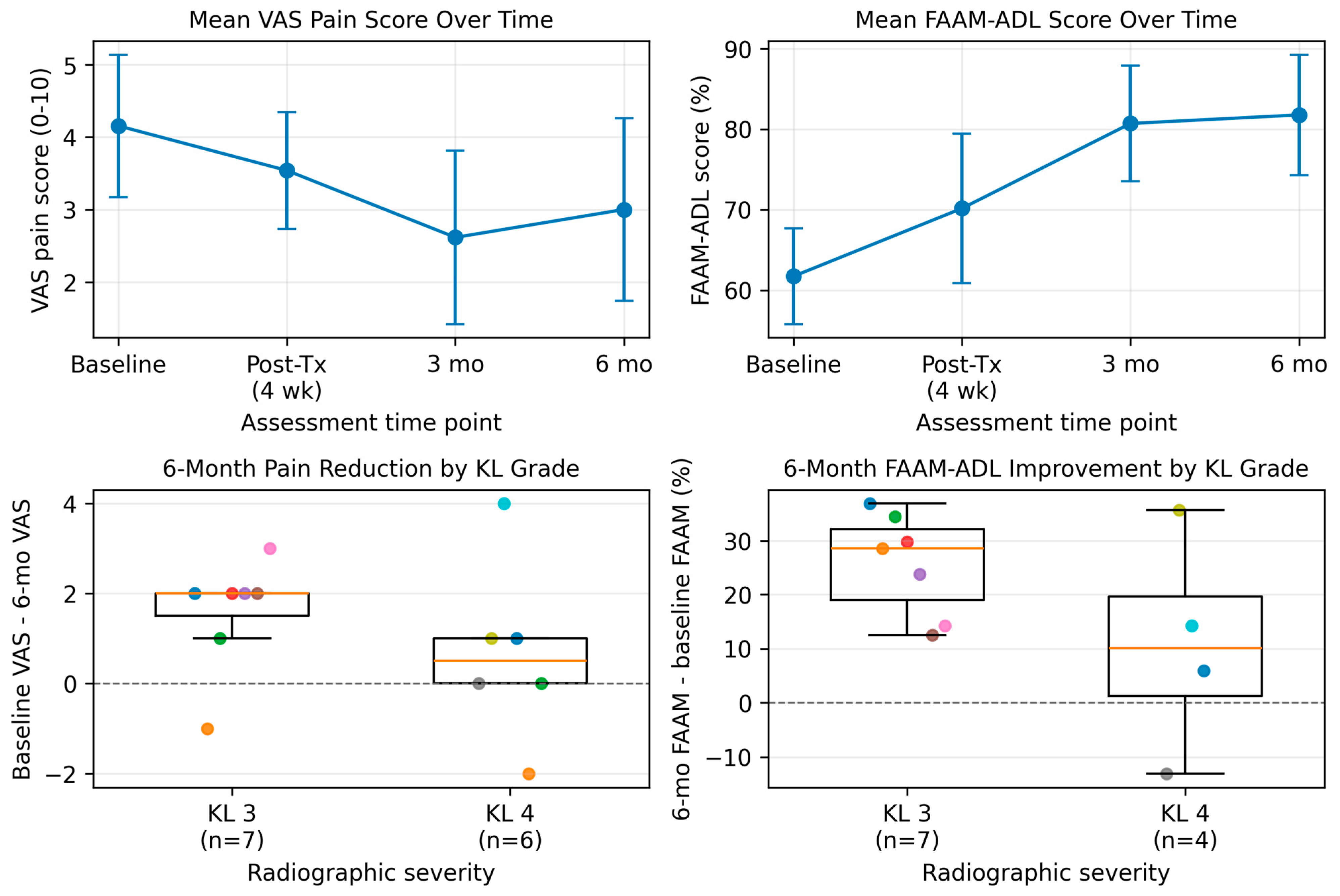
The overall repeated-measures ANOVA demonstrated a significant effect of time on VAS pain scores over the study period: F(3,36) = 3.93, p = 0.016. Mean VAS scores decreased from 4.15 ± 1.63 at baseline to a minimum of 2.62 ± 1.98 at 3 months, then increased to 3.00 ± 2.08 at 6 months. Bonferroni-adjusted pairwise comparisons among all time points showed that baseline-to-follow-up differences approached but did not consistently reach strict statistical significance. The strongest improvement occurred at 3 months (mean pain reduction, 1.54 points; 95% CI, 0.42 to 2.66; adjusted p = 0.067), while the 6-month time point showed a maintained but non-significant mean reduction (1.15 points; 95% CI, 0.17 to 2.14; adjusted p = 0.150).

3.3. Functional Outcomes (FAAM-ADL)

Functional improvement, measured by the FAAM-ADL subscale, was significant. The repeated-measures ANOVA demonstrated a robust main effect of time: F(3,30) = 13.76, p < 0.001. Mean available-case FAAM-ADL scores increased from 61.7% ± 9.9% at baseline to 70.2% ± 15.4% post-treatment, 80.7% ± 11.8% at 3 months, and 81.8% ± 11.1% at 6 months. Bonferroni-adjusted pairwise comparisons confirmed significant improvements at all post-treatment time points compared with baseline: post-treatment (p = 0.025), 3 months (p = 0.006), and 6 months (p = 0.008). The mean 6-month improvement among feet with complete 6-month FAAM-ADL data was +20.3 percentage points (95% CI, 10.0 to 30.6), exceeding the prespecified 8-point clinical significance threshold (Table 2).

3.4. Impact of Arthritis Severity

An exploratory analysis evaluated the influence of radiographic disease severity, classified by KL grade, on treatment response. The cohort included seven feet classified as moderate OA (KL grade 3) and six feet classified as severe/end-stage OA (KL grade 4). Patients with moderate OA demonstrated larger mean clinical improvements after EMTT than those with severe OA. Grade 3 feet achieved a mean 6-month functional improvement of +25.8 percentage points on FAAM-ADL (95% CI, 17.0 to 34.5), compared with +10.7 percentage points in grade 4 feet with complete FAAM-ADL data (95% CI, −21.5 to 42.9). Pain reduction was also more pronounced in the grade 3 cohort (+1.6 points; 95% CI, 0.4 to 2.7) than in the grade 4 cohort (+0.7 points; 95% CI, −1.4 to 2.7). Within-grade Spearman correlations between pain reduction and FAAM-ADL improvement were not statistically significant (KL grade 3: ρ = −0.39, p = 0.382; KL grade 4: ρ = 0.63, p = 0.368). Because of the small subgroup sizes, these findings should be interpreted as descriptive and hypothesis-generating (Table 3 and Figure 1).

4. Discussion

The present pilot study evaluated the preliminary effect of Extracorporeal Magnetotransduction Therapy (EMTT) in the management of midfoot osteoarthritis (OA), a condition that often presents a therapeutic challenge due to the complex biomechanics of the foot and limited noninvasive treatment options. In this small cohort, EMTT was not associated with treatment-related adverse events and was followed by clinically meaningful functional improvement over 6 months. The results are encouraging but should be interpreted as hypothesis-generating rather than definitive evidence of efficacy.

4.1. Efficacy and Clinical Impact

Our primary analysis demonstrated a significant overall time effect for VAS pain scores (p = 0.016), with the lowest mean pain observed at the 3-month follow-up (2.62 ± 1.98). However, Bonferroni-adjusted baseline-to-follow-up comparisons did not meet the conventional p < 0.05 threshold after adjustment across all time-point contrasts, with the 3-month comparison approaching significance (adjusted p = 0.067; mean reduction = 1.54 points). This reduction exceeded the prespecified clinical significance threshold and suggests that the observed pain change may be clinically meaningful despite limited statistical power. Functional outcomes showed a more robust pattern. FAAM-ADL scores improved significantly over time (p < 0.001), reaching 81.8% at 6 months, with a mean 6-month improvement of +20.3 percentage points among complete cases. This exceeded the 8-point FAAM-ADL MCID threshold and supports the possibility that EMTT may improve daily function in selected patients. The partial dissociation between pain and function suggests that functional restoration, potentially driven by improved gait tolerance, reduced stiffness, or better load distribution, may persist even when pain reduction plateaus. These findings align with emerging EMTT literature in other anatomical regions, where electromagnetic transduction has been associated with improved musculoskeletal function beyond the immediate treatment window [12].

4.2. Mechanism of Action

The observed clinical benefits may be related to the unique mechanism of EMTT. Unlike extracorporeal shockwave therapy (ESWT), which relies on acoustic mechanical energy, EMTT uses electromagnetic waves, similar to PEMF, with high-frequency oscillating magnetic fields (100–300 kHz) to penetrate deep tissues without thermal heating [11,12]. It has been hypothesized that electromagnetic stimulation influences cellular membrane potential and may support sodium–potassium pump activity in metabolically compromised cells [15,20]. In OA, this cellular modulation may reduce pro-inflammatory cytokine signaling and stimulate biological activity in subchondral bone and periarticular soft tissues [16,17,21]. The improvements observed in this cohort support the biological plausibility that EMTT may target inflammatory contributors to osteoarthritic pain; however, mechanistic confirmation was beyond the scope of this clinical pilot study.

4.3. Influence of Arthritis Severity (Kellgren–Lawrence Grade)

A notable exploratory observation from this pilot study is the trend toward severity-dependent response to EMTT. Feet with moderate osteoarthritis (KL grade 3) demonstrated larger mean improvements in both pain (+1.6 points) and function (+25.8 percentage points) than feet with severe, end-stage osteoarthritis (KL grade 4), which demonstrated more modest gains (+0.7 points pain reduction and +10.7 percentage points functional improvement among complete FAAM-ADL cases). The within-grade Spearman correlations between pain reduction and functional improvement were not statistically significant, and the subgroup sample sizes were too small for definitive inference. Still, the descriptive pattern suggests a potential therapeutic window of opportunity. In grade 4 disease, characterized by severe joint-space loss and bony deformity, the biological reserve of the joint may be insufficient to respond robustly to electromagnetic stimulation [20]. Conversely, grade 3 joints, which retain more joint structure, may be more suitable targets for noninvasive biologic modulation [22].

4.4. Limitations

The interpretation of these findings must be tempered by several limitations. First, the sample size (N = 8 patients, 13 feet) is small, limiting statistical power to detect smaller effect sizes or confirm subgroup differences. Second, because five participants contributed bilateral feet, treated-foot observations may not be fully independent. The foot-level approach was used because symptoms, KL grade, and treatment response were foot-specific, but future studies should account for patient-level clustering in the primary statistical model. Third, the lack of a sham-control group means that placebo and contextual effects cannot be ruled out, which is especially important in pain intervention studies [23,24]. The magnitude of functional improvement exceeded the FAAM-ADL MCID [19], but this does not establish causality in the absence of a control group. Fourth, 6-month FAAM-ADL data were unavailable for two grade 4 feet from one participant who proceeded to surgery and marked all FAAM-ADL items as not applicable; these feet were excluded from FAAM-ADL change analyses in accordance with the no-imputation approach and were retained as treatment failures clinically.
Additional limitations include the 6-month follow-up period, which is insufficient to determine long-term durability or the need for maintenance therapy. The dataset did not include height, weight, plantar contact area, or plantar pressure measurements, so BMI should be interpreted only as a broad patient-level descriptor rather than a foot-specific measure of mechanical loading. Future studies should include objective biomechanical variables, larger sample sizes, sham controls, and longer follow-up. The MAGNETOLITH device was provided by Curamedix; however, Curamedix did not have access to study results and had no role in data collection, analysis, interpretation, manuscript preparation, or the decision to publish.

5. Conclusions

This pilot study provides preliminary, hypothesis-generating evidence that Extracorporeal Magnetotransduction Therapy (EMTT) may be a safe, noninvasive treatment option for symptomatic midfoot osteoarthritis. EMTT was associated with clinically meaningful functional improvement and an overall time effect for pain, although adjusted pairwise pain comparisons did not consistently reach statistical significance. The descriptive data suggest that patients with moderate (KL grade 3) osteoarthritis may experience greater improvement than those with end-stage (KL grade 4) disease. Given the favorable safety profile observed in this small cohort, EMTT may warrant further evaluation within the conservative management algorithm for midfoot OA. Randomized, sham-controlled trials with larger cohorts and longer follow-up are needed to validate these findings and define optimal patient selection criteria.

Author Contributions

Conceptualization, E.B.; methodology, E.B.; software, A.V. and R.C.; validation, E.B., A.V. and R.C.; formal analysis, A.V. and R.C.; investigation, E.B.; data curation, A.V. and R.C.; writing—original draft preparation, R.C.; writing—review and editing, A.V., R.C. and E.B.; supervision, E.B.; project administration, E.B.; funding acquisition, E.B. All authors have read and agreed to the published version of the manuscript.

Funding

This research was funded by the Mayo Clinic’s Orthopedic Research Review Committee (ORRC) Research Support Program, internal department funds. No specific grant or award number was assigned to this funding. The funding source had no role in study design, data collection, data analysis, manuscript preparation, or the decision to submit the manuscript for publication.

Institutional Review Board Statement

The study was conducted in accordance with the Declaration of Helsinki, and approved by the Institutional Review Board (or Ethics Committee) of the Mayo Clinic in Rochester, MN (protocol code 24-002661 and date of approval: 29 October 2024).

Informed Consent Statement

Informed consent was obtained from all subjects involved in the study.

Data Availability Statement

The data underlying this article cannot be shared publicly because they contain protected health information and are subject to Mayo Clinic institutional data governance requirements. Request for access maybe considered on a case-by-case basis subject to institutional review an applicable regulatory approvals.

Acknowledgments

During preparation of this manuscript, the author(s) used Perplexity Pro (education pro) as a coding and drafting aid to assist with Python 3.14.3 syntax and statistical output checks. The authors reviewed and edited all generated output, verified the statistical results against the study dataset, and take full responsibility for the content of this publication.

Conflicts of Interest

Curamedix provided the EMTT device to our department for research purposes; however, they had no role in the design of the study; in the collection, analyses, or interpretation of data; in the writing of the manuscript; or in the decision to publish the results.

References

  1. Thomas, M.J.; Peat, G.; Rathod, T.; Marshall, M.; Moore, A.; Menz, H.B.; Roddy, E. The epidemiology of symptomatic midfoot osteoarthritis in community-dwelling older adults: Cross-sectional findings from the Clinical Assessment Study of the Foot. Arthritis Res. Ther. 2015, 17, 178. [Google Scholar] [CrossRef]
  2. Rao, S.; Baumhauer, J.F.; Nawoczenski, D.A. Is barefoot regional plantar loading related to self-reported foot pain in patients with midfoot osteoarthritis. Osteoarthr. Cartil. 2011, 19, 1019–1025. [Google Scholar] [CrossRef]
  3. Adams, S.B.; Garras, D.N.; Lee, S. Ankle arthritis: Part I. Joint preservation techniques and arthrodesis. In Orthopaedic Knowledge Update: Foot and Ankle 6; American Academy of Orthopaedic Surgeons: Rosemont, IL, USA, 2020; pp. 105–128. [Google Scholar]
  4. Rao, S.; Baumhauer, J.F.; Becica, L.; Nawoczenski, D.A. Shoe inserts alter plantar loading and function in patients with midfoot arthritis. J. Orthop. Sports Phys. Ther. 2009, 39, 522–531. [Google Scholar] [CrossRef] [PubMed]
  5. Halstead, J.; Chapman, G.J.; Gray, J.C.; Grainger, A.J.; Brown, S.; Wilkins, R.A.; Roddy, E.; Helliwell, P.S.; Keenan, A.M.; Redmond, A.C. Foot orthoses in the treatment of symptomatic midfoot osteoarthritis using clinical and biomechanical outcomes: A randomised feasibility study. Clin. Rheumatol. 2016, 35, 987–996. [Google Scholar] [PubMed]
  6. Nemec, S.A.; Habbu, R.A.; Anderson, J.G.; Bohay, D.R. Outcomes following Midfoot Arthrodesis for Primary Arthritis. Foot Ankle Int. 2011, 32, 355–361. [Google Scholar] [CrossRef] [PubMed]
  7. Reynolds, A.; Kumar, C.S. Outcomes and Complications of Midfoot Arthrodesis in a Cohort of 108 Patients. Orthop. Proc. 2023, 105-B, 4. [Google Scholar] [CrossRef]
  8. Buda, M.; Hagemeijer, N.C.; Kink, S.; Johnson, A.H.; Guss, D.; DiGiovanni, C.W. Effect of Fixation Type and Bone Graft on Tarsometatarsal Fusion. Foot Ankle Int. 2018, 39, 1394–1402. [Google Scholar] [CrossRef] [PubMed]
  9. Gougoulias, N.; Lampridis, V. Midfoot arthrodesis. Foot Ankle Surg. 2016, 22, 17–25. [Google Scholar] [CrossRef] [PubMed]
  10. Paolucci, T.; Pezzi, L.; Centra, A.M.; Giannandrea, N.; Bellomo, R.G.; Saggini, R. Electromagnetic Field Therapy: A Rehabilitative Perspective in the Management of Musculoskeletal Pain—A Systematic Review. J. Pain Res. 2020, 13, 1385–1400. [Google Scholar] [CrossRef] [PubMed]
  11. Gerdesmeyer, L.; Zielhardt, P.; Klüter, T.; Gollwitzer, H.; Gerdesmeyer, L.; Hausdorf, J.; Ringeisen, M.; Knobloch, K.; Saxena, A.; Krath, A. Stimulation of human bone marrow mesenchymal stem cells by electromagnetic transduction therapy—EMTT. Electromagn. Biol. Med. 2022, 41, 304–314. [Google Scholar] [CrossRef] [PubMed]
  12. Gerdesmeyer, L.; Tübel, J.; Obermeier, A.; Harrasser, N.; Glowalla, C.; von Eisenhart-Rothe, R.; Burgkart, R. Extracorporeal Magnetotransduction Therapy as a New Form of Electromagnetic Wave Therapy: From Gene Upregulation to Accelerated Matrix Mineralization in Bone Healing. Biomedicines 2024, 12, 2269. [Google Scholar] [CrossRef] [PubMed]
  13. Dong, Y.; Suryani, L.; Zhou, X.; Muthukumaran, P.; Rakshit, M.; Yang, F.; Wen, F.; Hassanbhai, A.M.; Parida, K.; Simon, D.T.; et al. Synergistic Effect of PVDF-Coated PCL-TCP Scaffolds and Pulsed Electromagnetic Field on Osteogenesis. Int. J. Mol. Sci. 2021, 22, 6438. [Google Scholar] [CrossRef] [PubMed]
  14. Kotnik, T.; Rems, L.; Tarek, M.; Miklavčič, D. Membrane Electroporation and Electropermeabilization: Mechanisms and Models. Annu. Rev. Biophys. 2019, 48, 63–91. [Google Scholar] [CrossRef] [PubMed]
  15. Mancini, M.; Vetrano, M.; Traversa, A.; Cauli, C.; Ceccarelli, S.; Malisan, F.; Vulpiani, M.C.; Maffulli, N.; Marchese, C.; Visco, V.; et al. Electromagnetic Transduction Therapy (EMTT) Enhances Tenocyte Regenerative Potential: Evidence for Senolytic-like Effects and Matrix Remodeling. Int. J. Mol. Sci. 2025, 26, 7122. [Google Scholar] [CrossRef] [PubMed]
  16. Zhou, S.; Wen, H.; He, X.; Han, X.; Li, H. Pulsed electromagnetic field ameliorates the progression of osteoarthritis via the Sirt1/NF-κB pathway. Arthritis Res. Ther. 2025, 27, 33. [Google Scholar] [CrossRef] [PubMed]
  17. Yang, X.; Guo, H.; Ye, W.; Yang, L.; He, C. Pulsed Electromagnetic Field Attenuates Osteoarthritis Progression in a Murine Destabilization-Induced Model through Inhibition of TNF-α and IL-6 Signaling. Cartilage 2021, 13, 1665S–1675S. [Google Scholar] [CrossRef] [PubMed]
  18. Tubach, F.; Ravaud, P.; Baron, G.; Falissard, B.; Logeart, I.; Bellamy, N.; Bombardier, C.; Felson, D.; Hochberg, M.; van der Heijde, D.; et al. Evaluation of clinically relevant changes in patient reported outcomes in knee and hip osteoarthritis: The minimal clinically important improvement. Ann. Rheum. Dis. 2005, 64, 29–33. [Google Scholar] [CrossRef] [PubMed]
  19. Martin, R.L.; Irrgang, J.J.; Burdett, R.G.; Conti, S.F.; Van Swearingen, J.M. Evidence of validity for the Foot and Ankle Ability Measure (FAAM). Foot Ankle Int. 2005, 26, 968–983. [Google Scholar] [CrossRef] [PubMed]
  20. Funk, R. Coupling of pulsed electromagnetic fields (PEMF) therapy to molecular grounds of the cell. Am. J. Transl. Res. 2018, 10, 1260–1272. [Google Scholar] [PubMed]
  21. Varani, K.; Vincenzi, F.; Pasquini, S.; Blo, I.; Salati, S.; Cadossi, M.; De Mattei, M. Pulsed Electromagnetic Field Stimulation in Osteogenesis and Chondrogenesis: Signaling Pathways and Therapeutic Implications. Int. J. Mol. Sci. 2021, 22, 809. [Google Scholar] [CrossRef] [PubMed]
  22. Sutbeyaz, S.T.; Sezer, N.; Koseoglu, B.F. The effect of pulsed electromagnetic fields in the treatment of cervical osteoarthritis: A randomized, double-blind, sham-controlled trial. Rheumatol. Int. 2006, 26, 320–324. [Google Scholar] [PubMed]
  23. Moseley, J.B.; O’Malley, K.; Petersen, N.J.; Menke, T.J.; Brody, B.A.; Kuykendall, D.H.; Hollingsworth, J.C.; Ashton, C.M.; Wray, N.P. A controlled trial of arthroscopic surgery for osteoarthritis of the knee. N. Engl. J. Med. 2002, 347, 81–88. [Google Scholar] [CrossRef] [PubMed]
  24. Zou, K.; Wong, J.; Abdullah, N.; Chen, X.; Smith, T.; Doherty, M.; Zhang, W. Examination of overall treatment effect and the proportion attributable to contextual effect in osteoarthritis: Meta-analysis of randomised controlled trials. Ann. Rheum. Dis. 2016, 75, 1964–1970. [Google Scholar] [CrossRef] [PubMed]
Figure 1. Subgroup longitudinal analysis of clinical outcomes following EMTT for midfoot osteoarthritis. Mean VAS pain scores (top left) and mean FAAM-ADL scores (top right) are shown over 6 months with 95% confidence intervals. Post-Tx indicates the 4-week assessment after the eighth EMTT session. Boxplots (bottom row) display individual treated-foot changes from baseline to 6 months categorized by radiographic severity. The FAAM-ADL change plot includes complete 6-month FAAM-ADL cases only (KL grade 3: n = 7; KL grade 4: n = 4).
Japma 116 00051 g001
Table 1. Demographic and clinical characteristics. Continuous variables are reported as mean ± SD with t-based 95% CI for the mean and minimum–maximum range. Categorical variables are reported as n (%). BMI = body mass index; KL = Kellgren–Lawrence.
CharacteristicValue
Patients, n8
Treated feet, n13
Bilateral midfoot OA, n (%)5 (62.5%)
Unilateral midfoot OA, n (%)3 (37.5%)
Age, years, mean ± SD (95% CI)68.1 ± 7.6 (61.8–74.5)
Age, years, minimum–maximum58–81
Female sex, n (%)8 (100%)
BMI, kg/m2, mean ± SD (95% CI)34.69 ± 7.43 (28.48–40.90)
BMI, kg/m2, minimum–maximum23.87–45.90
Radiographic severity, KL grade 3/grade 47 feet (53.8%)/6 feet (46.2%)
Table 2. Changes in pain (VAS) and functional scores (FAAM-ADL) from baseline to 6-month follow-up. Values are mean ± SD with t-based 95% CI for the mean. * Indicates statistically significant difference versus baseline (p < 0.05) after Bonferroni adjustment across all six pairwise time-point contrasts. The VAS analysis included 13 feet with complete data across all four time points. The FAAM-ADL repeated-measures ANOVA included 11 feet with complete data across all four time points; descriptive means use all available observations at each time point.
Outcome MeasureBaselinePost-Treatment
(4 Weeks)
3-Month Follow-Up6-Month Follow-UpOverall Main Effect of Time
VAS Pain Score
(0–10)
4.15 ± 1.633.54 ± 1.332.62 ± 1.983.00 ± 2.08F(3,36) = 3.93
p = 0.016
95% CI: 3.17–5.1495% CI: 2.73–4.3495% CI: 1.42–3.8195% CI: 1.74–4.26
n = 13n = 13n = 13n = 13
FAAM-ADL Score
(%)
61.7 ± 9.970.2 ± 15.4 *80.7 ± 11.8 *81.8 ± 11.1 *F(3,30) = 13.76
p < 0.001
95% CI: 55.8–67.795% CI: 60.9–79.595% CI: 73.5–87.995% CI: 74.3–89.2
n = 13n = 13n = 13n = 11
Table 3. Comparison of 6-month clinical improvement (ΔVAS and ΔFAAM-ADL) stratified by Kellgren–Lawrence (KL) arthritis grade. Values are mean ± SD with t-based 95% CI. Positive ΔVAS indicates pain reduction; positive ΔFAAM-ADL indicates functional improvement. Spearman ρ values reflect within-grade correlations between ΔVAS and ΔFAAM-ADL and are reported with two-sided p values. FAAM-ADL change in KL grade 4 is based on four feet because two grade 4 feet from one participant had all 6-month FAAM-ADL items marked not applicable after progression to surgery; no imputation was performed.
Radiographic
Severity
N for
ΔVAS
Mean Pain Reduction
ΔVAS (95% CI)
N for
ΔFAAM-ADL
Mean Functional Improvement
ΔFAAM-ADL (95% CI)
Spearman ρ
(p Value)
KL Grade 3
(Moderate)
7+1.6 ± 1.3 points
(0.4 to 2.7)
7+25.8 ± 9.5 points
(17.0 to 34.5)
−0.39
(p = 0.382)
KL Grade 4
(Severe)
6+0.7 ± 2.0 points
(−1.4 to 2.7)
4+10.7 ± 20.2 points
(−21.5 to 42.9)
0.63
(p = 0.368)
Disclaimer/Publisher’s Note: The statements, opinions and data contained in all publications are solely those of the individual author(s) and contributor(s) and not of MDPI and/or the editor(s). MDPI and/or the editor(s) disclaim responsibility for any injury to people or property resulting from any ideas, methods, instructions or products referred to in the content.