Article

Influence of the Hardness of Insoles on the Abductor Hallucis Muscle in Baxter’s Entrapment: A Cross-Over Randomized Study


by
Vanesa Pinto-Franco
1,
Marta Elena Losa Iglesias
1,
Israel Casado-Hernández
2,*,
Emmanuel María Navarro-Flores
3,
Daniel López-López
4,
Eva María Martínez-Jiménez
2,
Eduardo Pérez-Boal
5,
José Luis Muñoz-Sánchez
2 and
Ricardo Becerro-de-Bengoa-Vallejo
2
1
Faculty of Health Sciences, University Rey Juan Carlos, 28922 Alcorcón, Spain
2
Faculty of Nursing, Physiotherapy and Podiatry, University Complutense of Madrid, 28040 Madrid, Spain
3
Faculty of Nursing and Podiatry, Department of Nursing, University of Valencia, Research in Foot Health and Quality of life (INSAPICAVI), 46010 Valencia, Spain
4
Research, Health, and Podiatry Group, Department of Health Sciences, Faculty of Nursing and Podiatry, Industrial Campus of Ferrol, Universidade da Coruña, 15403 Ferrol, Spain
5
Faculty of Nursing and Physiotherapy, University of León, 24401 Ponferrada, Spain
*
Author to whom correspondence should be addressed.
J. Am. Podiatr. Med. Assoc. 2026, 116(2), 23241; https://doi.org/10.7547/23-241
Submission received: 30 December 2023 / Revised: 6 February 2024 / Accepted: 13 June 2024 / Published: 21 April 2026

Abstract

Background: The etiology and diagnosis of heel pain are complex and multifactorial, and it has been reported that Baxter’s entrapment is responsible for up to 20% of foot disorders. The most conservative treatment of Baxter’s entrapment has been the use of custom insoles. Electromyography was considered an effective test to assess muscle activity. The aim of this study was to test the use of insoles of different hardness on muscle activity of the abductor hallucis muscle in subjects with entrapment of the lateral branch of the external plantar nerve. Methods: A total of 18 subjects (seven women and 11 men) diagnosed with nerve entrapment of the first branch of the lateral plantar nerve were recruited. Muscle activity of the adductor hallucis muscle was analyzed with insoles of different hardness in static and dynamic situations using electromyographic evaluation. Results: The statistical analysis did not show statistically significant differences in the muscle activity of the abductor hallucis muscle in the static position, with and without plantar orthoses (p > 0.05); in contrast, in dynamic situations statistically significant differences were found between groups (p > 0.01). Conclusions: The use of a hard insole decreases the maximum peak muscular activity of the abductor hallucis muscle in subjects with Baxter’s nerve entrapment in walking conditions.

1. Introduction

Neuropathic plantar heel pain is characterized by involving branches of the posterior tibial nerve, the lateral plantar nerve and the nerve of the abductor digiti minimi (ADM) muscle [1,2]. Baxter et al. first described the isolated neurolysis of the nerve that supplies the ADM muscle and suggested entrapment neuropathy as an etiologic factor in heel pain [3]. Baxter’s neuropathy represents one of the more often overlooked causes of heel pain and has been reported to be responsible for up to 20% of this disorder [4,5]. Nerve entrapment can be caused by overuse, trauma, or injury from previous surgery; thus, the etiology of heel pain is complex and multifactorial [1]. However, mechanical overload was thought to be very important regarding injury development and was associated with several factors such as obesity, and the decreasing size and thickness of the heel pad; the biomechanics of gait disorders were also considered [6,7]. In the research carried out by Groof et al. it was concluded that the diagnosis was confusing because plantar fasciitis was one of the causes of ADM atrophy and, in turn, was also a cause of pain in the heel [8]. Custom insoles have been used for Baxter’s neuropathy treatment and, in cases where the insole treatment has been useless, in recalcitrant cases, the surgical release of the nerve was considered the best option to improve a better quality of life [9,10].
Recent research has demonstrated that the use of insoles and shoes have a high involvement in the lower limb muscles during the gait cycle, and to evaluate their electromyography has proved to be the best diagnostic instrument to analyze muscle gait disorders with high reliability [11,12]. The biomechanical literature has focused on gait analysis in standing and dynamic situations using different kinematic, kinetic and electromyographic diagnostic instruments (EMG) [13,14]. Scott et al. analyzed the influence of footwear on the electromyographic activity of a selection of muscles in the lower limb during walking [15]. In addition, Moisan et al. quantified the effects of two types of plantar orthoses on muscle activity during gait using electromyography to obtain the research data [16].
A previous study conducted by Casado-Hernández et al. analyzed the electromyographic activity of the muscles of the lower limb in healthy people using insoles of different hardness during motorcycling. They concluded that there was less electrical activity of the lower limb muscles using the hardest insoles [17,18].
There is a lack of scientific research on the influence of using insoles of different hardness on the muscle activity of the abductor hallucis (AH). Our hypothesis was that the hardness of the insoles decreases AH muscle activity compared to the use of soft insoles and walking barefoot in subjects with Baxter’s entrapment. The aim of our research was to analyze the muscle AH variation in subjects with Baxter’s entrapment using insoles of different hardness with electromyography.

2. Methods

2.1. Study Design

A crossover randomized study was carried out between September 2022 and February 2023 to analyze the effect of using insoles of different hardness on the abductor hallucis muscle in subjects diagnosed with Baxter’s nerve entrapment. This research was approved by the ethics committee of Clinical Research at the Hospital Clínico San Carlos of Madrid (3 February 2021, consent nº 21/048-EC P). All the subjects agreed to participate and signed the informed consent.
The recruited subjects were 11 women with a mean age of 50.86 ± 13.61 years, and 7 men with a mean age of 54.82 ± 8.13 years. Table 1 shows the sociodemographic characteristics where statistically non-significant differences were found.
All the subjects were screened previously by an expert clinician with more than 10 years’ experience. The inclusion criteria was as follows: (1) subjects diagnosed with Baxter’s nerve entrapment referring to the point of maximum pain on the medial edge of the heel at about five centimeters from its posterior aspect, extending towards the lateral aspect of the heel following the path of the nerve, and with the clinical findings: pain on palpation in the area of the entrapment between the deep fascia of the abductor of the first toe and the medial and caudal portion of the quadratus plantae muscle [19]. The exclusion criteria were: (1) subjects who suffered neuromuscular diseases, (2) subjects who had suffered a trauma injury or other physical disabilities or diseases that affect the lower limbs and their ability to walk, (3) pregnant women and (4) subjects who refused to sign the informed consent.
The sample size was calculated with the software from The Clinical and Biostatistical Epidemiology Unity, Complexo Hospitalario Universitario from La Coruña, Universidade A Coruña (www.fisterra.com). We set out testing differences in maximum plantar pressures with another study employing ethylene vinyl acetate (EVA) material. The maximum metatarsal pressure observed in subjects using a custom flat insole (CFI) and a contoured insole was 148.4 ± 35.2 and 147.2 ± 29.8 KPa, respectively [20]. To accomplish this, a statistical confidence of 95%, with a 2-tailed hypothesis test and a large effect size of 0.90, an α-error of 0.05, and a power of analysis of 0.80 (β error = 20%) obtained a minimum sample size of 12 subjects to be included in the observational group for our research [21].
The research was accomplished following the guidelines and list for Template for Intervention Description and Replication (TIDieR) [22].

2.2. Procedure

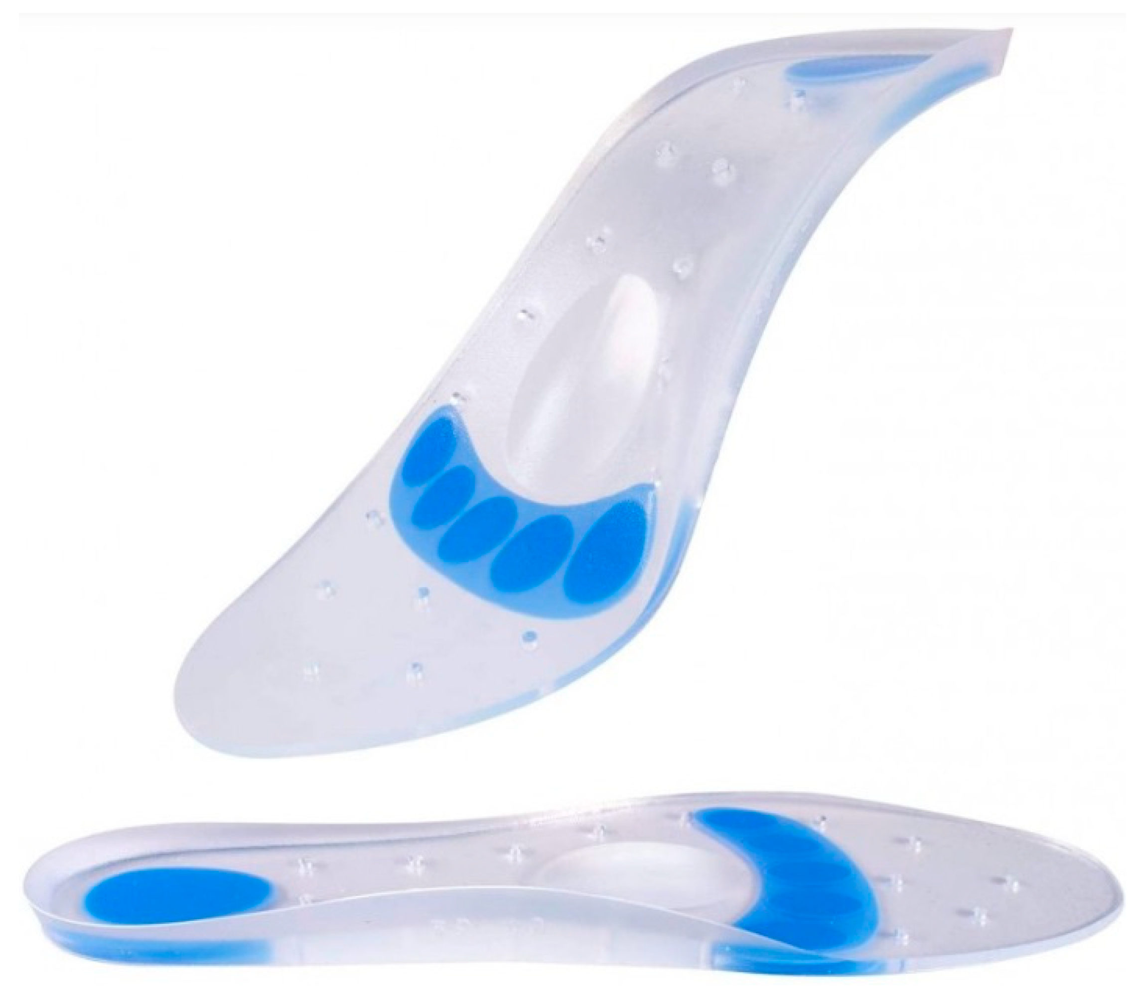
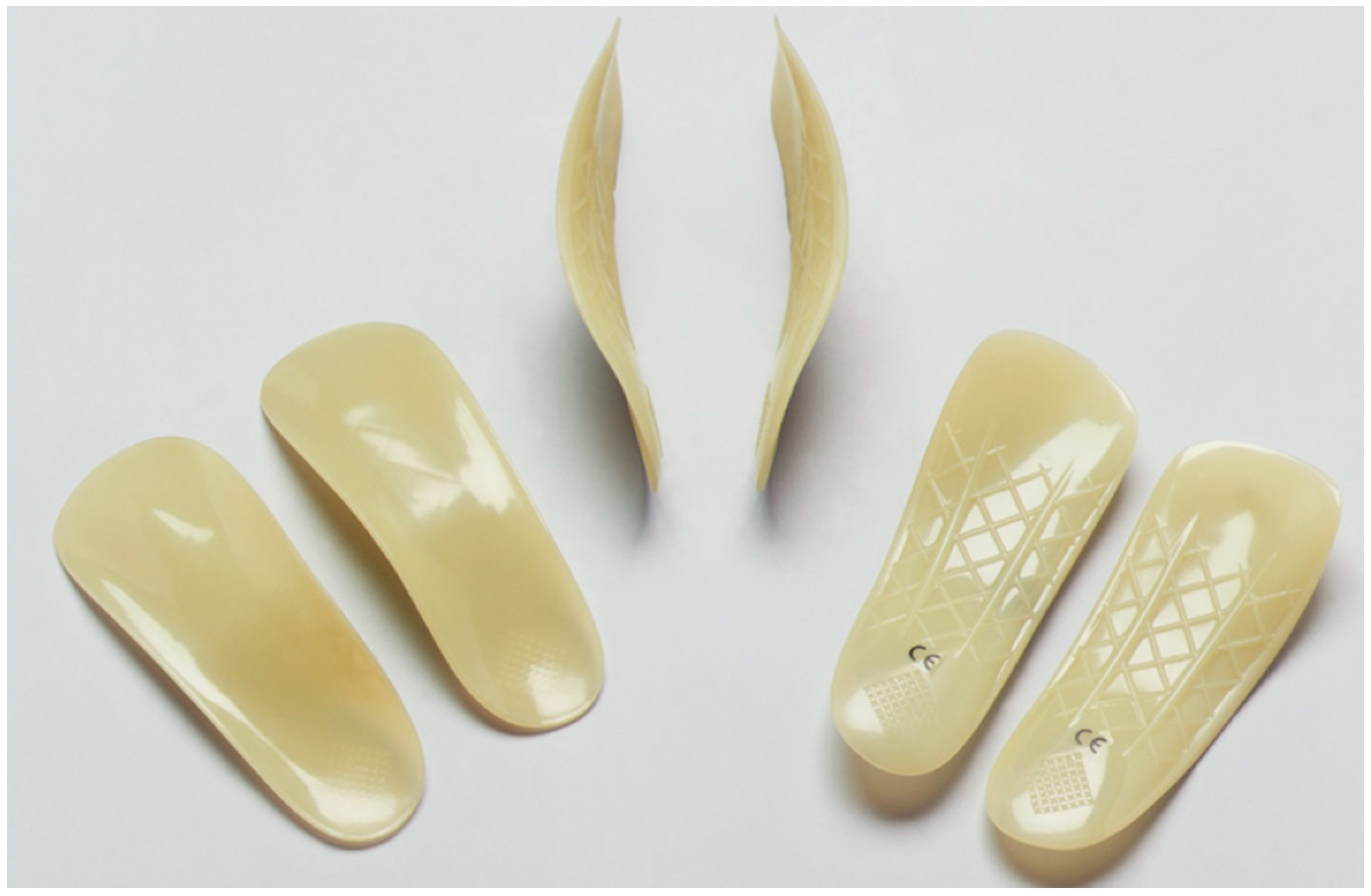
The experimental design of this analytical study was a crossover randomized trial based on a repetition of measurements of the muscle activity of the abductor hallucis and the abductor muscle of the fifth toe in static and dynamic situations during three conditions in a random way: barefoot, using soft insoles and using hard insoles. The soft insoles used were made of two different silicone densities: The insole was made of silicone with 5–9° Shore A and the posterior and medial side was filled with silicone of 10–14° Shore A (Figure 1). The hard insole was made of polypropylene of 58° Shore D (Figure 2).
The order of measurement in the different conditions (barefoot, soft plantar orthoses and hard plantar orthoses) was randomized for both static and dynamic situations using the Epidat 4.2 computer program (La Xunta de Galicia. Ministry of Health. Galicia. www.sergas.es/Saude-publica/EPIDAT).
The ambient conditions of the room were constant in terms of light and absence of noise. In a second test, the analysis was carried out under the same circumstances in a dynamic situation, using a treadmill with a constant speed of 4 km/h for one minute to avoid muscle fatigue and resting for five minutes between each condition. For the dynamic measurements, the plantar orthoses were fixed to the foot with a stretch underwrap elastic bandage to avoid wearing footwear that could interfere with the measurement of electrical activity [23].
Surface electrodes were used after skin preparation according to the guidelines established by the European SENIAM project (Surface Electromyography for the Non-Invasive Assessment of Muscles. http://www.seniam.org/) with cotton soaked in alcohol to minimize errors derived from impedance of the skin [24]. The electrodes were fixed to the skin using a stretch underwrap elastic bandage on the abductor hallucis (AH) from 3 cm from the origin on the postero-medial plantar aspect of the calcaneus longitudinally following the direction of the muscle and on the abductor of the fifth toe from the origin on the postero-lateral plantar aspect of the calcaneus longitudinally following the direction of the muscle.
To acquire the electromyographic data, DataLINK DLK900 was used (Biometrics Ltd., Newport, UK. www.biometricsltd.com) and surface electrodes were used for the data acquisition of the electrical activity of the APD and ADM with a Biometrics SX230FW surface electrode. The data were obtained and analyzed with Biometrics DataLINK software at 1000 Hz and filtered with 6–600 Hz.
The control data were processed in the maximum muscular activity during three consecutive periods in two seconds in the static condition and the maximum electrical activity of three periods of consecutive heel contacts was selected, in such a way that they maintained a certain homogeneity.
Pain scores were measured using the Visual Analog Pain Rating Scale (VAS). This scale has a reliability valued by an Intraclass Correlation Coefficient (ICC) of 0.97 [95% CI = 0.96 to 0.98] [24]. The scale ascends from 0 (no pain) to 10 (extreme pain).

2.3. Statistical Analysis

All data were analyzed for normality according to the Shapiro–Wilk test and data were regarded to be normally distributed if p > 0.05. The descriptive sociodemographic characteristics of the subjects presented the mean, median, body mass index (BMI), standard deviation (SD) and a 95% confidence interval (95% CI).
The intra-class correlation coefficients were computed to analyze reliability in each subject. To interpret ICC outcomes, we considered ICC outcomes between 0.20 and 0.40 as poor reliability, outcomes between 0.40 and 0.60 as fair, outcomes between 0.60 and 0.80 as good and, finally, outcomes between 0.80 and 1.00 were considered as excellent [25]. Portney and Watkins proposed that reliability coefficients greater than 0.90 were sufficient for clinical measurement [26].
The mean scores and the standard error of measurement (SEM) were calculated [27]. Bland and Altman’s formula was used to calculate the SEM as follow: SEM = SD × sqrt (1 − ICC) [28]. The minimal detectable change (MDC) was used to assess the minimal magnitude of change required to be 95% confident that the observed change between the 2 tests reflects the true change and not measurement error [29]. The MDC was calculated as: 1.96 × SEM × √2. To demonstrate the effect size of the comparisons, Cohen’s d coefficient was calculated. Cohen’s d effect size was rated as follows: values ≤0.20 indicate slight effects, values between 0.20 and 0.49 indicate fair effects, values between 0.50 and 0.79 indicate moderate effects, and values larger than 0.79 indicate large effects [30].
Regarding non-parametric data, the Mann–Whitney U test was used to analyze if between group comparisons showed statistically significant differences. For all tests, we considered p values < 0.05 to be statistically significant. All data were analyzed with the statistical software SPSS 21.0 for Windows (IBM, Chicago, IL, USA).

3. Results

The data were obtained from a sample size of 18 subjects composed of seven women and 11 men. Table 2 describes the reliability of the variables studied, highlighting almost perfect scores in dynamics in Muscle AH activity walking barefoot without insoles with an ICC = 0.887, in Muscle AH activity walking with soft insoles with an ICC = 0.949 and in Muscle AH activity walking with hard insoles with an ICC = 0.942. The variable Muscle AH activity barefoot hard insole static showed an almost perfect ICC = 0.930. Regarding the other static variables, all of them showed a good ICC.
Table 3 describes the differences between insoles of different hardness in static and walking conditions. Only the variable walking using soft insoles Vs hard insoles showed statistically significant differences with a p = 0.009. Regarding other conditions such as static and walking with insoles of different hardness, the other variables did not show statistically significant differences. Cohen’s d showed a large effect in the variable walking barefoot Vs soft insoles with a value 0.92 and the variable walking soft insoles Vs hard insoles with a value 0.94. The other variables showed a small effect.

4. Discussion

The aim of this research was to evaluate the electromyographic activity of the abductor hallucis muscle in Baxter’s entrapment with insoles of different hardness and compare the results in a static position and walking on a treadmill. As a novelty, this is the first research to investigate and analyze the effect on the muscle activity of the abductor hallucis using insoles of different hardness in subjects diagnosed with Baxter’s nerve entrapment in different conditions.
Recent research has compared electromyographic activity with different types of plantar orthoses but has focused on analyzing the activity of the following muscle groups: gluteus, gastrocnemius, vastus lateralis, tibial anterior and peroneus longus [16,17]. According to our research, the recent investigation that analyzed the relationship between the effect of using different insoles and the muscular activity of the abductor hallucis is a case report by Johnson et al. [31]. According to this study, acrylic resin insoles were used to analyze the muscular variation in the abductor hallucis in a weightlifter with Baxter’s neuropathy. The authors concluded that the entrapment was secondary to an excessive pronation during weightlifting and due to the stiffness of the insole, the compressive forces during weightlifting decreased and the muscle activity of the abductor hallucis too. In addition, these effects produced an instantaneous improvement in the subject’s symptoms. According to our research we that the best results in the static condition were found using insoles with a hardness of ICC = 0.930.
Currently, there is no clear consensus regarding conservative treatment of Baxter’s nerve entrapment and the choice of materials. On the other hand, there is homogeneity in the methods used to evaluate the muscle. Murley et al. [32] performed a systematic review on the effect of footwear, foot position and insoles on lower limb muscle activity during walking and running conditions. The conclusion of the systematic review was that using insoles produced an increase in the muscular activation of the tibialis anterior and peroneus longus and, in addition, could produce disorders in the muscles of the lower back. The literature collected in this research dealt with subjects walking and running. Regarding our research, data were collected both in static and walking situations and the result using soft insoles Vs hard insoles was statistically significant with a p value = 0.009. This result demonstrates that the use of hard insoles decreases the peak muscular activity of the abductor hallucis in subjects with Baxter’s entrapment improving their quality of life.
Casado et al. [17,18] analyzed the electromyographic variation in the lower limb, thigh and hip muscles using non-contoured insoles of different hardness made with polypropylene and aluminum in the metatarsal head and EVA 52° Shore A in motorcyclists and concluded that the maximum peak muscular activity produced by the rider on a motorcycle simulator decreased using hard insoles. Taking into consideration the similarity of the insoles used, according to our research, the use of hard insoles decreased the maximum peak activity of the abductor hallucis, but nevertheless, the research conducted by Casado et al. was with subjects on a motorcycle simulator and not walking or in a static position.
However, the findings of our study should be read in the context of some limitations that are acknowledged. First, all subjects that took part in the study were overweight. For future research it would be interesting to implement the same protocol in subjects with normal weight. Second, the data acquisition was obtained on a treadmill. For future research the effect of walking around in normal situations should be considered, such as up and down hills, and on different ground compositions. Nevertheless, this research has a great strength: it has been the first research to analyze the muscular activity variations in the abductor hallucis in subjects with Baxter’s nerve entrapment using different insoles.

5. Conclusions

The use of hard insoles decreases the maximum peak muscular activity of the abductor hallucis muscle in subjects with Baxter’s nerve entrapment in walking conditions. In addition, the reduction in the maximum peak muscular activity of the abductor hallucis may reduce the risk of individuals developing Baxter’s nerve entrapment and may help improve the quality of life of individuals who suffer from this disorder.

Author Contributions

Conceptualization, V.P.-F., I.C.-H., R.B.-d.-B.-V., M.E.L.-I., E.M.M.-J., D.L.-L., E.M.N.-F., J.L.M.-S. and E.P.-B., Data curation, D.L.-L. and I.C.-H., Formal analysis, V.P.-F., I.C.-H., E.M.N.-F., R.B.-d.-B.-V., M.E.L.-I., E.M.M.-J., D.L.-L. and E.P.-B., Investigation, V.P.-F., I.C.-H., R.B.-d.-B.-V., M.E.L.-I., E.M.M.-J., E.M.N.-F., D.L.-L. and E.P.-B. Methodology, V.P.-F., I.C.-H., R.B.-d.-B.-V., M.E.L.-I., E.M.M.-J., E.M.N.-F., D.L.-L. and E.P.-B., Supervision, V.P.-F., I.C.-H., R.B.-d.-B.-V., M.E.L.-I., E.M.M.-J., E.M.N.-F., D.L.-L. and E.P.-B., Writing—original draft, V.P.-F., I.C.-H., R.B.-d.-B.-V., M.E.L.-I., E.M.M.-J., E.M.N.-F., D.L.-L. and E.P.-B., Writing—review and editing, V.P.-F., I.C.-H., R.B.-d.-B.-V., M.E.L.-I., E.M.M.-J., E.M.N.-F., D.L.-L. and E.P.-B. All authors have read and agreed to the published version of the manuscript.

Funding

This research received no external funding.

Institutional Review Board Statement

The study was conducted in accordance with the Declaration of Helsinki, and approved by the Ethics Committee of Clinical Research at the Hospital Clínico San Carlos of Madrid (3 February 2021, consent nº 21/048-EC P).

Informed Consent Statement

Informed consent was obtained from all subjects involved in the study.

Data Availability Statement

The dataset supporting the conclusions of this article is available in the isracasa@ucm.es in the Research, Health and Podiatry Group. Department of Nursing. Faculty of Nursing, Physiotherapy and Podiatry. Complutense University of Madrid, Madrid, Spain.

Acknowledgments

The authors wish to express their deepest gratitude to all the participants of this study.

Conflicts of Interest

The authors declare no conflicts of interest.

References

  1. Ferkel, E.; Davis, W.H.; Ellington, J.K. Entrapment Neuropathies of the Foot and Ankle. Clin. Sports Med. 2015, 34, 791–801. [Google Scholar] [CrossRef] [PubMed]
  2. Tu, P.; Bytomski, J.R. Diagnosis of heel pain. Am. Fam. Physician 2011, 84, 909–916. [Google Scholar] [PubMed]
  3. Baxter, D.E.; Thigpen, C.M. Heel Pain—Operative Results. Foot Ankle Int. 1984, 5, 16–25. [Google Scholar] [CrossRef]
  4. Chundru, U.; Liebeskind, A.; Seidelmann, F.; Fogel, J.; Franklin, P.; Beltran, J. Plantar fasciitis and calcaneal spur formation are associated with abductor digiti minimi atrophy on MRI of the foot. Skelet. Radiol. 2008, 37, 505–510. [Google Scholar] [CrossRef]
  5. Przylucki, H.; Jones, C.L. Entrapment neuropathy of muscle branch of lateral plantar nerve: A cause of heel pain. J. Am. Podiatry Assoc. 1981, 71, 119–124. [Google Scholar] [CrossRef] [PubMed]
  6. Irving, D.B.; Cook, J.L.; Young, M.A.; Menz, H.B. Impact of chronic plantar heel pain on health-related quality of life. J. Am. Podiatr. Med. Assoc. 2008, 98, 283–289. [Google Scholar] [CrossRef] [PubMed]
  7. Thomas, J.L.; Christensen, J.C.; Kravitz, S.R.; Mendicino, R.W.; Schuberth, J.M.; Vanore, J.V.; Weil, L.S.; Zlotoff, H.J.; Bouché, R.; Baker, J. The diagnosis and treatment of heel pain: A clinical practice guideline-revision 2010. J. Foot Ankle Surg. 2010, 49, S1–S19. [Google Scholar] [CrossRef]
  8. Recht, M.P.; Grooff, P.; Ilaslan, H.; Recht, H.S.; Sferra, J.; Donley, B.G. Selective atrophy of the abductor digiti quinti: An MRI study. AJR Am. J. Roentgenol. 2007, 189, W123–W127. [Google Scholar] [CrossRef]
  9. Baxter, D.E.; Pfeffer, G.B. Treatment of chronic heel pain by surgical release of the first branch of the lateral plantar nerve. Clin. Orthop. Relat. Res. 1992, 279, 229–236. [Google Scholar] [CrossRef]
  10. Lui, T.H. Endoscopic decompression of the first branch of the lateral plantar nerve. Arch. Orthop. Trauma Surg. 2007, 127, 859–861. [Google Scholar] [CrossRef]
  11. Öztuna, V.; Ozge, A.; Eskandari, M.M.; Colak, M.; Gölpinar, A.; Kuyurtar, F. Nerve entrapment in painful heel syndrome. Foot Ankle Int. 2002, 23, 208–211. [Google Scholar] [CrossRef]
  12. Arenson, D.J.; Cosentino, G.L.; Suran, S.M. The inferior calcaneal nerve: An anatomical study. J. Am. Podiatry Assoc. 1980, 70, 552–560. [Google Scholar] [CrossRef]
  13. Landorf, K.B.; Keenan, A.M. Efficacy of foot orthoses. What does the literature tell us? J. Am. Podiatr. Med. Assoc. 2000, 90, 149–158. [Google Scholar] [CrossRef] [PubMed]
  14. Bonifácio, D.; Richards, J.; Selfe, J.; Curran, S.; Trede, R. Influence and benefits of foot orthoses on kinematics, kinetics and muscle activation during step descent task. Gait Posture 2018, 65, 106–111. [Google Scholar] [CrossRef] [PubMed]
  15. Scott, L.A.; Murley, G.S.; Wickham, J.B. The influence of footwear on the electromyographic activity of selected lower limb muscles during walking. J. Electromyogr. Kinesiol. 2012, 22, 1010–1016. [Google Scholar] [CrossRef]
  16. Moisan, G.; Cantin, V. Effects of two types of foot orthoses on lower limb muscle activity before and after a one-month period of wear. Gait Posture 2016, 46, 75–80. [Google Scholar] [CrossRef]
  17. Casado-Hernández, I.; Becerro-De-Bengoa-Vallejo, R.; Losa-Iglesias, M.E.; Martínez-Jiménez, E.M.; López-López, D.; Mazoteras-Pardo, V.; Romero-Morales, C.; Calvo-Lobo, C. Influence of different hardness custom foot insoles in the electromyography activity patterns of the thigh and hip muscles during motorcycling sport: A crossover study. Sensors 2020, 20, 1551. [Google Scholar] [CrossRef]
  18. Casado-Hernández, I.; Becerro-De-Bengoa-Vallejo, R.; Losa-Iglesias, M.E.; López-López, D.; Rodríguez-Sanz, D.; Martínez-Jiménez, E.M.; Calvo-Lobo, C. Electromyographic Evaluation of the Impacts of Different Insoles in the Activity Patterns of the Lower Limb Muscles during Sport Motorcycling: A Cross-Over Trial. Sensors 2019, 19, 2249. [Google Scholar] [CrossRef] [PubMed]
  19. Goecker, R.M.; Banks, A.S. Analysis of release of the first branch of the lateral plantar nerve. J. Am. Podiatr. Med. Assoc. 2000, 90, 281–286. [Google Scholar] [CrossRef]
  20. Bousie, J.A.; Blanch, P.; McPoil, T.G.; Vicenzino, B. Contoured in-shoe foot orthoses increase mid-foot plantar contact area when compared with a flat insert during cycling. J. Sci. Med. Sport 2013, 16, 60–64. [Google Scholar] [CrossRef]
  21. Kang, H. Sample size determination and power analysis using the G*Power software. J. Educ. Eval. Health Prof. 2021, 18, 17. [Google Scholar] [CrossRef]
  22. Hoffmann, T.C.; Glasziou, P.P.; Boutron, I.; Milne, R.; Perera, R.; Moher, D.; Altman, D.G.; Barbour, V.; Macdonald, H.; Johnston, M.; et al. Better reporting of interventions: Template for intervention description and replication (TIDieR) checklist and guide. BMJ 2014, 348, g1687. [Google Scholar] [CrossRef]
  23. Kim, M.K.; Kim, Y.H.; Yoo, K.T. Effects of shoe type on lower extremity muscle activity during treadmill walking. J. Phys. Ther. Sci. 2015, 27, 3833–3836. [Google Scholar] [CrossRef]
  24. Stegeman, D.; Hermens, H. Standards for surface electromyography: The European project Surface EMG for non-invasive assessment of muscles (SENIAM). Enschede Roessingh Res. Dev. 2007, 10, 108–112. [Google Scholar]
  25. Landis, J.R.; Koch, G.G. The Measurement of Observer Agreement for Categorical Data. Biometrics 1977, 33, 159. [Google Scholar] [CrossRef] [PubMed]
  26. Portney, L.G.; Watkins, M.P. Statistical measures of validity. In Foundations of Clinical Research: Applications to Practice; Pearson: London, UK, 2009. [Google Scholar]
  27. Fleiss, J.L. The Design and Analysis of Clinical Experiments; Wiley: New York, NY, USA, 2011. [Google Scholar]
  28. Bland, J.M.; Altman, D.G. Statistical methods for assessing agreement between two methods of clinical measurement. Lancet 1986, 1, 307–310. [Google Scholar] [CrossRef] [PubMed]
  29. Stratford, P.W.; Binkley, J.; Solomon, P.; Finch, E.; Gill, C.; Moreland, J. Defining the minimum level of detectable change for the Roland Morris questionnaire. Phys. Ther. 1996, 76, 359–365. [Google Scholar] [CrossRef] [PubMed]
  30. Cohen, J. A power primer. Psychol. Bull. 1992, 112, 155–159. [Google Scholar] [CrossRef]
  31. Johnson, E.R.; Kirby, K.; Lieberman, J.S. Lateral plantar nerve entrapment: Foot pain in a power lifter. Am. J. Sports Med. 1992, 20, 619–620. [Google Scholar] [CrossRef]
  32. Murley, G.S.; Landorf, K.B.; Menz, H.B.; Bird, A.R. Effect of foot posture, foot orthoses and footwear on lower limb muscle activity during walking and running: A systematic review. Gait Posture 2009, 29, 172–187. [Google Scholar] [CrossRef]
Figure 1. This figure shows transparent colored silicone insoles with a density of between 5 and 9° Shore A, and a blue colored density in the heel and metatarsal head areas of between 10 and 14° Shore A.
Japma 116 23241 g001
Figure 2. This figure shows polypropylene of 58° Shore D insoles.
Japma 116 23241 g002
Table 1. Anthropometric and sociodemographic characteristics.
VariablesFemale (n = 11)Male (n = 7)Total (n = 18)p Value
Mean ± SDMean ± SDMean ± SD
(IC95%)(IC95%)(IC95%)
Age (years)50.86 ± 13.6154.82 ± 8.1353.28 ± 10.400.993 *
(38.27–63.44)(49.35–60.28)(48.10–58.45)
Weigh (kg)75.60 ± 7.3579.61 ± 12.9778.05 ± 11.040.887 **
(68.80–82.40)(70.90–88.32)(72.56–83.54)
Heigh (cm)167.00 ± 3.92164.73 ± 10.88165.61 ± 8.740.989 *
(163.38–170.62)(157.42–172.04)(161.27–169.96)
BMI (kg/m2)27.07 ± 1.9729.22 ± 2.3528.38 ± 2.400.892 *
(25.25–28.89)(27.64–30.79)(27.19–29.58)
Abbreviations: SD: standard deviation; Min: minimum; Max: maximum; 95% CI: 95% confidence interval; cm: centimeters; kg: kilograms; BMI: Body Mass Index; kg/m2: kilograms/meter2. * t student test; ** Wilcoxon test; p value < 0.05 with a confidence interval of 95% was considered statistically significant.
Table 2. Reliability data for electromyography activity patterns of the abductor hallucis muscle in different conditions using different insoles.
VariablesMean ± SD (95% CI)ICC (95% CI)SEMSEM%MDC
Muscle AH activity barefoot static without insoles0.12 ± 0.01 (0.11–0.14)0.778 (0.587–0.902)0.0032.97%0.01
Muscle AH activity barefoot static with soft insole 0.10 ± 0.02 (0.05–0.15)0.684 (0.450–0.852)0.01414.25%0.041
Muscle AH activity barefoot static with hard insole static0.10 ± 0.01 (0.08–0.11)0.930 (0.856–0.971)0.0021.87%0.005
Muscle AH activity walking barefoot without insoles0.95 ± 0.07 (0.81–1.09)0.887 (0.774–0.952)0.0242.59%0.068
Muscle AH activity walking with soft insoles1.16 ± 0.02 (1.11–1.20)0.949 (0.892–0.979)0.0050.47%0.013
Muscle AH activity walking with hard insoles0.91 ± 0.06 (0.79–1.03)0.942 (0.879–0.976)0.0141.61%0.041
Abbreviations: AH: abductor hallucis; SD: standard deviation; ICC, intraclass correlation coefficient; SEM: standard error of the mean; MDC: minimal detectable change; 95% CI: 95% confidence interval.
Table 3. Comparison between different hardness insoles in static and walking conditions.
VariableBarefoot (N = 18)Soft Insole (N = 18)Hard Insole (N = 18)p Valuep Valuep Value
Mean ± SDMean ± SDMean ± SDBarefoot vs. Soft InsolesBarefoot vs. Hard insolesSoft Insoles vs. Hard Insoles
(95%CI)(95%CI)(95%CI)(d effect)(d effect)(d effect)
Static0.12 ± 0.010.10 ± 0.020.10 ± 0.010.3960.3960.266
(0.11–0.14)(0.05–0.15)(0.08–0.11)(0.30)(0.45)(0.00)
Walking0.95 ± 0.071.16 ± 0.020.91 ± 0.060.1630.1490.009
(0.81–1.09)(1.11–1.20)(0.79–1.03)(0.92)(0.38)(0.94)
Abbreviations: SD: standard deviation; 95% CI: 95% confidence interval; d effect: d cohen effect size.
Disclaimer/Publisher’s Note: The statements, opinions and data contained in all publications are solely those of the individual author(s) and contributor(s) and not of MDPI and/or the editor(s). MDPI and/or the editor(s) disclaim responsibility for any injury to people or property resulting from any ideas, methods, instructions or products referred to in the content.