1. Introduction
Hallux abducto valgus (HAV) deformity, with a prevalence ranging from 23% to 38%, is one of the most frequently corrected foot deformities through surgery [
1]. Factors such as the degree of the deformity along with patient symptoms are often considered with management of this deformity. Postcorrection relapse poses a common challenge, with reported rates reaching 25% [
2]. The risk of recurrence is influenced by various factors, with the choice of surgical technique playing a pivotal role in the deformity’s reduction capacity. For patients who have failed conservative treatment, have moderate-to-severe deformity, or demonstrate hypermobility at the first tarsometatarsal (TMT) joint, the Lapidus and its modified versions have become increasingly favorable treatment options [
3]. This intricate process involves fusion of the first TMT joint and can provide triplanar deformity correction for HAV. However, concerns persist regarding the potential for HAV recurrence, particularly when the arthrodesis lacks a stable internal fixation construct. In response to this, recent literature suggests the need to consider intermetatarsal and intercuneiform screw fixation in the correction of bunion deformity when performing the Lapidus procedure. Various researchers have proposed an intermetatarsal screw that extends from the base of the first metatarsal to the base of the second metatarsal or from the medial to the intermediate cuneiform. This screw acts to enhance the overall stability of the fusion site and reduce the risk of recurrence of HAV [
4]. However, radiographic studies that aim to assess the outcomes of the addition of a transverse screw for intermetatarsal fusion versus a metatarsal cuneiform screw or both remain scarce, and to our knowledge, there are no other studies comparing the radiographic stability of these constructs.
Therefore, the objective of this retrospective study was to evaluate and compare the outcomes of three distinct fusion constructs using the Lapidus procedure: transverse screw fixation, metatarsal cuneiform screw fixation, and combined transverse and metatarsal cuneiform screw fixation, with a specific focus on radiographic parameters such as measuring the intermetatarsal angle (IMA). We hypothesize that augmenting fixation of the first and second rays with transverse and metatarsal cuneiform screws will contribute to reduced overall recurrence rates of HAV and sustained maintenance of IMAs in the long term. Secondary outcomes of this study were to evaluate whether these constructs also reduce complication rates and improve patient satisfaction.
2. Methods
2.1. Study Design and Definitions
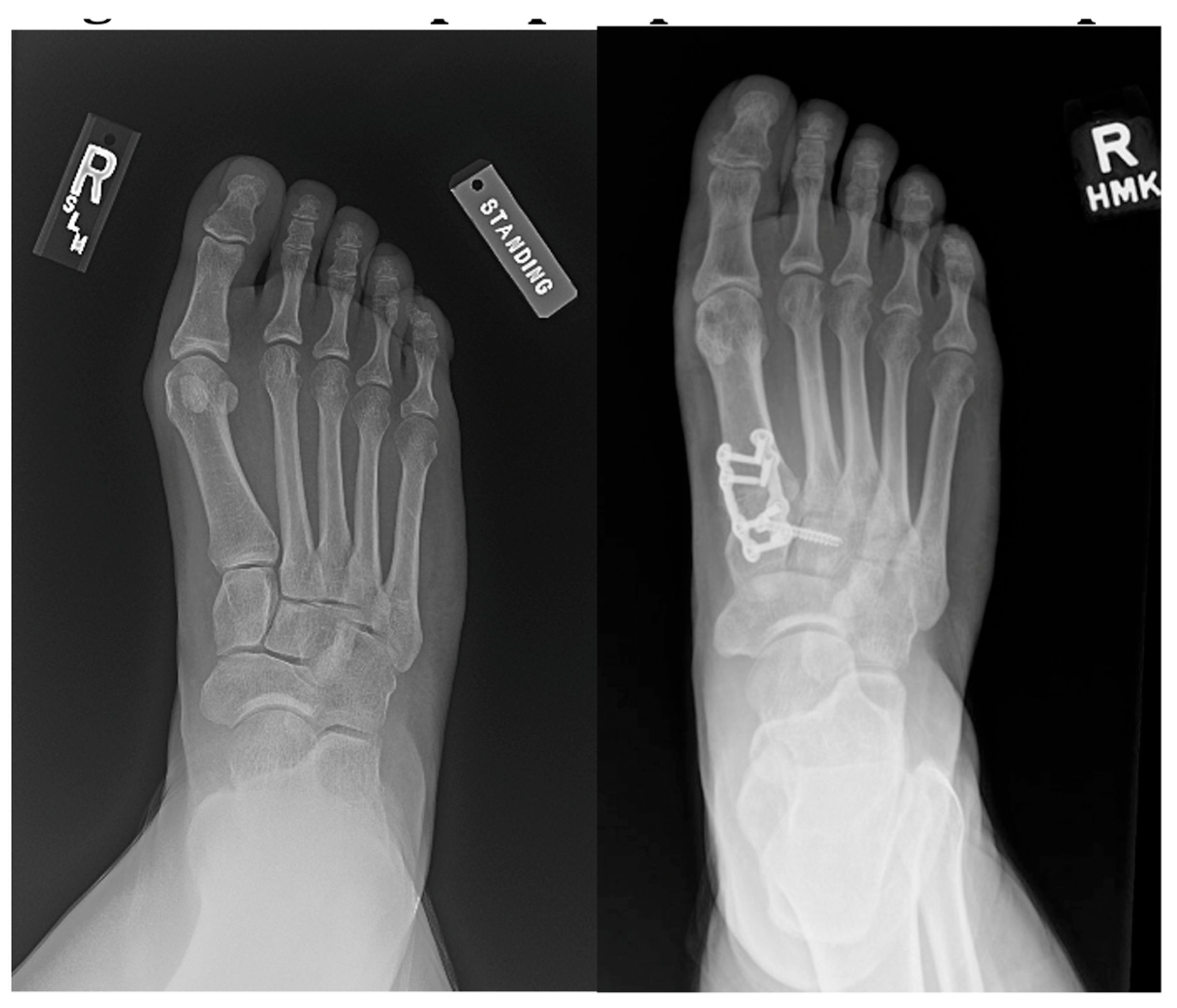
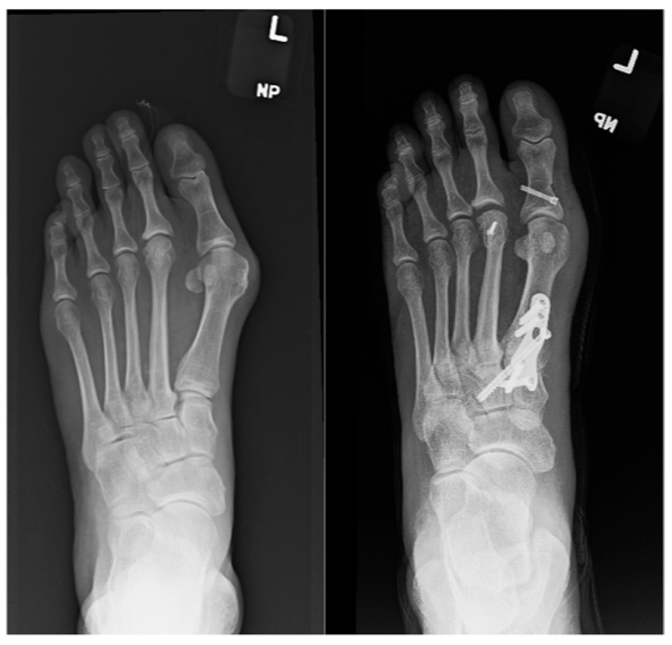
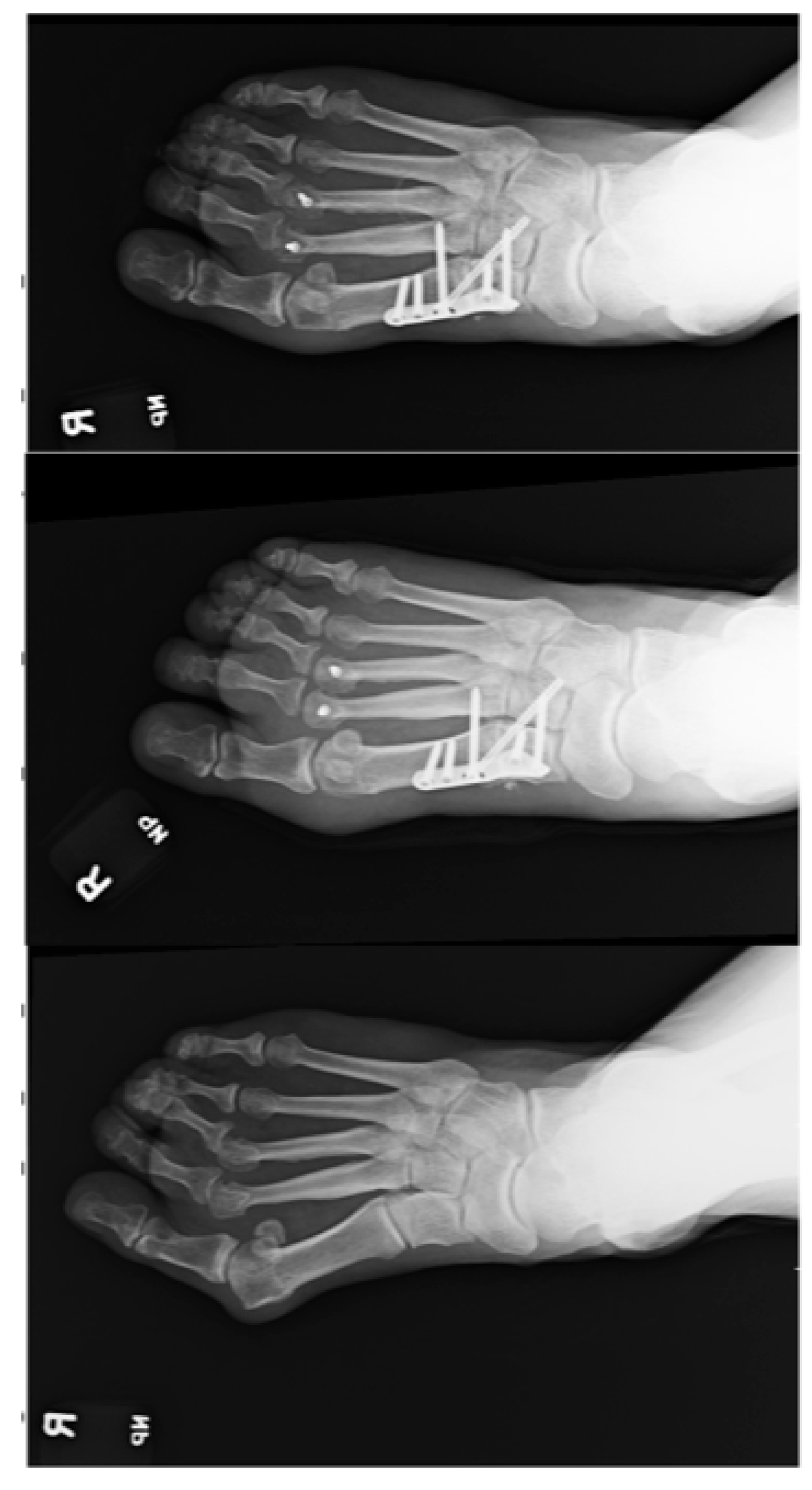
This study protocol obtained approval from the institutional review board and the local ethics committee at Trinity Health Livonia (Livonia, MI, USA) for medical record review of eligible patients. We conducted a retrospective enrollment of 26 patients (32 feet) who had painful moderate-to-severe HAV deformity. These patients underwent first TMT fusion using one of the following constructs within the past 3 years: transverse screw fixation, metatarsal cuneiform screw fixation, and combined transverse and metatarsal cuneiform screw fixation. Surgical intervention was indicated based on a combination of clinical assessment and radiologic examination finding. Specifically, an IMA of 11° to 14° was considered moderate, and an IMA exceeding 14° was categorized as severe HAV deformity. Deformity recurrence was defined as a significant increase in IMA (>5°) compared with the immediate postoperative measurement during final follow-up. The inclusion criterion was moderate-to-severe HAV deformity without arthritic changes to the first metatarsophalangeal joint, and the exclusion criteria consisted of previous surgery on the affected foot or the presence of any other foot or ankle disorders that could impact the patient’s outcome. It is imperative to emphasize that clearly defined inclusion and exclusion criteria were established to minimize bias and enhance the external validity of the study. Patients were categorized into three groups based on the chosen procedure: group 1, transverse screw fixation; group 2, metatarsal cuneiform screw fixation; and group 3, combined transverse and metatarsal cuneiform screw fixation. Of the 32 feet (26 patients) meeting the inclusion criteria, 11 were assigned to group 1, 8 to group 2, and 13 to group 3. Examples of each fixation type are shown in
Figure 1,
Figure 2 and
Figure 3. All of the surgeries were performed by two experienced surgeons at the Trinity Health Livonia facility. The selection of the procedure and fixation method were at the surgeon’s discretion based on the previous clinical assessment and intraoperative findings. Radiographs were taken to evaluate adequate fusion of the TMT joint and to measure the IMA preoperatively and immediately, 3 months, 6 months, and 1 year postoperatively. The range of follow-up in all of the groups was 6 months to 1 year, with mean follow-up of 6 months. The presence of postoperative complications was carefully assessed at each of these time intervals as well. We used the American Orthopaedic Foot and Ankle Society (AOFAS) scale to assess pain relief, function improvement, and patient satisfaction. The total score ranges from 0 to 100, with higher scores indicating improved foot and ankle function and reduced pain. This scoring system plays a crucial role in evaluating the effectiveness of treatments and surgeries, including procedures such as the Lapidus procedure, using different structural fixation techniques. The AOFAS scale scores were evaluated through postoperative surveys conducted at various intervals: 3 months, 6 months, and 1 year postoperatively. These surveys incorporated inquiries about pain relief, functional improvement, and overall satisfaction specific to the fusion structure used in the Lapidus procedure. Patient satisfaction with the Lapidus procedure was consistently high across all three fusion structures.
2.2. Surgical Technique
The preferred approach involved a medial longitudinal incision extending from the first cuneiform to the midshaft of the metatarsal. The first TMT joint was opened and prepared, then fixation was performed using either the Lapiplasty dorsal and medial plates with interfragmentary screw technique or the Paragon Lapidus dorsomedial plate and interfragmentary screw technique. A lateral release was performed involving the lateral joint capsule, metatarsal sesamoid ligaments, and adductor hallucis tendon. The subsequent steps were uniform across the three groups. The IMA was reduced to the appropriate angle and temporarily secured with Kirschner wires. Subsequent fluoroscopy confirmation was followed by the insertion of an interfragmentary lag screw to compress the former first TMT joint. Surgeon preference determined the use of a Paragon dorsomedial plate or Lapiplasty medial and dorsal plates to enhance stability in the first TMT joint. In all three groups, the first TMT joint underwent routine preparation for fusion, with the distinction being in the additional fixation: transverse screw, metatarsal cuneiform screw, or combined transverse and metatarsal cuneiform screw fixation. The procedure concluded with a medial capsulorrhaphy of the first metatarsophalangeal joint. In some cases, an Akin or Weil osteotomy was added if deemed necessary by the surgeon. Wound closure was performed in a layered fashion.
2.3. Statistical Analysis
The statistical analysis, using a one-way analysis of variance (ANOVA), aimed to discern variations in IMA reduction among the three distinct fixation technique groups at different time points (preoperatively and immediately, 3 months, 6 months, and 1 year postoperatively). Descriptive statistics illustrated mean ± SD IMAs for each group and time point. The global ANOVA test yielded a significant result, indicating statistically significant differences in IMA reduction among the groups. Post hoc comparisons with Bonferroni correction highlighted specific group differences, providing nuanced insights.
3. Results
This study found that first TMT fusion with the addition of transverse fixation using all three constructs was effective in maintaining correction of the HAV and restoring stability to the first TMT joint. Radiographic assessments demonstrated successful fusion, and patients reported improvements in pain and function as well as overall satisfaction with the procedure. Complication rates were within an acceptable range.
Table 1 provides a comprehensive overview of the descriptive statistics for the IMA, elucidating the outcomes across the three distinct fixation technique groups at various critical time points.
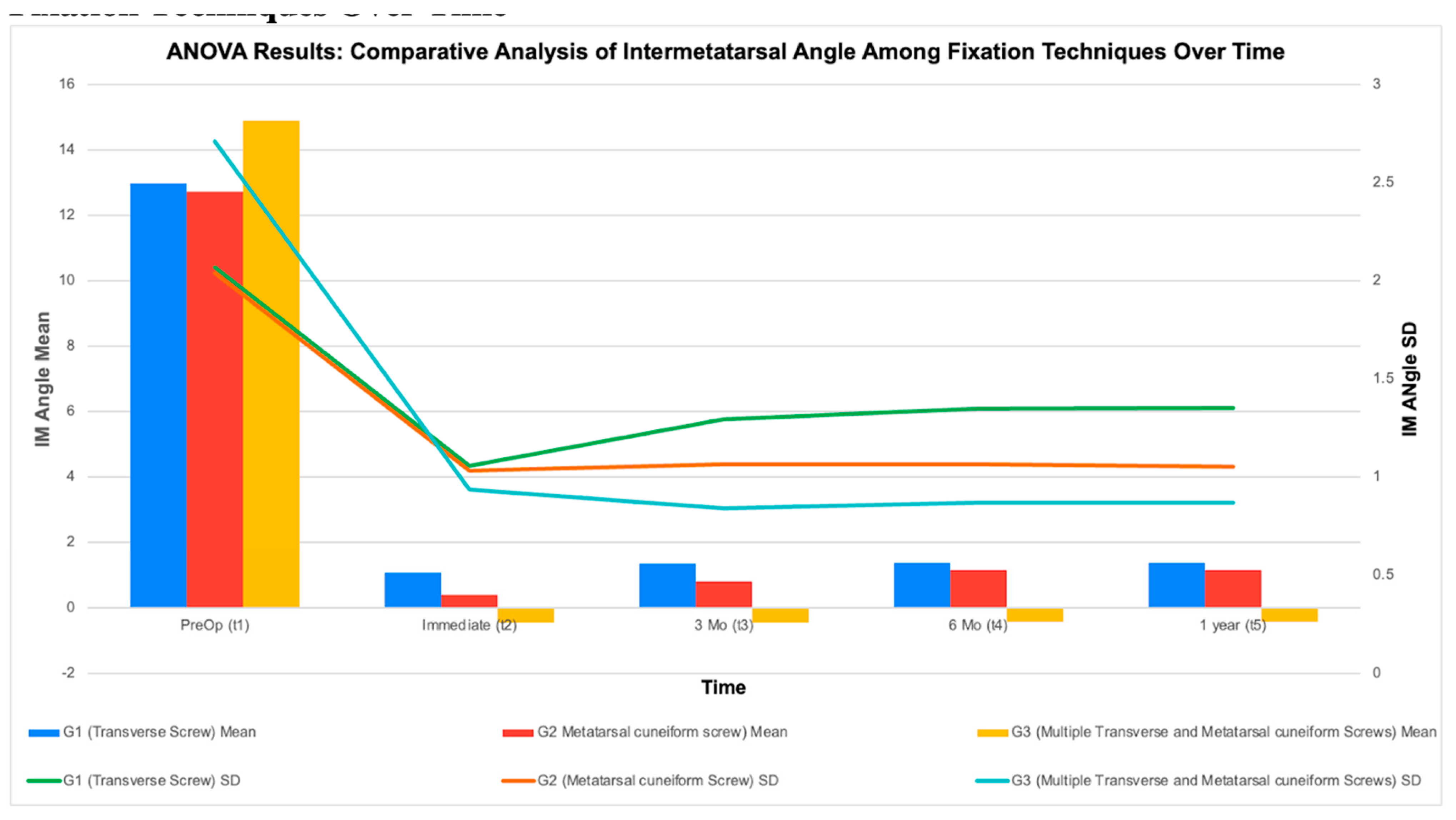
There was no significant difference in the preoperative IMA among the three groups (p > 0.99). The preoperative measurements (t1) established the baseline IMAs before surgical intervention, revealing mean values of 12.983 for group 1, 12.733 for group 2, and 14.91 for group 3. Immediate postoperative assessments (t2) indicated an initial statistically significant decrease in mean IMA, reflecting the immediate impact of the surgical procedures. Group 3 consistently demonstrated lower mean IMAs across all time points, suggesting a sustained and more effective reduction in IMAs compared with the other groups. On reaching the 3-month postoperative period (t3), IMAs continued to trend lower relative to preoperative values, with notable variations among the fixation techniques. The 6-month postoperative measurements (t4) reaffirmed the maintenance of lower IMAs compared with baseline, showcasing persistent distinctions among the fixation techniques. Importantly, at the crucial 1-year postoperative measurement (t5), group 3 stood out by maintaining a statistically significantly lower IMA 1 year postoperatively compared with groups 1 and 2. This sustained favorable outcome underscores the enduring effectiveness of the combined transverse and metatarsal cuneiform screw technique in group 3. These descriptive statistics serve as a crucial foundation for understanding the central tendencies and variations in IMAs over time. They not only lay the groundwork for subsequent statistical analyses but also provide valuable insights into the comparative effectiveness of the different fixation techniques, emphasizing the sustained positive outcomes associated with group 3 in the extended postoperative period.
Figure 4 demonstrates the ANOVA results, which robustly supported significant IMA reduction among the fixation technique groups, with group 3 demonstrating notably favorable results. Post hoc analyses clarified specific group differences, emphasizing the advantages of combined transverse and metatarsal cuneiform screw fixation in achieving and sustaining a more stable IMA correction after the Lapidus procedure. These findings contribute valuable insights into the comparative effectiveness of fixation techniques for hallux valgus deformity correction.
A secondary outcome of the study was to examine all of the complications during the follow-up time points for each patient. We found that there was a total of three complications among the 26 patients, with all three being in group 1. The complications consisted of wound healing difficulties, including a small abscess along the proximal incision at the 3-month follow-up, an area of necrosis along the incision line measuring 2 × 0.4 cm at the 3-month follow-up, and a small dehiscence with ulceration to incision at the 6-month follow-up. All of the complications were treated conservatively with local wound care and went on to heal uneventfully by the final follow-up. Nonunion was defined as lack of bony consolidation on radiographs 6 months postoperatively. There were no occurrences of delayed union or nonunion in any of the groups.
Another secondary outcome was to evaluate patient satisfaction using the AOFAS scale. As shown in
Table 2, AOFAS scale scores were calculated for three specific categories: overall satisfaction, pain relief, and function improvement. These mean scores provide a quantitative measure of patient-reported outcomes at various intervals postoperatively. The mean scores for overall satisfaction postoperatively were 95.33 for transverse screw fixation, 96.63 for metatarsal cuneiform screw fixation, and 98.58 for combined transverse and metatarsal cuneiform screw fixation. Group 3 (combined transverse and metatarsal cuneiform screw fixation) exhibited slightly superior satisfaction rates, sustained pain relief, and functional improvement compared with the other two methods, although not statistically significant. These findings imply that the choice of fusion structure may influence not only clinical outcomes but also patient-reported satisfaction and overall quality of life after the procedure.
4. Discussion
Recent literature suggests that the addition of a transverse screw to the traditional Lapidus procedure will allow for more stable fixation and improved maintenance of correction. To our knowledge, there is no available literature comparing different construct fixation techniques that incorporate the transverse screw to analyze which technique allows for the most stable construct with less recurrence of deformity.
One study by Feilmeier et al. [
5] compared the strength and stability of different transverse screw orientations in cadavers. They found that the first to second cuneiform screw did not provide any additional stability, the first to second metatarsal screw consistently reduced first-ray instability, and the first metatarsal to middle cuneiform screw provided intermediate results [
5]. The present study supports these results by showing that the transverse screw-only group (group 1) tended to have the least effective maintenance of correction compared with the other two groups, and maintenance of correction was significantly improved with combined transverse and first metatarsal to intermediate cuneiform fixation.
Another cadaveric study by Galli et al. [
6] measured sagittal plane mobility after traditional Lapidus fixation versus traditional fixation with the addition of a screw from the first metatarsal to the intermediate cuneiform. They found that the mean ± SD sagittal plane motion preoperatively was 7.45 ± 1.82 mm, which decreased to 4.41 ± 1.51 mm with traditional fixation and to 3.12 ± 1.06 mm with the addition of a first metatarsal to intermediate cuneiform screw. This improvement in stability was statistically significant with the additional screw. The increased stability with the additional screw is likely a large factor contributing to the favorable results of maintenance of IMA with additional transverse and metatarsal cuneiform screws in group 3.
Scheele et al. [
1] compared maintenance of IMA correction postoperatively with a traditional Lapidus fixation versus addition of a first to second metatarsal base screw. Although they found significantly larger improvement in the IMA immediately postoperatively, this difference was gone by the second follow-up, and they found no significant differences in maintenance of correction and recurrence rates between the two groups long term. Several other studies suggest that the addition of transverse or first metatarsal to cuneiform screws provides a significant decrease in the recurrence rate [
2,
7,
8]. This inconsistency in results suggests that certain constructs may provide improved results compared with others. The addition of a solely first metatarsal to second metatarsal base screw may not be the most stable construct long term.
Post hoc analyses of this study uncovered significant differences among group 1 (transverse screw), group 2 (metatarsal cuneiform screw), and group 3 (transverse and metatarsal cuneiform screws). Notably, group 3 exhibited a stronger initial reduction in the IMA, which became evident by the 3-month mark, maintaining significant distinctions at the final follow-up. Radiologic assessments indicated that group 3 maintained stable IMA values and exhibited a significantly lower increase in IMA over time compared with the other two groups (p < 0.05). These findings underscore the potential advantages of combined transverse and metatarsal cuneiform screw fixation in achieving and sustaining a more stable IMA correction after the Lapidus procedure. Although all of the groups with additional fixation to account for medial midfoot hypermobility had satisfactory results, we found that the most stable construct over time was with the addition of both a transverse screw and a metatarsal cuneiform screw. In all three groups, there were minimal complications, no incidences of nonunion, and excellent postoperative AOFAS scale scores.
Limitations of this study include a small sample size of 26 patients and 32 feet, its retrospective nature, and follow-up limited to 1 year postoperatively. Further research is indicated with longer follow-up and a larger sample size to determine long-term maintenance of correction with differing fixation constructs.
Author Contributions
Conceptualization, V.R.A. and R.A.; methodology, R.A., K.L., H.N. and K.C.; software, K.L., H.N. and K.C.; validation, K.L., H.N. and K.C.; formal analysis, K.L., H.N. and K.C.; investigation, K.L., H.N. and K.C.; resources, K.L., H.N. and K.C.; data curation, K.L., H.N. and K.C.; writing—original draft preparation, K.L., H.N. and K.C.; writing—review and editing, V.R.A.; visualization, K.L., H.N. and K.C.; supervision, R.A.; project administration, R.A. and V.R.A. All authors have read and agreed to the published version of the manuscript.
Funding
This research received no external funding.
Institutional Review Board Statement
The study was conducted in accordance with the Declaration of Helsinki and approved by the Institutional Review Board of Trinity Health Livonia Hospital, IRB #1. Protocol code E-23-1070 and date of approval 5/18/2023.
Informed Consent Statement
Patient consent was waived due to retrospective nature of study and the study posing no more than minimal risk to the participants as it only involves reviewing existing data. No new data is being collected from subjects.
Data Availability Statement
The original contributions presented in this study are included in the article. Further inquiries can be directed to the corresponding author(s).
Conflicts of Interest
The authors declare no conflicts of interest.
References
- Scheele, C.B.; Kinast, C.; Lenze, F.; Wimmer, J.; Beischl, S.; Harrasser, N. Radiographic evaluation of first tarsometatarsal joint arthrodesis for hallux valgus deformity: Does the fusion of the first to the second metatarsal base reduce the radiological recurrence rate? Foot Ankle Spec. 2024, 17, 382. [Google Scholar] [CrossRef]
- Bednarz, P.A.; Manoli, A. Modified Lapidus procedure for the treatment of hypermobile hallux valgus. Foot Ankle Int. 2000, 21, 816. [Google Scholar] [CrossRef]
- DeVries, J.G.; Granata, J.D.; Hyer, C.F. Fixation of first tarsometatarsal arthrodesis: A retrospective comparative cohort of two techniques. Foot Ankle Int. 2011, 32, 158. [Google Scholar] [CrossRef]
- Reed, L.; Luque-Sanchez, K.S.; Awad, S.K.; Mihas, A.K.; Young, S.M.; Patch, D.A.; Johnson, M.D. Intermetatarsal screw fixation reduced intermetatarsal angle following modified Lapidus procedures. Foot Ankle Orthop. 2022, 7, 2473011421S00901. [Google Scholar] [CrossRef]
- Feilmeier, M.; Dayton, P.; Kauwe, M.; Cifaldi, A.; Roberts, B.; Johnk, H.; Reimer, R. Comparison of transverse and coronal plane stability at the first tarsal-metatarsal joint with multiple screw orientations. Foot Ankle Spec. 2017, 10, 104. [Google Scholar] [CrossRef] [PubMed]
- Galli, M.M.; McAlister, J.E.; Berlet, G.C.; Hyer, C.F. Enhanced Lapidus arthrodesis: Crossed screw technique with middle cuneiform fixation further reduces sagittal mobility. J. Foot Ankle Surg. 2015, 54, 437. [Google Scholar] [CrossRef] [PubMed]
- Symeonidis, P.D.; Anderson, J.G. Original and modified Lapidus procedures: Proposals for a new terminology. J. Bone Jt. Surg. Am. 2021, 103, e15. [Google Scholar] [CrossRef] [PubMed]
- Fleming, J.J.; Kwaadu, K.Y.; Brinkley, J.C.; Ozuzu, Y. Intraoperative evaluation of medial intercuneiform instability after Lapidus arthrodesis: Intercuneiform hook test. J. Foot Ankle Surg. 2015, 54, 464. [Google Scholar] [CrossRef] [PubMed]
| Disclaimer/Publisher’s Note: The statements, opinions and data contained in all publications are solely those of the individual author(s) and contributor(s) and not of MDPI and/or the editor(s). MDPI and/or the editor(s) disclaim responsibility for any injury to people or property resulting from any ideas, methods, instructions or products referred to in the content. |
© 2026 by the authors. Published by MDPI on behalf of the American Podiatric Medical Association (APMA). Licensee MDPI, Basel, Switzerland. This article is an open access article distributed under the terms and conditions of the Creative Commons Attribution (CC BY) license.