Article

The Immediate Effect of KINESIO TAPE® on Static Plantar Foot Pressure and Force in Young Females with Flexible Flatfoot: A Pilot Study

1
Department of Biomechanics, Faculty of Physical Therapy, Modern University of Technology and Information, Cairo 117112, Egypt
2
Department of Biomechanics, Faculty of Physical Therapy, Cairo University, Giza 12612, Egypt
3
Department of Physical Therapy and Health Rehabilitation, Jouf University, Sakaka, AlJouf Region 72388, Saudi Arabia
4
Physical Therapy Department, University of St. Augustine for Health Sciences, San Marcos, CA 92069, USA
5
Department of Developmental Disorders in Pediatrics and Its Surgery, Faculty of Physical Therapy, Cairo University, 7 Ahmed Elziat Street, Ben Elsarayat, El Dokki, Giza 12612, Egypt
*
Author to whom correspondence should be addressed.
J. Am. Podiatr. Med. Assoc. 2026, 116(2), 14; https://doi.org/10.3390/japma116020014
Submission received: 9 May 2025 / Revised: 15 July 2025 / Accepted: 1 August 2025 / Published: 31 March 2026

Abstract

Background: Flatfoot is a condition brought on by trauma, persistent foot stress, obesity, and poor biomechanics. These factors result in the development of a flat foot, collapse of the foot arch, and malfunction of the posterior tibial tendon. This study aimed to assess the immediate effects of Kinesio Tape on static plantar foot pressure and force in young females with flexible flatfoot. Methods: A pilot study (pre-experimental study design) with a convenience sample of 20 female subjects from a university with flexible flatfoot (age = 20.1 ± 1.3 years, weight = 91.8 ± 14.4 kg, height = 162.2 ± 6.3 cm, BMI = 34.9 ± 5, foot posture index (FPI) = 8.8 ± 2.1) was selected. The TekScan MatScan® system was used to measure the static plantar forces and pressures, foot contact area, and the mediolateral displacement of COF over time while standing (Boston, MA, USA) before and immediately after the application of Kinesio Tape (KT). Results: While there were no statistically significant changes in the foot peak or total pressure, paired-sample t-tests showed a statistically significant reduction in foot contact area (p < 0.05) and a statistically significant increase in midfoot maximum force (p < 0.05) following the application of KT. Furthermore, after applying KT, there was a statistically significant decrease in the mediolateral COF velocity, indicating greater lateral displacement of COF (p < 0.05). Conclusions: The results of this study concluded that Kinesio Tape was a useful intervention method for immediately redistributing pressure and forces in young females with flexible flat feet.

1. Introduction

Kinesio Tape® (KT) is a therapeutic tape used to treat numerous musculoskeletal clinical disorders in various contexts, including physical therapy, sports medicine, and athletic training [1]. KT is made of 100% elastic cotton with a heat-activated adhesive on the underside [2]. This tape may be stretched longitudinally to 30–40% of its resting length and measures about the thickness of the epidermis, mimicking the characteristics of human skin [3]. Kinesio Tape® has been claimed to improve function and/or reduce pain instead of providing structural support by facilitating skeletal muscle action through tactile and proprioceptive input [4,5,6]. The posterior tibialis is the muscle most capable of supinating the foot and preventing the incidence of flatfoot [7].
Clinical evaluation of foot posture is routine, and findings of excessive pronation or supination may affect judgements about therapies like orthotics or taping. Some research has revealed a connection between specific lower limb injury risks and foot position [3]. A pronated foot position was linked to a higher risk of developing lower limb overuse injuries, such as medial tibial stress syndrome, Achilles tendinopathy, and patellofemoral pain syndrome, according to a comprehensive study by Neal et al. [8].
The prevalence of flexible flatfoot was found to be higher among female university students than their male counterparts in several previous studies [9,10]. The fact that women typically have smaller bones and less muscular mass may be the cause of this higher incidence in females, given that both elements are beneficial in the preservation of the foot’s arches [11]. This is the reason our study concentrated on female subjects instead of males.
Taping techniques may help correct static foot posture in people with excessive foot pronation due to their suggested effects in the correction of foot position [12,13]. For example, low-dye (LD) taping was shown to relate to changes in the midfoot and forefoot’s peak plantar pressure, resulting in less foot pronation. However, the degree of change seen with LD taping was quite slight, and more investigation is required to elucidate its therapeutic implications [13]. On the other hand, an existing study on the capability of KT to alter foot forces and pressure in static situations, however, suggests that it is limited despite its widespread use. The majority of earlier studies demonstrated that KT had negative effects on flatfoot correction, which may be attributed to a number of constraints with regard to the use of KT and the measurement of force and pressure variables [3,14]. For example, taping just one foot rather than both could compromise measurement accuracy since the taped foot may target the faceplate [3]. In order to increase measurement accuracy, this problem was taken into account in the current investigation by taping both feet at the same time. A static foot posture is riskier than dynamic foot posture because it puts more strain on the foot’s structure [15]. Static posture inhibits blood flow and causes muscle damage [15]. In addition, the nerves, ligaments, blood vessels, and tendons can all be impacted by a static posture. A static posture is one of the potential ergonomic risk factors for work-related musculoskeletal disorders (WMSDs) [15]. For this reason, the current study examined static foot position instead than dynamic foot position.
In 2014, Luque-Suarez [16] reported that there was no difference between people who did and did not have their feet taped with KT when using the foot posture index (FPI) [17,18] to describe the level of foot pronation, but recommended that more research be done on people who experience pain and excessive pronation in order to better understand this problem. By choosing subjects with significant foot pronation, the current study may close this gap and shed light on the mechanical impact of KT on pressure and force distribution in subjects with flexible flat feet.
Chang et al. [19] investigated KT and the low-dye taping method with white fabric athletic tape using a platform plantar pressure apparatus [20,21]. When they used cloth tape, they discovered an increase in peak pressure under the fifth metatarsal and a decrease in peak pressure under the toes. No statistically significant difference was noticed when they applied KT, but the study collected data during level walking rather than in a static situation. The current study tried to focus on static foot position and the effect of KT on plantar foot pressure and force distribution. As far as we are aware, there has not been any research published yet that examines how KT affects pronated foot pressure and forces during static situations. Most of the previous studies were conducted during dynamic situations [22,23]. The goal of this study was to ascertain if KT has an effect on static plantar foot pressure and force in young females with flexible flatfoot.

2. Materials and Methods

2.1. Study Design

A pre-experimental research design (pre-test–post-test intervention study) was used to detect the differences in foot plantar pressures and forces, foot contact area, and mediolateral center of force (COF) velocity in one group of adult female subjects with flexible flatfoot. Only one group was used in the current study to avoid any foot variability due to weight bearing that may affect the pre-intervention variables, especially after using KT.

2.2. Participants

A convenience sample of 25 female subjects (age 20.1 ± 1.3 years, weight 91.8 ± 14.4 kg, height 162.2 ± 6.3 cm, BMI = 34.9 ± 5, FPI = 8.8 ± 2.1) with foot pronation (according to the FPI6) was drawn from the general Jouf University population. Five subjects dropped out: 3 subjects did not meet the inclusion criteria and 2 subjects declined to participate from the beginning, as shown in the flow chart (Figure 1). Before starting the study, each subject underwent a physical examination to ensure they met certain inclusion criteria: (1) they should be female subjects, as the prevalence of flatfoot is more common in females than in males [24,25]; (2) they should be aged from 18 to 25 years; (3) their body weight should range between 70 and 130 kg; (4) they should have at least a +6 score in the FPI in a weight-bearing position. Subjects were excluded if: (1) individuals were unable to stand or walk normally due to a history of a musculoskeletal or neuro-musculoskeletal problems; (2) they had a history of foot or lower-extremity injuries or surgeries within the previous six months; (3) they had any balance problems (4) they were pregnant; (5) they practiced specific sports or activities rather than ADL. Before taking part in the study, each participant read and signed a written informed consent form. The study was carried out in line with the Declaration of Helsinki for the safety of human contributors. In addition, this study received ethical approval from the Ethics Committee of Jouf University (approval number: 6-05-45) and clinical trial registration No. PACTR202407474074446. All participants had the right to withdraw from participation at any time and without any restriction.

2.3. Blinding

Blinding took place throughout the data collection and analysis stage for both the therapist and participants by providing each participant with a code during the data entry and analysis.

2.4. Instrumentation

Each subject was checked to see if they met the required FPI6 score. The FPI6 involves rating six distinct foot characteristics that are connected to either a supinated or pronated foot posture when standing. Pronation is shown by positive values, while supination is indicated by negative numbers. The six feature scores are then added together. The FPI6 has been demonstrated to have reasonable validity, moderate inter-rater reliability, and good intra-rater reliability [23,24].
The static plantar forces and pressures, as well as the mediolateral displacement of COF over time, were measured using the TekScan MatScan® system (Boston, MA, USA). The system is made up of a 5 mm thick floor mat (432 368 mm), 2288 resistive sensors (1.4 sensors/cm2), a 40 Hz data sample rate, and foot pressure software on a laptop for data acquisition. The TekScan MatScan® system demonstrates generally moderate to good reliability for static and dynamic plantar pressure forces and pressure measurement [26,27]. Pressure mats might be a good choice for spotting significant changes in mediolateral COF displacement [28].

2.5. Procedure

The participants’ gender, height, and weight were recorded after they had signed the informed consent form. BMI was calculated for each participant. One of the examiners evaluated and noted the participants’ FPI6 score for their dominant foot using the procedures outlined by Redmond et al. [29]. The toe rising “Jack” test was used to detect if the medial longitudinal arch of the foot was flexible or not: The subjects stood in a neutral, relaxed position. Next, the examiner passively flexed the big toe’s first metatarsal joint.
The TekScan MatScan® system was located in front of each participant. All participants were barefoot. First, a new file was opened for each participant after the software had received the subject’s information. The participant’s weight was entered into the TekScan system software for calibration purposes, and then the patient was asked to stand on one leg on the platform until the calibration was completed. Each participant was asked to stand straight (with equal weight distribution on both feet) and barefoot on the pressure platform before the application of KT. The main variables were therefore measured during static standing. Using the TekScan analysis software, the plantar pressure, midfoot forces, foot contact area, and mediolateral COF displacement over time (velocity) were detected and reported for the dominant foot. These factors were chosen to detect the effect of KT on flexible flatfoot alignment after its application.
After the standing trials, first, both the participants’ feet and legs were cleaned with alcohol wipes, and any excessive hair was shaved. Both the participants’ feet and legs were taped using a method described by Kase. [30]. A Certified Kinesio Tape Practitioner (T.J.) applied Kinesio Tape®-Classic (Kinesio USA Corporation, Albuquerque, NM, USA) to each participant to assure consistency.
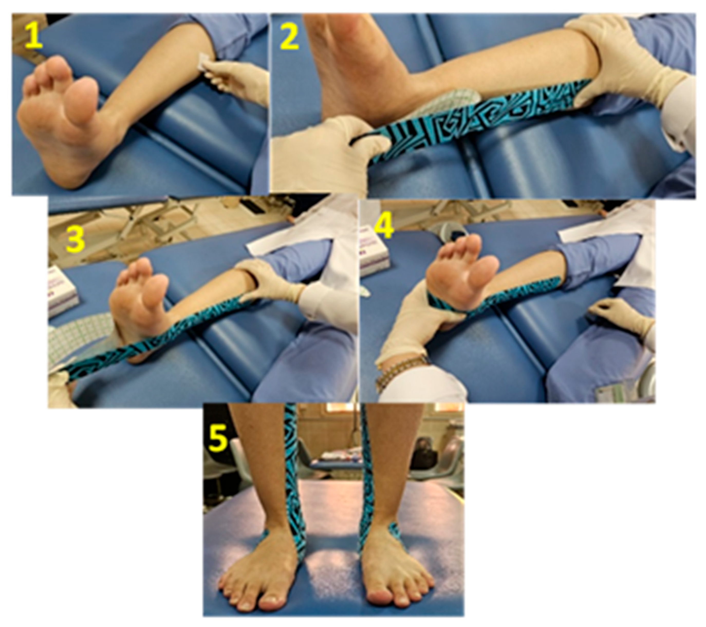
The tibialis posterior muscle was facilitated by the tape. Each participant sat long-seated on a taping table with their ankle in a neutral position, or with zero degrees of dorsiflexion, for the taping operation. The taping was applied to both feet and legs to avoid the subject targeting a certain foot, but the required data was collected from the dominant foot only. The first strip of tape was applied with the base on the medial proximal third of the tibia and the midsection of the tape passed under the medial longitudinal arch, while the tail ended on the lateral side of the foot. The midsection of the tape was subjected to moderate tension (50% of the possible length), whereas the base and tail of the tape received no tension, as shown in Figure 2.
The participant was then asked to stand on the pressure platform immediately after the application of KT to measure the main variables of interest from the dominant foot. The tape was taken off after the second set of measurements, and any prior markings were cleaned with alcohol. Three trials were captured before and after the application of KT so that one could be selected to be analyzed by the software.

2.6. Statistical Analysis

Data exploration was conducted to detect any outliers. The Shapiro–Wilk test, histograms, and Q-Q plots confirmed the homogeneity of the data. Descriptive statistics (mean and standard deviation [SD]) were calculated for demographic data (weight, height, age, BMI) and the variables of interest (foot peak pressure, foot total pressure, midfoot force, contact area of foot, mediolateral COF displacement over time). Paired-sample t-tests were used to compare the taped and untaped conditions for the same group. An alpha level of 0.05 was set. All statistical tests were performed using the IBM SPSS® software package, version 20 (IBM, Armonk, NY, USA).

3. Results

3.1. Descriptive Statistics

The mean and standard deviation of the following variables—age, height, weight, body mass index (BMI), and foot pressure index (FPI)—were ascertained using descriptive statistics, as shown in Table 1.
Paired-Sample t-Test Pre- and Post-KT Intervention:
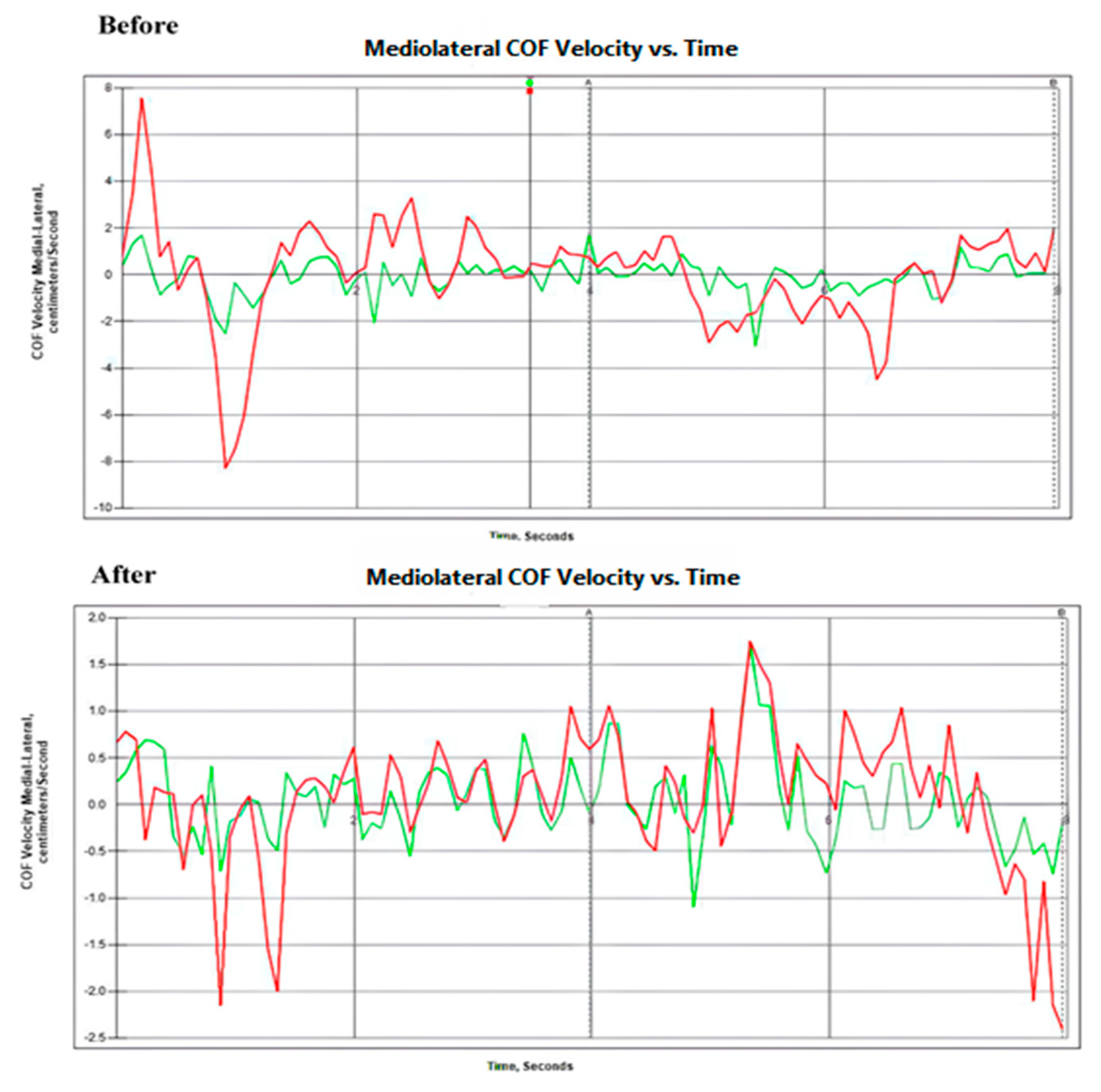
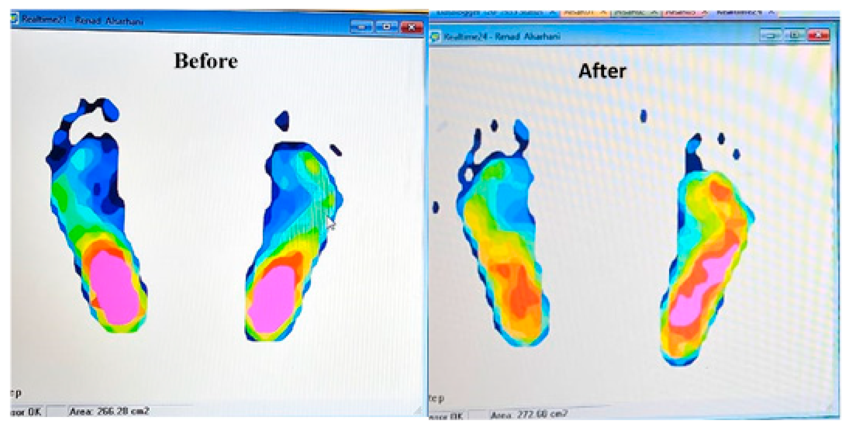
The paired-sample t-tests revealed no significant changes in foot peak pressure and total foot pressure pre-and post-intervention with KT (p = 0.755, p = 0.649), with no and weak effect sizes, respectively. On the other hand, there was a significant reduction in the foot contact area and mediolateral force velocity between pre- and post-intervention measurements (p ˂ 0.05). Alternatively, there was a statistically significant increase in the maximum midfoot force. The foot contact area and midfoot maximum force showed a medium effect size, while the mediolateral COF velocity showed a high effect size (Table 2). Figure 3 indicates the mediolateral COF velocity before and after KT application, while Figure 4 shows the plantar foot pressure distribution before and after KT application.

3.2. Discussion

The main purpose of the current study is to investigate the immediate effect of KT on foot plantar pressure and force in young females with flexible flatfoot. The current study’s findings showed that there were no statistically significant changes in foot peak pressure and total foot pressure immediately following the KT intervention, with no and weak effect sizes, respectively. However, immediately following KT application, there was a statistically significant decrease in the foot contact area and mediolateral COF velocity and a statistically significant increase in the maximum midfoot force. The mediolateral COF velocity displayed a high effect size, but the foot contact area and midfoot maximal force displayed a medium effect size. This indicates the immediate effect of KT application in decreasing the contact area of the foot, reflected in the increased maximum midfoot force without significant changes in the magnitude of foot pressure. Moreover, the COF velocity showed a statistically significant shift from the medial to the lateral side with a high effect size, which suggests the role of KT in reducing the load on the medial longitudinal arch of the foot and decreasing foot pronation. This intervention can also be done when there is little subtalar mobility to elevate the arch and lessens the strain on the plantar aponeurosis. Likewise, as the tape pulls the lateral aspect of the foot medially, the height of the medial arch rises.
The results of the study conducted by Karthikeyan et al. [31] support the results of the current study, showing that following the application of Kinesio Tape, foot pressure, pain, foot contact area, and foot pronation on the bilateral leg considerably decreased, and range of motion improved. However, this study differed from the current study in that our KT intervention resulted in foot pressure redistribution instead of significantly lowering foot pressure.
On the other hand, the results of the study performed by Cornwall et al. [32] showed no statistically significant changes in foot plantar pressure during walking, with a low effect size, which agrees with the results of the current study, which showed no statistically significant change in foot plantar pressure (total and peak pressures), with low effect sizes. At the same time, the previous study did not show any effect of KT on foot posture or frontal plane rearfoot motion. This may be because the study performed its measurements during dynamic situations and was not concentrated on static foot posture. In addition, the authors attributed the inability to detect changes between the pre- and post-KT situations to the low power in their study. Moreover, the results of a study performed by Ilie et al. [33] implied that taping application may be beneficial for normalizing plantar pressures in children with flat feet. However, these effects are limited to acute use of the tape and correspond to immediate needs. This conclusion is consistent with the conclusion of our results, although the previous study was performed on a child population.
The immediate improvement in flexible flatfoot after the application of the KT is supported by a study conducted by Wang et al. [34], who reported that using Kinesio Tape to support the transverse arch for static stability facilitates the force exerted by the tibialis posterior for dynamic stability and decreases foot pronation immediately after application in individuals with functional flatfoot. In addition, another study conducted by Siu et al. [35] confirmed the role of KT in facilitating the tibialis posterior and reinforcing the transverse arch in individuals with flexible flatfoot immediately after application.
Cornwall et al. [32] confirmed that a lateral shift in the foot plantar contact area or force would have been expected if the KT had been able to promote activity in the posterior tibialis muscle, hence reducing excess foot pronation. This happened in our study, as shown through the decrease in contact area and the lateral shifting of the COF. KT application tends to redistribute the foot pressure rather than changing it by adjusting the foot posture and changing the foot contact area and forces.
Another study, conducted by Tahmasbi et al. [36], confirmed the role of KT in immediately stimulating the tibialis posterior muscle (an essential dynamic stabilizer of the medial longitudinal arch), which tends to improve foot posture and at the same time increase the maximum total force of the stance phase. This result is in agreement with the results of our study, which showed an increase in midfoot force without a change in plantar foot pressure due to decreasing the foot contact area and reducing foot pronation; however, this previous study was done on a dynamic rather than a static situation.
This current study has some limitations that need to be considered. The study population included only female subjects without consideration of gender differences, so the result of this study can only be generalized to a similar population. In addition, it would be advantageous to conduct the current study again with a larger sample size to verify whether the impact of KT on plantar pressure and forces has any therapeutic significance. Moreover, it is recommended to conduct a future study to detect the long-term effect of KT.

4. Conclusions

The results of the current study successfully demonstrate that KT statistically alters the foot contact area, midfoot force, and COF displacement over time without changing the total and peak foot pressure in static standing posture, which indicates the important role of KT in the redistribution of plantar foot pressure after correcting the contact area and the force of the foot. In addition, the current study confirms the correction of foot posture by KT by displacing the COF laterally. Because of the moderate and large effect size of the differences between the KT and no-KT conditions, KT is recommended to modify plantar foot pressure and forces in flexible flatfoot subjects.

Author Contributions

Conceptualization, M.A., A.A., G.A., W.A., F.A., and S.A.; methodology, M.A., A.A.A., A.A., G.A., W.A., F.A., and S.A.; software, M.A.; validation, M.A.; formal analysis, M.A., A.A.A., A.A., G.A., W.A., F.A., and S.A.; investigation, M.A., A.A., G.A., W.A., F.A., and S.A.; resources, M.A., A.A., G.A., W.A., F.A., and S.A.; data curation, M.A., A.A.A., A.A., G.A., W.A., F.A., and S.A.; writing—original draft preparation, M.A., A.A.A., A.A., G.A., W.A., F.A., S.A., and M.K.; writing—review and editing, M.A., A.A.A., A.A., G.A., W.A., F.A., S.A., and M.K.; visualization, M.A., A.A.A., A.A., G.A., W.A., F.A., S.A., and M.K.; supervision, M.A., A.A.A., A.A., G.A., W.A., F.A., and S.A.; project administration, M.A., A.A.A., A.A., G.A., W.A., F.A., and S.A. All authors have read and agreed to the published version of the manuscript.

Funding

All authors declare that there is no financial support for this work.

Institutional Review Board Statement

The study was conducted in accordance with the Declaration of Helsinki, and approved by the Institutional Review Board of Jouf University (Approval No. 6-05-45 and 17 March 2024).

Informed Consent Statement

Informed consent was obtained from all subjects involved in the study. Written informed consent has been obtained from the patient(s) to publish this paper.

Data Availability Statement

The data presented in this study are available on request from the corresponding author due to privacy.

Acknowledgments

The authors wish to thank Jouf University for providing us with suitable equipment to conduct this study. Finally, special thanks to all people who participated in this current study.

Conflicts of Interest

All authors disclosed no conflicts of interest that may have influenced either the conduct or the presentation of the research.

References

  1. Cai, C.; Au, I.; An, W.; Cheung, R. Facilitatory and inhibitory effects of Kinesio tape: Fact or fad? J. Sci. Med. Sport 2016, 19, 109–112. [Google Scholar] [CrossRef] [PubMed]
  2. Keenan, K.A.; Akins, J.S.; Varnell, M.; Abt, J.; Lovalekar, M.; Lephart, S.; Sell, T.C. Kinesiology taping does not alter shoulder strength, shoulder proprioception, or scapular kinematics in healthy, physically active subjects and subjects with Subacromial Impingement Syndrome. Phys. Ther. Sport 2017, 24, 60–66. [Google Scholar] [CrossRef] [PubMed]
  3. Cornwall, M.W.; Jain, T.K.; Holmgren, S.; Dorri, A.; Young, C. The effect of KINESIO TAPE® on static foot posture, plantar pressure, and rearfoot motion in individual with pronated feet. Int. J. Sports Phys. Ther. 2019, 14, 368–375. [Google Scholar] [CrossRef]
  4. Aguilar, M.B.; Abián-Vicén, J.; Halstead, J.; Gijon-Nogueron, G. Effectiveness of neuromuscular taping on pronated foot posture and walking plantar pressures in amateur runners. J. Sci. Med. Sport 2016, 19, 348–353. [Google Scholar] [CrossRef] [PubMed]
  5. Griebert, M.C.; Needle, A.R.; McConnell, J.; Kaminski, T.W. Lower-leg Kinesio tape reduces rate of loading in participants with medial tibial stress syndrome. Phys. Ther. Sport 2016, 18, 62–67. [Google Scholar] [CrossRef]
  6. Lim, E.C.W.; Tay, M.G.X. Kinesio taping in musculoskeletal pain and disability that lasts for more than 4 weeks: Is it time to peel off the tape and throw it out with the sweat? A systematic review with meta-analysis focused on pain and also methods of tape application. Br. J. Sports Med. 2015, 49, 1558–1566. [Google Scholar] [CrossRef]
  7. Bolgla, L.A.; Malone, T.R. Plantar fasciitis and the windlass mechanism: A biomechanical link to clinical practice. J. Athl. Train 2004, 39, 77–82. [Google Scholar]
  8. Neal, B.S.; Griffiths, I.B.; Dowling, G.J.; Murley, G.S.; Munteanu, S.E.; Smith, M.M.F.; Collins, N.J.; Barton, C.J. Foot posture as a risk factor for lower limb overuse injury: A systematic review and meta-analysis. J. Foot Ankle Res. 2014, 7, 55. [Google Scholar] [CrossRef]
  9. Eluwa, M.A.; Omini, R.B.; Kpela, T.; Ekanem, T.B.; Akpantah, A.O. The incidence of pes planus amongst Akwa Ibom State students in the University of Calabar. Internet J. Forensic. Sci. 2009, 3, 1–5. [Google Scholar]
  10. Aenumulapalli, A.; Kulkarni, M.M.; Gandotra, A.R. Prevalence of flexible flat foot in adults: A cross-sectional study. J. Clin. Diagn. Res. JCDR1 2017, 11, AC17. [Google Scholar] [CrossRef]
  11. Jh, H. The foot as a support. Acta Anat. 1955, 25, 34–45. [Google Scholar]
  12. Guner, S.; Alsancak, S. Kinesiotaping Techniques to Alter Static Load in Patients With Foot Pronation. J. Chiropr. Med. 2020, 19, 175–180. [Google Scholar] [CrossRef] [PubMed]
  13. O’SUllivan, K.; Kennedy, N.; O’NEill, E.; Ni Mhainin, U. The effect of low-dye taping on rearfoot motion and plantar pressure during the stance phase of gait. BMC Musculoskelet. Disord. 2008, 9, 111. [Google Scholar] [CrossRef]
  14. Park, S.-Y. Effectiveness of Arch Support Taping is Subjects With Excessive Foot Pronation: A Meta-analysis. Phys. Ther. Korea 2019, 26, 70–76. [Google Scholar] [CrossRef]
  15. Carneiro, P.; Braga, A.C.; Barroso, M. Work-related musculoskeletal disorders in home care nurses: Study of the main risk factors. Int. J. Ind. Ergon. 2017, 61, 22–28. [Google Scholar] [CrossRef]
  16. Luque-Suarez, A.; Gijon-Nogueron, G.; Baron-Lopez, F.J.; Labajos-Manzanares, M.T.; Hush, J.; Hancock, M.J. Effects of kinesiotaping on foot posture in participants with pronated foot: A quasi-randomised, double-blind study. Physiotherapy 2014, 100, 36–40. [Google Scholar] [CrossRef]
  17. Oleksy, Ł.; Mika, A.; Łukomska-Górny, A.; Marchewka, A. Foot Posture Index (FPI-6) w badaniu stóp u dzieci i młodzieży–rzetelność testu powtarzanego przez tego samego badającego. Rehabil. Med. 2010, 14, 18–28. [Google Scholar]
  18. Redmond, A.C.; Crane, Y.Z.; Menz, H.B. Normative values for the Foot Posture Index. J. Foot Ankle Res. 2008, 1, 6. [Google Scholar] [CrossRef]
  19. Chang, Y.W.; Hung, W.; Wu, H.W.; Hsu, H.C. Effect of Non-Elastic White Tape and Kinesio Tape on Foot Pressure During Level Walking. In Proceedings of the 26 International Conference on Biomechanics in Sports, Seoul, Republic of Korea, 14–18 July 2008. [Google Scholar]
  20. Hafer, J.F.; Lenhoff, M.W.; Song, J.; Jordan, J.M.; Hannan, M.T.; Hillstrom, H.J. Reliability of plantar pressure platforms. Gait Posture 2013, 38, 544–548. [Google Scholar] [CrossRef]
  21. Brenton-Rule, A.; Mattock, J.; Carroll, M.; Dalbeth, N.; Bassett, S.; Menz, H.B.; Rome, K. Reliability of the TekScan MatScan® system for the measurement of postural stability in older people with rheumatoid arthritis. J. Foot Ankle Res. 2012, 5, 21. [Google Scholar] [CrossRef]
  22. Kuni, B.; Mussler, J.; Kalkum, E.; Schmitt, H.; Wolf, S.I. Effect of kinesiotaping, non-elastic taping and bracing on segmental foot kinematics during drop landing in healthy subjects and subjects with chronic ankle instability. Physiotherapy 2016, 102, 287–293. [Google Scholar] [CrossRef]
  23. Vicenzino, B.; Franettovich, M.; McPoil, T.; Russell, T.; Skardoon, G.; Bartold, S.J. Initial effects of anti-pronation tape on the medial longitudinal arch during walking and running. Br. J. Sports Med. 2005, 39, 939–943. [Google Scholar] [CrossRef] [PubMed]
  24. Nagano, K.; Okuyama, R.; Taniguchi, N.; Yoshida, T. Gender difference in factors affecting the medial longitudinal arch height of the foot in healthy young adults. J. Phys. Ther. Sci. 2018, 30, 675–679. [Google Scholar] [CrossRef] [PubMed]
  25. Askary Kachoosangy, R.; Aliabadi, F.; Ghorbani, M. Prevalence of flat foot: Comparison between male and female primary school students. Iran. Rehabil. J. 2013, 11, 21–24. [Google Scholar]
  26. Zammit, G.V.; Menz, H.B.; Munteanu, S.E. Reliability of the TekScan MatScan®system for the measurement of plantar forces and pressures during barefoot level walking in healthy adults. J. Foot Ankle Res. 2010, 3, 11. [Google Scholar] [CrossRef]
  27. Giacomozzi, C. Appropriateness of plantar pressure measurement devices: A comparative technical assessment. Gait Posture 2010, 32, 141–144. [Google Scholar] [CrossRef]
  28. Goetschius, J.; Feger, M.A.; Hertel, J.; Hart, J.M. Validating Center-of-Pressure Balance Measurements Using the MatScan® Pressure Mat. J. Sport Rehabil. 2018, 27, 1–5. [Google Scholar] [CrossRef]
  29. Redmond, A.C.; Crosbie, J.; Ouvrier, R.A. Development and validation of a novel rating system for scoring standing foot posture: The Foot Posture Index. Clin. Biomech. 2006, 21, 89–98. [Google Scholar] [CrossRef]
  30. Kase, K. Clinical therapeutic applications of the Kinesio (! R) Taping method. Albuquerque 2003. [Google Scholar]
  31. Karthikeyan, J.; Singh, K.; Govind, S.; Mahalingam, K.; Vamsi, S.; Annamalai, P. To compare the effectiveness of taping and arch support on the flexible flat foot on a random population. Indian J. Forensic. Med. Toxicol. 2020, 14, 825–7832. [Google Scholar]
  32. Cornwall, M.W.; McPoil, T.G.; Fair, A. The effect of exercise and time on the height and width of the medial longitudinal arch following the modified reverse-6 and the modified augmented low-dye taping procedures. Int. J. Sports Phys. Ther. 2014, 9, 635–643. [Google Scholar]
  33. Ilie, E.; Rusu, L.; Geambesa, M. The acute effect of dynamic taping on plantar distribution in children with flat foot. Hum. Mov. New Paradig. Chang. World 2022, 83–94. [Google Scholar] [CrossRef]
  34. Wang, J.-S.; Um, G.-M.; Choi, J.-H. Immediate effects of kinematic taping on lower extremity muscle tone and stiffness in flexible flat feet. J. Phys. Ther. Sci. 2016, 28, 1339–1342. [Google Scholar] [CrossRef][Green Version]
  35. Siu, W.-S.; Shih, Y.-F.; Lin, H.-C. Effects of Kinesio tape on supporting medial foot arch in runners with functional flatfoot: A preliminary study. Res. Sports Med. 2019, 28, 168–180. [Google Scholar] [CrossRef]
  36. Tahmasbi, A.; Shadmehr, A.; Moghadam, B.A.; Fereydounnia, S. Does Kinesio taping of tibialis posterior or peroneus longus have an immediate effect on improving foot posture, dynamic balance, and biomechanical variables in young women with flexible flatfoot? Foot 2023, 56, 102032. [Google Scholar] [CrossRef] [PubMed]
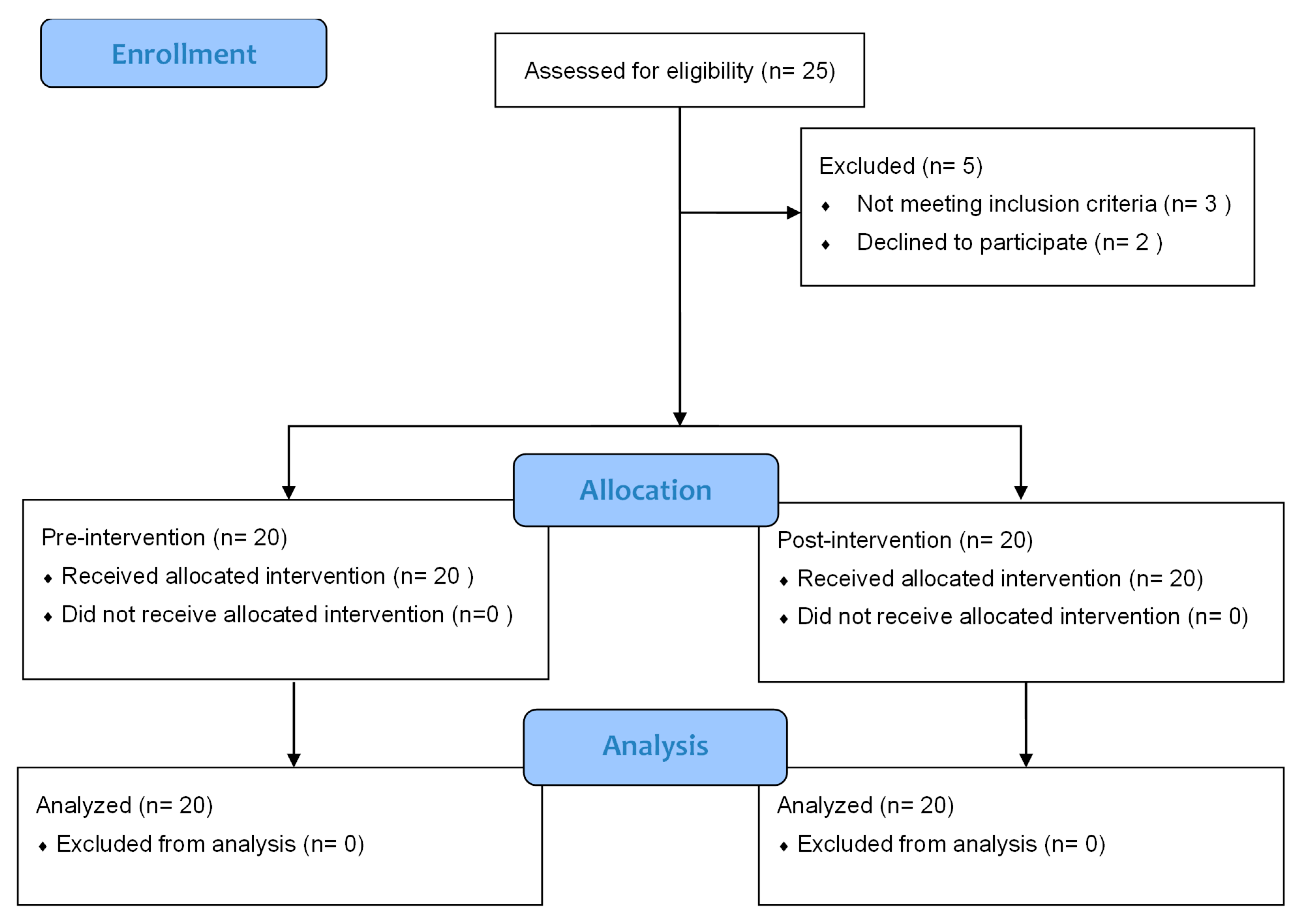
Figure 1. Flow chart of participants (Consort 2010 flow diagram).
Japma 116 00014 g001
Figure 2. Application of KT for flexible flatfoot: (1) Cleaning of the application area; (2) proximal base of the KT applied to the medial proximal third of the tibia without tension; (3) the midsection of the tape was subjected to moderate tension; (4) the tape was passed under the medial longitudinal arch; (5) final position of the KT.
Japma 116 00014 g002
Figure 3. Mediolateral COF velocity before and after KT application: the red line indicates the COF velocity of the right foot; the green line indicates the COF velocity of the left foot.
Japma 116 00014 g003
Figure 4. Pressure distribution before and after KT application, indicating pressure displacement on the lateral side of the foot after KT application; red and pink colors indicate high pressure, while light blue and blue colors indicate lower pressure.
Japma 116 00014 g004
Table 1. Descriptive statistics of demographic data.
VariablesMinimumMaximumMean ± SD
FPI66 11 8.8 ± 2.05
Age, years18 22 20.1 ± 1.33
Body height, cm153 172 162 ± 6.25
Body weight, kg72 120 91.8 ± 14.42
BMI, kg/cm226 45 34.9 ± 4.97
Abbreviation: BMI: body mass index; SD: standard deviation; cm: centimeter; kg: kilogram.
Table 2. Pre–post measurement comparison for foot pressures, forces, and contact area.
VariablesPre-Measurement
(n = 20)
(Mean ± SD)
Post-Measurement
(n = 20)
(Mean ± SD)
p-Valuet-ValueEffect Size
Foot peak pressure, kg/cm21.19 ± 0.18 1.18 ± 0.15 0.755 0.317 0.06 *
Total foot pressure, kg/cm21.01 ± 0.14 1 ± 0.13 0.649 0.462 0.165 **
Foot contact area, cm2115.76 ± 13.44 109.75 ± 11.81 0.001 ^ 4.982 0.475 ***
Midfoot force, kg21.6 ± 7.51 24.35 ± 7.69 0.001 ^ −5.117 0.362 ***
Mediolateral COF velocity, cm/s3.92 ± 2.57 2.08 ± 1.89 0.001 ^ 5.450 0.816 ****
Abbreviations: COF: Center of force. ^ Significance Level: p-value < 0.05. * No effect size. ** Weak effect size. *** Medium effect size. **** High effect size.
Disclaimer/Publisher’s Note: The statements, opinions and data contained in all publications are solely those of the individual author(s) and contributor(s) and not of MDPI and/or the editor(s). MDPI and/or the editor(s) disclaim responsibility for any injury to people or property resulting from any ideas, methods, instructions or products referred to in the content.