Neuropathic foot ulcerations can lead to devastating complications such as loss of limb or life.[
1] Diabetes mellitus (DM) is the most common cause of peripheral neuropathy,[
2] which causes individuals to lose sensation at pressure points on the plantar foot, leading to repetitive microtrauma, breakdown of tissue, and subsequent diabetic foot ulcer (DFU).[
3] Relative to other ambulatory clinical cases during 2007-2013, DFUs were associated with a 3.4 times higher odds of a direct emergency department referral or inpatient admission.[
4] In 2010, the mean adjusted cost per patient hospitalized with a primary diagnosis of DFU was
$13,258.[
5] Reducing the vertical and shear forces by off-loading the DFU is critical for healing.[
6] Whereas neuropathic DFUs are well studied, there are other causes of neuropathic plantar foot ulcers, such as alcohol use disorder, spinal disorders, or chemotherapy,[
2]
,[7–9] that are not as well studied, but these individuals should similarly benefit from off-loading.
The International Working Group on the Diabetic Foot (IWGDF) recommends use of a total-contact cast (TCC) or a nonremovable knee-high walker with an appropriate foot–device interface to off-load plantar neuropathic forefoot and midfoot ulcerations.[
6] However, TCCs are not commonly applied. According to a 2005 survey of 895 private practices, academic practices, and clinics in the United States that actively treated DFUs, only 1.7% of centers reported using TCCs most of the time in the treatment of plantar DFUs.[
10] Applying the TCC is time-consuming, requires an individual with adequate training or experience to apply the cast, may result in difficulty sleeping and bathing, and does not allow inspection of the wound.[
10] Furthermore, an 8-year retrospective review of patients seen at the Cleveland Clinic noted that 23% of patients (approximately 4% of casts) developed iatrogenic complications after application of a TCC, with a new ulcer, most commonly on the heel, followed by blisters, maceration, and abrasions being the most common.[
11] As second and third options, the IWGDF recommends a removable knee-high or ankle-high off-loading device with an appropriate foot–device interface. However, adherence to removable off-loading devices is low; in a study by Armstrong et al,[
12] individuals wore the device only during 28% of the total steps of the day. Greater adherence to using the off-loading device is correlated with DFU healing.[
13] Yet, postural instability is a significant barrier, resulting in decreased adherence to wearing a removable off-loading device.[
13]
Appropriately fitting conventional or standard therapeutic footwear combined with felted foam padding is considered the fourth choice by the IWGDF for off-loading ulcerations.[
6] Felt off-loading padding can be readily fashioned to fit the foot. It can be placed directly on the foot or applied to a shoe, with similar healing times when used to treat neuropathic DFUs.[
14] Fleischli et al[
15] found that wearing a ¼-inch felt and ¼-inch polyethylene off-loading pad with an aperture partially around the ulcer in a postoperative shoe reduced peak pressure in the forefoot compared with wearing a rubber-soled canvas oxford by 48%, which was significantly greater than the pressure reduction when wearing a postoperative shoe alone, which reduced peak pressure by 36%. Similarly, Birke et al[
16] demonstrated that wearing a surgical shoe with ¼-inch felt and an aperture at the site of the previous neuropathic ulcer under a metatarsal head reduced peak pressure by 48% compared with walking barefoot. Neither deficient adherence nor postural instability have been cited with the use of felt padding. Despite the use of felted foam to off-load DFUs since the 1960s,[
17] few studies have examined the effect of felt padding on DFU or neuropathic plantar foot ulcer healing rates and times. A recent systematic review found two controlled trials and one cohort study with a high risk of bias that evaluated felt foam off-loading for the treatment of a DFU.[
18] The most recent study was published in 2016 and included only 15 participants.[
19] The off-loading effect of felt padding has been shown to be comparable with that of an off-loading device[
20]; however, the quality of evidence has been low to moderate.[
6,
18,
21]
The present study is unique because it describes the use of multilayer felt padding off-loading techniques, assesses felt padding characteristics (ie, thickness, shape, and position), and evaluates visit characteristics (ie, frequency and number of visits) in relation to healing rates of neuropathic plantar foot ulcers. We hypothesize that the healing times will be faster than the times from previously published studies using a removable off-loading device without felt padding.
Research Design and Methods
This retrospective study was approved by the institutional review board at Massachusetts General Hospital (Boston). International Statistical Classification of Diseases and Related Health Problems diagnostic codes L97.4X and L97.5X were queried in the Research Patient Data Registry to identify individuals who qualify. The senior investigator’s (S.K.) clinic log was screened for patients with plantar forefoot and midfoot ulcerations treated with felt padding between August 1, 2016, and July 15, 2020.
Inclusion Criteria
Patients with a forefoot or midfoot plantar neuropathic ulceration, aged 18 years or older, treated with at least ½-inch felt padding were included in the present study. The presence of neuropathy was defined by physical examination findings demonstrating inability to identify a 10-g monofilament over at least two sites on the foot and/or decreased vibratory sensation when tested over the great toes with a 128-Hz tuning fork.
Exclusion Criteria
Patients were excluded if they had the presence of atrophic skin, heavily draining wound, undrained abscess, or systemic signs of infection. Patients were also excluded if the study ulceration was not located along the plantar aspect of the foot or if the study ulceration was located on the toes or heel.
Felt Padding Application
All of the patients were followed by one doctor of podiatric medicine (S.K.), who also determined the shape and position of the felt pads. Generally, ¼-inch adhesive felt pads (Gill Podiatry Supply and Equipment Co, Strongsville, Ohio) were cut out based on a tracing of the plantar foot extending from the heel to the metatarsophalangeal joint region. The number of layers of padding, the position of the felt pad, and the size of the aperture were determined by S.K.’s clinical experience, according to the size and depth of the wound, degree of deformity, and the patient’s weight, activity level, balance, and previous response to the padding. If the ulceration was located under a metatarsal head, one to two layers of adhesive felt padding may be applied under the metatarsal shafts. Specifically, for ulcerations located under the second, third, and fourth metatarsal heads and under the midfoot, typically an aperture was cut out extending three-quarters around the periphery of the ulcer approximately to ¼ inch beyond the borders of the ulcer. For ulcerations under the first metatarsal head, the aperture extended proximal and lateral to the ulceration, and for ulcerations under the fifth metatarsal head, the aperture extended proximal and medial to the ulceration, with an additional one to three layers of ¼-inch felt applied over the initial layer.
The felt padding was then secured to the foot with 2-inch Medipore tape (3M, Saint Paul, Minnesota), with care taken to avoid wrapping the tape circumferentially around the foot. Off-loading of the ulcer may be estimated by having the patient stand and take a step on a sterile drape, observing off-loading of the ulcer before applying a dressing. The dressing was then applied over the wound and secured with paper tape. The patient or visiting nurse may then replace the dressing and inspect the ulcer without disturbing the off-loading felt padding. The wounds were debrided as needed, and the felt padding was replaced every 1 to 2 weeks while treating the ulcerations. In some cases, a template with a sample of the felt padding was given to the patient to continue applications at home. Patients were advised that attention must be given to avoid overfilling the aperture with the dressing because this may decrease the effectiveness of the aperture.[
23] A removable off-loading device or a postoperative shoe was then dispensed.
Data Collection
Study data were collected and managed using the Research Electronic Data Capture (REDCap) electronic data capture tools hosted at Massachusetts General Hospital.[
22] Demographic characteristics, including age, sex, weight, body mass index, ethnicity, and race, were recorded. Medical and social histories, including hemoglobin A
1c values in the 3 months before and after the initial visit, neuropathy etiology, tobacco use, the presence of pertinent chronic conditions (ie, peripheral artery disease, congestive heart failure, hypertension, hyperlipidemia, renal disease, Charcot’s neuroarthropathy, and depression), and laboratory results (including albumin and creatinine levels), were recorded.
Foot ulcer history and measurements recorded included ulcer classification using the Wagner scale, number of ulcers, the study ulcer duration at the initial visit, laterality, adverse events, and ulcer surface area at the initial visit, as well as by 6, 12, and 24 weeks. If a patient had more than one ulcer, the largest ulcer was used for the study. Ulcers were measured using a ruler, and surface area was calculated using the formula for an ellipse: (Length/2) × (Width/2) × 3.14. Ulcer healing was defined as complete epithelialization without drainage. Treatment history, including podiatric medical visit frequency before, during, and after the study ulceration, was recorded based on up to four visits before identification of the ulcer and four visits subsequent to healing of the ulcer.
Characteristics of treatment that were recorded were thickness, shape, and positions of the felt pads, and shoe or off-loading device used. These were recorded based on the most commonly used felt pad technique and off-loading device throughout the treatment.
Statistical Analysis
Differences between the efficiency of off-loading treatments in patients with and without DM were assessed in terms of the following variables: ulcer surface area at initial visit, healing time, number of visits to heal, and time to heal for the group that healed within 12 weeks. Each of these variables was assessed by two-sample t test, with mean values for each group and resulting P values reported. In addition, differences in treatment efficacy were assessed with respect to percentage of patients in each treatment cohort healed at 6, 12, and 24 weeks. Pairwise differences in percentage healed were assessed by Fisher exact test. Finally, the time to reach 50% surface area reduction was assessed using a Kaplan-Meier curve and a P value determined using the log-rank test.
Results
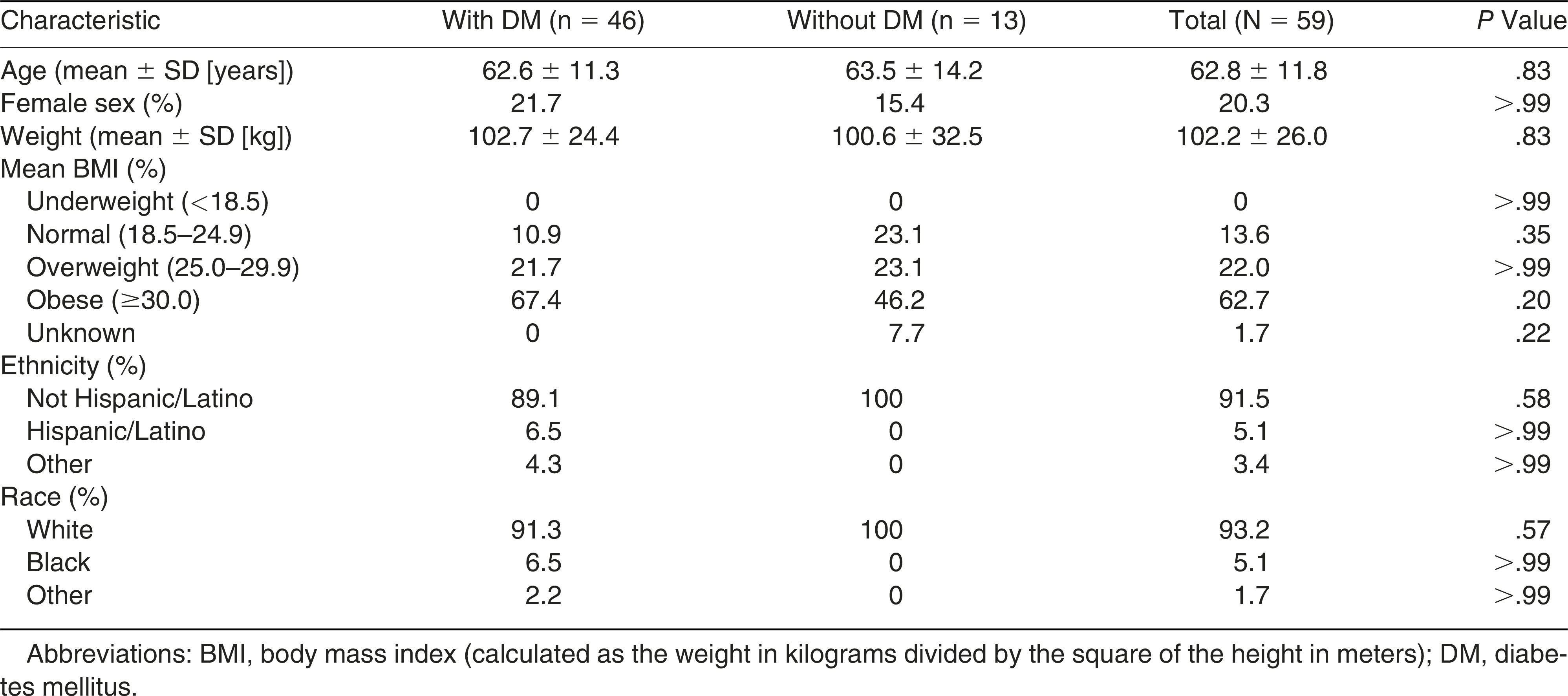
Sixty-two patients met the inclusion and exclusion criteria. Three patients were excluded because they were lost to follow-up for more than 30 days and did not receive additional felt padding according to medical record notes. Therefore, the study included 59 patients. Forty-six patients had DM (78.0%), and the remaining patients had a neuropathic foot ulcer due to causes other than DM (eg, spinal disorder, alcohol abuse, and idiopathic). The characteristics of the study population are summarized in
Table 1.
Table 1.
Patient Demographic Characteristics.
Table 1.
Patient Demographic Characteristics.
The mean hemoglobin A1c value of patients with DM was 8.0% (range, 5.3%–12.9%). Patients had comorbid hypertension (81.4%), hyperlipidemia (66.1%), chronic kidney disease (44.1%), depression (25.4%), congestive heart failure (20.3%), Charcot’s neuroarthropathy (20.3%), and peripheral artery disease (16.9%). Mean albumin and creatinine levels were 3.96 mg/dL and 1.60 mg/dL, respectively.
Foot Ulcer Characteristics and History
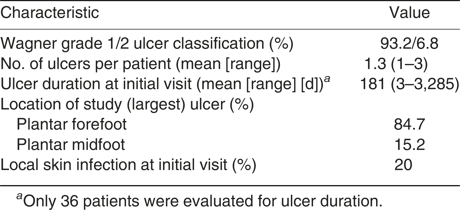
Most patients (89.8%) had a history of hemorrhagic callus. The median podiatric medical visit frequency before development of study ulcer was 56.9 days compared with 11.8 days during ulcer days and 23.5 days during postulcer days. Additional foot ulcer characteristics are summarized in
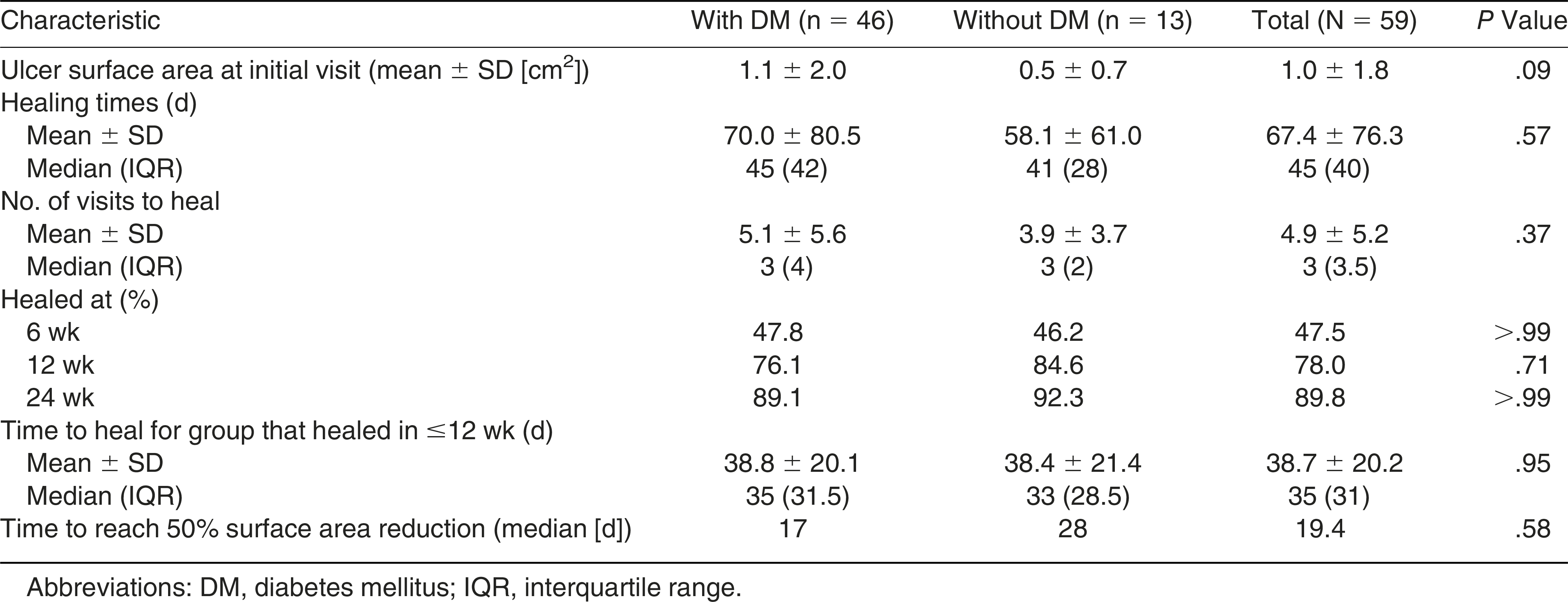
Table 2. Twenty percent of patients had a local skin infection at the initial visit that was treated with oral or topical antibiotics. The DFU healing times are summarized in
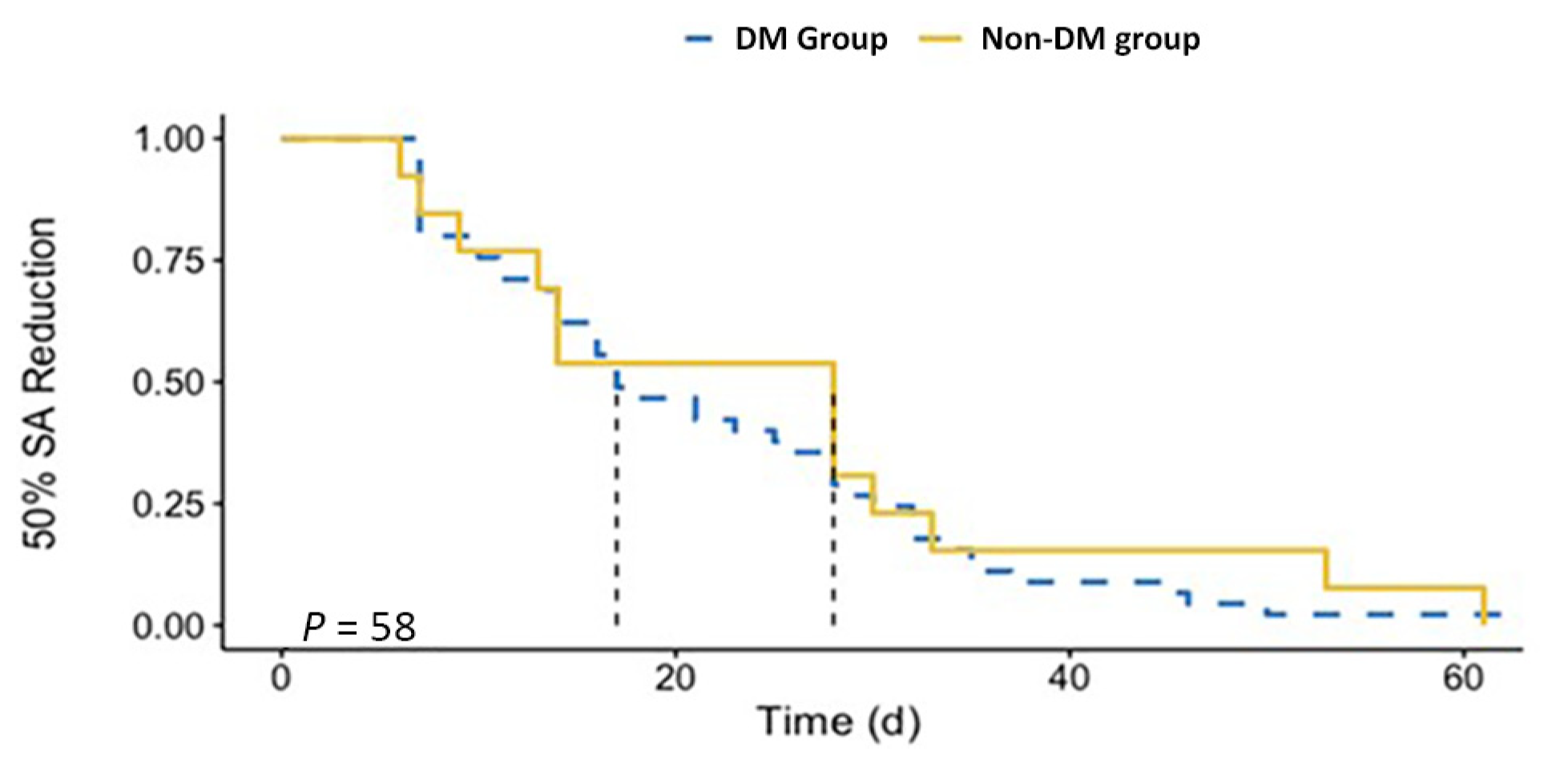
Table 3. Using a Kaplan-Meier curve, the median time to 50% surface area reduction of foot ulcer was estimated to be 17 days for the group with DM and 28 days for the group without DM (
Fig. 1).
Table 2.
Neuropathic Foot Ulcer Characteristics of the 59 Study Patients.
Table 2.
Neuropathic Foot Ulcer Characteristics of the 59 Study Patients.
Table 3.
Neuropathic Foot Ulcer Healing Characteristics.
Table 3.
Neuropathic Foot Ulcer Healing Characteristics.
Figure 1.
Kaplan-Meier curve showing the percentage of patients achieving 50% surface area (SA) reduction of foot ulcer, over time, grouped by diabetes mellitus (DM) status. Median time to 50% SA reduction is shown as dotted lines, with P value assessed by log-rank test.
Figure 1.
Kaplan-Meier curve showing the percentage of patients achieving 50% surface area (SA) reduction of foot ulcer, over time, grouped by diabetes mellitus (DM) status. Median time to 50% SA reduction is shown as dotted lines, with P value assessed by log-rank test.
Median (interquartile range) healing time for forefoot ulcers (n = 50) was 36.5 (37.5) days compared with 149 (188) days for midfoot ulcers (n = 9) (P = .02). For midfoot ulcers, patients with DM had a mean ± SD healing time of 152.3 ± 103.8 days and patients without DM had a mean ± SD healing time of 143 ± 144.3 days. For forefoot ulcers, patients with DM had a mean ± SD healing time of 55.3 ± 67.2 days and patients without DM had a mean ± SD healing time of 42.6 ± 26.1 days. For midfoot and forefoot ulcers, the difference in healing time was not significant between diabetic and nondiabetic patients (P = .94 and P = .34, respectively). The group with diabetic neuropathic foot ulcers and the group with neuropathy due to other causes did not significantly differ by patient demographics (P > .05), healing times (P = .57), number of visits to heal (P = .37), or percentage healed at 6, 12, and 24 weeks.
Foot Ulcer Treatment
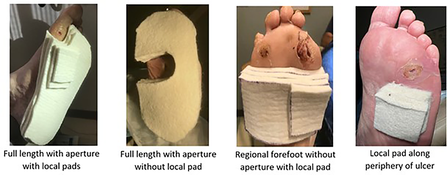
Figure 2 shows photographs of felt padding techniques. Apertures were U- or C-shaped. A minimum of two and a maximum of five ¼-inch felt pads were applied to the periphery of the ulcer with the following distribution of the thickness of the pads: 21% (0.50 inches), 43.5% (0.75 inches), 24.2% (1.0 inch), and 11.3% (1.25 inches). The distribution of the felt pad positions and styles was as follows: full length with aperture with (62.7%) and without (16.9%) local pad, regional forefoot with (6.8%) and without (1.7%,) aperture, local periphery of ulcer (5.1%), regional midfoot with aperture (3.4%), and a full length without aperture with (1.7%) and without (1.7%) local pad.
Figure 2.
Photographs of representative padding of forefoot and midfoot ulcerations. Full-length pads extended from the heel to the metatarsophalangeal joints. Regional pads covered the forefoot or midfoot. Local pads were applied along the immediate periphery of the ulcer.
Figure 2.
Photographs of representative padding of forefoot and midfoot ulcerations. Full-length pads extended from the heel to the metatarsophalangeal joints. Regional pads covered the forefoot or midfoot. Local pads were applied along the immediate periphery of the ulcer.
In addition to the felt padding, most patients adjunctively used a form of off-loading device: knee-high removable off-loading device (42.4%), standard postoperative sandal (35.6%), ankle-high removable off-loading device (11.9%), and TCC (1.7%). The remainder of patients used felt padding with a shoe (6.8%) and crutches or a knee scooter (1.7%).
The total number of clinic visits was 287, and felt padding was applied at each visit except on two occasions because the ulcers were very small. In only five of the 287 visits, it was noted that an off-loading pad was not on the patient’s foot when the patient returned to the clinic for reevaluation, suggesting a very high adherence rate. Adverse events attributed to use of the felt padding included six cases of skin tear (10.2% of patients, 2.1% of felt applications) and one case of contact dermatitis (1.7% of patients, 3.5% of felt applications). There were no infections resulting from application of the off-loading padding. No emergency surgeries or admissions to the hospital were required during treatment as a result of the felt padding. During treatment for the ulceration, one patient required a skin graft and one patient required admission to the hospital for osteomyelitis of a metatarsal head.
Discussion
Although TCCs and nonremovable knee-high cast walkers are considered the gold standard in the treatment of plantar diabetic neuropathic ulcerations, they are not frequently applied.[
10,
24,
25] Removable off-loading devices may be as effective, but due to the devices being readily removable, the effectiveness is limited by low adherence. Multilayered felt padding can off-load ulcerations with a removable off-loading device because it can continue to provide off-loading when the removable off-loading device is not worn. Therefore, its primary effectiveness may be the result of off-loading when an individual does not adhere to use of the off-loading device.
The mean ± SD size of the ulcers in this study was 1.0 ± 1.8 cm
2, which, although small, may reflect the typical size seen in a general podiatric medical practice. It was noted that in 1,088 patients with new foot ulcers from the Eurodiale consortium (a network of 14 European diabetic foot centers), 37% had an ulcer size at inclusion of less than 1.0 cm
2.[
26] The mean time to heal for patients with felt off-loading in the present study was 67.4 days, which is similar to other studies using felt foam padding to treat neuropathic DFUs (79.6 and 75.2 days).[
20,
27] Furthermore, in comparing the present results with those of other studies using removable off-loading devices, it was noted that 58% of ulcers with a mean ± SD initial surface area of 1.29 ± 1.09 cm
2 treated with a bivalve TCC and a felt aperture pad were healed at 12 weeks.[
28] The percentage healed at 12 weeks in the present study was higher at 78.0%, which may be due to the smaller ulcer size. In two studies that looked at removable off-loading devices without felt padding, the percentage healed in the intention-to-treat group at 12 weeks was 65.0% and 51.9% for neuropathic ulcers with a mean ± SD size of 1.4 ± 1.4 cm
2 and 2.0 ± 1.1 cm
2, respectively.[
25,
29] The present study found a higher percentage healed at 12 weeks, possibly partly due to the adjunctive use of felt padding with a removable off-loading device.
A previous felt off-loading study noted a minor skin tear caused by the dressing tape in 6% of patients, wound maceration in 20%, and tinea pedis in 9%, but most felt off-loading studies did not discuss complications.[
21] The adverse events in this study included skin tear and contact dermatitis. Skin tears have occurred when the patient or medical staff removed the felt pads. Alcohol or Uni-Solve (Smith & Nephew, London, United Kingdom) may facilitate removing the felt pads atraumatically. Skin preparation may be applied to the skin before application of the felt pads. If a skin tear occurs while removing the pads, a small border foam dressing may be applied. Another common complication was shifting of the pads. This has been attributed to the pads becoming wet or insufficient tape to secure the pads. It was postulated that there may be impaired circulation if the tape is too tight.[
17,
30] If greater than ¼- or ½-inch (after removing the shoe insole) felt off-loading padding is applied to the patient’s foot while wearing a postoperative shoe or removable cast walker but due to snow, heavy rain, or concern with safety driving, the patient wears a regular shoe with the padding on the foot, the patient may develop abrasions or ulcerations over the toes or dorsal foot. The patient may experience ankle instability wearing the multilayer felt padding while wearing a postoperative shoe. The postoperative shoe may be modified by stiffening the heel counter, or an ankle-high removable cast walker may be worn. A contralateral shoe lift such as EVENup (OPED Medical Inc, Buford, Georgia) may also be used to address the leg length discrepancy.[
6]
This study has several strengths. There were relatively few exclusions in the patient population, making it more generalizable to the general population. For instance, ischemic and infected ulcers were not excluded. Furthermore, this study examined felt off-loading in a larger study size (n = 59) than previous studies (n = 27 and 31),[
19,
20] allowing for more accurate estimates of mean healing time. In addition, this study was performed at a large hospital where most patients also are treated by their primary care physician and other specialists. If the patient developed a complication requiring hospitalization or surgery, it would likely be noted in the electronic medical record. Because the felt padding was generally reapplied by the investigator, adherence to wearing the felt padding could be determined.
This study also has a few limitations. This is an unblinded retrospective study with no control group. The study was performed at one institution primarily by one physician, limiting generalization. Adherence to wearing the removable cast walker or postoperative shoe was not measured. Adherence to using the felt padding was estimated to be 98.3% based on clinical notes in this study. Because patients were often monitored longer than weekly, the actual healing time of the ulcer may have occurred anytime between the visit at which the healed ulcer was identified and the previous visit, resulting in overestimation of the ulcer healing time and underestimation of the percentage healed at 6, 12, and 24 weeks.[
31] Due to COVID-19 restrictions, patients missed follow-up evaluations during Spring 2020.
Overall, felt off-loading padding is easily fabricated using a template that is peeled off the adhesive side of the felt padding.[
22] The template may be scanned into the patient’s electronic medical record, and photographs may be included in the medical record for reference in future applications. The initial pad is applied to the foot, with subsequent layers applied individually over the initial layer, set back slightly from the underlying pad. If the patient feels comfortable driving only while wearing a standard shoe and cannot defer driving, then the patient can generally wear one layer of felt off-loading padding in a lace-up, extra-depth shoe or two layers of padding if the shoe insole is removed. The visiting nurse or patient may replace the padding between visits if the padding compresses excessively or becomes soiled. Due to ease of application, telemedicine may be used to assist the visiting nurse or patient in reapplying the padding.
Future studies may be performed to determine the optimal shape, thickness, and location of the padding to off-load an ulcer at specific locations based on plantar tissue stress primarily resulting from vertical force, horizontal (shear) stress, and frequency of weightbearing activity.[
32] Pressure may be measured on the skin around the immediate periphery of the wound to determine whether there is an increase in the pressure gradient that may result in soft-tissue shearing.[
33] In addition, an “edge effect” can occur, where there is an increase in vertical stress at the periphery of the aperture pad contiguous with an area of lower vertical stress, which may increase shear forces and result in peripheral tissue injury as the metatarsal head moves proximately during the propulsive phase of gait. When deep tissue slides under superficial tissue, shear stress occurs.[
34] Subsurface shear stress may also be evaluated.[
32,
35] Future studies may also determine whether the felt pads cause deterioration of gait. Furthermore, other studies may involve Podimetrics SmartMat (Podimetrics, Somerville, Massachusetts), Siren Socks (Siren Care Inc, San Francisco, California), or other remote patient monitoring devices that identify a localized area of increased skin temperature along the plantar forefoot. When a remote patient monitoring system identifies a localized increase in skin temperature on the foot, it has been recommended that the patient off-loads and reduces ambulation by 50%.[
36] However, if the patient applies off-loading felt until there is a decrease in skin temperature, it may not be necessary to reduce ambulation, which would be desirable for diabetic patients. In addition, a cost analysis may be performed considering the cost of the felt, tape, and time required for the assistant to cut the pads and apply the pads to the foot, and patient-reported outcome measures may be used to assess the patient’s perception of the felt off-loading approach to treating ulcerations.
Conclusion
Felt padding provides off-loading when a patient removes their off-loading device; therefore, it is an important adjunctive tool in the management of neuropathic plantar foot ulcers. Although felt padding is not the gold standard for off-loading neuropathic plantar foot ulcers, its use with off-loading devices may result in better healing rates compared with removable off-loading devices alone.
Acknowledgments: Carolyn Cooper for her assistance in the design and fabrication of the felt off-loading pads. The Massachusetts General Hospital Podiatry Research Group for their feedback on the final paper: Adam Landsman, DPM, PhD; Sara Rose, DPM; Gurneet Khangura, DPM; Jennifer Skolnik, DPM; Timothy Cheung, PhD; and Shanay Fischer, BS.
Financial Disclosure: None reported.
Conflict of Interest: None reported.