As CN progresses, it goes through an active hyperemic phase that eventually becomes inactive, and the foot/ankle consolidates. The period in which the disease progresses from active to inactive is where instability, deformity, dislocation, and ulceration may occur depending on what deforming forces are stressing the affected pathologic area. There is no consensus, even among experts, as to the optimal time for reconstruction [
8]. Currently, the gold standard in treatment for active CN is total-contact casting to off-load the foot and ankle. The goal is to prevent progression of deformity while maintaining a plantigrade, stable, ulcer-free foot [
9]. However, in selected patients, early in the disease process may be the best time to surgically intervene, before significant architectural collapse and consolidation in a maligned position.
The purpose of the present study was to retrospectively review 30 reconstructions in 30 patients with CN who underwent early primary staged reconstruction of the midfoot or ankle in an active Charcot process secondary to gross instability with impending ulceration, deformity with ulceration, or frank dislocation. The procedure was performed with a staged protocol using multiplanar circular ring external fixation in the active phase and internal fixation after the acute event had subsided or the ulceration was healed. We are not aware of any patient series using a staged technique in the active stage of CN with definitive internal fixation.
Materials and Methods
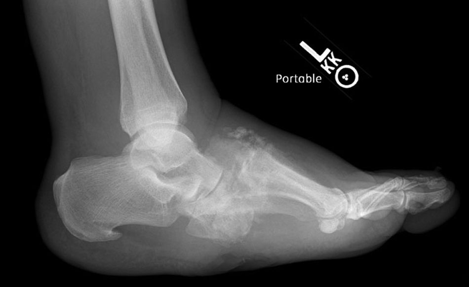
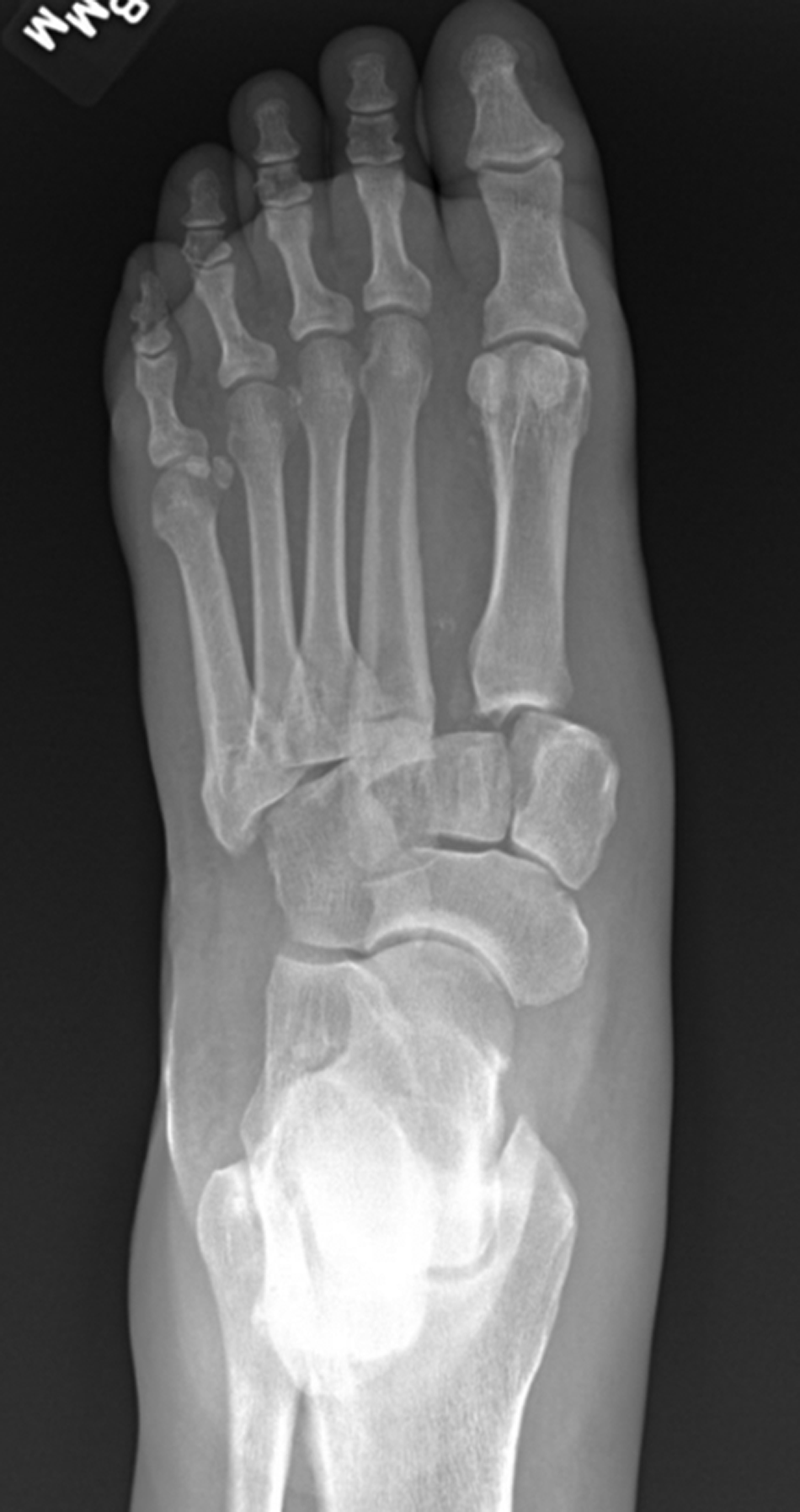
Formal institutional review board approval was obtained, and the electronic medical records of the senior author (P.R.B.) were retrospectively reviewed from 2006 to 2019 for patients who underwent primary staged surgical reconstruction for active midfoot and/or ankle CN. We included patients with diabetic peripheral neuropathy and active midfoot or ankle CN with or without ulcerations on presentation. Active CN was diagnosed clinically and radiographically. On clinical examination, patients demonstrated a red, hot, swollen foot/ankle compared with the unaffected side. Radiographs demonstrated joint effusion, deformity, subluxation, frank dislocation, and periarticular bone debris (
Figs. 1 and
2). Active CN was diagnosed by the lead surgeon (P.R.B.) based on radiographic and clinical evaluation, most notably in conjunction with each other. Clinical examination findings were based on the author’s experience with management of CN, in conjunction with findings on plain radiographs. Temperature changes were determined by examination and physical examination compared with the contralateral limb rather than by thermometer. In patients who presented with an ulceration, presence of osteomyelitis was evaluated by a combination of signs such as probe-to-bone test, complete blood cell count, erythrocyte sedimentation rate, C-reactive protein level, and microbiological analysis after bone biopsy during application of the external fixator. The location of CN was classified by using the Brodsky-Trepman classification scheme (
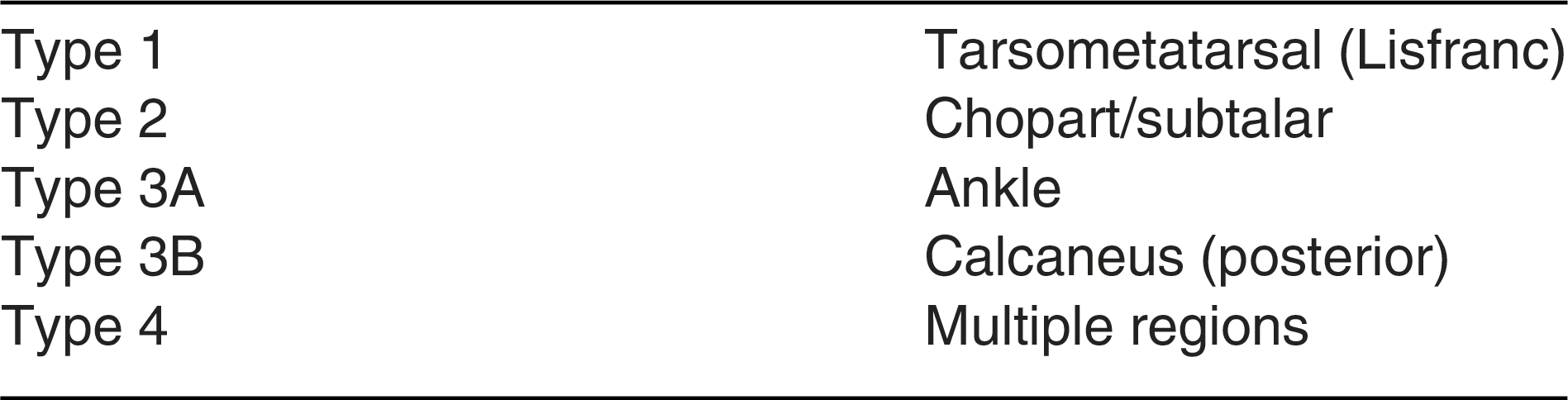
Table 1).
Figure 1.
Lateral radiograph of dorsally dislocated acute Charcot’s neuroarthropathy presenting with ulceration.
Figure 1.
Lateral radiograph of dorsally dislocated acute Charcot’s neuroarthropathy presenting with ulceration.
Figure 2.
Anteroposterior radiograph of acute Charcot’s neuroarthropathy with lateral displacement of the midfoot.
Figure 2.
Anteroposterior radiograph of acute Charcot’s neuroarthropathy with lateral displacement of the midfoot.
Table 1.
Brodsky-Trepman Classification Scheme
Table 1.
Brodsky-Trepman Classification Scheme
The exclusion criteria were patients who had previous attempts at nonsurgical treatment but were still in the active phase, recurrent “acute on chronic” CN, and inactive CN. Indications for surgical intervention were made by one of us (P.R.B.). The main indications were presence of an unstable deformity with ulceration, frank joint dislocation, a midfoot deformity without ulceration, and a negative cuboid height on lateral radiographs as described by Wukich et al [
10] and a nonplantigrade foot. The primary outcomes of interest were limb salvage rate, presence of ulceration at final follow-up, and, finally, if patients were able to walk with or without assistance.
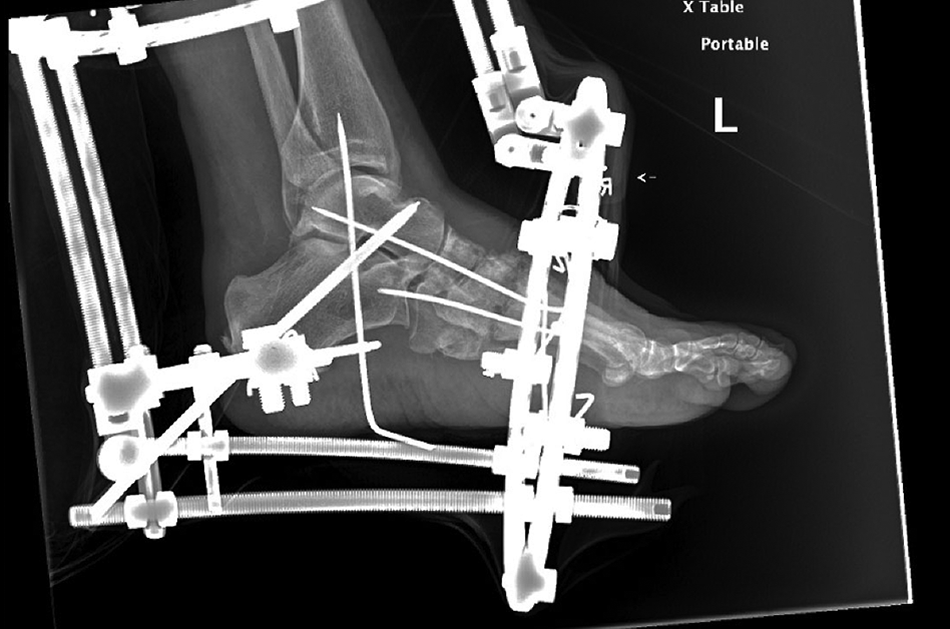
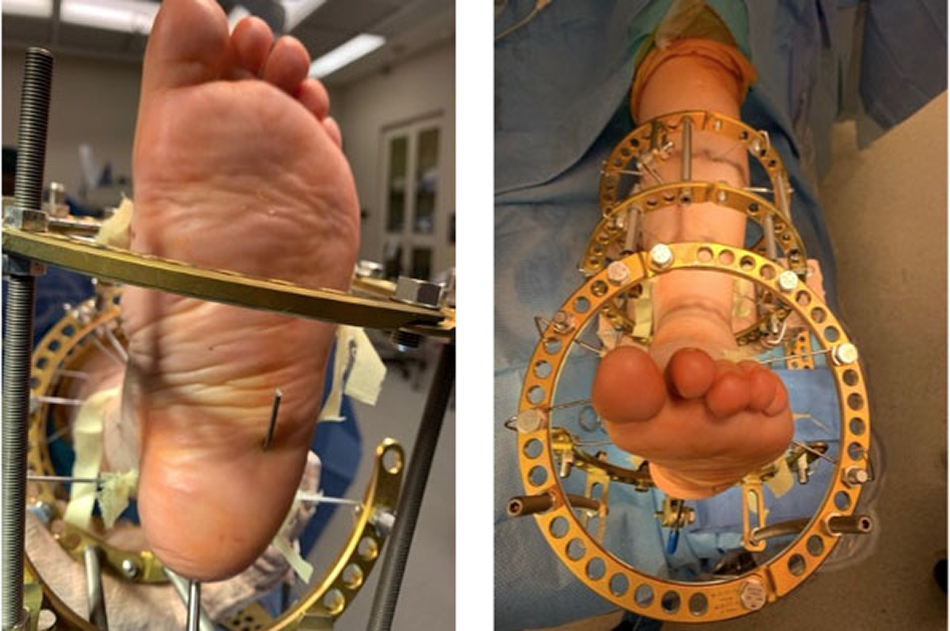
The index procedure consisted of closed reduction of the deformity with application of a multiplanar circular ring external fixator. Small accessory incisions, transarticular pin fixation, temporary pin to bar distraction, and percutaneous tendo Achillis lengthening were performed as necessary (
Figs. 3 and
4). The goals of the external fixation are to reduce the deformity to a more anatomical position, optimize the soft-tissue envelope, allow ulcerations to heal if present, and achieve skeletal stabilization, which in turn allows the Charcot process to become inactive. The external fixator was maintained for approximately 2 months. Once removed, the patients were placed in a nonweightbearing short-leg cast for 2 weeks to maintain stability and allow the pin sites to heal.
Figure 3.
Radiograph of the initial multiplanar circular ring external fixator with transarticular pins for active Charcot’s neuroarthropathy.
Figure 3.
Radiograph of the initial multiplanar circular ring external fixator with transarticular pins for active Charcot’s neuroarthropathy.
Figure 4.
Clinical photographs of a patient’s foot in the initial multiplanar circular external fixator for active Charcot’s neuroarthropathy.
Figure 4.
Clinical photographs of a patient’s foot in the initial multiplanar circular external fixator for active Charcot’s neuroarthropathy.
The definitive procedures for internal hardware varied depending on the location of the affected area and the year the final fixation was performed. The main surgeon followed ideas of the “Super Construct” as proposed by Sammarco [
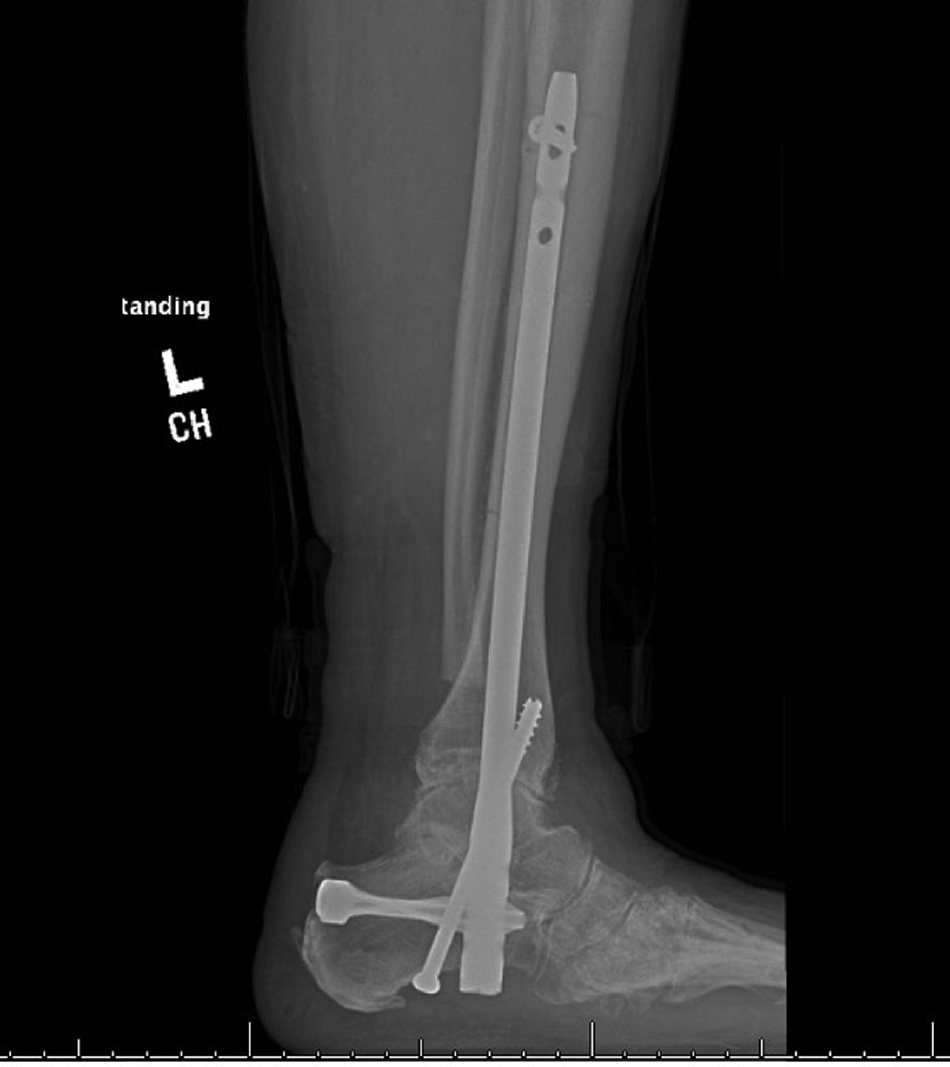
11]. One of the key focuses of this construct is fusion beyond the zone of injury. This concept helped with construction of the mainstay of our patients. To extend beyond the zone of injury of a midfoot CN, fixation was achieved via tibiotalocalcaneal arthrodesis with fixation provided by an intramedullary nail (
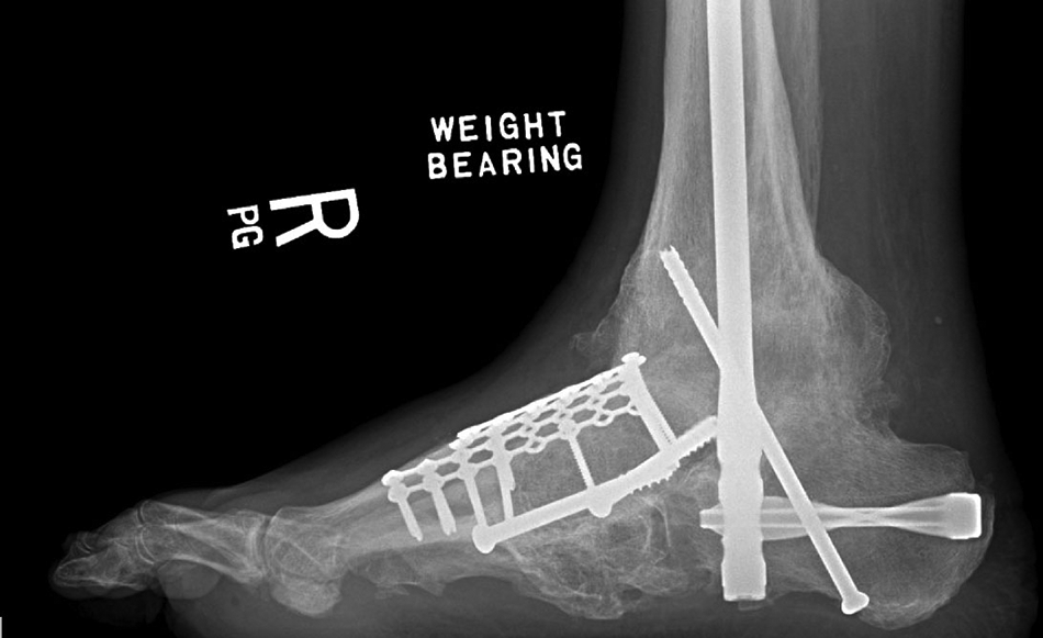
Fig. 5). Depending on involvement, dislocation, or sagittal fault at the talonavicular joint, a decision was made to include this in the final fixation. Fixation of the talonavicular joint consisted of use of large cannulated screws with a dorsal spanning plate or staples (
Fig. 6).
Figure 5.
Lateral radiograph status post intramedullary fixation with augmentation screw in a patient who presented with dislocated midfoot Charcot’s neuroarthropathy.
Figure 5.
Lateral radiograph status post intramedullary fixation with augmentation screw in a patient who presented with dislocated midfoot Charcot’s neuroarthropathy.
Figure 6.
“Pan foot arthrodesis.” Fusion of the tibiotalar, subtalar, and talonavicular joints.
Figure 6.
“Pan foot arthrodesis.” Fusion of the tibiotalar, subtalar, and talonavicular joints.
Discussion
There is a great paucity in the literature in terms of reconstruction in the active stage of CN. Recently, there have been studies regarding the conservative treatment of CN. For instance, Gratwohl et al [
12] examined 159 patients who received conservative treatment for CN. They found that there was a limb salvage rate of 92.9% for these patients. Where the present study differs is that our patients underwent surgical reconstruction based on findings from our primary surgeon (P.R.B.) only, which included the presence of an unstable deformity with ulceration, frank joint dislocation, a midfoot deformity without ulceration, and a negative cuboid height on lateral radiographs as described by Wukich et al [
10] and a nonplantigrade foot. If patients were in the active stage and did not have these findings, they were treated conservatively. This treatment greatly differs from that of Gratwohl et al [
12], who stated that their patients underwent conservative treatment for a “noncollapsed plantigrade CN foot” and that patients who needed surgical correction for deformity, as the present patient population underwent, were not analyzed in their study. Because our study included only Charcot patients with the previously mentioned findings, we cannot compare our study to that of Gratwhol et al, which only treated noncollapsed feet conservatively.
Simon et al [
13] first described the effectiveness of arthrodesis in acute midfoot CN. They operatively reduced and successfully fused all of the 14 patients in their study during the active Eichenholtz stage 1. Operating in the active phase did not lengthen time to heal but in fact seemed to expedite the reversal of the destructive process. They hypothesized that this quicker reversal of the destructive phase could be due to anatomical restoration and stabilization. This study included only patients with midfoot disease. Mittlmeier et al [
6] retrospectively reviewed 22 patients with midfoot (n = 9) or hindfoot (n = 17) CN who underwent primary surgical reconstruction and reorientation arthrodesis due to instability, nonplantigrade foot position, and deformity with ulceration or impending ulceration. Of the 22 patients studied, only four were assigned active stage 1 CN based on the Eichenholtz classification. Due to the heterogeneity of this patient cohort, it was impossible to identify whether the patients with active CN did well in this study [
6].
Myerson et al [
14] reported outcomes of 85 patients with CN of the midfoot, including eight patients with acute neuroarthropathy who were treated with open reduction and arthrodesis. Their criterion for surgery was severe dislocation that was unstable and manually reducible. These patients were hospitalized, and surgery was delayed until skin wrinkling occurred. They used single-stage rigid internal fixation with interfragmentary screw compression. These patients remained stable at an average follow-up of 28 months [
14]. Lamm et al [
15] presented a staged approach to Charcot’s diabetic foot reconstruction in 2010 with the use of external fixation and intramedullary fixation. They described gradual osseous realignment with Taylor spatial frame followed by minimally invasive arthrodesis using percutaneous intramedullary screws. They reported the results of 11 feet reconstructed in this manner. There were no recurrent ulcerations, and they achieved a plantigrade foot with no deep infections at an average follow-up of 22 months. Only one patient was in active stage 1 CN [
15].
The treatment protocol in the present study is a hybrid of techniques previously described for surgical reconstruction of active CN. Reduction during the active phase negates the need for gradual distraction, and thus a static ringed fixator can be used successfully. We follow external fixation with rigid internal fixation, most commonly intramedullary nail fixation or a combination of intramedullary fixation and plating. The fixation decision was based on previous studies, as well as super construct principles laid out by Sammarco [
11]. We believe that the ankle must be included with Charcot’s reconstruction to allow for fusion beyond the zone of injury. In addition, not all of the final fixation included the midfoot, although a primary amount of the pathology occurred there. This finding is supported by Shibata et al [
16] in their study of CN in the leprotic ankle fracture population. They noted that fusion rates were lower in patients who had a longer rigid lever arm. They stated that they believed this caused more stress at the ankle joint. We believe this to be true as well, which led to many fixations of the present acute CN primarily as a tibiotalocalcaneal arthrodesis with just an intramedullary nail. This also allowed us to avoid areas of possible infected bone, especially for patients who presented with midfoot CN collapse and ulceration. Shibata et al [
16] also stated that there was a noted increase in destruction of adjacent joints due to abnormal motion of the ankle joint. This further cemented the idea that final fixation must include the ankle joint, even when treating isolated midfoot CN. Furthermore, Shibata et al [
16] noted that patients had a more successful arthrodesis in earlier stages of Eichenholtz rather than in stage 3, which is the stage in which most reconstructions take place in studies.
Surgery in the acute setting for CN has been regarded as worrisome owing to complications associated with healing and wounds. The present study presented the staged reconstruction approach to help combat these complications. We noted that patients who present with acute CN have a higher chance of a reducible deformity compared with patients in Eichenholtz stage 3, when most patients are regarded to undergo surgical intervention. Another point is that the first modality of treatment for patients with noted acute CN is immobilization. After that, a decision is made about whether internal fixation is required for the deformity. At that point, patients not only have noted local osteopenia secondary to unopposed RANK-L [
17] but also experience disuse osteopenia. With a staged reconstruction, we can realign the foot to the proper position and allow it to consolidate in this new position.
Operation in the acute stage, we believe, can aid in decreasing the major amputation risk in patients diagnosed as having CN. Patients who develop CN are at higher risk for the development of ulceration. Sohn et al [
18] evaluated patients in a Veterans Administration setting and noted that those with CN developed ulceration 34% of the time in follow-up and had an amputation rate of 14.7%. Of the present patients, none developed an ulceration as a result of their reconstruction, and neither did any of the patient need to undergo a minor amputation. In terms of major amputation, it was noted that 10% of the patients in this study required a major amputation. This is favorable compared with the major amputation rate of Nilsen et al [
19] of 14.9%. As stated previously, the present minor amputation rate after reconstructions was noted to be 0%, which, compared with the minor amputation rate of 25.7% in the study by Nilsen et al [
19], is promising. However, none of the patients in the study by Nilsen et al [
19] underwent a surgical intervention; all of their patients were noted to be treated via weightbearing in a Charcot’s restraint orthotic walker.
All three of the present patients who underwent major amputation were secondary to infection of the lower extremity. Of note, every patient who underwent a major amputation presented with a wound or a minor amputation before undergoing reconstructive surgery. Major amputations were due to continued recalcitrant osteomyelitis, although each patient was treated with long-term intravenous antibiotics and initial debridement and resection of bone.
One patient who originally presented with midfoot CN presented to the emergency department 2 months after internal fixation with worsening redness and drainage from the area of the ulceration. She left the hospital against medical advice before being transferred to the lead surgeon’s hospital. When she finally arrived, radiographs showed osteolytic changes to the talus and the calcaneus. Discussion was had with the patient, who elected amputation of the lower extremity. Another patient, 2 years after his tibiotalocalcaneal fusion, presented with septic ankle arthritis and underwent subsequent hardware removal. Four months after the hardware removal, the patient had worsening of deformity in the degree of valgus ankle, with osteolytic changes to the talus and the calcaneus. Due to inability to walk on the affected limb, the patient elected for a below-the-knee amputation. Finally, the third patient underwent final fixation via intramedullary nail for his treated midfoot CN that initially presented with ulceration, abscess, and acute deformity. Three years after the patient’s internal fixation, he presented with ulceration to the plantar foot. After months of local wound care and antibiotic drug therapy, the patient arrived at the emergency department with redness, discharge, and edema to the operative extremity. The decision was made for removal of the hardware followed by subsequent below-the-knee amputation.
Nevertheless, the major amputation rate of patients who presented with ulceration was noted to be favorable compared with that of Wukich et al. [
20]. In the study by Wukich et al [
20], patients had a major amputation rate of 37.2% when there was a diagnosis of osteomyelitis present with concomitant CN. In the present cohort, 50% of patients (n = 15) presented with active CN complicated by an ulceration and osteomyelitis. The major amputation rate in patients who presented with ulceration at the initial visit along with active CN was 20% (n = 3 of 15). The present study showed that patients with ulceration had a 40% healing rate of ulceration after intervention (n = 8 of 15). The biggest end point for this study was to reduce major limb amputation, which in the present cohort would have a 12 times increased risk of amputation in the future [
18]. Limb salvage for these patients was obtained with reduction of significant deformity. Of the patients who healed after intervention, none had recurrence of ulceration. This is favorably compared with Armstrong et al [
21], who showed 1-year recurrence of a diabetic foot ulcer of 40% and 5-year recurrence of 65%.
Of interest, early reconstruction of Charcot’s deformities in patients without ulceration is not only becoming more prevalent but also can be more cost-efficient from a US payer perspective. Albright et al [
22] determined quality-adjusted life years (QALYs) and incremental cost-effectiveness ratios (ICERs) of surgical reconstruction compared with other treatment options, such as bracing and amputation. Based on these data, patients who underwent reconstruction for CN without an ulceration showed a 1.63 QALY gained and an ICER of
$14,340 per QALY compared with bracing/conservative treatment. Reconstruction was also effective for patients from a QALY standpoint of 1.04 and an ICER of
$26,220 per QALY compared with bracing for patients who underwent reconstruction for an uninfected ulceration. This study pointed out that when ulcerations became infected, they began to show a decrease in QALYs compared with other treatment options, such as amputation or bracing. However, this study helps show how early reconstruction, especially in the acute phase of CN, can be beneficial not only from a limb salvage perspective but also from an economic and cost-effective perspective.
There are several limitations of this study. First, the presence of selection bias is evident for patients receiving early surgical reconstruction versus nonsurgical treatment. Currently, there is sparse evidence to support surgical reconstruction in active CN, let alone a standardized protocol to guide the decision for when a patient is ideal for early surgical intervention. Also, not every patient with acute CN who presented to the clinic underwent a staged reconstruction. The patients who underwent reconstruction were those who had an unstable foot, deformity, ulceration, or impending ulceration. Second, no objective measurements were obtained in this study. Although we achieved 90% limb salvage, we cannot objectively report on the physical or functional components of these patients. Another limitation to this study is that we did not record any radiographic parameters regarding reduction of deformity. Although we feel that this is important, we believed that the most important measurement for this patient population was a plantigrade foot so that a patient could wear shoes. Third, the areas of deformity were heterogeneous, but they compare with many other CN reconstruction papers that have been published.