Next Article in Journal
Evaluation of YouTube Information Quality About Pes Planus
Previous Article in Journal
Evaluation of Prosthesis Satisfaction, Psychological Status, and Quality of Life in Lower-Extremity Amputee Patients: A Pilot Study
 
 
Journal of the American Podiatric Medical Association is published by MDPI from Volume 116 Issue 1 (2026). Previous articles were published by another publisher in Open Access under a CC-BY (or CC-BY-NC-ND) licence, and they are hosted by MDPI on mdpi.com as a courtesy and upon agreement with American Podiatric Medical Association.
Article

Dermatophytomas in Onychomycosis: A Scoping Review of Prevalence, Diagnosis, and Treatment

by
Shari R. Lipner
1,*,
Tracey Vlahovic
2,
Mahmoud A. Ghannoum
3,
Boni Elewski
4 and
Warren S. Joseph
5
1
Weill Cornell Medicine, 1305 York Ave, 9th Floor, New York, NY 10021
2
Temple University School of Podiatric Medicine, Philadelphia, PA
3
Case Western Reserve University, Cleveland, OH; University Hospitals Cleveland Medical Center, Cleveland, OH
4
University of Alabama at Birmingham School of Medicine, Birmingham, AL
5
JAPMA, Rockville, MD; Arizona College of Podiatric Medicine, Midwestern University, Glendale, AZ
*
Author to whom correspondence should be addressed.
J. Am. Podiatr. Med. Assoc. 2024, 114(2), 22161; https://doi.org/10.7547/22-161
Published: 1 March 2024

Abstract

Background: Dermatophytoma, also described as a longitudinal streak/spike, is a form of onychomycosis that presents as yellow/white streaks or patches in the subungual space, with dense fungal masses encased in biofilm. This scoping review of the literature was conducted to address a general lack of information about the epidemiology, pathophysiology, and treatment of dermatophytomas in onychomycosis. Methods: A search was performed in the PubMed and Embase databases for the terms “longitudinal spike” or “dermatophytoma.” Outcomes of interest were definition, prevalence, methods used for diagnosis, treatments, and treatment efficacy. Inclusion and exclusion of search results required agreement between two independent reviewers. Results: Of a total of 51 records, 37 were included. Two reports provided the first unique definitions/clinical features of dermatophytomas. Overall, many descriptions were found, but one conclusive definition was lacking. Prevalence data were limited and inconsistent. The most frequently mentioned diagnostic techniques were clinical assessment, potassium hydroxide/microscopy, and fungal culture/mycology. Oral terbinafine and topical efinaconazole 10% were the most frequently mentioned treatments, followed by topical luliconazole 5% and other oral treatments (itraconazole, fluconazole, fosravuconazole). In studies with five or more patients without nail excision, cure rates were highest with efinaconazole 10%, which ranged from 41% to 100% depending on the clinical and/or mycologic assessment evaluated. Other drugs with greater than or equal to 50% cure rates were topical luliconazole 5% (50%), oral fosravuconazole (57%), and oral terbinafine (67%). In studies that combined oral terbinafine treatment with nail excision using surgical or chemical (40% urea) methods, cure rates ranged from 50% to 100%. Conclusions: There is little published information regarding dermatophytomas in onychomycosis. More clinical research and physician education are needed. Although dermatophytomas have historically been considered difficult to treat, the efficacy data gathered in this scoping review have demonstrated that newer topical treatments are effective, as are oral antifungals in combination with chemical or surgical methods.

Onychomycosis is a fungal infection of the nail bed or plate caused by dermatophytes or nondermatophyte molds (NDMs) or yeasts.[1,2] Onychomycosis can occur in toenails or fingernails but is much less common in fingernails.[3] Treatment can be difficult, and achieving complete cure may take 12 months or more in toenails as nail growth is slow.[4] Some patients may present clinically with yellow or white streaks or patches in the subungual space and, pathologically, there may be dense fungal masses encased in a layer of biofilm.[5,6] This form of onychomycosis has been called a dermatophytoma or a longitudinal streak or spike.[5,6,7,8] Dermatophytomas are not well known to podiatrists and dermatologists as a type of onychomycosis.
Historically, dermatophytomas have been shown to be difficult to treat, with poor treatment response to oral drugs.[5,6,9] However, data on treatment efficacy are limited as patients with dermatophytomas tend to be excluded from clinical trials of onychomycosis treatments.[10] The prevalence is unclear as there have been few published articles on dermatophytomas, and many are case reports. To our knowledge, there are no published systematic or scoping reviews on the topic of dermatophytomas. The aim of this scoping review was to examine the literature for information regarding the epidemiology, pathophysiology, and treatment of dermatophytomas in onychomycosis.

Methods

This scoping review was conducted according to the methods described by Peters et al.[11]

Search Strategy

A search for articles was performed in the PubMed and Embase (Ovid) databases on December 6, 2021. No restrictions on language, publication year, or study type were applied to the searches because of the limited number of publications on this topic. The following search syntax was used: PubMed: “longitudinal spike”[all fields] OR “dermatophytoma”[all fields]; Embase: manuscripts: all(dermatophytoma) OR all(“longitudinal spike”) and Embase (yes) NOT MEDL(yes); abstracts: all(dermatophytoma) OR all(“longitudinal spike”).

Selection of Reports

Inclusion criteria consisted of studies conducted in humans or in vitro and full manuscripts published in English. Exclusion criteria included duplicate search hits (defined as any records appearing in both the PubMed and Embase searches or any abstracts that matched a full published manuscript), abstracts not published in English, and nonrelevant reports (ie, no mention of onychomycosis or dermatophytoma). The abstracts of all records identified were reviewed to determine whether they met inclusion criteria. Of those that met criteria, the full report was obtained and further reviewed to ensure inclusion criteria were still met. At each stage of the record/report selection process, the two reviewers had to agree independently on the inclusion or exclusion of each.

Outcomes of Interest

For this scoping review of dermatophytomas, the outcomes of interest were definition; prevalence (ie, data extracted from studies on patients with onychomycosis that also included one or more patients with dermatophytoma); methods used for diagnosis; treatments (oral, topical, surgical, other); and treatment efficacy.

Data Extraction and Analyses

Data for each outcome of interest were extracted from reports and entered into tables by each reviewer. Prevalence estimates were calculated as the number of patients with dermatophytoma divided by the total number of patients with onychomycosis, multiplied by 100. Efficacy data were extracted from reports that provided at least one efficacy result for five or more patients for oral or topical drug(s) and/or surgical/chemical nail removal for onychomycosis. All data were presented as provided within the report (ie, percentages, means or medians, P values); no additional analyses were performed. Discordance in extracted data was resolved by consensus between the two reviewers.

Results

Study Inclusion

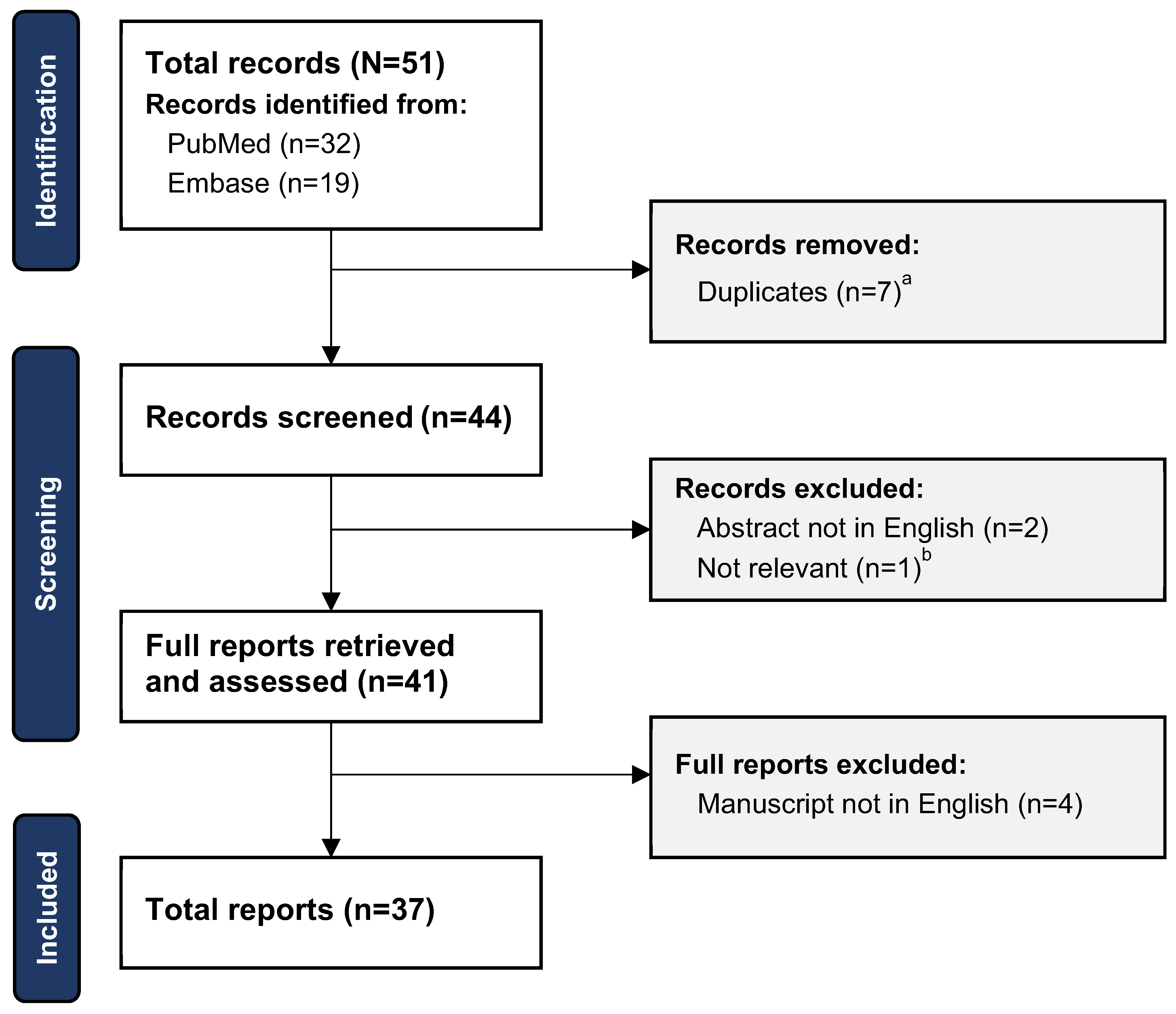
Of a total of 51 records from the initial search (32 from PubMed and 19 from Embase), 37 were included in this review (seven duplicates removed; seven records excluded [six not in English, one not relevant]) (Fig. 1). Publication years ranged from 1998 to 2021.
Figure 1. Preferred Reporting Items for Systematic Reviews and Meta-Analyses—extension for Scoping Reviews (PRISMA-ScR) flow chart of study selection process. aDuplicate search hits were defined as any records appearing in both the PubMed and Embase searches or any abstracts that matched a full published manuscript. bThe record contained no mention of onychomycosis or dermatophytoma and had been conducted in canine small intestines.
Japma 114 22161 g001

Study Characteristics

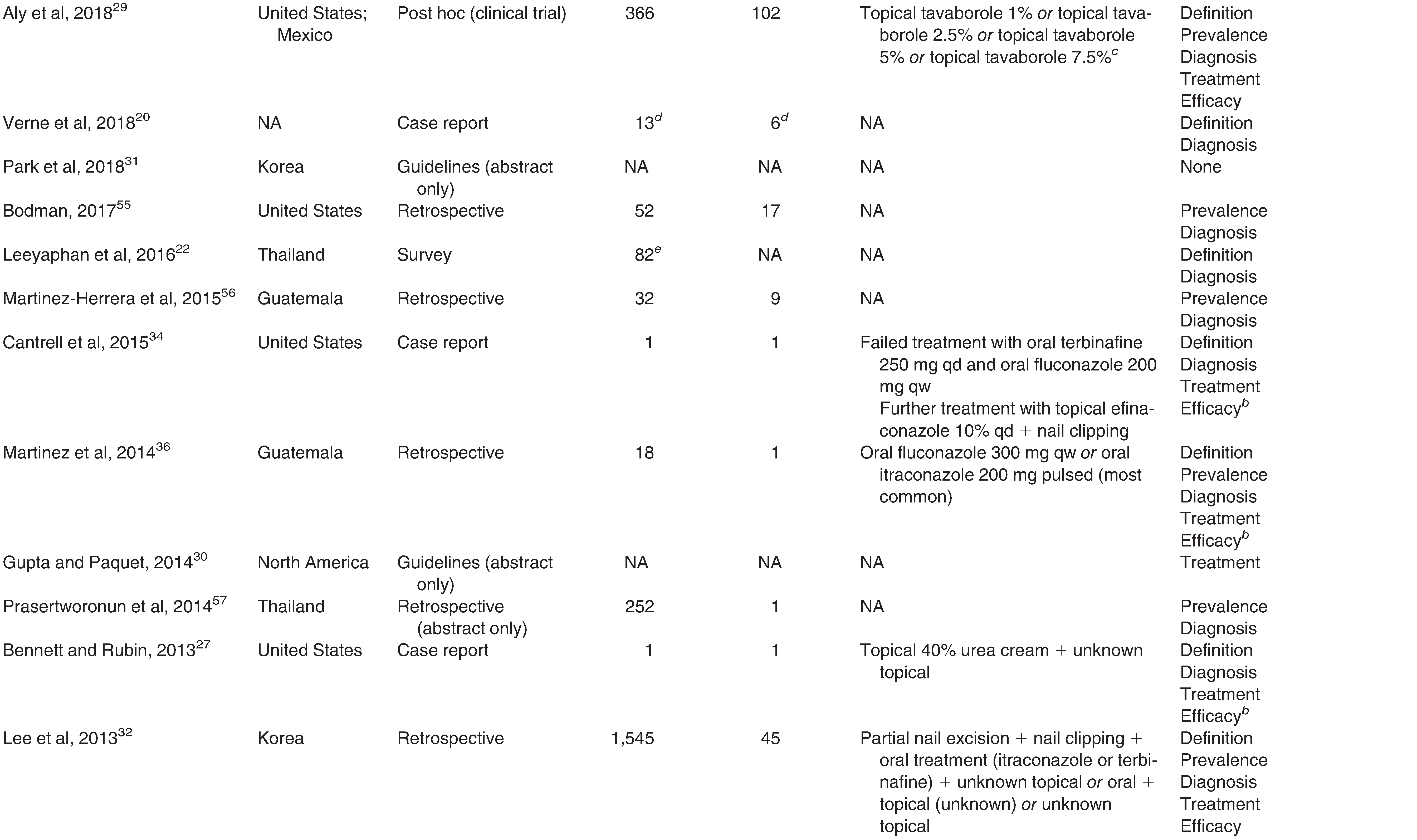
Study characteristics are shown in Table 1. The countries/regions with the largest number of reports were United States/North and Central America (n = 10) and Japan (n = 6). Most reports were case studies (n = 8), retrospective studies (n = 8), reviews (n = 6), or open label/prospective studies (n = 4). Of the six review articles, all were on onychomycosis, but none focused specifically on dermatophytomas.[12,13,14,15,16,17]
Table 1. Overview of the 37 Reports Included in the Scoping Review, by Year of Publication 
Japma 114 22161 i001
Japma 114 22161 i002
Japma 114 22161 i003

Definition

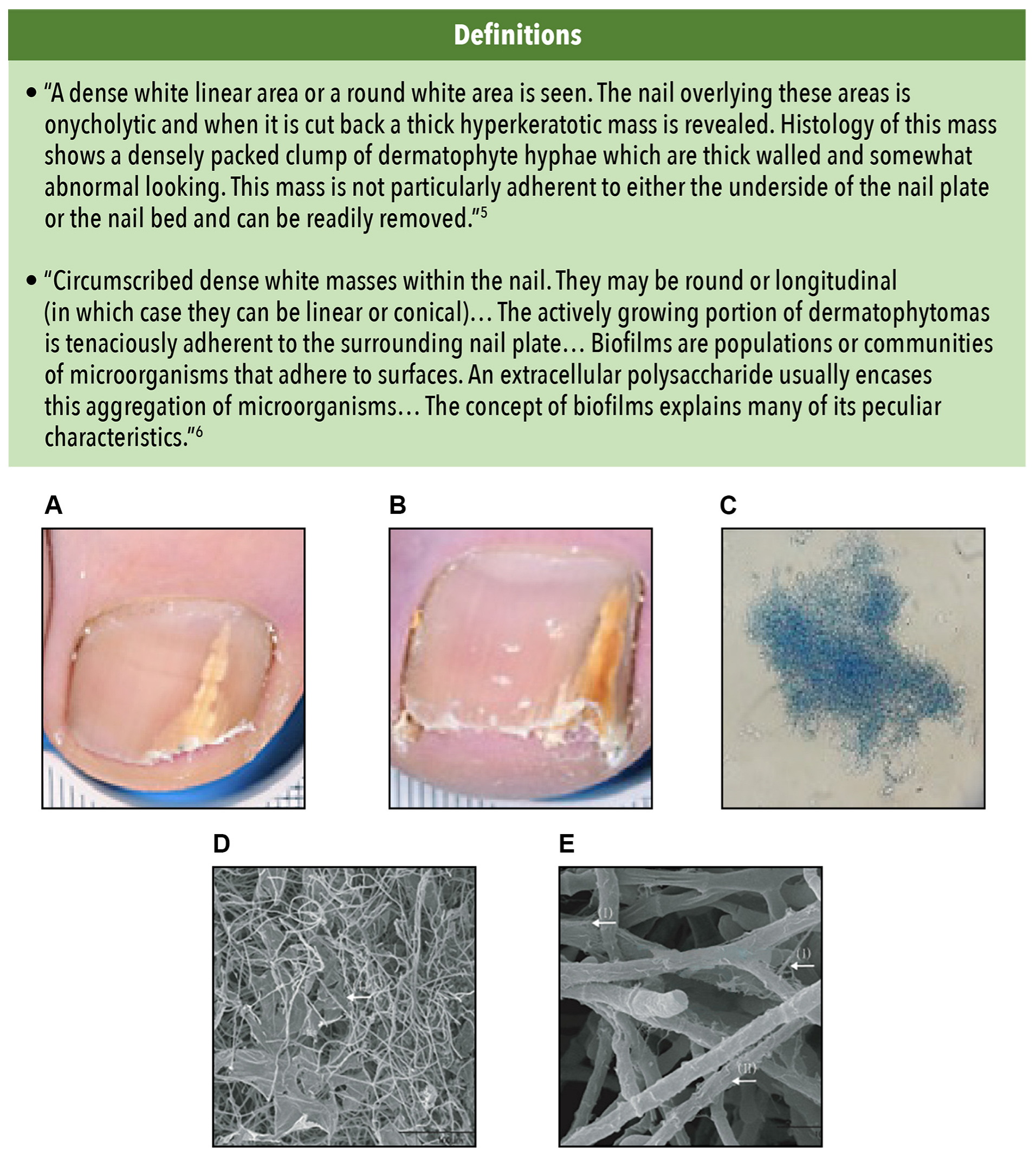
Part of the challenge of diagnosing (and therefore treating) dermatophytomas is the lack of standardized descriptions for this condition. Historically, two reports have provided unique definitions of dermatophytoma[5,6] (Fig. 2). The first mention, which was in 1998,[5] stated that subungual dermatophytomas present as a dense white linear or round onycholytic area in the nail with densely packed fungal masses underneath; they are not adherent to the underside of the nail plate or bed. This definition was amended in 2002 to note that these masses are adherent to the nail plate; it was also expanded to include biofilms.[6] Dermatophytomas present clinically with dense round or longitudinal white thick/hyperkeratotic masses in the subungual space and pathologically with adherent fungal masses encased in biofilms. Biofilms were defined as communities of micro-organisms that adhere to surfaces and aggregate, forming a fungal mass and producing an extracellular polysaccharide. They are more resistant to traditional treatments.[6] Several causative fungal agents of onychomycosis have demonstrated the ability to form biofilms in vitro and in vivo.[18,19]
Figure 2. Dermatophytomas: definitions and clinical examples. A and B, Examples of distal, lateral subungual onychomycosis complicated by dermatophytoma. C, From a dermatophytoma: abundant fungal filament compacted forming a fungal ball (potassium hydroxide, x40). D and E, Scanning electron microscopic images of the biofilms of T mentagrophytes at x500 (D) and x5000 (E). Architecture observed include a thin “blanket-like” layer covering the spaces between hyphae (D) and a fine “mesh-like” layer wrapping the hyphae filaments (E). Reproduced from Wang C, Cantrell W, Canavan T, et al: Successful treatment of dermatophytomas in 19 patients using efinaconazole 10% solution. Skin Appendage Disord 5: 304, 2019. Copyright 2019 Karger Publishers, Basel, Switzerland[10]; Chen B, Sun Y, Zhang J, et al: In vitro evaluation of photodynamic effects against biofilms of dermatophytes involved in onychomycosis. Front Microbiol 10: 1228, 2019. Copyright 2019[40]; and Castro LÁ, Álvarez MI: Nail dermatophytoma in HIV-infected patients in Cali, Colombia. J Mycol Med 31: 101172, 2021. Copyright 2021 Elsevier Masson SAS. All rights reserved.[54].
Japma 114 22161 g002
Most reports that provided a definition used wording similar to one or both reports noted above; many added the color yellow to the clinical description. Seven reports described dermatophytomas as a “fungal ball” of filaments and spores under the nail plate.[17,20,21,22,23,24,25] Yellow or white, linear or round areas of onychomycosis were also referred to as longitudinal spikes, bands, streaks, and/or striae in two reports from Japan,[8,23,25,26] two case reports,[21,27] one treatment guideline,[28] one survey,[22] three reviews,[13,15,17] two open label studies,[9,10] one post hoc clinical trial,[29] and one report of an onychomycosis severity scoring system.[7] Examples of dermatophytomas and biofilms are provided in Figure 2.

Prevalence

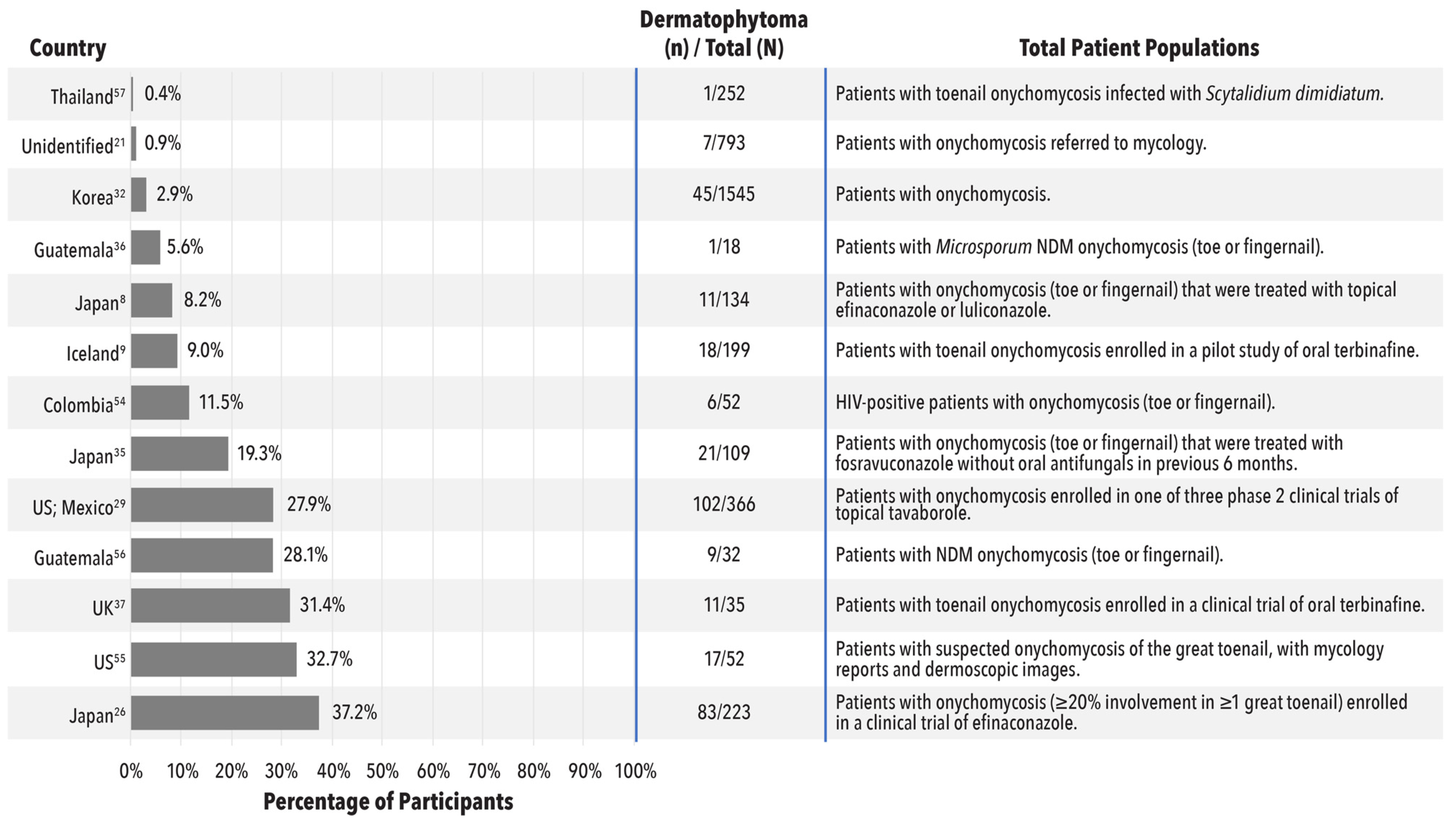
Of the 13 articles included in this review from which prevalence could be calculated, values ranged from 0.4% to 37.2% (Fig. 3). Interestingly, although patients with dermatophytomas are often excluded from clinical trials, of the three clinical trials included in this scoping review, dermatophytoma prevalence rates were among the highest, ranging from 27.9% to 37.2%. It should be noted, however, that the patient populations from which dermatophytoma prevalence was calculated differed greatly between the publications. For example, total populations ranged from 18 to 1,545, with the larger patient populations comprising individuals from clinical trials, individuals from hospital clinics, individuals with confirmed onychomycosis, or individuals referred to mycology. Some of the patient groups also had specific disease characteristics, such as concomitant human immunodeficiency virus (HIV) infection or onychomycosis caused by NDMs or Microsporum. In addition, the years in which articles were published varied; although most were between 2010 and 2021, one article was published over 20 years ago in 2003. Finally, the observed variability could be attributable to the lack of consensus on the definition or description of a dermatophytoma. Together, these issues could have contributed to the varied estimates of prevalence, making comparisons difficult.
Figure 3. Dermatophytoma prevalence estimates (13 reports).[8,9,21,26,29,32,35,36,37,54,55,56,57] HIV, human immunodeficiency virus; KOH, potassium hydroxide; NDM, nondermatophyte mold.
Japma 114 22161 g003

Diagnosis

None of the guidelines on onychomycosis (three reports) provided information on the diagnosis of dermatophytomas.[28,30,31] The two commentaries provided a description of the clinical features,[5,6] and one retrospective study provided a detailed summary of the clinical presentation of 45 patients with dermatophytomas.[32]
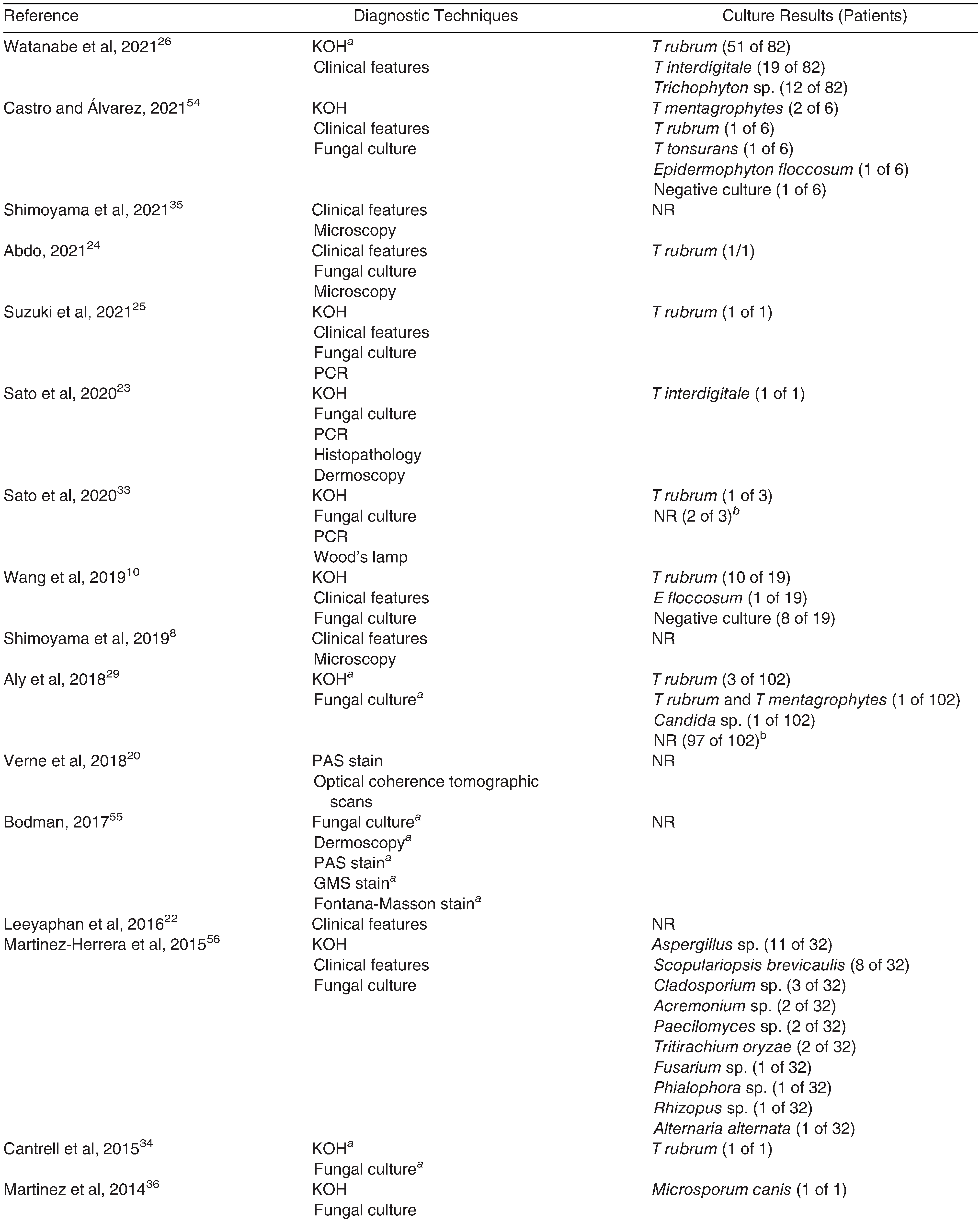
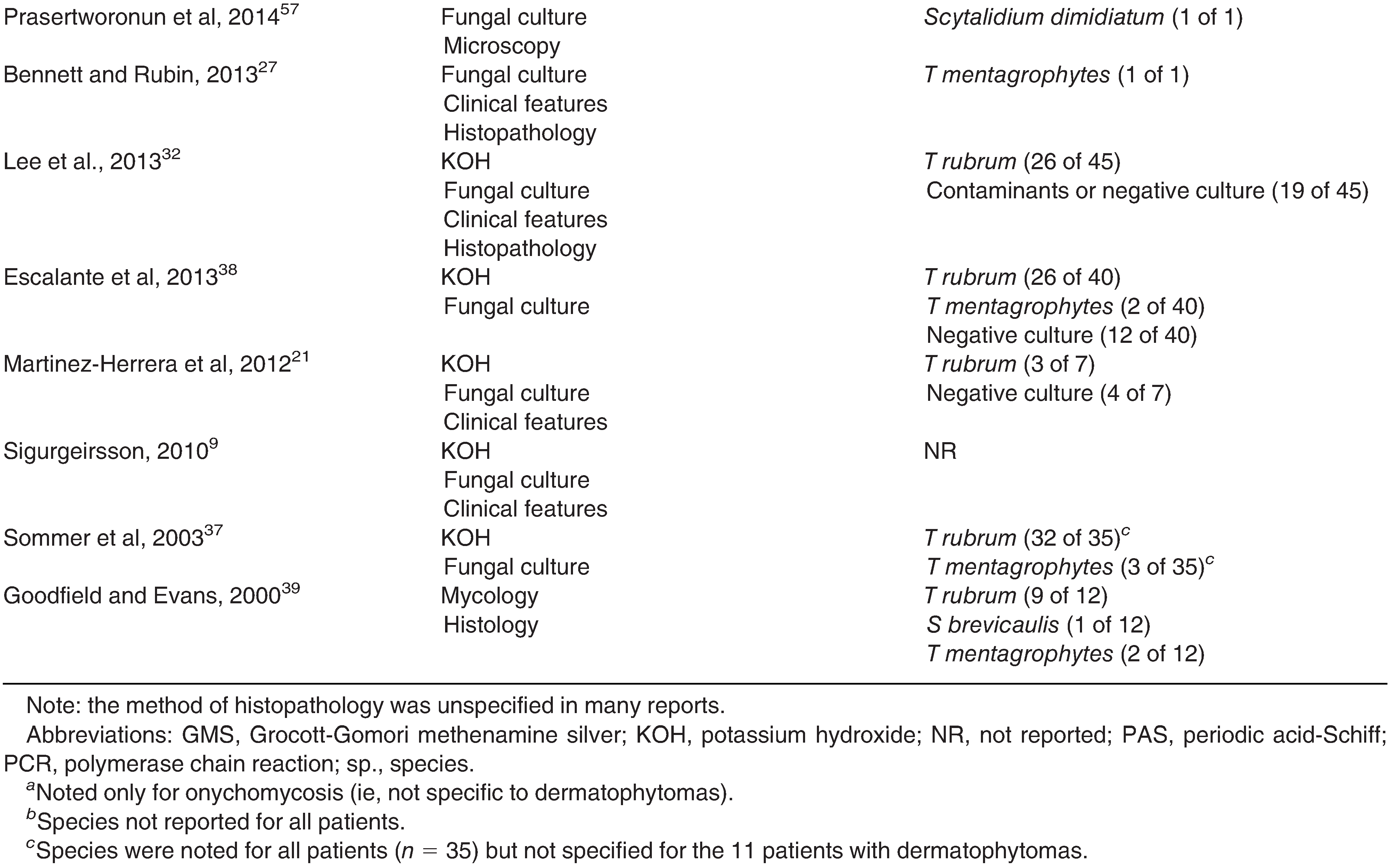
Of the 24 reports that noted any tools for diagnosis of onychomycosis and/or dermatophytomas (Table 2), more than three-quarters of studies used at least two diagnostic techniques. The most frequently mentioned techniques were fungal culture/ mycology (n = 19), potassium hydroxide (KOH; n = 15), and assessment of clinical features (n = 13). Several studies used other techniques alone or in combination with the above-mentioned techniques: histopathology (method not identified)/periodic acid-Schiff stain/Grocott-Gomori methenamine silver stain/Fontana-Masson stain (n = 5), microscopy (method not identified) (n = 4), polymerase chain reaction (PCR) test (n = 3), dermoscopy (n = 2), optical coherence tomographic scans (n = 1), or Wood’s lamp (n = 1). The most frequently combined techniques, described in 14 publications, were potassium hydroxide plus fungal culture. Of these, half of the publications also reported assessing clinical features (with or without additional measures, such as PCR and histopathology). The remaining seven publications used KOH plus fungal culture alone (n = 5) or in combination with dermoscopy, histopathology, Wood’s lamp, and/or PCR (n = 2).
In a survey on dermatophytoma diagnosis and management conducted in Thailand[22] in which 82 physicians were shown a “typical” photograph of a dermatophytoma, 92% were able to diagnose the patient as having onychomycosis, but only 2.4% suggested adding debridement to treatment—a practice the authors propose was needed for dermatophytoma cure.
A total of 18 articles reported specific culture results for 247 people (Table 2). Of these, the fungal genus noted most frequently was Trichophyton, which was present in 85% of individuals (209 of 247). T rubrum was the most common species overall, found in 67% of all cases (166 of 247); the next most common species were T interdigitale (8%; 20 of 247) and T mentagrophytes (4%; 10 of 247). Nondermatophyte molds constituted 14% of all cases (35 of 247).
Table 2. Diagnostic Techniques (24 Reports) and Culture Results (18 Reports) 
Japma 114 22161 i004
Japma 114 22161 i005

Treatments

Three reports were guidelines regarding the treatment of onychomycosis.[28,30,31] Of these, two had recommendations specifically for the treatment of dermatophytomas, which included mechanical/chemical nail avulsion combined with topical/oral treatment[16] or abrasion or surgical/chemical nail removal combined with topical treatment.[28] It should be noted, however, that the recommendations for nail removal with oral or topical treatments were published before 2010— before the approval of newer topical formulations such as efinaconazole and tavaborole.
There were 19 other reports that described a pharmaceutical treatment of dermatophytomas (Table 1). The majority were case reports (n = 7),[21,23,24,25,27,33,34] and the remaining were retrospective studies (n = 5),[8,32,35,36,37] open label/prospective studies (n = 4),[9,10,38,39] post hoc analyses (n = 2),[26,29] or in vitro studies (n = 1).[40] Treatments are listed in Table 1. Doses or dosing regimens were not always provided.
Oral treatments were noted in nine reports.[9,25,32,34,35,36,37,38,39] Terbinafine was used most often (n = 6 reports) followed by itraconazole, fluconazole, and fosravuconazole (n = 2 each). Topical treatments were noted in 11 reports.[8,10,21,23,24,26,27,29,32,33,34] The majority reported the use of efinaconazole 10% (n = 4) or luliconazole 5% (n = 3); the remaining were unidentified topical treatments (n = 2), tavaborole 5%, ciclopirox olamine 1% (n = 1), or bifonazole 1% (n = 1). In five reports, oral or topical treatment was combined with nail avulsion/excision using either surgical techniques (n = 2)[32,39] or topical urea 40% cream (n = 3)[21,27,38]; a topical urea was also given as monotherapy in one of these reports.[38] Debridement or nail clipping were used in four reports[23,24,32,33,34] in combination with topical or oral treatments. One report noted the use of photodynamic therapy in vitro to determine biofilm variability and drug susceptibility of various onychomycosis-causing fungal strains.[40]

Treatment Efficacy

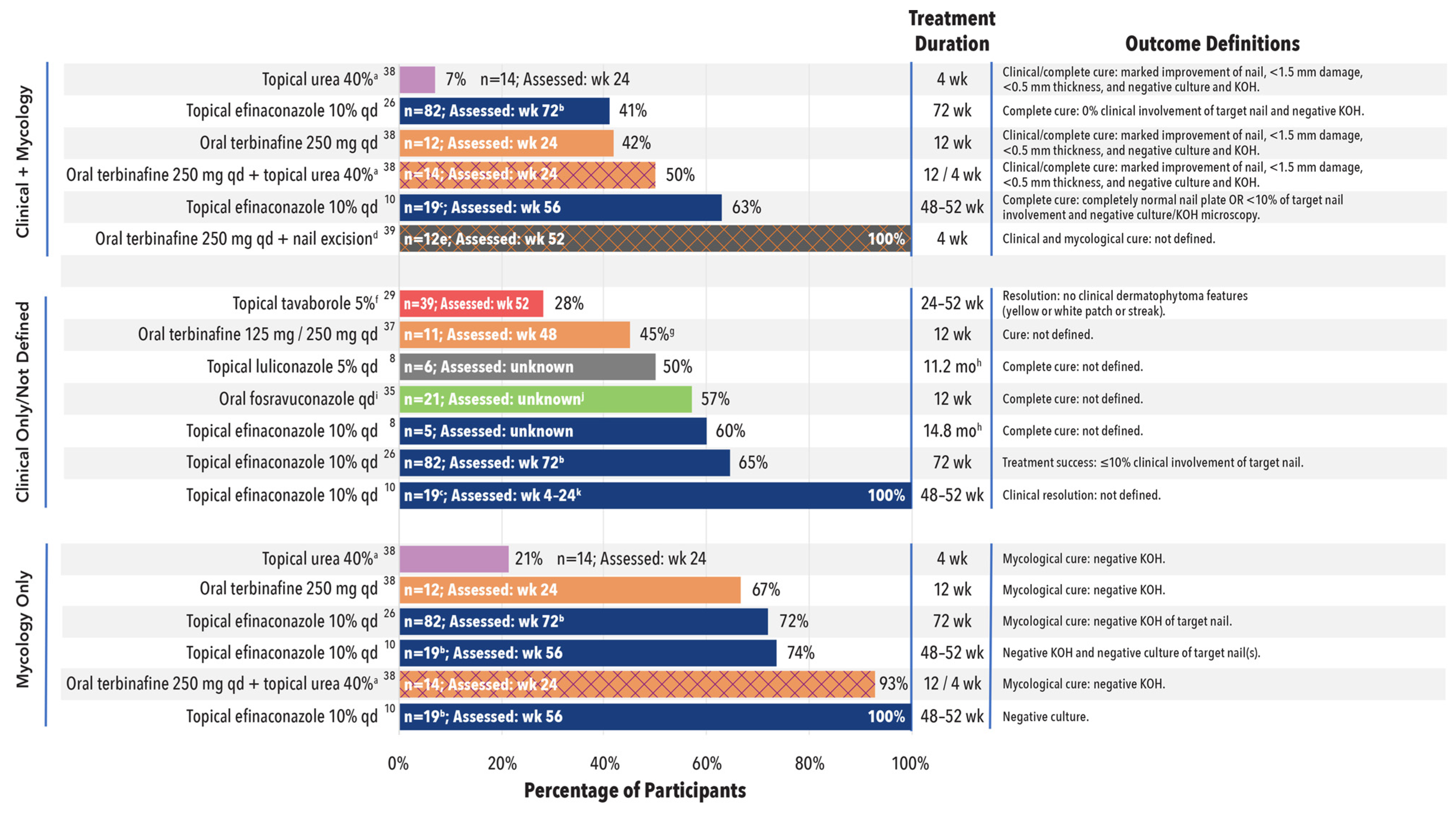
Although clinical/complete/mycologic cure, improvement, resolution, and/or recurrence were frequently reported as efficacy outcomes, the definitions varied greatly across publications. Furthermore, data for patients with dermatophytoma were not always provided for every measure. Only eight reports on dermatophytoma treatments in five or more participants included one or more measures of treatment efficacy and are summarized here and in Figure 4.[8,10,26,29,35,37,38,39] Across the studies, treatment durations were 4 weeks for urea 40%, 12 weeks for oral treatments, and approximately 24 to 72 weeks for topical treatments, with assessments ranging from 4 to 72 weeks.
Figure 4. Efficacy results of dermatophytoma treatments (eight reports).[8,10,26,29,35,37,38,39] aUrea cream was applied with occlusion for 4 weeks. bFinal evaluation, week 72, or date of completion of administration. cOne participant had two target nails. dNail excision performed by means of hot cautery (seven had partial nail removal and five had full nail removal). One participant had two nails involved. Publication did not explicitly detail that all 12 participants had dermatophytoma, though it was stated that “Histological examination revealed the appearance of large amounts of fungal material, much of it demonstrating thick-walled hyphae ... consistent with the recent description of a dermatophytoma.” eSix participants had failed a prior treatment regimen of oral itraconazole or terbinafine (no dosing or treatment time provided) and six participants were previously untreated. fData combined from three phase II studies with various doses and dosing regimens of tavaborole; only data for tavaborole 5% are shown here. gParticipants achieving cure: 125 mg, n = 1; 250 mg, n = 4. hTreatment duration was provided as a mean. iDaily dose was equivalent to 100 mg oral ravuconazole. jMean time to complete cure was 34.3 weeks. kResolution was achieved between weeks 4 and 24 for all participants; mean and median times to resolution were 16 weeks. KOH, potassium hydroxide; qd, once per day.
Japma 114 22161 g004
Among studies in which outcomes were defined using both clinical and mycologic results (ie, more stringent definitions), the highest cure rate was reported in one study of oral terbinafine plus full/ partial nail excision (100%),[39] followed by topical efinaconazole 10% (63%),[10] a study of oral terbinafine with and without chemical nail excision with topical urea 40% (50% and 42%, respectively),[38] and another study with efinaconazole 10% (41%).[26] Topical urea alone had the lowest efficacy, with only 7% of participants achieving a clinical cure.[38] When outcomes were defined using only clinical results (or were not clearly defined), the highest rates were observed in three studies of topical efinaconazole 10% (range, 60%–100%)[8,10,26] versus one study each of oral fosravuconazole,[35] topical luliconazole 5%,[8] oral terbinafine,[29] and topical tavaborole 5%[37] (range, 28%–57%). None of these studies included nail excision or clipping.
In studies where outcomes were defined by means of mycologic results, higher rates of negative KOH and/or negative culture were seen in two studies (one with two different mycologic assessments) of efinaconazole (72%, 74%, and 100%)[10,26] and oral terbinafine with nail excision using topical urea 40% (93%)[38] than in one study of oral terbinafine (67%) or topical urea (21%) alone.[38]
Recurrence rates, which were reported in two studies, were 0% (zero of 19) for efinaconazole by study end at week 56,[10] and 31.6% (six of 19) for tavaborole during a 180-day washout (after 180 days of treatment).[29] In addition, one publication (not included in Fig. 4) noted that there was no recurrence in 15 patients with 1-year follow up data who were treated with oral itraconazole or terbinafine, a topical lacquer, and partial nail removal/surgical clipping.[32] The follow-up periods used in these studies, however, may not have been long enough to fully determine recurrence rates.
Overall, clinical plus mycologic, clinical/undefined, and mycologic cure rates greater than 50% for dermatophytomas were observed with topical efinaconazole 10%, oral fosravuconazole, and oral terbinafine with or without nail excision; cure rates were higher for oral terbinafine when combined with chemical or surgical nail excision.

Discussion

The term “dermatophytoma” was defined by Roberts and Evans[5] in 1998, with an expanded definition in 2002 by Burkhart et al.[6] Nearly a quarter of a century after its first definition, however, this scoping review found only 37 nonduplicate, English-language reports that mentioned dermatophytomas in the title, abstract, or keywords. Although definitions, prevalence, diagnosis, and treatment of dermatophytomas have been included in these publications, there is an overall dearth of studies and descriptions of dermatophytomas. This may make it challenging for physicians to have a clear understanding of this condition and how to properly diagnose and treat it.
In the original reports, dermatophytomas were defined by their clinical features of dense white linear or round areas with pathologic features of densely packed hyphae/biofilms.[5,6] Terms frequently mentioned in other publications not included in the first reports included yellow; fungal ball; and longitudinal spike, band, streak, or striae. Although there are many descriptions of dermatophytomas provided, one conclusive definition is still lacking.
Epidemiologic data for dermatophytomas, including prevalence, are also limited and inconsistent. Of the publications included in this scoping review, prevalence ranged from 0.4% to 37.2%. This wide variation may be attributable in part to the different patient populations included in the studies, making comparisons difficult. For example, populations in these prevalence estimates included patients referred to mycology; patients enrolled in clinical studies; individuals with onychomycosis caused by NDMs or Microsporum; HIV-positive patients; and patients from countries all over the Americas, Asia, and Europe. The large range and lack of data may also be the result of underdiagnosis of dermatophytomas by physicians because of insufficient knowledge of clinical features or use of microscopy.[21,22]
In terms of diagnosis of dermatophytomas/onychomycosis, the most frequently mentioned techniques were fungal culture/mycology, KOH, and assessment of clinical features. Caution must be taken when assessing clinical features, however, as a small survey published in 2016 showed that most physicians were able to correctly diagnose the features of onychomycosis from a photograph of a dermatophytoma but not the dermatophytoma itself.[22] Improved education on the definition and clinical features of dermatophytomas may improve the ability of physicians to properly diagnose this condition. In addition, the use of diagnostic tests (KOH/fungal culture) along with clinical features is recommended to improve the accuracy of diagnosis.[41]
When examining the treatments used for dermato-phytomas, oral terbinafine and topical efinaconazole 10% were mentioned most frequently, followed by topical luliconazole 5% and the oral agents itraconazole, fluconazole, and fosravuconazole. Surgical or chemical nail avulsion/excision or debridement were also mentioned, typically in combination with either an oral or topical treatment. An additional six onychomycosis treatment guidelines or recommendations that were not found in our main search contained at least one mention of dermatophytoma.[41,42,43,44,45,46] Five of these reports generally noted that dermatophytoma may be difficult to treat and/or may require adjunctive treatments.[41,43,44,45,46] Three did not indicate a specific treatment[42,44,46]; of those that did, two reports recommended nail avulsion/excision,[43,45] and one recommended topical efinaconazole 10%.[41] An additional case report on a patient with dermatophytoma not found in the original search noted nail debridement in combination with oral terbinafine and an unidentified topical agent.[47]
Although the older treatment guidelines retrieved in the main search recommended partial nail removal or debridement combined with an oral or topical treatment,[28,30] more recent publications have indicated that some topical and oral treatments are effective against dermatophytomas without nail excision (Fig. 4). This may render the more invasive nail excision unnecessary. For example, in the 2021 therapeutic recommendations,[41] topical efinaconazole 10% was recommended as a first-line medication for dermatophytoma treatment. Although avulsion and debridement were not mentioned specifically for dermatophytoma, the consensus was that avulsion was generally not recommended, although debridement could be an adjunctive treatment with an oral or topical treatment.[41]
When examining the efficacy of treatments used for dermatophytomas, clinical/complete/mycologic cure, improvement, resolution, and/or recurrence were reported as outcomes, but the definitions of these endpoints varied greatly. In addition, the data for patients with dermatophytoma were not always provided for every outcome. This may be partly a result of dermatophytomas being an incidental finding or exclusionary criterion in onychomycosis studies. The studies with the greatest number of patients with dermatophytomas were those that examined the topicals tavaborole 5% (n = 102)[29] and efinaconazole 10% (n = 83).[26]
In studies with five or more patients that provided at least one efficacy outcome, the highest cure rates for dermatophytomas—using more stringently defined outcomes (ie, clinical plus mycologic cure)—were observed with oral terbinafine plus full or partial nail excision and topical efinaconazole 10%. For those outcomes that were not defined or only included clinical or mycologic cure, efinaconazole 10%, oral terbinafine with nail excision, and oral fosravuconazole had the highest rates. Across all efficacy assessments, cure rates were highest with efinaconazole 10%, which ranged from 41% to 100% with 48 to 72 weeks of treatment. Other drugs with greater than or equal to 50% cure rates were topical luliconazole 5% (50%), oral fosravuconazole (57%), and oral terbinafine (67%). Oral terbinafine cure rates were higher when combined with chemical or physical nail excision (50%–100%) than when given as monotherapy (42%–67%). Avulsion/excision, however, is no longer generally recommended for onychomycosis treatment.[41]
Historically, dermatophytomas have been considered recalcitrant to oral treatments.[5,6] Although some oral treatments were observed to be efficacious in treating dermatophytomas in the present review, a study on predictors of treatment failure in onychomycosis showed that patients with dermatophytoma were less likely to achieve mycologic cure after 72 weeks of oral terbinafine.[9] This was also demonstrated in a case study of a patient with a dermatophytoma who had failed two different oral treatments (terbinafine and fluconazole) and later achieved complete resolution with topical efinaconazole 10% and nail clipping.[34] Finally, there are safety concerns with the use of oral agents such as drug-drug interactions and liver toxicity.[48]
In the present review, the topical treatment efinaconazole 10% showed higher clinical and/or mycologic cure rates than the oral terbinafine alone (ie, without nail excision). In addition, topical efinaconazole has fewer safety concerns than oral treatments.[49] When treating onychomycosis, the nail can present a substantial barrier, and achieving penetration and therapeutic drug levels can be difficult.[48] Biofilms may also account for the historically poor treatment response in dermatophytomas.[6] However, non–lacquer-based topicals such as efinaconazole have been observed to have good penetration into and under the nail plate to reach and spread through the subungual space.[50,51,52] In an in vitro study, efinaconazole also demonstrated better activity against biofilms than oral terbinafine or fluconazole.[53] These results, in addition to its positive safety profile, may have led to the recent recommendation that topical efinaconazole 10% should be used for dermatophytoma treatment.[41] Overall, although dermatophytomas have historically been considered to have a poor treatment response, results shown in this review demonstrate successful treatment is possible with topical or oral treatments.
This scoping review had limitations in the search and subsequent analysis of publications. Articles not indexed in Embase or PubMed, including articles in online publications such as Podiatry Today, would not have been identified and included in the review. Furthermore, publications that may have discussed dermatophytomas without including the term in the title, abstract, or keywords would not have appeared in our search. The populations evaluated in the studies also varied greatly, making prevalence estimates and comparisons difficult. For example, several studies were limited to very specific patient populations (ie, HIV-positive patients, onychomycosis caused by NDMs or Microsporum) or were case studies of only one patient. Differences in study designs and a lack of standard terms and definitions for the commonly used measures of treatment efficacy (eg, mycologic, complete, or clinical cure) further limited the accuracy of direct treatment comparisons. For these reasons, specific details on patient populations (Fig. 3) and outcome definitions (Fig. 4), when provided, were included within this scoping review.
Although there are limited publications on dermatophytoma patients, this scoping review provides the first research synthesis on this topic, with the goal of providing information on both what is known and gaps in our knowledge/research. The results of this scoping review demonstrated that a standardized description, and physician education, may be required to allow for better diagnosis and management of dermatophytomas. The most effective US Food and Drug Administration–approved treatments for dermatophytomas without nail excision appear to be topical efinaconazole followed by oral terbinafine in combination with chemical or surgical methods; this mirrors the onychomycosis treatment recommendations published in 2021 noting efinaconazole as an ideal first-line topical treatment for patients with dermatophytomas, with avoidance of nail avulsion/excision whenever possible.[41] However, more studies with larger numbers of patients are needed to determine the best treatments for dermatophytomas. The inclusion of patients with dermatophytomas in clinical trials of onychomycosis treatments would help provide additional information. Finally, standardized efficacy outcomes in clinical trials of onychomycosis and dermatophytoma treatments would allow for more accurate treatment comparisons.

Conclusions

There is little published information regarding the form of onychomycosis known as dermatophytoma or longitudinal streak. More clinical research and physician education is needed to properly understand, diagnose, and treat dermatophytomas. Although dermatophytomas have historically been considered difficult to treat, the efficacy data gathered in this scoping review show that newer topicals are an effective treatment, such that nail excision/avulsion can be avoided whenever possible.

Acknowledgements

Manuscript preparation support was provided by Lynn M. Anderson, PhD and Jacqueline Benjamin, PhD from Prescott Medical Communications Group, a Citrus Health Group, Inc. company (Chicago, IL) with financial support from Ortho Dermatologics. Ortho Dermatologics is a division of Bausch Health US, LLC.

Financial Disclosures

This study was funded by Ortho Dermatologics, a division of Bausch Health US, LLC.

Conflict of Interest

Shari R. Lipner has served as a consultant for Ortho Dermatologics, Hoth Therapeutics, Hexima, BelleTorus Corporation, and Verrica. Tracey C. Vlahovic has served as investigator and speaker for Ortho Dermatologics. Mahmoud Ghannoum has acted as a consultant or received contracts from Scynexis, Inc, Bausch & Lomb, Pfizer, and Mycovia. Boni Elewski has provided clinical research support (research funding to University) for AbbVie, Anaptys-Bio, Boehringer Ingelheim, Bristol-Myers Squibb, Celgene, Incyte, LEO Pharma, Lilly, Merck, Menlo, Novartis, Pfizer, Regeneron, Sun Pharma, Ortho Dermatologics, Vanda; and as consultant (received honorarium) from Boehringer Ingelheim, Bristol Meyers Squibb, Celgene, LEO Pharma, Lilly, Menlo, Novartis, Pfizer, Sun Pharma, Ortho Dermatologics, Verrica. Warren Joseph has served as consultant and speaker for Ortho Dermatologics.

Note

Dr. Joseph was not involved in the review or acceptance of this article.

References

  1. GHANNOUM, MA; HAJJEH, RA; SCHER, R; ET, AL. A large-scale North American study of fungal isolates from nails: the frequency of onychomycosis, fungal distribution, and antifungal susceptibility patterns. J Am Acad Dermatol 2000, 43, 641. [Google Scholar] [CrossRef] [PubMed]
  2. GUPTA, AK. STEC N: Recent advances in therapies for onychomycosis and its management. F1000Res 2019, 8, F1000 Faculty Rev–968. [Google Scholar] [CrossRef] [PubMed]
  3. GUPTA, AK; JAIN, HC; LYNDE, CW. ET AL: Prevalence and epidemiology of onychomycosis in patients visiting physicians’ offices: a multicenter Canadian survey of 15,000 patients. J Am Acad Dermatol 2000, 43, 244. [Google Scholar] [CrossRef]
  4. GUPCHUP, GV; ZATZ, J. Structural characteristics and permeability properties of the human nail: a review. J Cosmet Sci 1999, 50, 363. [Google Scholar]
  5. ROBERTS, DT; EVANS, EG. Subungual dermatophytoma complicating dermatophyte onychomycosis. Br J Dermatol 1998, 138, 189. [Google Scholar] [CrossRef] [PubMed]
  6. BURKHART, CN; BURKHART, CG; GUPTA, AK. Dermatophytoma: recalcitrance to treatment because of existence of fungal biofilm. J Am Acad Dermatol 2002, 47, 629. [Google Scholar] [CrossRef]
  7. CARNEY, C; TOSTI, A; DANIEL, R; et al. A new classification system for grading the severity of onychomycosis: onychomycosis severity index. Arch Dermatol 2011, 147, 1277. [Google Scholar] [CrossRef]
  8. SHIMOYAMA, H; KUWANO, Y; SEI, Y. Retrospective survey of treatment outcomes of efinaconazole 10% solution and luliconazole 5% solution for onychomycosis in our facility. Med Mycol J 2019, 60, 95. [Google Scholar] [CrossRef]
  9. SIGURGEIRSSON B: Prognostic factors for cure following treatment of onychomycosis. J Eur Acad Dermatol Venereol 2010, 24, 679. [CrossRef]
  10. WANG, C; CANTRELL, W; CANAVAN, T; et al. Successful treatment of dermatophytomas in 19 patients using efinaconazole 10% solution. Skin Appendage Disord 2019, 5, 304. [Google Scholar] [CrossRef]
  11. PETERS, MDJ; MARNIE, C; TRICCO, AC. ET AL: Updated methodological guidance for the conduct of scoping reviews. JBI Evid Synth 2021, 19, 3. [Google Scholar]
  12. GUPTA AK: Onychomycosis in the elderly. Drugs Aging 2000, 16, 397. [CrossRef]
  13. GUPTA, AK; BARAN, R. Ciclopirox nail lacquer solution 8% in the 21st century. J Am Acad Dermatol 2000, 43, S96. [Google Scholar] [CrossRef]
  14. GUPTA, AK. Ciclopirox nail lacquer: a brush with onychomycosis. Cutis 2001, 68, 13. [Google Scholar]
  15. GUPTA, AK; BARAN, R; SUMMERBELL, R. Onychomycosis: strategies to improve efficacy and reduce recurrence. J Eur Acad Dermatol Venereol 2002, 16, 579. [Google Scholar] [CrossRef] [PubMed]
  16. GUPTA, AK; BUCKLEY, L. The limitations in the treatment of onychomycosis. J Am Acad Dermatol 2009, 60, AB117. [Google Scholar]
  17. SHEMER, A; DANIEL, R; RIGOPOULOS, D; et al. Variability in systemic treatment efficacy for onychomycosis: information that clinical studies do not impart to the office dermatologist. Skin Appendage Disord 2018, 4, 141. [Google Scholar] [CrossRef] [PubMed]
  18. COSTA-ORLANDI, CB; SARDI, JC; SANTOS, CT. ET AL: In vitro characterization of Trichophyton rubrum and T. mentagrophytes biofilms. Biofouling 2014, 30, 719. [Google Scholar] [CrossRef] [PubMed]
  19. GUPTA, AK; DAIGLE, D. CARVIEL JL: The role of biofilms in onychomycosis. J Am Acad Dermatol 2016, 74, 1241. [Google Scholar] [CrossRef]
  20. VERNE, SH; CHEN, L; SHAH, V. ET AL: Optical coherence tomography features of dermatophytoma. JAMA Dermatol 2018, 154, 225. [Google Scholar] [CrossRef]
  21. Martinez-Herrera E, Moreno-Coutin˜ o G, Ferna´ ndez-MARTI´NEZ RF, ET AL: Dermatophytoma: description of 7 cases. J Am Acad Dermatol 2012, 66, 1014. [CrossRef]
  22. LEEYAPHAN C, BUNYARATAVEJ S, PRASERTWORANUN N, ET AL: Dermatophytoma: an under-recognized condition. Indian J Dermatol Venereol Leprol 2016, 82, 188.
  23. SATO, T; HORIKAWA, H; YAMAZAKI, K. Mold mite infestation in toenail dermatophytoma with pustular psoriasis. J Dtsch Dermatol Ges 2020, 18, 251. [Google Scholar] [CrossRef]
  24. ABDO, HM. Excellent response of dermatophytoma and nail splitting to nail plate debridement plus topical ciclopirox olamine 1% solution. Skin Appendage Disord 2021, 7, 127. [Google Scholar] [CrossRef] [PubMed]
  25. SUZUKI, T; SATO, T; KASUYA, A. ET AL: A case of tinea faciei, tinea corporis, and tinea unguium with dermatophytoma successfully treated with oral fosravuconazole L-lysine ethanolate. Med Mycol J 2021, 62, 1. [Google Scholar] [CrossRef] [PubMed]
  26. WATANABE, S; IOZUMI, K; ABE, M; ET, AL. Clinical effectiveness of efinaconazole 10% solution for treatment of onychomycosis with longitudinal spikes. J Dermatol 2021, 48, 1474. [Google Scholar] [CrossRef]
  27. BENNETT, D; RUBIN, AI. Dermatophytoma: a clinicopathologic entity important for dermatologists and dermatopathologists to identify. Int J Dermatol 2013, 52, 1285. [Google Scholar] [CrossRef]
  28. Lecha M, Effendy I, Feuilhade de Chauvin M, et al: Treatment options—development of consensus guidelines. J Eur Acad Dermatol Venereol 2005, 19 (suppl 1), 25. [CrossRef]
  29. ALY, R; WINTER, T; HALL, S; ET, AL. Topical tavaborole in the treatment of onychomycosis complicated by dermatophytoma: a post-hoc assessment of phase II subjects. J Drugs Dermatol 2018, 17, 347. [Google Scholar]
  30. GUPTA, AK; PAQUET, M. Management of onychomycosis in North America in 2014. J Am Acad Dermatol 2014, 70, AB91. [Google Scholar]
  31. PARK, J; NAM, JH; LEE, JH. ET AL: Korean guideline for the diagnosis and treatment of onychomycosis: a consensus update by The Korean Society for Medical Mycology. Med Mycol 2018, 56, S95. [Google Scholar]
  32. LEE, MH; SUH, MK; Ha, GY. A clinico-mycological study of onychomycosis with dermatophytoma. Korean J Med Mycol 2013, 18, 21. [Google Scholar] [CrossRef]
  33. SATO, T; ASAHINA, Y; TOSHIMA, S. ET AL: Usefulness of Wood’s lamp for the diagnosis and treatment follow-up of onychomycosis. Med Mycol J 2020, 61, 17. [Google Scholar] [CrossRef]
  34. CANTRELL, W; CANAVAN, T; ELEWSKI, B. Report of a case of a dermatophytoma successfully treated with topical efinaconazole 10% solution. J Drugs Dermatol 2015, 14, 524. [Google Scholar]
  35. SHIMOYAMA, H; YO, A; SEI, Y. ET AL: Treatment outcome with fosravuconazole for onychomycosis. Mycopathologia 2021, 186, 259. [Google Scholar] [CrossRef]
  36. MARTI´NEZ, E; AMEEN, M; TEJADA, D; et al. Microsporum spp. onychomycosis: disease presentation, risk factors and treatment responses in an urban population. Braz J Infect Dis 2014, 18, 181. [Google Scholar] [CrossRef] [PubMed]
  37. SOMMER S, SHEEHAN-DARE RA, GOODFIELD MJ, ET AL: Prediction of outcome in the treatment of onychomycosis. Clin Exp Dermatol 2003, 28, 425.
  38. Escalante K, Marti´nez E, Torres-Guerrero E, et al: Onychomycosis with dermatophytoma. A comparison among the results of treatments with oral terbinafine, topical 40% urea in monotherapy and combination therapy. Dermatol Klin 2013, 15, 67.
  39. GOODFIELD, MJD. EVANS EGV: Combined treatment with surgery and short duration oral antifungal therapy in patients with limited dermatophyte toenail infection. J Dermatol Treatment 2000, 11, 259. [Google Scholar] [CrossRef]
  40. CHEN, B; SUN, Y; ZHANG, J; ET, AL. In vitro evaluation of photodynamic effects against biofilms of dermatophytes involved in onychomycosis. Front Microbiol 2019, 10, 1228. [Google Scholar] [CrossRef]
  41. LIPNER, SR; JOSEPH, WS; VLAHOVIC, TC. ET AL: Therapeutic recommendations for the treatment of toenail onychomycosis in the US. J Drugs Dermatol 2021, 20, 1076. [Google Scholar]
  42. GUPTA, AK; SIBBALD, RG; ANDRIESSEN, A. ET AL: Toenail onychomycosis—a Canadian approach with a new transungual treatment: development of a clinical pathway. J Cutan Med Surg 2015, 19, 440. [Google Scholar] [CrossRef] [PubMed]
  43. AMEEN, M; LEAR, JT; MADAN, V; et al. British Association of Dermatologists’ guidelines for the management of onychomycosis 2014. Br J Dermatol 2014, 171, 937. [Google Scholar] [CrossRef]
  44. SEEBACHER, C; BRASCH, J; ABECK, D. ET AL: Onychomycosis. Mycoses 2007, 50, 321. [Google Scholar] [CrossRef]
  45. ROBERTS, DT; TAYLOR, WD; BOYLE, J. Guidelines for treatment of onychomycosis. Br J Dermatol 2003, 148, 402. [Google Scholar] [CrossRef]
  46. VLAHOVIC, TC; JOSEPH, WS; SCHER, RK; et al. Diagnosis and management of onychomycosis perspectives from a joint podiatric medicine-dermatology roundtable. JAPMA 2016, 106, 155. [Google Scholar] [CrossRef]
  47. VLAHOVIC T: When a patient presents with linear streaks in a nail. Podiatry Today 2015, 28, 12.
  48. DEL ROSSO, JQ. The role of topical antifungal therapy for onychomycosis and the emergence of newer agents. J Clin Aesthet Dermatol 2014, 7, 10. [Google Scholar] [PubMed]
  49. VLAHOVIC, TC. GUPTA AK: Efinaconazole topical solution (10%) for the treatment of onychomycosis in adult and pediatric patients. Expert Rev Anti Infect Ther 2022, 20, 3. [Google Scholar] [CrossRef]
  50. SAKAMOTO, M; SUGIMOTO, N; KAWABATA, H. ET AL: Transungual delivery of efinaconazole: its deposition in the nail of onychomycosis patients and in vitro fungicidal activity in human nails. J Drugs Dermatol 2014, 13, 1388. [Google Scholar]
  51. ZEICHNER, JA; GOLD, LS; KOROTZER, A. Penetration of (14C)-efinaconazole topical solution, 10%, does not appear to be influenced by nail polish. J Clin Aesthet Dermatol 2014, 7, 34. [Google Scholar] [PubMed]
  52. ELEWSKI, BE; POLLAK, RA; PILLAI, R. ET AL: Access of efinaconazole topical solution, 10%, to the infection site by spreading through the subungual space. J Drugs Dermatol 2014, 13, 1394. [Google Scholar]
  53. COSTA-ORLANDI, CB; MARTINEZ, LR; BILA, NM. ET AL: Nitric oxide-releasing nanoparticles are similar to efinaconazole in their capacity to eradicate trichophyton rubrum biofilms. Front Cell Infect Microbiol 2021, 11, 684150. [Google Scholar] [CrossRef]
  54. CASTRO LA´, LVAREZ MI. Nail dermatophytoma in HIV-infected patients in Cali, Colombia. J Mycol Med 2021, 31, 101172. [Google Scholar] [CrossRef]
  55. BODMAN MA: Point-of-care diagnosis of onychomycosis by dermoscopy. JAPMA 2017, 107, 413. [CrossRef] [PubMed]
  56. Marti´nez-Herrera EO, Arroyo-Camarena S, Tejada-Garci´a DL, ET AL: Onychomycosis due to opportunistic molds. An Bras Dermatol 2015, 90, 334. [CrossRef]
  57. PRASERTWORONUN N, MUANPRASART C, LEEYAPHAN C, ET AL: Clinical manifestations of outpatients infected with Scytalidium dimidiatum: a retrospective study at Siriraj Hospital, Thailand. J Am Acad Dermatol 2014, 70, AB108.