Plantar heel pain (PHP)/plantar fasciitis usually presents as a chronic condition that is considered to be degeneration in the proximal plantar aponeurosis with microtears due to repeated tension in the plantar fascia [
1,
2,
3]. The main complaint of people with chronic PHP (CPHP) is the initial step pain felt prominently in the medial side of the heel. Typically, pain subsides after some steps but may worsen after prolonged load-bearing activities [
3]. Chronic PHP has a remarkable negative effect on foot-specific and general health–related quality of life [
4]. In addition, kinesiophobia is associated with poorer foot function [
5].
Most patients with PHP respond to nonsurgical treatments with several modalities [
6,
7]. In the literature, there is a consensus that extracorporeal shockwave therapy (ESWT), manual therapy, and stretching exercises (SEs) are safe and effective [
8,
9], with level of evidence A [
3,
9].
Low-energy ESWT applications cause microtrauma in the damaged tissue, producing localized hyperemia through sound waves [
8]. According to a sytematic review by Sun et al [
10], ESWT might be related to improvements in soft-tissue healing, reduction of calcification, inhibition of pain receptors, or denervation to achieve pain relief and neovascularization. In the treatment of PHP, studies have indicated that ESWT is an effective [
3,
9,
10,
11] and reliable [
9,
12] treatment method. It has also been stated that there is a need for multicentered, well-designed, higher-quality randomized controlled studies with sufficient numbers of participants [
10].
As a manual therapy method, instrument-assisted soft-tissue mobilization (IASTM) treatment had been reported to be effective against pain and secondary disability due to pain in musculoskeletal diseases [
13]. Graston Technique (GT) applies longitudinal pressure along the tissue fibers through specially designed instruments, creating microtraumas in the damaged areas, scar tissue destruction, and adhesion loosening. In addition, collagen synthesis and connective tissue remodeling are stimulated [
13,
14,
15,
16]. Studies revealing the effectiveness of GT in CPHP are usually case studies with no control group, and evidence levels are low [
6,
17].
In CPHP management, several research studies compare ESWT, myofascial release, and other methods [
9,
18,
19,
20]. To our knowledge, there is no study comparing the effectiveness of ESWT and GT interventions. Hence, we aimed to compare the effectiveness of ESWT and GT interventions combined with SEs on pain, functional status, quality of life, and kinesiophobia in patients with CPHP.
Study Design
A prospective, double-blind, randomized controlled trial was performed at the Yeditepe University Orthopedics and Traumatology Clinic and the Bahcesehir University Faculty of Health Sciences Physiotherapy and Rehabilitation Laboratory (both in Istanbul, Turkey) between December 1, 2018, and March 31, 2020, in accordance with the Declaration of Helsinki. This study was approved by the Medipol University Non-Interventional Clinical Research Ethics Committee (Istanbul). All of the participants were informed about the interventions and the potential adverse effects and signed the informed consent form.
The participants who fulfilled the criteria were randomized to one of the three parallel groups. Group 1 received ESWT and SEs (n = 23) and group 2 was managed by GT and SEs (n = 23). A third group received SEs only and served as the control group (CG) (n = 23). The online randomization web service Research Randomizer (
https://www.randomizer.org/) was used for distribution of the groups. Simple randomization procedures (computerized random numbers) were conducted, and sequentially numbered index cards with the random assignment were prepared by an investigator (F.S.) with no clinical involvement in the study. The index cards were folded and placed in sealed, opaque envelopes. Then, the investigator opened each envelope and assigned the participants to their groups. The interventions were performed by the same physical therapists (GT by P.P. and ESWT by E.T.C.), and the outcome measures were administered by another therapist (D.K.C.). Because the interventionists were aware of the allocated arm, the outcome assessor was blinded to the allocation procedure.
Participants
The study included 87 feet of 69 patients with CPHP (mean age, 47 years; age range, 22–65 years; 28 women and 38 men). Inclusion criteria were age 18 to 60 years, symptomatic for at least 3 months, with a history of initial step pain (visual analog scale [VAS] score ≥5) and still symptomatic after at least three nonsurgical treatments (including nonsteroidal anti-inflammatory drug use and cortisone injection). Patients with a history of foot surgery, stress fracture, tarsal tunnel syndrome, infection, neurologic problems, tumorous conditions, coagulation disorder, and pregnancy were excluded.
Outcome Measures
A senior orthopedic surgeon (U.S.) evaluated all of the participants initially. A predetermined structured questionnaire (age, sex, height, weight, symptom duration, sociodemographic conditions, presence of chronic diseases, and previous foot surgery) was completed through face-to-face interviews by the same investigator (U.S.). Outcome measures were completed at the pretreatment, posttreatment (4-week), and 8-week follow-up visits (face to face), and 6-month follow-up assessment was performed by telephone interview by the same blinded assessor (D.K.C.). Only Foot Function Index (FFI) scores and initial step pain status were evaluated at 6-month follow-up. The primary outcome was VAS-initial step pain. Secondary outcomes were VAS-activity pain, FFI, 12-item Short-Form Health Survey (SF-12), and Tampa Scale for Kinesiophobia scores.
Visual Analog Scale
For initial step and activity pain, the patients marked on a scale from 0 (no pain) to 100 mm (most severe pain). The marked point was measured with a millimeter ruler [
21]. The minimal clinical significance was found to be 13 mm for chronic conditions [
22].
Foot Function Index
The FFI is a self-assessment scale consisting of 23 items measuring the impact of foot disorders in terms of pain, disability, and activity limitation. The higher the score, the worse the individual's condition. It has been shown that the Turkish version of the FFI, used in this study, is sensitive to changes, valid, and reliable [
23]. The following minimal important differences were found for FFI: 12 for pain, 7 for disability, and 7 for total FFI [
24].
12-Item Short-Form Health Survey
The questionnaire consists of 12 statements organized into various subscales, including physical and mental domains. The total score ranges from 0 to 100, with higher scores indicating better status [
25].
Tampa Scale for Kinesiophobia
Kinesiophobia was evaluated using the Tampa Scale for Kinesiophobia, a 17-item questionnaire that evaluates a person’s fear of movement or (re)injury. The total score ranges from 17 to 68, with higher scores indicating higher degrees of kinesiophobia. In 2011, the scale was translated into Turkish, and a test-retest reliability study was conducted by Yilmaz et al [
26].
Patient Education Program
The patient education program was provided to inform about CPHP (including symptoms and complications) and to give instructions (through E.T.C.) before randomization. The exercise programs were introduced as “hands-on” instruction (calf- and plantar fascia–specific SEs) initially by E.T.C [
27].
SE Protocol
The SEs for the plantar fascia and the gastrocnemius and soleus muscles were assigned to all of the groups as part of their home program twice a day (7 d/wk). For each region, SEs were performed with three repetitions, and intermittently and 30 seconds of holding time were preferred in their program.
Self-stretching plantar fascia exercises were performed by placing the affected leg crosswise on the contralateral leg in the seated position. Patients held the base of their toes with one hand and dorsiflexed the distal metatarsophalangeal joints until they felt tension in the arch of the foot [
27,
28,
29].
Self-stretching of the calf muscles was performed with the patients standing and leaning against the wall, with the affected leg behind. With the knee flexed, the soleus muscle was stretched; with the knee extended, the gastrocnemius muscle was stretched. During the exercises, participants were careful to keep the heel on the floor [
27,
30].
ESWT Protocol
With the patient in the prone position, the ankle was supported with a cylindrical pillow. Low-dose ESWT was applied to painful areas in the plantar fascia and tuber calcanei once a week for 4 weeks with the BTL-6000 shockwave therapy device (BTL Industries Ltd, Newcastle, United Kingdom), with an application dose of 10 Hz, 2.5 bar, 2,000 shockwave [
31].
GT Protocol
The technique was applied by a GT-certified therapist with 13 years of experience in orthopedic rehabilitation. The plantar fascia and the gastrocnemius and soleus muscles were treated with GT instruments twice a week for 4 weeks. The GT instruments and mobilization techniques to be used were determined according to the GT manual, and the applied protocol is demonstrated in
Table 1 [
32]. Before the intervention, superficial heat was applied for 10 min with a hot pack to the plantar fascia and the gastrocnemius and soleus muscles. After GT treatment was completed, participants performed SEs in the clinic to ensure collagen reorganization, as suggested by the protocol [
33].
Table 1.
Graston Technique Application Protocol
Table 1.
Graston Technique Application Protocol
Sample Size
Power analysis of the study was with 80% reliability, alpha level of 0.05, minimal clinical significance of 1.3 cm [
22], standard deviation of 0.5 as 21 patients per group and 63 patients in total by using G*Power 3.1.9.7 software (Universität Kiel, Kiel, Germany). Considering the 10% dropout rate, the number of patients required for recruitment was calculated to be 69.
Statistical Analysis
IBM SPSS Statistics for Windows, Version 20.0 (IBM Corp, Armonk, New York) was preferred. The Kolmogorov-Smirnov test was performed to assess the distribution of the data. Differences between groups in baseline characteristics (age, body mass index, and symptom duration) were assessed through independent-samples
t tests and χ
2 tests (sex). Changes in the mean (95% confidence interval) variable scores within the groups were assessed through paired-samples
t tests. Repeated-measures analysis of variance (rANOVA) was conducted with time (pretreatment, posttreatment, and follow-up) as a within-subject variable and group (group 1, group 2, or CG) as a between-subjects variable to analyze the effect of the interventions on the primary and secondary outcomes. The effect sizes (ESs) were determined, as suggested by Kazis et al[
34], by dividing the changes in mean baseline and follow-up scores by the baseline standard deviation. The ESs of 0.2, 0.5, and 0.8 were considered small, moderate, and large, respectively. The significance level was set at
P < .05. Once the differences between the mean scores were determined, the least significant difference (LSD) post hoc test was used with a Bonferroni correction. Depending on the repetition of the outcome measures, significance was accepted as
P = .05/3 =.016 or
P = .05/4 = .0125.
Results
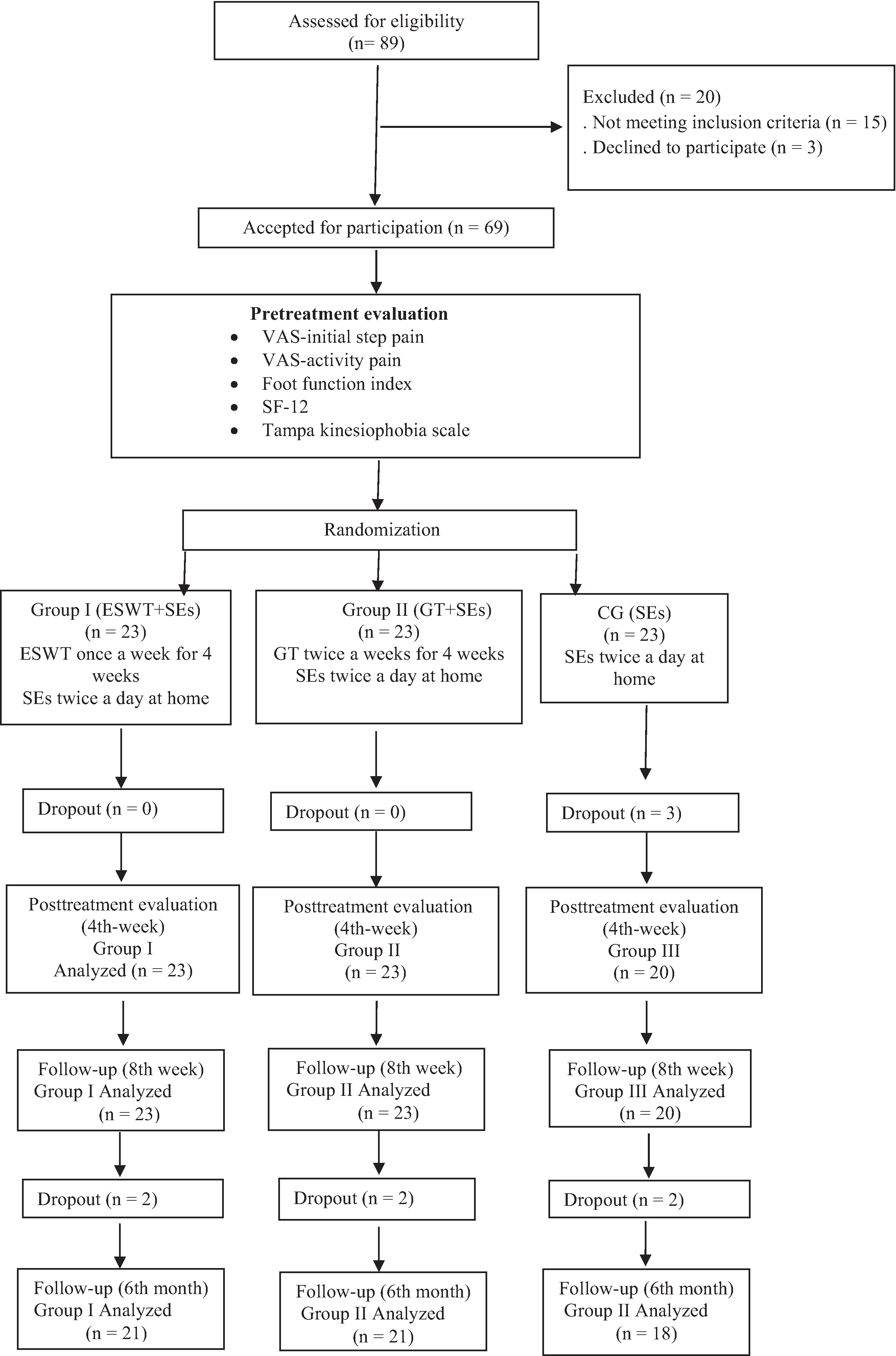
Twenty-three participants in group 1 (29 feet), 23 in group 2 (29 feet), and 20 in the CG (26 feet), a total of 66 participants (84 feet), completed the study; three CG participants dropped out. At baseline, all of the groups were homogenous in terms of demographics and symptom duration (
P > .05) (
Table 2). Twenty-one patients in group 1 (27 feet), 21 in group 2 (26 feet), and 18 in the CG (23 feet) participated in the 6-month follow-up interview. A flow diagram of the study is shown in
Figure 1.
Figure 1.
Flow diagram of the study. ESWT, extracorporal shockwave therapy; GT, Graston Technique; SE, stretching exercise; SF-12, 12-item Short-Form Health Survey; VAS, visual analog scale.
Figure 1.
Flow diagram of the study. ESWT, extracorporal shockwave therapy; GT, Graston Technique; SE, stretching exercise; SF-12, 12-item Short-Form Health Survey; VAS, visual analog scale.
Table 2.
Baseline Variables
Table 2.
Baseline Variables
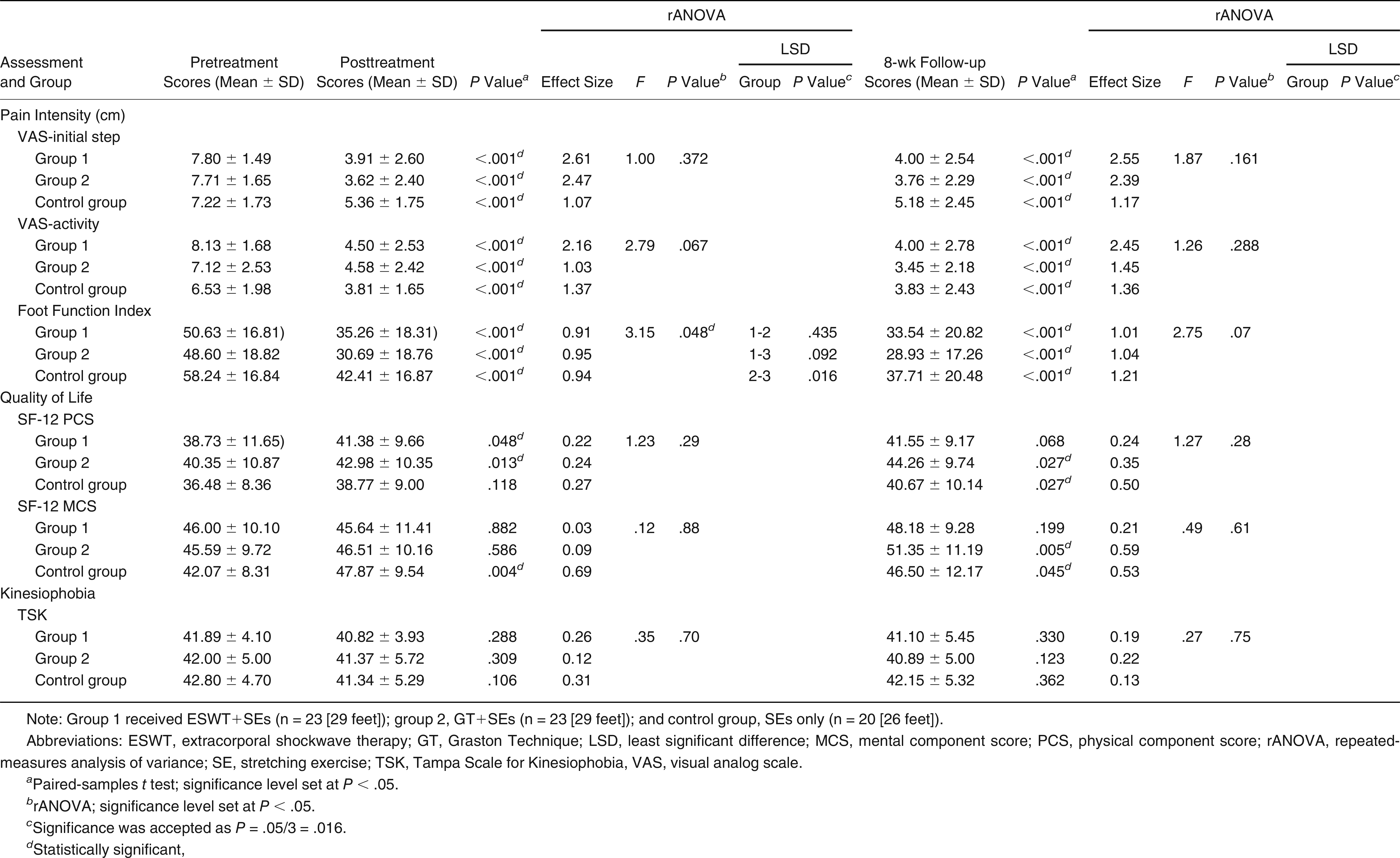
All of the groups demonstrated significant improvement in VAS-initial step pain, VAS-activity pain, and FFI scores from baseline to posttreatment (
P < .001 for all groups) and from baseline to 8-week (
P < .001 for all groups) (
Table 3) and 6-month (
P < .001 for all groups) follow-up (
Table 4 and
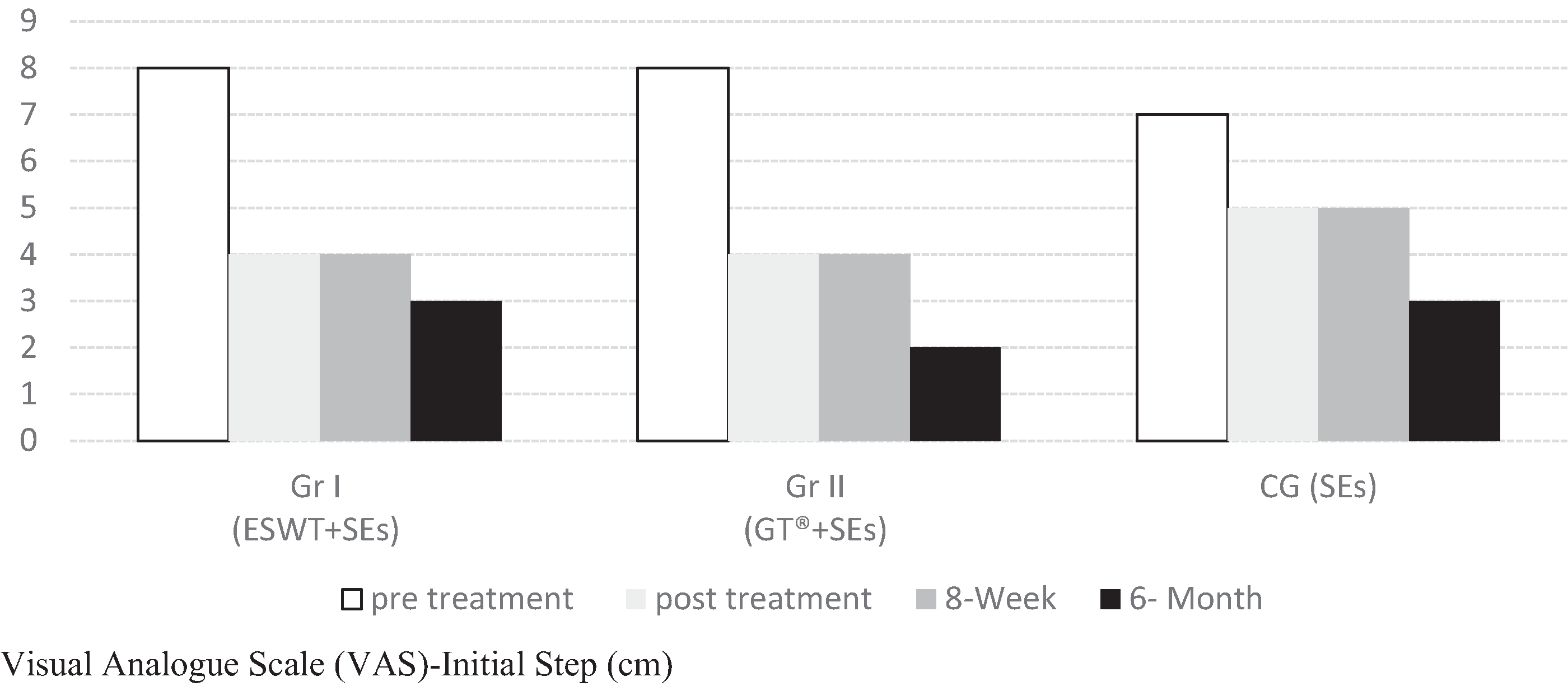
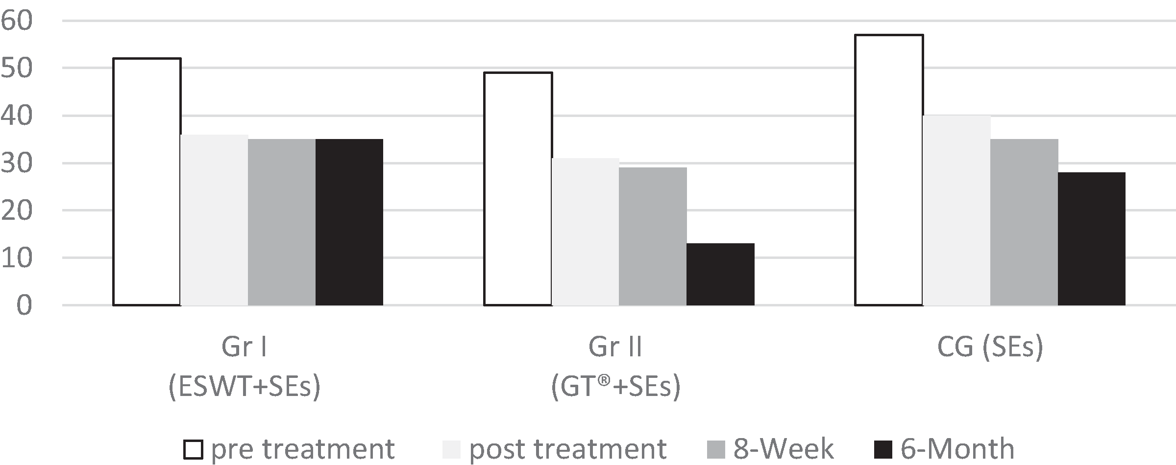
Figs. 2 and
3). The overall group × time interaction for rANOVA was not significant for VAS-initial step pain (posttreatment:
F = 1.00;
P = .372; 8-week follow-up:
F = 1.87;
P = .161) and VAS-activity pain (posttreatment:
F = 2.79;
P = .067; 8-week follow-up:
F = 1.26;
P = .288 [
Table 3]; VAS-initial step pain at 6-month follow-up:
F = 2.20;
P = .117 [
Table 4]). According to the ES calculation, although both groups 1 and 2 were more effective than the CG in VAS-initial step pain posttreatment (ES = 2.61, 2.47, and 1.07, respectively) and at 8-week follow-up (2.55, 2.39, and 1.17, respectively), ESWT+SEs was most effective in VAS-activity pain posttreatment and at 8 weeks. The ESs of all of the groups were similar in terms of FFI posttreatment and at 8 weeks. At 6-month follow-up, the ES of group 2 was found to be highest in VAS-initial step pain.
Figure 2.
Comparison of visual analog scale (VAS) initial step pain scores between groups at 6-month follow-up. ESWT, extracorporal shockwave therapy; GT, Graston Technique; SE, stretching exercise.
Figure 2.
Comparison of visual analog scale (VAS) initial step pain scores between groups at 6-month follow-up. ESWT, extracorporal shockwave therapy; GT, Graston Technique; SE, stretching exercise.
Figure 3.
Comparison of Foot Function Index (FFI) scores between groups at 6-month follow-up. ESWT, extracorporal shockwave therapy; GT, Graston Technique; SE, stretching exercise.
Figure 3.
Comparison of Foot Function Index (FFI) scores between groups at 6-month follow-up. ESWT, extracorporal shockwave therapy; GT, Graston Technique; SE, stretching exercise.
Table 3.
Comparison of Pain Intensity, Foot Function Index, Quality of Life and Kinesiophobia Between Groups
Table 3.
Comparison of Pain Intensity, Foot Function Index, Quality of Life and Kinesiophobia Between Groups
Table 4.
Comparison of Initial Step Pain and FFI Between Groups at 6 Months
Table 4.
Comparison of Initial Step Pain and FFI Between Groups at 6 Months
The overall group × time interaction for rANOVA was significant for FFI from baseline to posttreatment (F = 3.15; P = .048) and from baseline to 6-month follow-up (F = 6.33; P = .003). The mean ± SD difference in FFI scores in group 2 and the CG from baseline to after treatment was –17.90 ± 17.85 and –15.83 ± 8.61, respectively, in favor of group 2 (P = .048); after the LSD test, P = .016. However, after the LSD test, P = .002 between groups 1 and 2 and P = .007 between group 2 and the CG in favor of group 2 for 6-month follow-up.
Although SF-12 physical component scores improved posttreatment in groups 1 and 2 (
P = .048 and
P = .013, respectively), significant improvement was observed in group 2 and the CG compared with pretreatment at the 8-week evaluation (
P = .027 for both). Looking at the ES values, all of the treatment groups had a small effect on posttreatment (ES = 0.22, 0.24, 0.27, respectively) and 8-week (ES = 0.24 and 0.35 for groups 1 and 2, respectively) recovery except for the CG, where improvement at 8 weeks was moderately effective (ES = 0.50). Although significant improvement in SF-12 mental component score was observed only in the CG posttreatment (
P = .004), the ES was medium (ES = 0.69). In the 8-week results, significant improvement was observed in group 2 (
P = .005) and the CG (
P = .04), and the ESs were small for group 1 (ES = 0.21) and medium for the other groups (ES = 0.59 and 0.53, respectively). No statistically significant improvement was observed in kinesiophobia scores in any of the groups posttreatment and at 8 weeks (
Table 3).
Discussion
This randomized controlled study compared the ESWT and GT interventions in CPHP management. The findings reveal remarkable improvements in pain relief and functional outcomes in all of the groups. However, the ES for initial step pain in groups 1 and 2 was better than that in the CG posttreatment and in follow-up. For activity pain, ESWT+SEs (group 1) seemed to be the most effective. Although the mean FFI score statistically significantly improved in all of the groups, none was superior to the others. There was also no statistically significant difference between the mean kinesiophobia scores of the groups posttreatment and during follow-up.
Stretching exercises have been reported to be an effective, safe, and early-applied method with the highest level of evidence in PHP treatment [
9]. In the PHP/plantar fasciitis clinical practice guideline, clinicians recommended plantar fascia–specific and gastrocnemius and soleus muscle SE programs to reduce pain in the short term (1 week to 4 months) [
3]. Kamonseki et al [
30] found that SEs are directly associated with functional status in patients with plantar fasciitis. According to the present results, the mean pain status (initial step and activity) and FFI level improved in the CG, which is consistent with the literature. Although only the physical component score of the SF-12 increased during follow-up, statistically significant improvements were seen in the mental component score of the SF-12 posttreatment and during follow-up in the CG. Although it is well-known that SEs are effective for patients with CPHP, ESWT+SEs and GT+SEs had superior ESs in pain level compared with SEs only. Thus, considering the higher ES value for pain level, we may suggest the combined therapies especially for patients with CPHP to reduce the pain intensity level for initial step and activity.
Extracorporeal shockwave therapy is another treatment option that is frequently recommended in the literature. Rompe et al [
29] compared the effects of ESWT and those of ESWT+SEs on pain (initial step pain, worst pain, and FFI pain subscale) and function (FFI sum score). In that study, one group received ESWT only (n = 73) once a week for 3 weeks and the others participated in ESWT and plantar fascia–specific SEs (n = 79). All of the patients were assessed at baseline and at 2, 4, and 24 months. According to this study, the FFI sum score had significant improvement in the ESWT+SEs group compared with the ESWT-only group (
P < .001). The group that performed ESWT and SEs outperformed the other group in all of the evaluations. The treatment efficacy continued at 4-month follow-up, but no difference was observed between the two groups at 24 months [
29]. In contrast to the study by Rompe et al [
29], the present results indicate that the ESWT+SEs combination was found to be more effective than SEs only in reducing initial step and activity pain. Still, there was no difference between the groups in terms of functional status. To our knowledge, there is no study investigating quality of life and kinesiophobia scores in the literature. In the present study, although ESWT+SEs had a remarkable improvement in SF-12 physical component scores, this effect did not last until follow-up, with no change in the kinesiophobia score.
Manual therapy includes joint mobilization or soft-tissue mobilization (deep-tissue massage or myofascial release). In the literature, myofascial release performed on the calf and plantar fascia has been found to be effective in the treatment of CPHP [
27,
35,
36,
37]. Instrument-assisted soft-tissue mobilization treatment performed in combination with SEs has been recommended to increase the flexibility of the plantar fascia and to provide collagen reorganization [
17]. Studies using IASTM for plantar fasciitis are limited to case studies [
17,
37], a case series [
6], and a pilot study performed with a small number [
38]. The results of these studies showed improvement in pain and function [
6,
17,
37]. Because IASTM was not performed alone in any of these studies and there were no control groups, it was not possible to evaluate the contribution of IASTM to the treatment. In a feasibility study examining the effectiveness of IASTM treatment using GT in PHP, a group that was given stretching and strengthening exercises and a home program (n = 6) was compared with a group on which GT was performed in addition to these exercises (n = 5). After eight treatment sessions (two sessions × 4 weeks) and follow-up after 90 days, clinically important changes in the IASTM group and moderate-to-large between-group ESs suggested that further research is warranted to determine whether these trends are meaningful [
38]. The results of the present study are in line with the literature in that pain, function, and quality of life physical scores improved posttreatment and during follow-up in group 2. In the present study, the ES of VAS-initial step pain was much higher in group 2 compared with the CG group, and there was no difference in ES values of VAS-activity pain and FFI scores. The SF-12 physical component scores improved posttreatment and during follow-up, whereas no change was observed in kinesiophobia scores. In addition, FFI scores were better at 6 months in group 2 compared with group 1 and the CG.
Considering these findings, we believe that attempts to further optimize nonoperative treatment modalities in patients with CPHP are warranted. Furthermore, the number of randomized controlled studies with blinded assessors is limited, and there are few studies that compared combined conservative management protocols, which are common in clinical practice. To our knowledge, the present study is the first prospective randomized controlled study comparing three methods of nonoperative treatment for CPHP by evaluating pain level, functional capacity, and kinesiophobia. As far as we know, this is the first study in which the GT intervention was conducted with a sufficient number of patients, a randomized controlled research design, and 6-month follow-up. We considered that the GT intervention and SEs lasting 4 weeks have the potential to improve FFI scores at 6 months compared with pretreatment assessment. The present study contributes to the literature in these aspects.
There are a few limitations in this study. First, changes in body weight were not monitored. Second, the physical activity status of the participants was not evaluated with an objective method. High body mass index and restricted activity level may have a relationship with functional status. Further studies are needed to monitor these risk factors influencing the outcomes after the treatment. We think that it is important for future studies to expand patient education programs by taking other risk factors into account and via long-term follow-up. In the present study, we reached the calculated sample size. If the number of participants increases, the consistency of the outcomes in the ESWT+SEs and GT+SEs groups may be identified better compared with SEs only.
Conclusions
The results of this study show that all treatment protocols (ESWT+SEs, GT+SEs, and SEs only) have similar effects on functional capacity and pain level in the short term. It was observed that the effectiveness of ESWT and GT treatments on initial step pain and the efficiency of ESWT on activity pain created the highest improvement in the short term. In addition, we believe that the management strategies in PHP that must lead to improvements in functional capacity, quality of life, and kinesiophobia, as well as pain level, can be considered satisfactory. In conclusion, we suggest using the GT combined with SEs protocol for the management of CPHP due to effectiveness on pain relief and on functional status for long-term effect in light of the findings. Further studies with larger sample sizes, long-term follow-up, and risk factor monitoring (eg, body mass index, physical activity level) will provide a more tailored management strategy.