Plantar fasciitis is one of the common causes of painful foot syndromes. Although its cause is not known exactly, it is believed to originate from tension of the fibers and foot abnormalities that cause inflammation. Extreme and forceful activities and use of ill-fitting shoes may also be causes.[
1] Plantar fasciitis is characterized by pain in the heel emerging in the morning or after prolonged sitting.[
2,
3]
Nonsteroidal anti-inflammatory medications, heel cushions or orthoses, physical therapy, tensioning exercises, corticosteroid and plasma-rich platelet injections, or fasciotomy can be used in the treatment of plantar fasciitis.[
4,
5] Intralesional botulinum toxin A and plantar fascia or gastrocnemius and soleus muscle tensioning exercises can also be applied.[
1] Recently, extracorporeal shockwave therapy (ESWT) has been reported to provide significant relief in approximately 80% of the patients.[
6,
7] The exact mechanism of action of ESWT is not completely understood, but it is believed to stimulate the healing process of the body.[
6,
8,
9,
10] Extracorporeal shockwave therapy is preferred because of advantages such as a noninvasive approach, usability, and short-term application. In addition, adverse effects such as pain, redness, edema, and ecchymosis are transient and rarely seen.[
11]
The response to treatment for plantar fasciitis has been evaluated by several scales in the literature. However, these scales subjectively assess the disease activity and response to treatment. Objective assessment can be made by using magnetic resonance imaging (MRI) findings, including an increase in the thickness of the plantar fascia, calcaneal bone marrow edema, and calcaneal spur (osteophyte) formation.[
12] Moreover, MRI has an advantage for distinguishing inflammatory disease from neuropathic disease. However, only a few reports have investigated the therapeutic effects of ESWT based on MRI findings.[
9] The purpose of this study is to investigate the therapeutic efficacy of ESWT in plantar fasciitis by comparing pretreatment and posttreatment MRI findings.
Material and Methods
Patients with plantar fasciitis diagnosed by history, physical examination, and MRI findings were included in this study. A history of pain in the soles of the feet when getting up from bed in the morning, plantar fascia sensitivity on physical examination, and a lack of findings suggestive of other diseases were considered adequate for physical diagnosis. All patients provided informed consent, and Bakirköy Dr. Sadi Konuk Training and Research Hospital Ethic Committee approved this study.
The inclusion criteria were age 20 to 60 years, presence of symptoms for at least 6 weeks, diagnosis of plantar fasciitis with or without coexistent calcaneal spurs on plain film radiographic evaluation, lack of an improvement with medical treatment, and adequate intellectual capacity and education (education of primary school or more to understand and answer the forms that are used in the evaluations). The exclusion criteria included presence of diabetes mellitus, malignancy, active infections, history of mixed collagen tissue diseases, trauma, skin lesions, presence of infection or an open wound, neuropathy, radiculopathy, disordered peripheral circulation, warfarin use due to coagulopathy, arthropathy, congenital or acquired lower-extremity deformations, fracture sequelae at the lower extremities, orthoses, internal plaque screw fixator, cardiac pacemaker, metal implant at the application site, and pregnancy that could pose a problem for physiotherapy application.
The age, sex, profession, and level of education were determined for all of the patients. The total daily duration of standing visual analog scale (VAS) was used in the evaluation of pain level. The VAS scores at getting up from bed in the morning, at rest, and during activity were evaluated before and 3 months after treatment. The duration of walking without heel pain at standing up after a rest was evaluated with a 6-point rating scale before and 3 months after treatment. The ESWT was applied once weekly for three sessions. The device (Master-puls MP100-SWISS; Karl Storz Lithotripsy-America Inc, Kennesaw, Georgia) was adjusted to a frequency of 12 to 15 Hz, 2 to 3 bars, and 2,500 pulses. Mild local pain and redness were observed during ESWT application, but adverse effects requiring intervention did not occur.
After the clinical diagnosis of plantar fasciitis, MRI was performed with a 1.5-T whole-body scanner (Avanto; Siemens, Erlangen, Germany). Leonardo software (Leonardo Workstation syngo 1 MMWP-VE52A; Siemens, Germany) was used for imaging. The same monitors were used to score images. The MRI protocol consisted of sagittal, axial, and coronal T1-weighted and T2-weighted images with and without fat saturation and short tau inversion recovery images (
Table 1). We did not use contrast material.
Table 1.
Specifications of Magnetic Resonance Imagaing.
Table 1.
Specifications of Magnetic Resonance Imagaing.
The MRIs before and after ESWT treatment were reviewed to assess the post-ESWT changes in soft-tissue and bone marrow edema, the thickness of the proximal plantar fascia, and the presence of heel spurs. The radiologists did not know whether the images were from after or before treatment. All of the images (pre and post) for each participant were scored in random order. Two experienced radiologists (E.H. and S.A.) assessed the images by consensus. The MRIs were scored semiquantitatively. The radiologists used a ruler within the software to measure the thickness of the fascia. Measurements were performed 1 cm from the insertion site of the fascia calcaneus, and thickness of 3 mm or greater was accepted as being abnormal. Bone marrow and soft-tissue edema were diagnosed according to hyperintense signals of short tau inversion recovery images. No repeatability study was undertaken for scoring images. This was one of the limitations of this study.
Statistical Analysis
The statistical analysis of this study was performed with NCSS 2007 statistical software (NCSS LLC, Kaysville, Utah). Descriptive statistical analysis of data was performed. The McNemar test was used for repeated comparisons of qualitative data. The paired-sample t test was used to assess the pretreatment and posttreatment MRI findings. The results were evaluated at a significance level of P < .05.
Results
A total of 30 patients (30 feet) with a diagnosis of plantar fasciitis were involved in this study. All of the patients except one were women (96.7%). The mean ± SD age of patients was 45.23 ± 8.57 years. The demographic characteristics of the patients are outlined in
Table 2. Twenty-one of the 30 patients (70%) were standing for more than 5 hours a day. The morning, activity, and resting VAS pain scores showed a significant decrease at the posttreatment clinical evaluation compared with the pretreatment values (
P = .0001,
P = .0001,
P = .008, respectively) (
Table 3). The significant increase in the number of patients who could walk less than 5 min without pain according to the 6-point rating scale compared with pretreatment assessment are shown in
Table 3 (
P < .0001). The posttreatment Foot and Ankle Outcome Score (FAOS) values showed a statistically significant increase in comparison with pretreatment values (
Table 4). There were no significant correlations between body mass index (calculated as the weight in kilograms divided by the square of the height in meters) and pretreatment morning, activity, and resting VAS pain scores; 6-point rating scale scores; and FAOS values (
P > .05).
Table 2.
Demographic Characteristics of the 30 Study Patients.
Table 2.
Demographic Characteristics of the 30 Study Patients.
Table 3.
VAS and 6-Point Rating Scale Scores Before and 3 Months After Treatment.
Table 3.
VAS and 6-Point Rating Scale Scores Before and 3 Months After Treatment.
Table 4.
FAOS Values Before and 3 Months After Treatment.
Table 4.
FAOS Values Before and 3 Months After Treatment.
All of the MRI findings except osteophytes showed a significant improvement compared with pretreatment values (
P = .0001,
P = .0001,
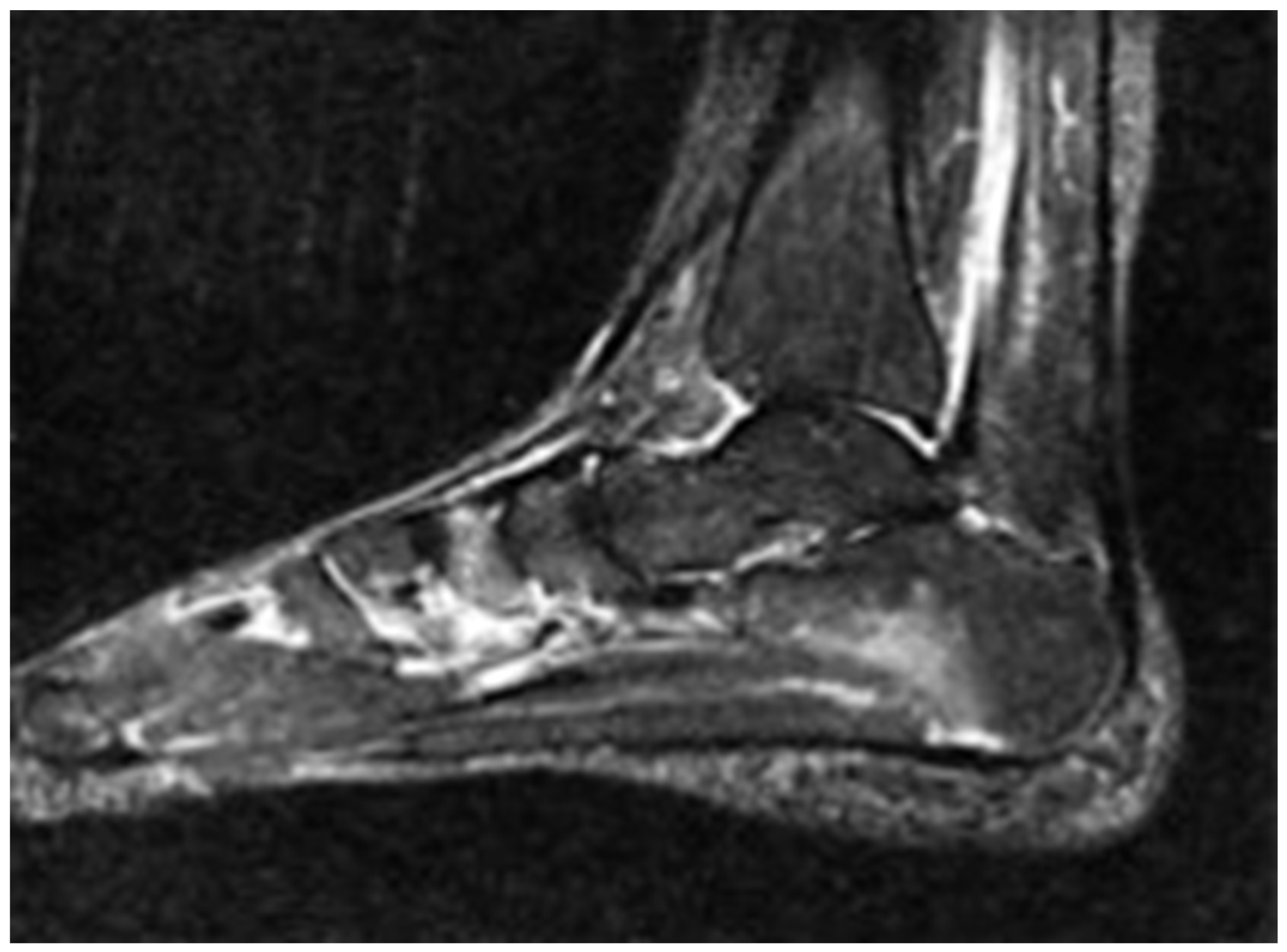
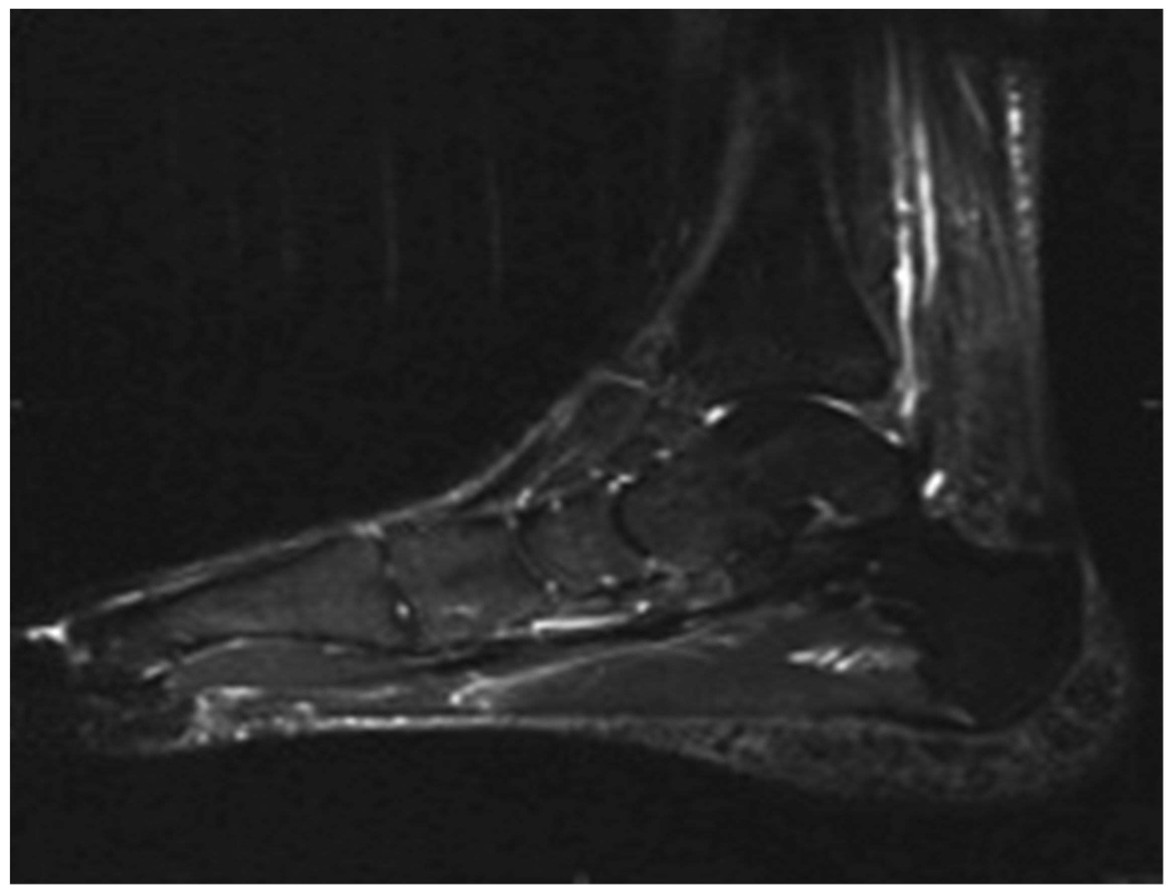
P = .013). Before treatment, 23 of the 30 patients had thickening of the plantar fascia; this number decreased to 11 after treatment (
Figure 1 and
Figure 2). The MRI findings of plantar fascia are thickening of the plantar fascia and edema of neighboring soft tissue and bone marrow. The number of patients with soft-tissue edema and bone marrow edema before and after treatment were 30 and 9, and 19 and 9, respectively. Thus, a significant decrease was observed in plantar fascia thickness and soft-tissue and bone marrow edema. There were osteophytes in 17 patients before treatment, and this number increased to 18 after treatment (
Table 5).
Table 5.
Pretreatment and Posttreatment Magnetic Resonance Imaging Findings in the 30 Study Patients.
Table 5.
Pretreatment and Posttreatment Magnetic Resonance Imaging Findings in the 30 Study Patients.
Discussion
Plantar fasciitis is a painful and limiting disorder that may affect the daily activities of a patient. The main treatment for plantar fasciitis is conservative.[
12] Nonsteroidal anti-inflammatory drug therapy, heel cushions, various physical medicine modalities, corticosteroid injections, and stretching exercises of the plantar fascia are among these conservative options. Extracorporeal shockwave therapy was approved for the treatment of plantar fasciitis in 2000 by the US Food and Drug Administration.[
13,
14]
Extracorporeal shockwave therapy has also been shown to be an effective treatment modality for myofascial pain syndrome. Its effects in the treatment of musculoskeletal disorders such as fracture, pseudoarthrosis, calcific tendinitis, and plantar fasciitis have been proven.[
12] In the literature, studies have reported increased healing in the growth process of bone, tendon, and surrounding tissues by ESWT. This has been observed in experiments on mice at moderate doses of ESWT in subchondral and cartilage tissues in the early phases.[
15]
Extracorporeal shockwave therapy is an effective, easy, and safe treatment modality. Additionally, it may be an alternative to surgical treatment, with its low and nonsignificant complication rates and low treatment costs, and it may be an alternative to other physical treatment modalities, with its faster efficacy.[
16] In addition, ESWT has the advantages of being a painless application, noninvasive, and well tolerated by patients, with few adverse effects.[
5,
16,
17] For these reasons, ESWT has been increasingly used in the treatment of soft-tissue inflammations in recent years.
The present study evaluated the efficacy of ESWT in the treatment of chronic plantar fasciitis. Moderate- to high-intensity ESWT was found to be effective for this clinical entity.[
16] Also, ESWT was demonstrated to be effective in the short- and medium-term in the treatment of plantar fasciitis in the study by Tuna.[
18] Also, the length of the epin calcanei was found not to have any effects on the heel pain and efficacy of ESWT.[
18,
19] We also found similar results in the present study. Although there are studies and meta-analyses that have shown the efficacy of ESWT in the treatment of plantar fasciitis, others could not show a significant difference between ESWT and other treatment modalities.[
20,
21] According to Kudo et al,[
10] this could be explained by differences in technical characteristics (the design of the device, shock intensity, frequency, number of sessions, and different placebo-controlled drug treatment options), patient populations, disease severity, and study designs.
Few studies have reported the correlation between clinical outcome and radiologic changes in patients with plantar fasciitis treated with ESWT. We analyzed MRI findings of plantar fasciitis before and 3 months after treatment. We found statistically significant improvement of MRI findings with ESWT. Similarly, Liang et al[
21] found a prominent improvement in pain complaints of patients after ESWT and showed a correlation between this improvement and decreased thickness of the plantar fascia. Correspondingly, MRI has been found to be useful for determining the response to ESWT.[
2,
13,
16]
Although no radiologic change in the number of osteophytes after ESWT has been shown (except in one patient, whose number of osteophytes increased), patients’ complaints about heel spurs decrease significantly.[
22]
The results of this study should be considered in light of the following limitations. No reliability study was undertaken for scoring images; therefore, some of these results may be due to repeatability error. In addition, the number of patients was low, and no long-term radiologic follow-up occurred.
Conclusions
Extracorporeal shockwave therapy is a treatment method that produces positive results regarding pain and function in patients with plantar fasciitis. Magnetic resonance imaging is useful not only for diagnosis but also for evaluation of the response to plantar fasciitis treatment. Further studies to explore these findings are now recommended.