Retronychia (‘‘retro’’—Latin for backward; ‘‘onychia’’—Greek for nail) is a newly reported condition describing a combination of proximal nail plate ingrowth into the proximal nail fold associated with multiple generations of nail plate misalignment beneath the proximal nail folds causing chronic paronychia.[
1,
2] It is also called
proximal ingrown nail and was first described by de Berker and Renall in 1999.[
1] It is generally not recognized at an early stage and is often inadequately treated. Retronychia mainly affects the great toe unilaterally, although some cases of bilateral involvement have been reported. It may also, but less frequently, affect the nails of the hands, especially the thumbs and index fingers.[
2,
3] This recently described disorder is rarely reported, and its pathogenesis is not well-known. We report a case of retronychia associated with distal onycholysis in a patient with a medical history of congenital malalignment of the toenails, with emphasis on the pathogenesis of the disease and the surgical treatment.
Observation
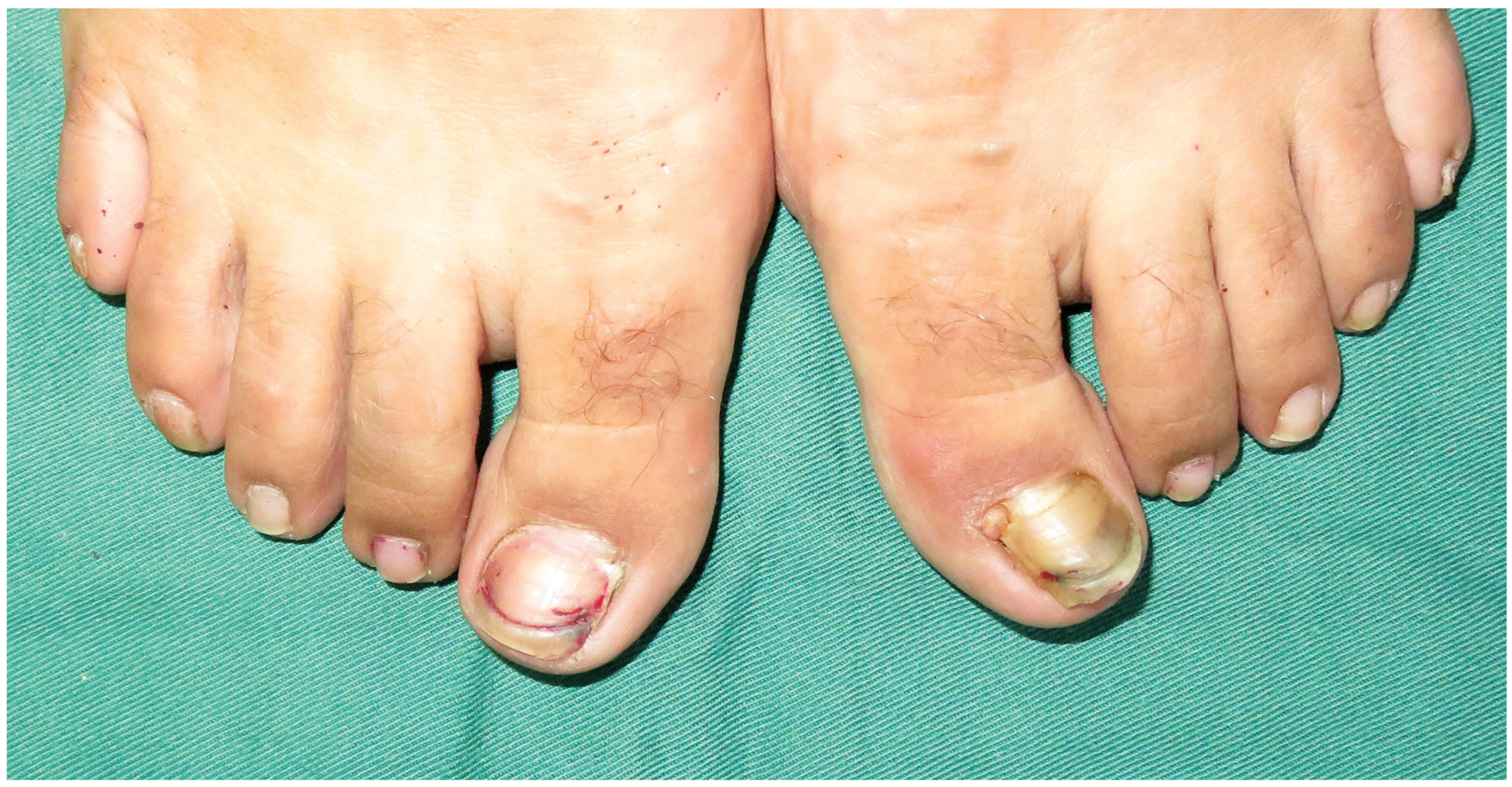
A 34-year-old woman with a medical history of congenital malalignment of the toenails presented with a 9-month history of pain and discharge of the left toenail. She reported that the left toenail ceased to grow. The condition had been previously treated with antibiotics and antifungals but had not shown improvement. Clinical examination revealed yellowish discoloration, distal onycholysis, and onychomadesis of the left toenail associated with marked swelling and erythema of the periungual skin. Some granulation tissue protruded at the corner between the lateral and proximal nail folds (
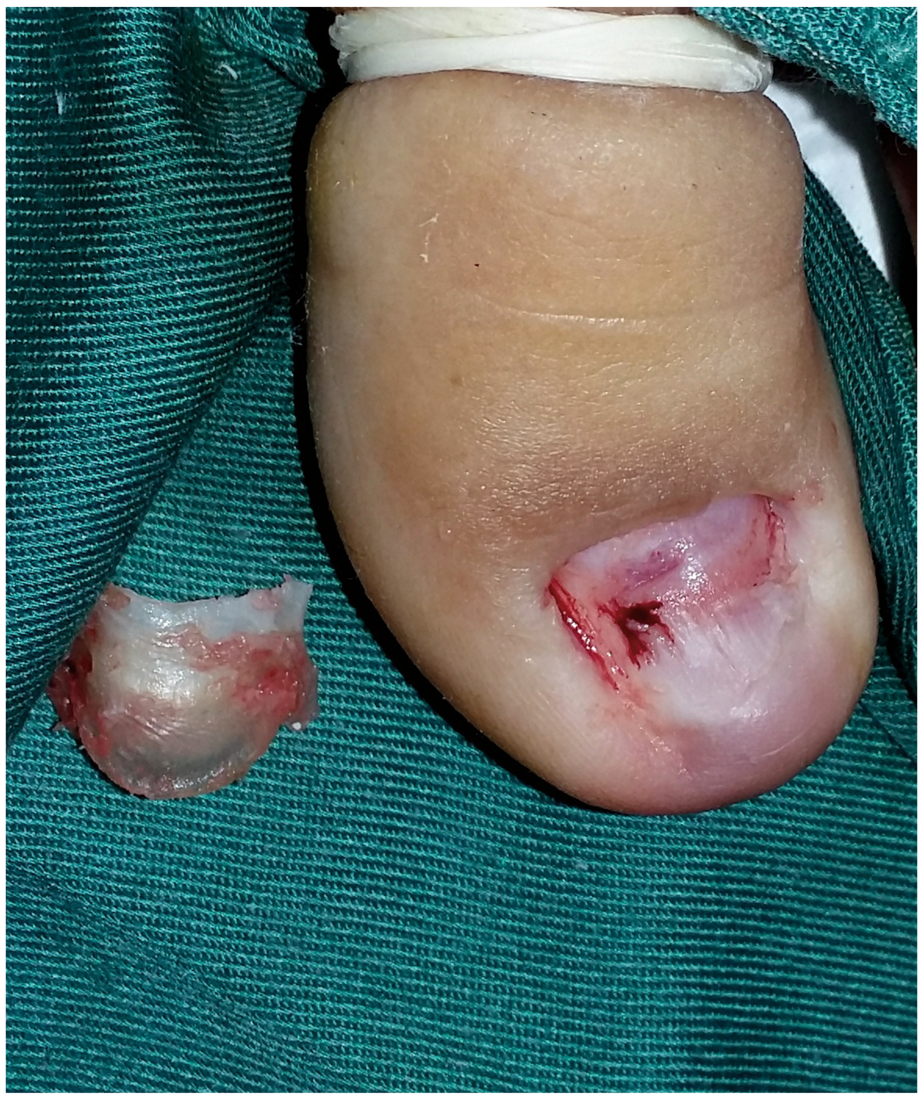
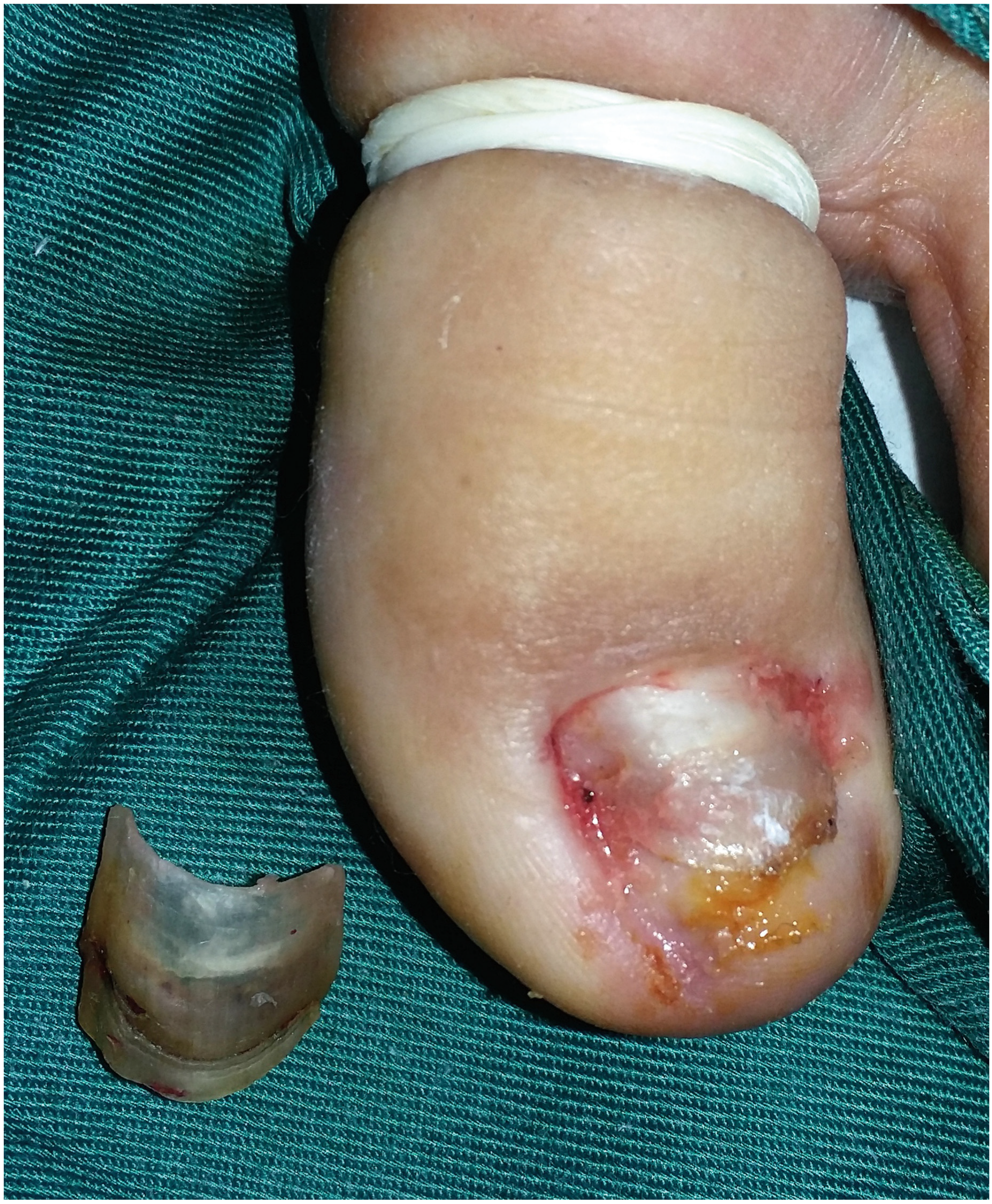
Figure 1). Examination of the right toenail showed onychomadesis and Beau lines. Based on these clinical findings, the diagnosis of retronychia was made. Surgical avulsion of the nail, under local anesthetic, was performed using an anterior approach. The nail exhibited a markedly thickened proximal end caused by the presence of three nail layers. Avulsion of only the two upper nail layers was performed (
Figure 2 and
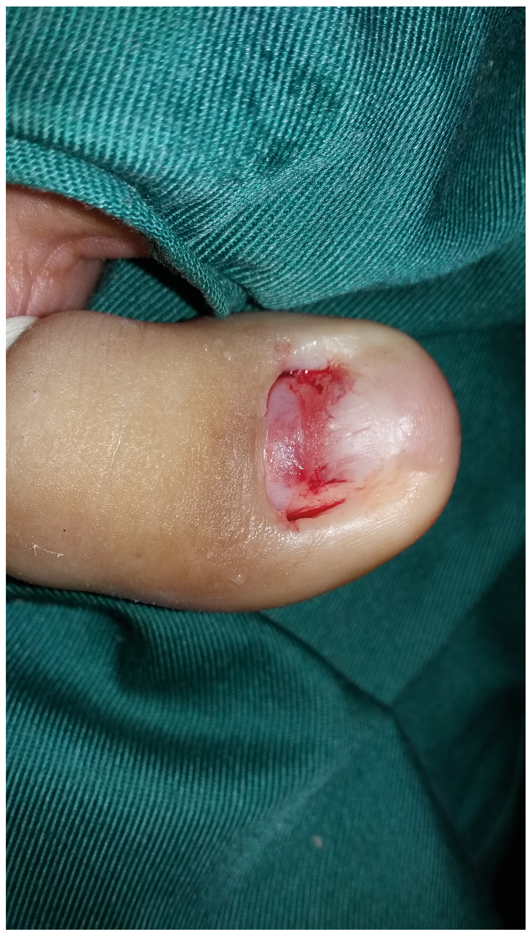
Figure 3). The underlying nail was whitish with a healthy shine, so we decided to conserve it (
Figure 4). A week after surgery, the patient was seen and reported no more pain. Three months later, 50% of the nail had regrown normally and the patient was free of symptoms.
Discussion
The frequent traumatization of the great toes probably explains why retronychia generally affects these digits. In fact, trauma is the most common trigger.[
3,
4] This mechanical factor of an acute physical (shoe trauma, hiking, toe trauma, jogging, dancing, asymmetrical gait nail unit syndrome) or systemic (thrombophlebitis, osteoarthritis) nature disrupts the longitudinal growth of the nail.[
4] As seen in the present patient, lateral deviation of the great toe represents a potential predisposing factor.[
3] The precipitating event for the present patient was probably the congenital malalignment of the toenails causing lateral deviation of the toe and repeated distal trauma. Retronychia usually affects young women (20–30 years old), especially those wearing ill-fitting shoes, and sportspeople.[
5] A direct consequence of repeated trauma is the loss of attachment of the nail plate to the matrix. So retronychia starts with disruption of the longitudinal growth of the nail. With the growth of a new nail plate, the old one is pushed upward and backward. This leads to stacking of the nail plates, embedding of the top nail into the ventral aspect of the proximal nail fold, and to chronic inflammation of the periungual skin. Retronychia and onychomadesis share a common pathophysiologic basis consisting of slowing or cessation of nail plate production at the nail matrix. This similar mechanism explains the association of these clinical findings in the present patient.[
4,
5,
6,
7]
It was first supposed that the old nail remained firmly attached to the nail bed and could not be pushed out by the new nail plate, aggravating retronychia.[
2,
7] However, in the present patient, a marked distal onycholysis was found. We believe that retronychia is caused by two mechanisms: a repetitive disruption of the nail matrix, leading to stacking of the nail plates, and a nail plate not attached enough to the nail bed and continually pushed into the proximal nail fold, leading to an emergence of granulation tissue that helps the growth of bacteria and prevents spontaneous resolution of the condition.[
4,
7,
8]
The diagnosis of retronychia is made clinically. The five most important criteria for diagnosis are disruption of the longitudinal growth of the nail, the rise of the proximal nail fold by the nail plate, pachyonychia, dyschromia, and chronic persistent paronychia.[
7] Although the diagnosis of retronychia can be based on clinical findings, ultrasound can be used as a noninvasive confirmatory test and in the differential diagnosis of chronic paronychia.[
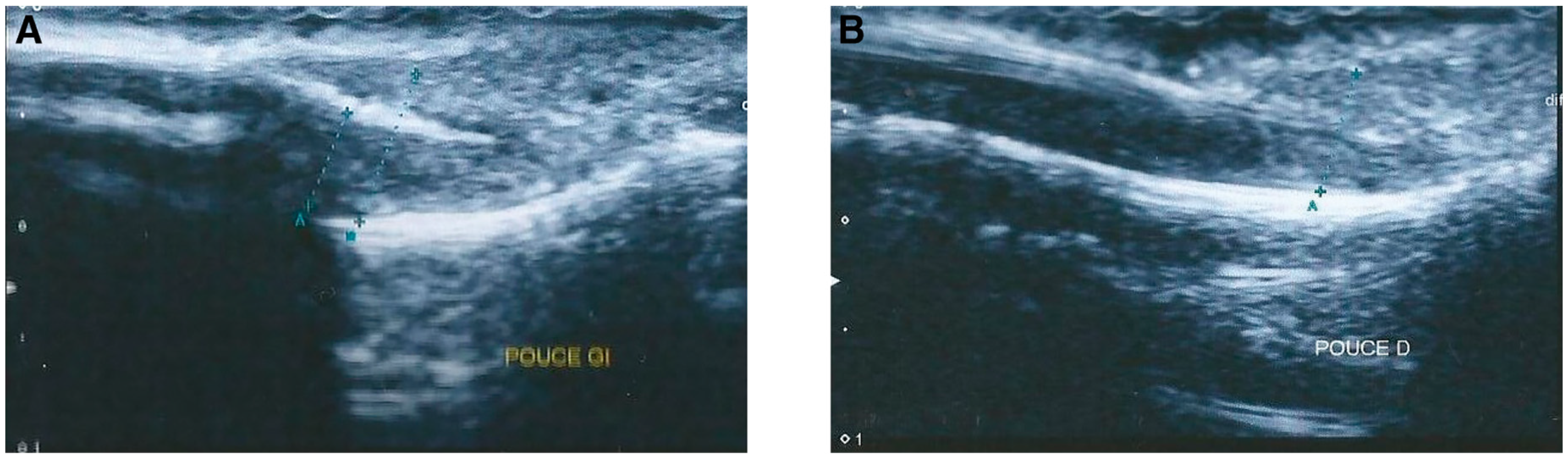
3] Ultrasound could be useful particularly in difficult cases. It shows the stacking of two or more nail plates, and a shorter distance between the root of the nail plate and the base of the distal phalanx, compared with the healthy contralateral nail (
Figure 5).[
3]
The main differential diagnosis includes candidal or bacterial infection, psoriatic arthritis, subungal cysts and tumors such as Bowen’s disease, keratoacanthomas, squamous cell carcinomas, enchondromas, and amelanotic malignant melanomas, as well as processes associated with medications such as retinoids, cyclosporine, protease inhibitors, and epidermal growth factor receptor inhibitors.[
3,
6]
Management of retronychia consists of total or proximal nail plate avulsion with a proximal approach.[
5,
7,
8] It confirms the diagnosis and leads to rapid pain relief. Conservative treatment with topical corticosteroids and antibiotics represents another option but is associated with a high relapse rate and longer follow-up.[
3,
4,
5] Nail avulsion is a fast and curative treatment that should be considered as first-line therapy for retronychia. If the underlying nail is whitish, nonpainful, and appears healthy—as found in the present patient—avulsion of only the top nail plates can be performed.[
6,
7] Prevention of retronychia is important, focusing on the importance of properly fitting footwear.[
4] Knowledge of retronychia is still limited among practitioners, which can lead to diagnostic errors and therapeutic delay.