Next Article in Journal
Effect of Asymmetrical Load Carrying on Joint Kinetics of the Lower Extremity During Walking in High-Heeled Shoes in Young Women
Previous Article in Journal
A Novel Method of Quantifying Gait Deviations Using Plantar Pressure Patterns
 
 
Journal of the American Podiatric Medical Association is published by MDPI from Volume 116 Issue 1 (2026). Previous articles were published by another publisher in Open Access under a CC-BY (or CC-BY-NC-ND) licence, and they are hosted by MDPI on mdpi.com as a courtesy and upon agreement with American Podiatric Medical Association.
Article

Cost-Effectiveness of Becaplermin Gel on Diabetic Foot Ulcer Healing

by
Curtis R. Waycaster
1,2,*,
Adrienne M. Gilligan
1,2 and
Travis A. Motley
3
1
Smith & Nephew Inc, 3909 Hulen Street, Fort Worth, TX 76107
2
Department of Pharmacotherapy, University of North Texas Health Sciences Center, Fort Worth, TX
3
University of North Texas Health Sciences Center, Bone and Joint Institute, Fort Worth, TX
*
Author to whom correspondence should be addressed.
J. Am. Podiatr. Med. Assoc. 2016, 106(4), 273-282; https://doi.org/10.7547/15-004
Published: 1 July 2016

Abstract

Background: A comparison of the cost-effectiveness of becaplermin plus good wound care (BGWC) versus good wound care (GWC) alone in treating patients with diabetic foot ulcers (DFUs) may enable physicians and health-care decision makers in the United States to make better-informed choices about treating DFUs, which currently contribute to a substantial portion of the economic burden of diabetes. Methods: Data from three phase III trials were used to predict expected 1-year costs and outcomes, including the average percentage reduction from baseline in wound surface area (WSA), the direct costs of DFU therapy, and the cost per cm2 of WSA reduction. Results: At 20 weeks, the BGWC group had a statistically greater probability of complete wound closure than the GWC group (50% versus 35%; P = .015). Based on reported WSA reduction rates, DFUs in the BGWC group were predicted to close by 100% at 27 weeks, and those in the GWC group were predicted to close by 88% at 52 weeks. The GWC group had higher total estimated 1-year direct cost of DFU care ($6,809 versus $4,414) and higher cost per cm2 of wound closure ($3,501 versus $2,006). Conclusions: Becaplermin plus good wound care demonstrated economic dominance compared with GWC by providing better clinical outcomes via faster reduction in WSA and higher rates of closure at a lower direct cost.

Diabetes mellitus is a serious, chronic disease with a prevalence in the United States that reached approximately 29.1 million (21.0 million diagnosed and 8.1 undiagnosed) people (9.3% of the total population) in 2012.[1] The prevalence of diabetes is growing at an exponential rate in the United States and throughout the world.[2] Chronic hyperglycemia in patients with diabetes may lead to peripheral neuropathy, a microvascular complication that can accelerate peripheral vascular disease and other conditions.[3] These conditions may contribute to the development of diabetic foot ulcers (DFUs), which occur in approximately 15% to 25% of patients with diabetes.[4-7]
On average, medical expenditures are two to three times higher in patients with diabetes compared with individuals without diabetes.[8] In the past 10 years, the estimated total (direct and indirect) annual expenditures for diabetes management have increased from $132 billion in 2002 to more than $245 billion in 2012.[1,9] Diabetic foot ulcers contribute to a substantial portion of the economic burden of diabetes. Sedory Holzer and colleagues estimated that the average cost of an uncomplicated DFU was approximately $13,000, and the cost of a limb amputation was almost $77,000 in 2012.[10] The average length of hospital stay for patients with a DFU was 59% longer than for diabetic patients without ulcers, mostly because of the need for amputations.[6] Approximately 85% of lower-extremity amputations in patients with diabetes are preceded by a DFU.[11-14] The estimated annual burden of illness for diabetic patients with DFU compared with those without DFU is significantly greater, resulting in an additional $11,710 in incremental annual health-care costs for Medicare beneficiaries and $16,883 for those with private insurance.[15]
The three major components in treating DFU are debridement, infection control, and offloading.[16] Despite good wound care practice, many DFUs either heal slowly or fail to heal, thereby increasing the risk of infections, hospitalization, and subsequent amputations. Approximately 30% of DFUs evolve into chronic, full-thickness ulcers that may become difficult to treat or that may result in infection and gangrene, two common causes of amputation in patients with DFUs.[4,6,17] If standard care alone fails to achieve epithelialization, surgical options for closure may be limited because of the number of comorbidities within this patient population.[18] Advanced therapies, such as platelet-derived growth factors (PDGFs), cellular/tissue derived products, hyperbaric oxygen therapy, and negative pressure wound therapy, may be the only means of rapidly and effectively attaining wound closure.[19]

Platelet-Derived Growth Factors and Wound Healing

Platelet-derived growth factors are endogenous proteins that have growth-promoting capabilities and can assist with the wound-healing process.[20-23] The core protein of PDGF is composed of polypeptide chains A and B and forms PDGF-AA, PDGF-BB, and PDGF-AB isoforms.[20,21] However, the effects of PDGF differ over the course of the wound-healing process.[24] Shortly after injury, in the hemostasis and inflammatory phases, PDGF is released from platelets and endothelial cells to serve as a chemotactic signal for neutrophils, macrophages, and fibroblasts to the damaged cells.[24] Continuous stimulation of these cells leads to endogenous production of PDGF, provisional extracellular matrix synthesis, fibroblast proliferation, and collagen production to transition the wound into the proliferative phase.[24] Finally, PDGF signaling orchestrates active collagen turnover and cross-linking and contributes to wound remodeling and epithelialization.[24] In preclinical studies, PDGF-BB has been shown to promote the formation of granulation tissue at wound sites and to stimulate wound healing.[25]
Studies have shown that autologous releasates and genetically engineered products, such as recombinant human PDGF-BB becaplermin (Smith & Nephew Inc, Fort Worth, TX), have clinical benefits for patients with DFUs.[26-28] Becaplermin was one of the first advanced therapies indicated for diabetic neuropathic foot ulcer.[29] By retaining the biologic activity of naturally occurring PDGF, becaplermin helps promote proliferation of cells involved in the wound-healing process.[30] Studies have shown that becaplermin stimulates chemotaxis and mitogenesis of neutrophils, fibroblasts, monocytes, and other components that create the cellular basis of chronic wound healing.[31-33] Becaplermin is dispersed in a sodium carboxymethylcellulose-based gel for topical administration. The addition of the gel provides the benefit of a moist wound environment for healing.[34,35]
Multiple clinical trials have demonstrated the clinical efficacy of becaplermin gel on measures such as wound surface area (WSA) reduction and healing.[26,27,29,36] Two multi-center, prospective clinical trials that included patients (20 and 113, respectively) with large diabetic neuropathic plantar ulcers found that treatment using becaplermin gel in conjunction with standard care accelerated the time to complete wound closure by 41.8% (50 versus 86 days) and 40.6% (57 versus 96 days), respectively.[37,38] The improved healing rates, shorter healing times, and reduction in WSA associated with becaplermin gel have important implications for the direct medical costs of treating DFUs. Cost-effectiveness studies on the direct cost of care for DFU therapy included healing as the only primary outcome and did not include other potential surrogate endpoints, such as cost per cm2 of WSA reduction.[39,40] Previous work by Margolis et al[41] found that percentage change in WSA can be used as a surrogate marker for complete wound healing.
Access to health economic data will enable physicians and health-care decision makers in the United States to make better informed choices about treating DFUs. In addition, these data may draw attention to the importance of treatment patterns in the management of DFUs. Therefore, the primary objective of this study was to compare the cost-effectiveness of treating DFUs with good wound care (GWC) versus treating with becaplermin plus good wound care (BGWC). Specific outcomes of interest included WSA reduction rates, the direct costs of DFU therapy, and the cost per cm2 of WSA reduction over the episode of DFU care.

Methods

Study Participants, Design, and Intervention

Data for this study were derived from three phase III clinical trials. [26,27,29,36] A summary of the study designs can be found in Table 1. Study 1 was a pivotal, multicenter, double-blind, placebo-controlled comparison of the safety and efficacy of becaplermin and placebo gel. A total of 382 patients with DFUs from 1 to 40 cm2 were randomly assigned to treatment with becaplermin gel 30 μg/g (n = 132), becaplermin gel 100 μg/g (n = 123), or placebo gel (n = 127).[27] Study 2 was a multicenter, evaluator-blinded, controlled trial of 172 patients with DFUs from 1 to 10 cm2 who were treated with placebo gel (n = 70), good wound care alone (n = 68), or becaplermin gel 100 μg/g (n = 34).[26] Study 3 was a multicenter, randomized, evaluator-blinded, controlled trial that included 252 patients with DFUs from 1 to 40 cm2. The study compared the safety and efficacy of becaplermin gel 100 μg/g (n = 128) and good wound care alone (n = 122).[29] Two patients in study 3 did not proceed further than signing the consent form and therefore were eliminated from the analysis.
Table 1. Summary of Phase III Study Designs
Japma 106 00273 t01
Data from the becaplermin gel 30 μg/g treatment group in study 1 were excluded from the economic analysis because the dose was never commercially available. The efficacy of BGWC and GWC was evaluated based on the combined total number of patients receiving becaplermin gel 100 μg/g (N = 285) and GWC (N = 190) across the three studies. Despite differences in sample size across the BGWC and GWC treatment groups, statistical similarity across the three studies in baseline demographic and clinical characteristics has been previously established using meta-analytic methods and were not statistically different (P > .05).[29]

Economic Analysis

The WSA reduction rates were used to predict the expected costs and outcomes of wound healing for BGWC and GWC over a 1-year time horizon. The WSA reduction rates allowed for the time-course prediction of wound closure. In addition to reduction rates of WSA, specific outcomes of interest included the direct costs of DFU therapy and the cost per cm2 of WSA reduction. Once the wound closed, costs for DFU treatment ceased. A sensitivity analysis was performed on the number of tubes of becaplermin gel used, which ranged from one tube to the maximum recommended treatment of three tubes.[42] A sensitivity analysis is the process of changing the value of an input parameter; in this instance becaplermin use and costs associated with use were varied to assess the magnitude of its effect on the final results. The costs for becaplermin gel and DFU patient evaluation and management were derived from standard cost references.[43] Becaplermin use was calculated using the manufacturer's recommended dosing algorithm.[44]

Time Horizon

A 1-year time horizon was chosen to allow sufficient time to capture all costs associated with DFU treatment that continued to accrue until wound closure was achieved.

Clinical Outcomes Definition

The clinical outcome for this study was wound healing based on WSA reduction rates in the BGWC and GWC groups.[24,25,27,35] Data were extrapolated to 52 weeks assuming cumulative reduction rates over time using a nonlinear modeling algorithm for WSA reduction described by Cardinal et al.[45] The impact of contraction on wound healing may explain why the exponential decay model is accurate in terms of correlation significance, low residual error, and predictability for healing wounds.45Although there is significant clinical value for the use of linear healing rates and wound healing trajectories, neither method is an approximation to a standard mathematical function.[46,47] The exponential decay curve used in this study is defined with the following equation: y = a * ebx, where y equals the WSA at time x; a and b are the parameters for initial wound size in cm2 and healing rates, respectively; and x represents the corresponding time in weeks for each wound measurement. The algorithm returned the estimated values for change in WSA over time used in the economic analysis. Healing rates were determined from the aforementioned phase III clinical trial data using JMP Statistical Discovery software developed by SAS Institute Inc, Cary, North Carolina.

Economic Outcomes Definition

The perspective of the analysis was that of the payer, specifically, the third-party payer. Given that the annual direct cost of care for Medicare beneficiaries with DFUs exceeds $33,000 for total reimbursement of all Medicare services,[48] the third-party payer of interest was the Centers for Medicare and Medicaid Services (CMS). Maximum-allowable costs set by CMS were used as proxies for assessing the total cumulative cost of care. Only direct medical costs of care were considered in the economic analysis. All costs were reported in 2013 US dollars, and no discounting was performed because the duration of the economic analysis was a single year. Derivation of costs is displayed in Table 2. Costs for selective sharp debridement, evaluation and management visits for new and established patients, and ankle brachial index testing were considered. Becaplermin gel costs were based on the 2013 wholesale acquisition cost ($812 per tube). The costs of offloading were not included in the economic analysis. The CMS does not provide reimbursement of offloading devices for the treatment of DFU, except the total contact cast (TCC).[49] Because TCC was not used in any of the phase III pivotal trials, costs for offloading devices were not included in the economic analysis because they are a direct cost to the patient and not the third-party payer.
Table 2. Costs Associated With Resource Use
Japma 106 00273 t02

Results

Clinical Outcomes

The analysis included data from a total of 475 patients from the phase III clinical trials, with 190 patients in the GWC group and 285 in the BGWC group. Patient demographics and ulcer characteristics by treatment group are displayed in Table 3. Baseline clinical characteristics and patient demographics in both groups were statistically similar. Most patients were male (71.2%), and the mean ± SD age of the overall patient population was 58.9 ± 11.4 years. The mean ± SD patient weight was 215 ± 53.2 pounds. The mean ± SD transcutaneous oxygen tension was 56.3 ± 21.5 mm Hg. Mean ± SD wound size and duration were 2.7 ± 3.6 cm2 and 56.3 ± 80.9 weeks, respectively.
Table 3. Baseline Patient Demographic and Ulcer Characteristics (Intent-to-Treat Population)
Japma 106 00273 t03
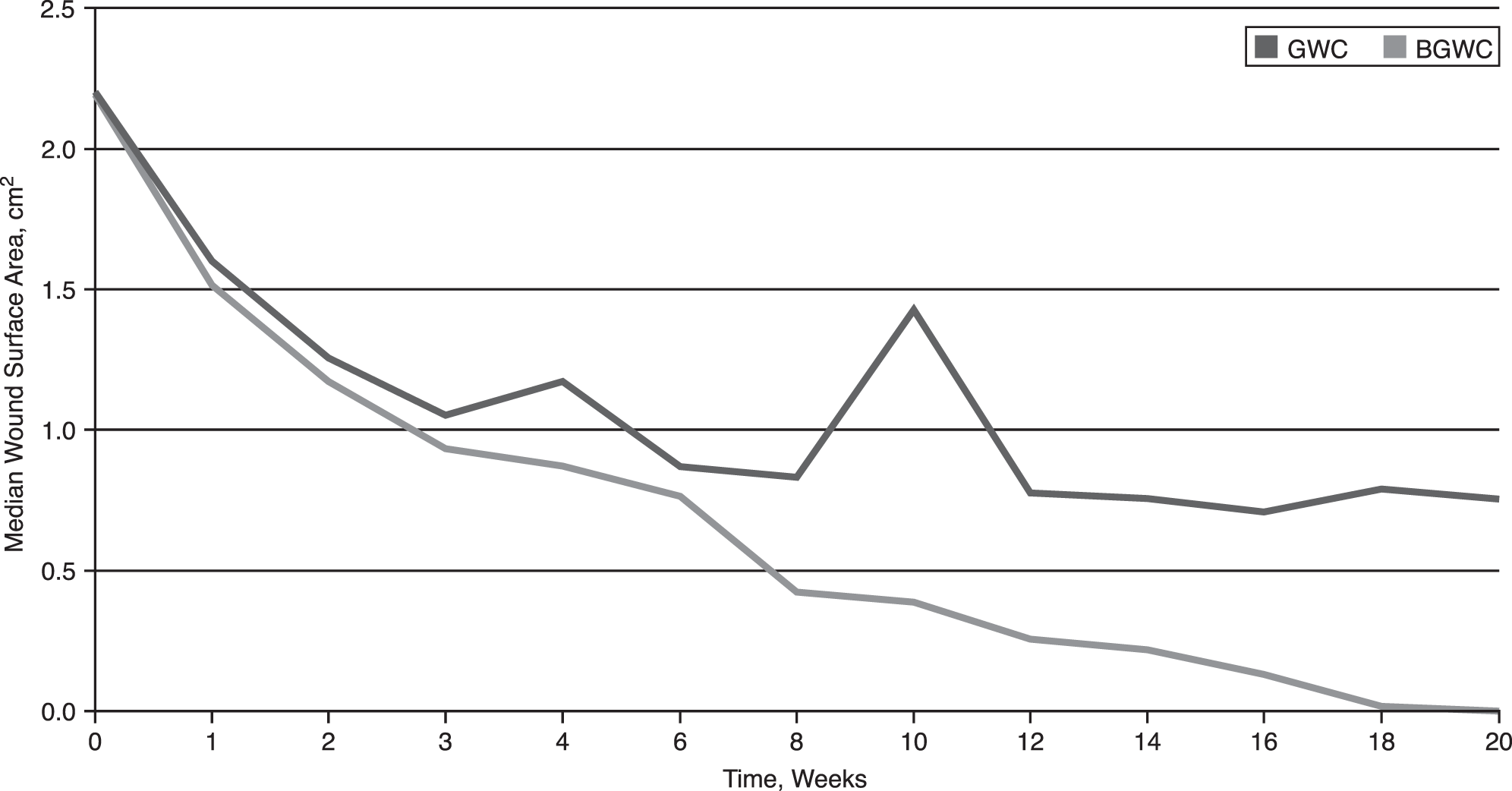
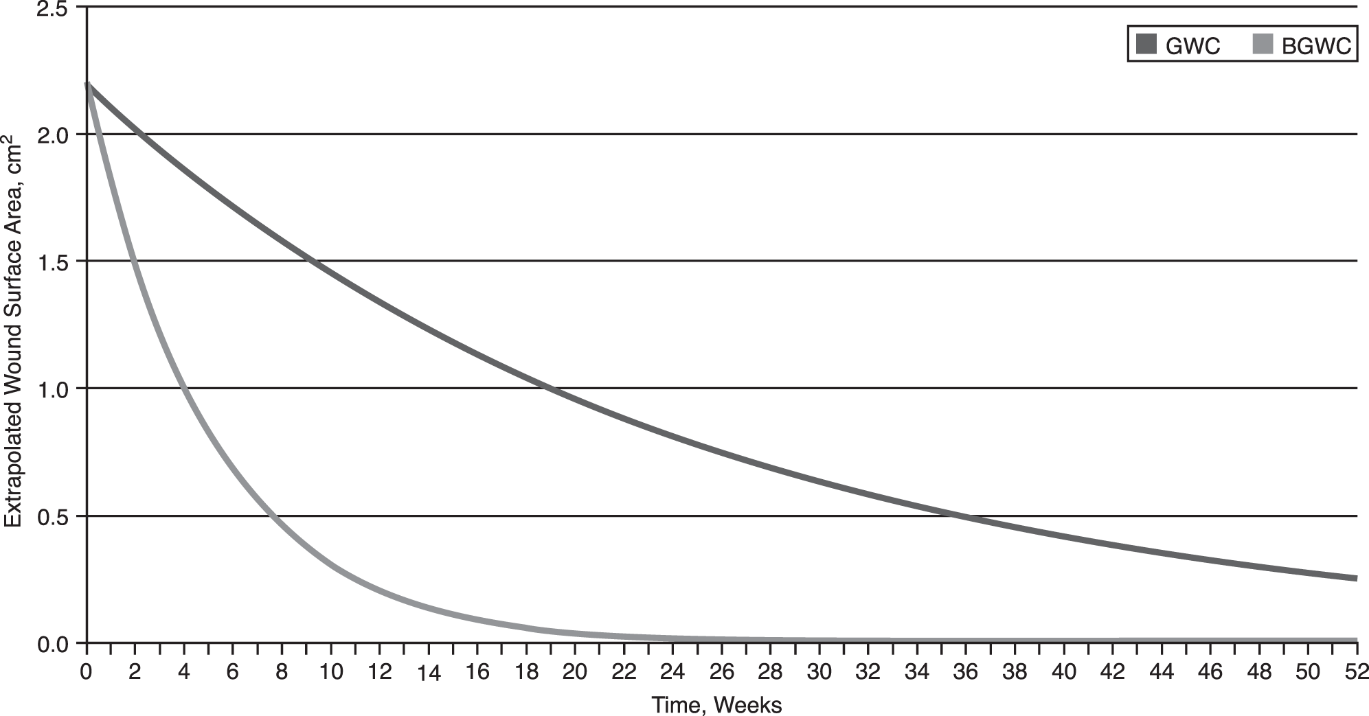
Median baseline WSA was 2.2 cm2 in both groups. At 20 weeks, median WSA was 0.8 cm2 for the GWC group and 0 cm2 for the BGWC group (Fig. 1). The BGWC group had a statistically greater probability of complete wound closure at 20 weeks compared with the GWC group (50% versus 35%; P = .015). Based on reported WSA reduction rates and extrapolation to 52 weeks, DFUs in the BGWC group were expected to close 100% by 27 weeks, and those in the GWC group were expected to close 88% by 52 weeks (Fig. 2). These rates of healing indicate that BGWC-treated DFUs achieve closure approximately 25 weeks (6 months) earlier than DFUs treated with GWC alone.
Figure 1. Reduction of median wound surface area (WSA) from baseline to 20 weeks with good wound care (GWC) and becaplermin gel plus good wound care (BGWC) treatment.
Japma 106 00273 f01
Figure 2. Extrapolated log reduction of wound surface area (WSA) from baseline to 52 weeks with good wound care (GWC) and becaplermin gel plus good wound care (BGWC) treatment. Data were extrapolated to 52 weeks assuming cumulative reduction rates over time using a nonlinear modeling algorithm for WSA reduction.
Japma 106 00273 f02

Economic Results

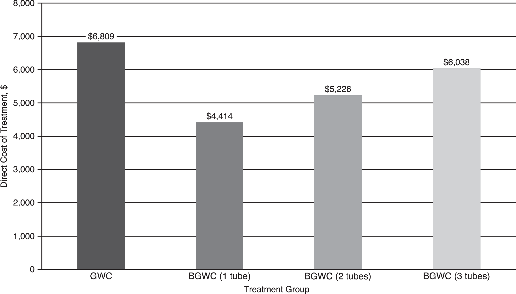
The total costs for BGWC remained lower than the total cost of care for GWC alone, regardless of the number of tubes of becaplermin gel used for BGWC. The total expected direct costs per episode of DFU care were estimated at $6,809 with GWC treatment. Costs for BGWC treatment ranged from $4,414 with one tube of becaplermin gel to $6,038 with three tubes. Becaplermin plus good wound care treatment resulted in an estimated annual cost savings of $771 to $2,395, depending on the amount of becaplermin needed to achieve wound closure (Fig. 3). When costs were compared by wound-closure rates, the cost to achieve a 1-cm2 reduction in WSA ranged from $2,006 to $2,745 in the BGWC group compared with $3,501 in the GWC treatment group. Becaplermin plus good wound-care treatment resulted in an estimated cost savings of $756 to $1,495 per episode of DFU care.
Figure 3. Direct costs for good wound care (GWC) and becaplermin gel plus good wound care (BGWC) treatments.
Japma 106 00273 f03

Discussion

Chronic DFUs often require considerable time to heal and are associated with an increased risk of infections, recurrence, hospitalization, amputations, and other cost drivers that result in severe and costly outcomes.[3] The financial burden of DFUs is influenced by late management of diabetes, resources needed for compliance with standard care (eg, offloading and infection control), complexity of treatment regimens in patients with osteomyelitis, high recurrence and amputation rates, and high morbidity and mortality rates following amputation.[50] The results of this study show that the use of becaplermin gel as an adjunct to GWC can accelerate wound closure and that the total estimated 1-year cost of BGWC therapy was lower than the cost of GWC. Together, these results indicate that BGWC therapy is both more clinically efficacious and less costly than GWC therapy alone.
One of the largest varying costs in advanced DFU therapy regimens is the initial cost of care (ie, the cost of becaplermin gel). Although the cost of adding becaplermin gel to GWC was approximately three times the cost of GWC alone, the therapeutic effect of BGWC was approximately 49% greater than for GWC when wound closure was measured across the entire episode of care. Becaplermin plus good wound care had substantially better closure at 20 weeks (50% versus 35%), faster WSA reduction at 20 weeks (2.2 versus 1.4 cm2), and a significantly greater probability of closure (100% at 27 weeks versus 88% at 52 weeks). It is important to note that prior to initiation of therapy the overall average wound duration was greater than 1 year, with a large standard deviation (56.3 weeks, SD = 80.9). Therefore, these results should be interpreted with caution, as there is large variance in both DFU treatment groups.
In addition to faster closure and WSA reduction, the expected direct cost per episode of care for DFU was lower in the BGWC group compared with GWC alone ($4,414 versus $6,809), a cost savings of up to approximately $2,400 over 1 year. Despite the development of sophisticated mathematical analyses to model the kinetics of wound closure, acceptance among clinicians and regulatory agencies remains limited. Findings from the current investigation using cost per cm2 reduction in WSA using an exponential decay algorithm indicate how the addition of becaplermin to DFU standard treatment can reduce the total direct cost of DFU care to the payer. Using an exponential decay model provides the ability to quantify closure rates as coefficient estimates from a standard statistical test, allows for hypothesis testing on treatment efficacy, and may avoid the limitations of using a binary endpoint (closed or unclosed) at an arbitrarily selected point in time.[45] The model is also an easy-to-use application and does not require mathematical transformation of the actual wound measurements. Furthermore, the simplicity of the model would enable health-care providers to incorporate the algorithm in clinical decision-making at the bedside.[45]
Currently, quality measures in wound care are not well-established. The Tax Relief and Health Care Act was passed in 2006, which authorized a pay-for-performance program called the Physician Quality Reporting Initiative (PQRI), wherein payment is linked to whether the health-care provider performs certain tasks in a given time frame for patients with chronic comorbidities.[51] Although a PQRI measure exists relating to diabetic “foot care” via performing a peripheral neuropathy evaluation and prescribing appropriate footwear, offloading of an existing DFU is not a PQRI measure. As the population continues to age and the prevalence of diabetes and obesity increase, it will be important for third-party payers, such as Medicare, Medicaid, and other government agencies, to evaluate the efficacy and effectiveness of treatment patterns and quality measures in wound care.
These results demonstrate the economic value of becaplermin gel for the treatment of DFUs in outpatient care settings. It is important to note that patients treated with BGWC received becaplermin at the beginning of the trials as an adjunct to GWC and not 4 to 6 weeks after GWC therapy failed. Results from this study suggest that earlier therapy with advanced treatments such as PDGF may be more efficacious and less costly over the long term, even though they are initially more expensive and have higher acquisition costs. A cost analysis by Lantis, et al[42] reported that the average cost to patients with prescription benefits for a course of becaplermin therapy is less than $50 in an inner-city tertiary care facility; therefore, early initiation could potentially lead to lower overall health costs by improving wound-healing outcomes through faster healing and faster closure.
The cost effectiveness of becaplermin has been demonstrated both in the United States and in Europe.[39,40,42,52,53] Becaplermin as an adjunct to GWC has yielded favorable cost-effectiveness ratios in Canada,[39] France,[40] Sweden,[40,52] Switzerland,[40] the United Kingdom,[40] and the United States.[42,53,54] Studies inside and outside the United States have found that treatment with becaplermin has resulted in additional ulcer-free days and weeks and reduced long-term expenses, including office visits, dressings, recurrence rates, and amputation.[39,52,54] Reduced long-term expenses, particularly fewer infections, will yield large savings from fewer hospital days and fewer admissions, readmissions, or both.[55] Advanced therapies, such as becaplermin, that promote rapid and complete healing and reduce the need for expensive surgical procedures could affect these costs substantially. Future research should focus on evaluating the cost-effectiveness of advanced therapies in countries outside the United States and Europe.[56]
Certain limitations should be considered when interpreting findings from this study. The specific amount of becaplermin use was unknown and was estimated using the recommended dosing formula from the product labeling.[44] Whereas the model suggests that WSA reduction follows an exponential decay/growth process, it does not imply that the overall wound healing trajectory follows an exponential decay pattern. Additional research is needed to develop a mathematical formula that accurately fits the entire process of wound closure. In addition, the results of this analysis cannot be applied to other standard care methods, health-care settings, or the treatment of wounds of other etiologies. Furthermore, only the direct costs of care were included in the analysis; indirect and intangible costs were not assessed. Decision makers in other health-care settings and systems should investigate the cost effectiveness of advanced treatment modalities in their own setting. Finally, even though the probability of mortality secondary to malignancy was not measured in the economic model, it should be noted that becaplermin gel contains a black box warning.

Conclusions

The results of this analysis suggest that once-daily treatment with becaplermin gel 100 μg/g increases the incidence of complete healing relative to GWC alone. Treatment of DFU with becaplermin gel resulted in improved clinical outcomes and substantial cost savings despite the additional acquisition cost for becaplermin gel. Health-care providers and decision makers should take into account both the acquisition cost of becaplermin and the total cost of care when making decisions about allocation of financial resources. Furthermore, this study supports using surrogate endpoints such as WSA reduction to measure the cost-effectiveness of wound care therapies. Future investigations of the effectiveness of becaplermin as an adjunct to GWC in routine clinical practice and patient-reported outcomes, such as health-related quality of life, are needed to complement the existing cost-effectiveness data and help reimbursement agencies make informed coverage decisions.

Financial Disclosure

This study, medical writing, and editorial support were funded by Smith & Nephew Inc.

Conflicts of Interest

Drs. Waycaster and Gilligan are employees of Smith & Nephew Inc.

References

  1. American Diabetes Association: National Diabetes Statistics Report 2014. Centers for Disease Control. Available at: http://www.cdc.gov/diabetes/pubs/statsreport14/national-diabetes-report-web.pdf. Accessed 23 July 1, 2014.
  2. Boulton AJ, Vileikyte L, Ragnarson-Tennvall G, et al: The global burden of diabetic foot disease. Lancet366: 1719, 2005.
  3. Frykberg RG, Zgonis T, Armstrong DG, et al: Diabetic foot disorders: a clinical practice guideline (2006 revision). J Foot Ankle Surg 45(5 Suppl): S1,2006.
  4. Palumbo PJ, Melton LJ: “Peripheral vascular disease and diabetes,” inDiabetes In America, edited byMIHarrisand RFHamman, p 1, National Institutes of Health, Bethesda,1985.
  5. Reiber GE: “Epidemiology of foot ulcers and amputations in the diabetic foot, “In:The Diabetic Foot, edited byJHBowkerand MAPfeifer, p 13, Mosby, St. Louis,2001.
  6. Reiber GE, Boyko EJ, Smith DG: “Lower extremity foot ulcers and amputations in diabetes,” inDiabetes in America, 2nd Ed, edited byMIHarris, CCowie, MPStern, p 409, NIH Publication No. 95-1468,1995.
  7. Frykberg RG, Habershaw GM, Chrzan JS: “Epidemiology of the diabetic foot: ulcerations and amputations,” inContemporary Endocrinology: Clinical Management of Diabetic Neuropathy, edited byAVeves, p 273, Humana Press, Totowa,1998.
  8. American Diabetes Association: Economic costs of diabetes in the US in 2007. Diabetes Care31: 596, 2008.
  9. American Diabetes Association: Economic costs of diabetes in the US in 2002. Diabetes Care26: 917, 2003.
  10. Sedory Holzer SE, Camerota A, Martens L, et al: Costs and duration of care for lower extremity ulcers in patients with diabetes. Clin Ther20: 169, 1998.
  11. Pecoraro RE, Reiber GE, Burgess EM: Pathways to diabetic limb amputation: basis for prevention. Diabetes Care13: 513, 1990.
  12. Larsson J, Agardh CD, Apelqvist J, et al: Long-term prognosis after healed amputation in patients with diabetes. Clin Orthop Relat Res350: 149, 1998.
  13. American Diabetes Association: Consensus development conference on diabetic foot wound care. Diabetes Care22: 1354, 1999.
  14. Margolis DJ, Allen-Taylor L, Hoffstad O, et al: Diabetic neuropathic foot ulcers and amputation. Wound Repair Regen13: 230, 2005.
  15. Rice JB, Desai U, Cummings AKG, et al: Burden of diabetic foot ulcers for Medicare and private insurers. Diabetes Care37: 651, 2014
  16. Steed DL, Donohoe D, Webster MW, et al: Effect of extensive debridement and treatment on the healing of diabetic foot ulcers. Diabetic Ulcer Study Group. J Am Coll Surg183: 61, 1996.
  17. Wieman TJ, Griffiths GD, Polk HC, et al: Management of diabetic midfoot ulcers. Ann Surg15: 627, 1992.
  18. Teng YJ, Li YP, Wang JW, et al: Bioengineered skin in diabetic foot ulcers. Diabetes Obes Metab12: 307, 2010.
  19. Mulder G, Armstrong DG, Seaman S: Standard appropriate and advanced care: medical legal considerations for diabetic lower extremity wounds. Wounds26: 1069, 2003.
  20. Ross R, Raines EW, Bowen-Pope DF: The biology of platelet-derived growth factor. Cell46: 155, 1986.
  21. Ross R: Platelet-derived growth factor. Lancet1: 1179, 1989.
  22. Pierce GF, Mustoe TA, Altrock BW, et al: Role of platelet-derived growth factor in wound healing. J Cell Biochem45: 319, 1991.
  23. Kiritsy CP, Lynch AB, Lynch SE: Role of growth factors in cutaneous wound healing: a review. Crit Rev Oral Biol Med4: 729, 1993.
  24. Bennett NT, Schultz GS: Growth factors and wound healing: Part II. Role in normal and chronic wound healing. Am J Surg166: 74, 1993.
  25. Grotendorst GR, Martin GR, Pencey D, et al: Stimulation of granulation tissue formation by platelet-derived growth factor in normal and diabetic rats. J Clin Invest76: 2323, 1985.
  26. Wieman TJ: Clinical efficacy of becaplermin (rhPDGF-BB) gel. Becaplermin Gel Studies Group. Am J Surg 176 (2A Suppl): 74S,1998.
  27. Wieman TJ, Smiell JM, Su Y: Efficacy and safety of a topical gel formulation of recombinant human platelet-derived growth factor-BB (becaplermin) in patients with chronic neuropathic diabetic ulcers. A phase III randomized placebo-controlled double-blind study. Diabetes Care21: 822, 1998.
  28. Knighton DR, Ciresi KF, Fiegel VD, et al: Classification and treatment of chronic nonhealing wounds: successful treatment with autologous platelet-derived wound healing factors (PDWHF). Ann Surg204: 332, 1986.
  29. Smiell JM, Wieman J, Steed DL, et al: Efficacy and safety of becaplermin (recombinant human platelet-derived growth factor-BB) in patients with nonhealing, lower extremity diabetic ulcers: a combined analysis of four randomized studies. Wound Rep Reg7: 335, 1999.
  30. Steed D: “Modulating wound healing in diabetes,” inThe Diabetic Foot, edited byJBowker, MPfeiffer, p 395, Mosby, St. Louis,2001.
  31. Falanga V, Shen J: “Growth factors, signal transduction and cellular responses,” inCutaneous Wound Healing, edited byVFalanga, p 81, Martin Dunitz Ltd, London,2001.
  32. Robson M, Smith P: “Topical use of growth factors to enhance healing,” inCutaneous Wound Healing, edited byVFalanga, p 379, Martin Dunitz Ltd, London,2001.
  33. Hogge J, Krasner D, Nguyen HC, et al: The potential benefits of advanced therapeutic modalities in the treatment of diabetic foot wounds. JAPMA90: 57, 2000.
  34. Rovee DT, Kurowsky CA, Labun J: Local wound environment and epidermal healing. Arch Dermatol106: 330, 1972.
  35. D'Hemecourt PA, Smiell JM, Hugill JV, et al: The effect of topically applied sodium carboxymethyl cellulose gel on non healing lower extremity ulcers in patients with diabetes. Wounds10: 69, 1998.
  36. Steed DL, The Diabetic Ulcer Study Group: Clinical evaluation of recombinant human platelet-derived growth factor for the treatment of lower extremity diabetic ulcers. J Vasc Surg21: 71, 1995.
  37. Bhansali A, Venkatesh H, Dutta P, et al: Which is the better option: recombinant human PDGF-BB 0.01% gel or standard wound care, in diabetic neuropathic large plantar ulcers off-loaded by a customized contact cast?Diabetes Res Clin Pract83: 13, 2009.
  38. Hardikar JV: Efficacy of recombinant human platelet derived growth factor (rhPDGF) based gel in diabetic foot ulcers: a randomized, multicenter, double-blind, placebo-controlled study in India. Wounds17: 141, 2005.
  39. Sibbald RG, Torrance G, Hux M, et al: Cost-effectiveness of becaplermin for nonhealing neuropathic diabetic foot ulcers. Ostomy Wound Manage49: 76, 2003.
  40. Ghatnekar O, Persson U, Willis M, et al: Cost effectiveness of becaplermin in the treatment of diabetic foot ulcers in four European countries. Pharmacoeconomics19: 767, 2001.
  41. Margolis DJ, Gelfand JM, Hoffstad O, et al: Surrogate end points for the treatment of diabetic neuropathic foot ulcers. Diabetes Care26: 1696, 2003.
  42. Lantis JCII, Boone D, Gendics C, et al: Analysis of patient cost for recombinant human platelet-derived growth factor therapy as the first-line treatment of the insured patient with a diabetic foot ulcer. Adv Skin Wound Care22: 167, 2009.
  43. American Medical Association: Current Procedural Terminology. American Medical Association. Available at: http://www.ama-assn.org/ama/pub/physician-resources/solutions-managing-your-practice/coding-billing-insurance/cpt.page. Accessed June 25, 2014.
  44. Regranex Directions for Use. Smith & Nephew Biotherapeutics, Inc.Available at: http://www.regranex.com/. Accessed June 25, 2014.
  45. Cardinal M, Phillips T, Eisenbud DE, et al: Nonlinear modeling of venous leg ulcer healing rates. BMC Dermatology9: 1, 2009.
  46. Gillman T: Wound outcomes: the utility of surface measures. Low Extrem Wounds3: 125, 2004.
  47. Robson MC, Hill D, Woodske M, et al: Wound healing trajectories as predictors of effectiveness of therapeutic agents. Arch Dermatol135: 773, 2000.
  48. Margolis DJ, Malay DS, Hoffstad OJ, et al: Economic burden of diabetic foot ulcers and amputations: Data points #3. Agency for Healthcare Research and Quality, Rockville, 2011.
  49. Fife CE, Carter MJ, Walker D: Why is it so hard to do the right thing in wound care?Wound Rep Reg18: 154, 2010.
  50. Allenet B, Paree F, Lebrun T, et al: Cost-effectiveness modeling of Dermagraft for the treatment of diabetic foot ulcers in the French context. Diabetes Metab26: 125, 2000.
  51. US Government GPO: Tax Relief and Health Care Act of 2006. United States Congress, 2006. Available at: http://www.gpo.gov/fdsys/pkg/PLAW-109publ432/pdf/PLAW-109publ432.pdf. Accessed December 8, 2014.
  52. Persson U, Willis M, Odegaard K, et al: The cost-effectiveness of treating diabetic lower extremity ulcers with becaplermin (Regranex): a core model with an application using Swedish cost data. Value Health3(Suppl 1): S39, 2000.
  53. Albert S: Cost-effective management of recalcitrant diabetic foot ulcers. Clin Podiatr Med Surg19: 483, 2002.
  54. Kantor J, Margolis DJ: Treatment options for diabetic neuropathic foot ulcers: a cost-effectiveness analysis. Dermatol Surg27: 347, 2001.
  55. Warriner RAIII, Driver VR: The true cost of growth factor therapy in diabetic foot ulcer care. Wounds (Supplement): S1, 2006.
  56. Papanas N, Maltezos E: Becaplermin gel in the treatment of diabetic neuropathic foot ulcers. Clin Interv Aging3: 233, 2008.