Percutaneous distal osteotomy (PDO) is an effective procedure for treating mild-to-moderate hallux valgus deformity [
1,
2,
3,
4]. Done under fluoroscopic visualization, the procedure involves a complete distal metatarsal osteotomy performed through a 1-cm medial incision at the metatarsal neck. The realignment is stabilized by the insertion of a 2-mm Kirschner wire from distal to proximal into the medullary canal of the first metatarsal [
1]. The most commonly reported benefits of the procedure include shorter operating times, a decreased risk of complications related to the minimal surgical exposure and higher patient satisfaction, with clinical outcomes comparable to the more popular open techniques. The major advantage of performing a PDO is that it should have a limited effect on the postoperative range of motion of the hallux because it is an extra-articular operation. However, complications such as avascular osteonecrosis, nonunion, malunion, and recurrence of the deformity have been previously reported [
5]. Nonunion of the first metatarsal after hallux valgus surgery is a rare complication that requires surgical management.
We present a case of a 42-year-old woman who developed a pseudarthrosis of the first metatarsal after percutaneous retrocapital distal osteotomy of the first metatarsal for a mild hallux valgus deformity. The operative treatment consisted of debridement of fibrous nonunion with plating followed by application of pulsed electromagnetic fields (PEMF) with an external device.
Case Report
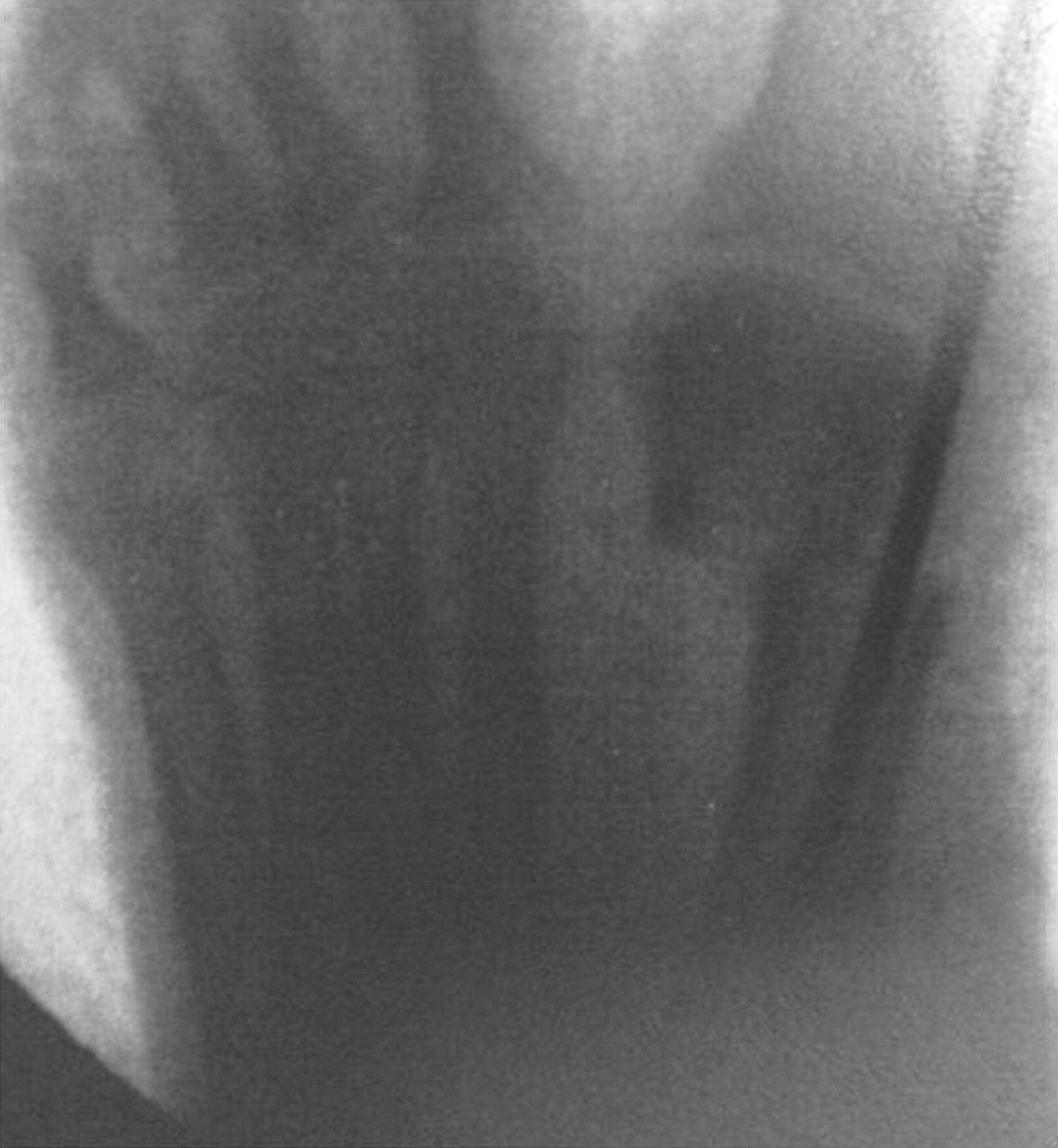
A 42-year-old woman was referred to our clinic with complaints of painful walking with metatarsalgia. Six months before presentation, she underwent a PDO of the first metatarsal of the right foot for a mild hallux valgus deformity. In reviewing the operative records, no complications during or immediately after metatarsal osteotomy were noted (
Fig. 1). The Kirschner wire was removed 5 weeks postoperatively. Partial weightbearing was advised with a postoperative shoe; after 6 weeks, full weightbearing was allowed. Six months later she still had persistent pain on the anteromedial aspect of the right foot during walking without a history of trauma since the procedure. Conservative treatment with physical therapy, activity modification, and foot orthotic had failed to relieve metatarsalgia.
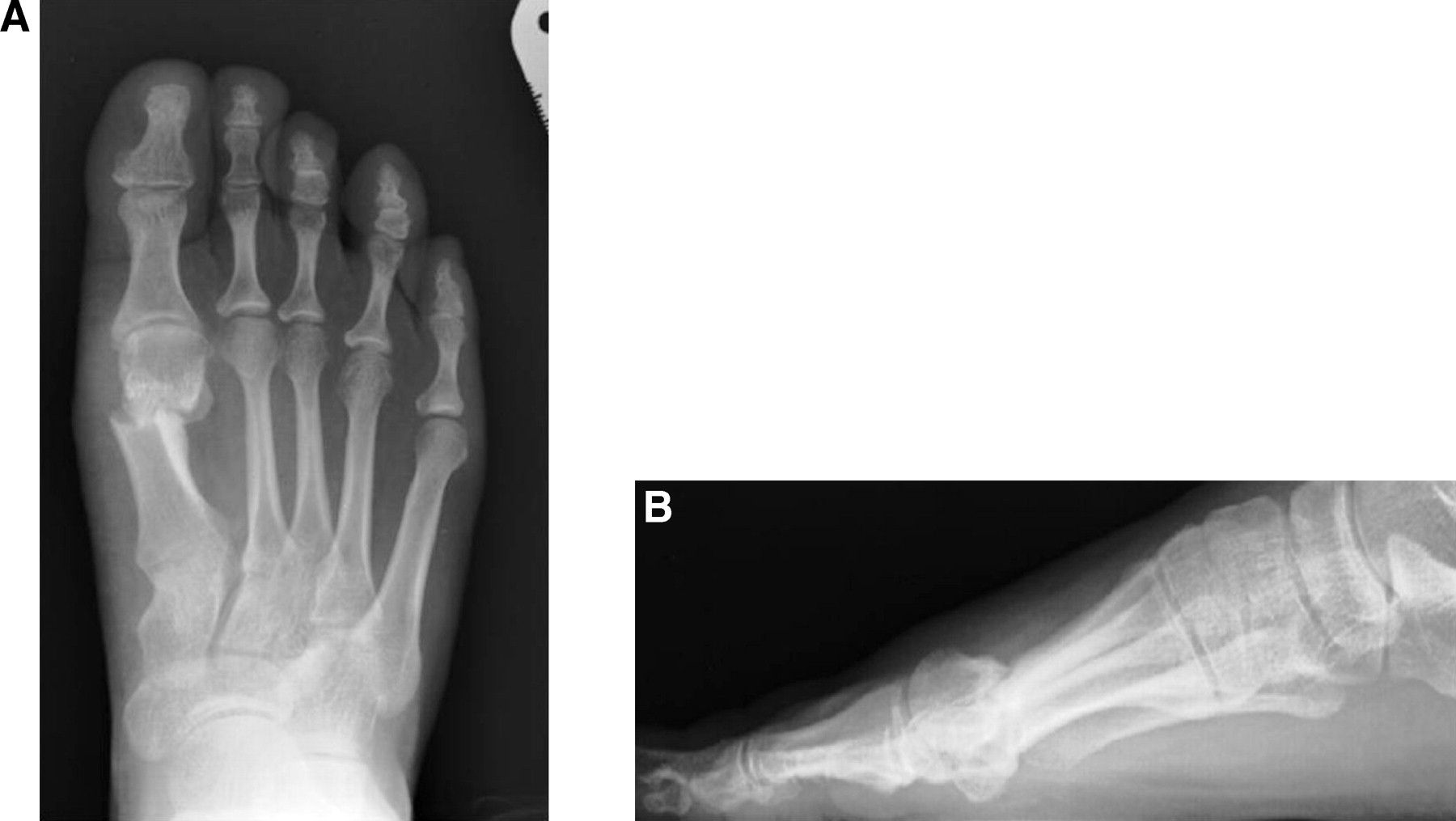
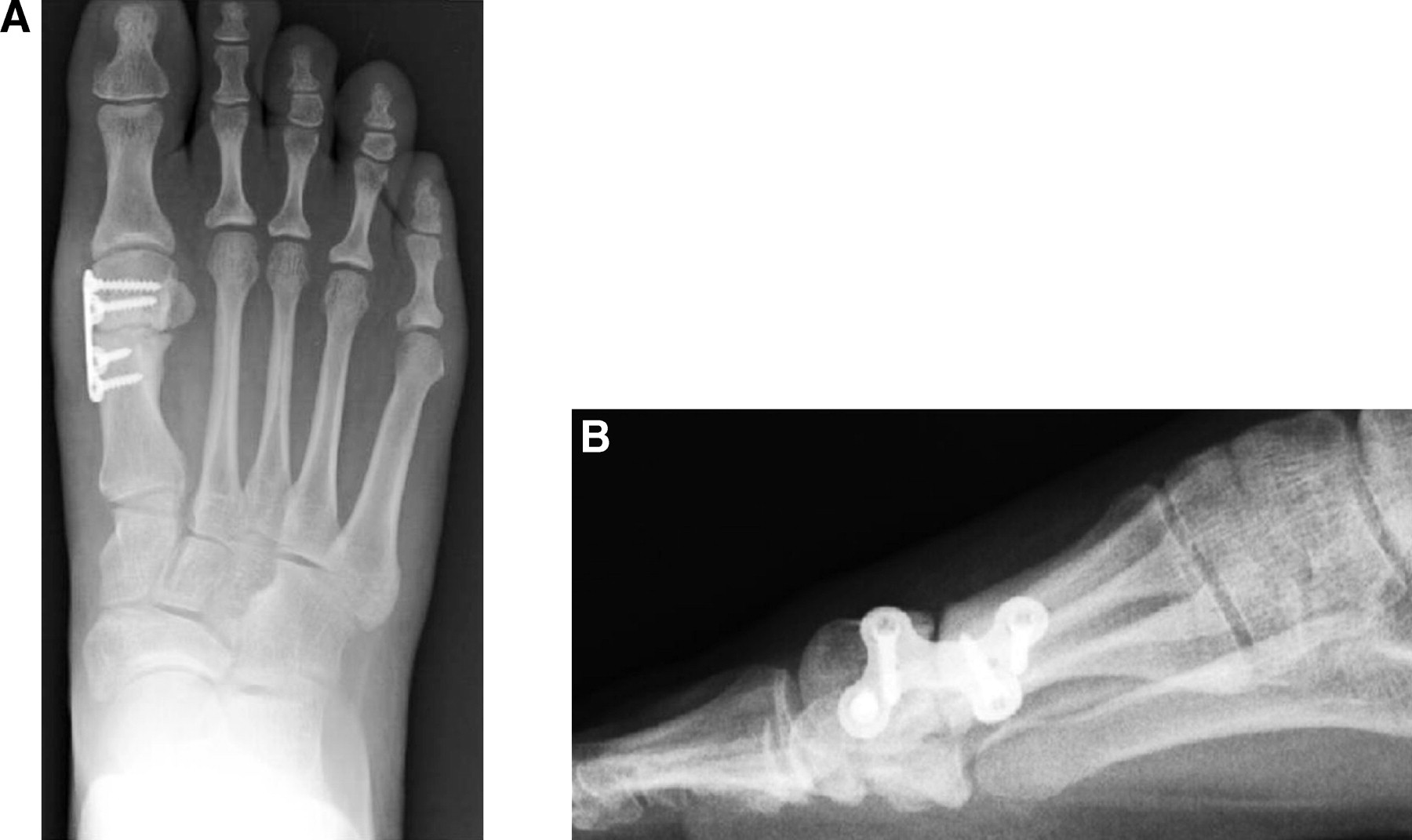
On physical examination, there was painful limited range of motion of the first metatarsophalangeal joint with 40° of dorsiflexion and 30° of plantarflexion. No signs of infection were observed. Palpation of the first metatarsal head was painful. Radiographic examination showed signs of non-union with dorsal displacement of the metatarsal head (
Fig. 2). First metatarsal length of the right foot was 44 mm and 45 mm on the left foot. Magnetic resonance imaging was performed in order to exclude an avascular necrosis of the metatarsal head.
Figure 1.
Intraoperative fluoroscopic image showing the Kirschner wire used to stabilize the metatarsal head.
Figure 1.
Intraoperative fluoroscopic image showing the Kirschner wire used to stabilize the metatarsal head.
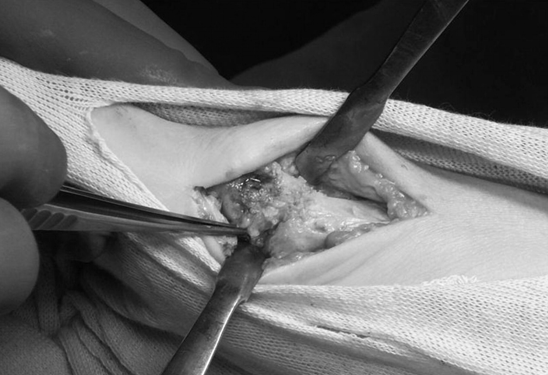
Through a dorsomedial incision, the first metatarsophalangeal joint and the osteotomy site were inspected (
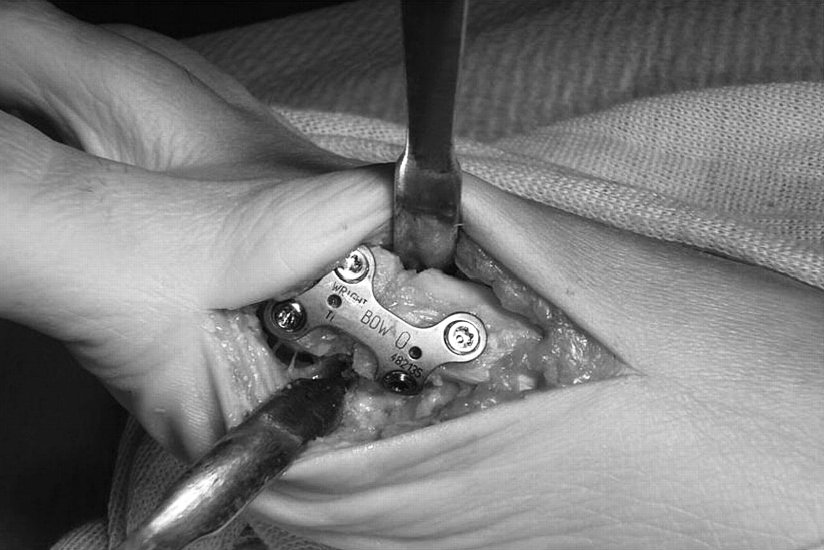
Fig. 3). The osteotomy site was identified, and granulation and fibrous tissues were removed with a curette. A high-speed burr was used to freshen the sclerotic bone of the cortical bone ends. The ankle tourniquet was released to observe punctuate bleeding from both ends. Internal fixation was achieved with a medial locking plate (Bow; Wright Medical, Arlington, TN) repositioning the first metatarsal head plantarly (
Fig. 4).
Early postoperative mobilizations started on the first postoperative day, and full weightbearing was allowed with a postoperative shoe for 6 weeks. Full weightbearing without limitations was permitted at 6 weeks when radiographs showed absence of malalignment (
Fig. 5). The PEMF stimulation started 7 days after surgery and was maintained until initial bone consolidation was observed. The device used was BIOSTIM (IGEA, Carpi, Italy), which generated pulses of 1.3 ms duration with 75 Hz frequency and an induced electric field of 3.5 ± 0.5 mV as measured in a standard coil probe [
6].
The use of PEMF provided an effective treatment for delayed unions and nonunion of the lesser metatarsal [
7].
Figure 2.
Dorsoplantar (A) and lateral (B) weightbearing radiographs showing the nonunion of the first metatarsal with elevation of the metatarsal head.
Figure 2.
Dorsoplantar (A) and lateral (B) weightbearing radiographs showing the nonunion of the first metatarsal with elevation of the metatarsal head.
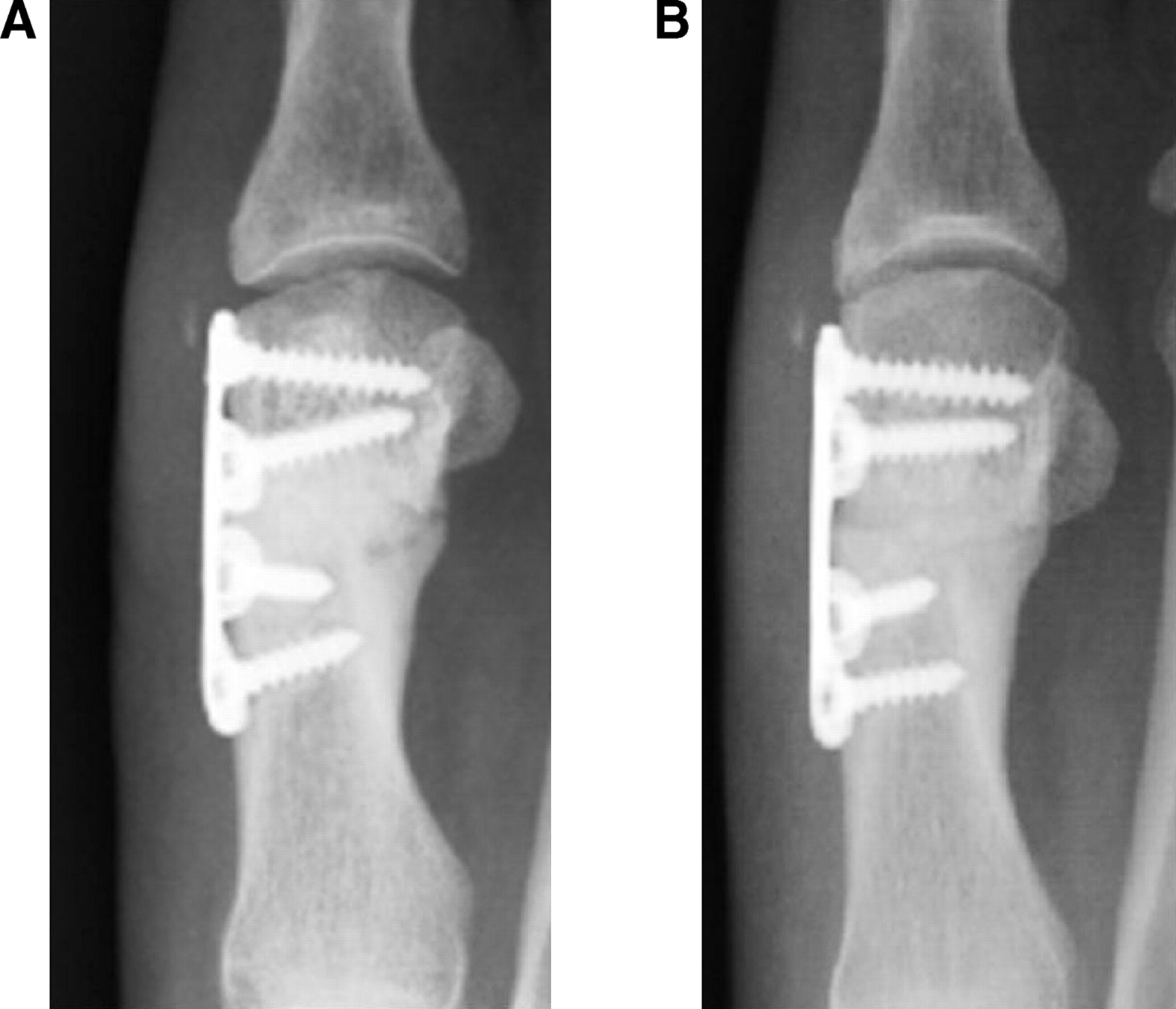
The hallux metatarsophalangeal-interphalangeal score according to the system of the American Orthopaedic Foot and Ankle Society was 44 points before surgery and 100 points at the last follow-up (14 months). Radiographically, initial bone healing was observed at 3 months follow-up (
Fig. 6). Nonunion was considered healed at 6 months follow-up. At 12 months follow-up, the patient was pain free and had resumed running.
Figure 3.
Intraoperative picture demonstrating non-union of the first metatarsal head.
Figure 3.
Intraoperative picture demonstrating non-union of the first metatarsal head.
Figure 4.
A locking plate with four screws was applied medially. The metatarsal head was repositioned more plantarly.
Figure 4.
A locking plate with four screws was applied medially. The metatarsal head was repositioned more plantarly.
Discussion
Nonunions represent a challenging problem and often result in significant pain and disability. Many factors have been identified as contributing to the development of nonunions, which include infection, inadequate fixation, medical comorbidities, and comminution [
8].
Nonunion as a consequence of distal metatarsal osteotomy for the treatment of hallux valgus represents a rare complication. Inadequate fixation at the time of surgery is considered the most frequent cause of nonunion [
9]. Leutloff described the case of a patient affected by pseudarthrosis after first distal metatarsal osteotomy treated with Kirschner wire fixation and local inoculation of autolougous corticospongiose material collected from the pelvic crest [
10].
Figure 5.
Dorsoplantar (A) and lateral (B) weightbearing radiograph 1 month after surgery.
Figure 5.
Dorsoplantar (A) and lateral (B) weightbearing radiograph 1 month after surgery.
Minimally invasive distal first metatarsal osteotomy with a percutaneous technique was first described by Bösch et al [
11] in 1990, and satisfactory results were reported in a 7-to-10–year follow-up study [
1]. Portaluri [
12] achieved an 89% satisfaction rate with the Bösch technique and stated that the advantages of this technique included short operation time and low incidence of complications. Sanna and Ruiu [
13]. reported excellent results in a long-term follow-up study of percutaneous distal first metatarsal osteotomies. Magnan et al [
4]. reported that the patients were satisfied following 107 (91%) of 118 percutaneous distal first metatarsal osteotomies. Maffulli et al [
3]. reported no difference between the Scarf technique and minimal incision subcapital osteotomy of the first metatarsal for treating patients with mild-to-moderate hallux valgus deformity.
Figure 6.
Bone consolidation at 3 months (A) and 6 months (B) postoperatively.
Figure 6.
Bone consolidation at 3 months (A) and 6 months (B) postoperatively.
Disadvantages of the percutaneous technique are the long learning curve and the lack of predictability of head displacement [
5]. Magnan et al [
4]. stated that inadequate fixation of the osteotomy, while performing a PDO, is avoided by driving the Kirschner wire into the base of the first metatarsal. However, doubts about the stability of fixation still persist. In a series of 13 patients treated with a PDO for a mild-to-moderate hallux valgus, Kadakia et al reported an unacceptable rate of complications, such as osteonecrosis, nonunion, malunion and recurrence [
5]. The authors concluded that the intraoperative correction was commonly lost after removal of the intramedullary Kirschner wire.
In this patient, the radiographic examination did not show an atrophic nonunion, suggesting that excessive motion was the main etiologic factor for nonunion rather than impaired vascularity. We choose the operative treatment with locking plate stabilization without bone grafting, considering the acceptable length of the first metatarsal and good vascularity of the bone ends. The locking plate has been applied on the medial aspect of the first metatarsal shaft in order to prevent dorsal impingement during active dorsiflexion of the hallux, with the tendon. We did not consider plate removal because it was well tolerated by the patient.
In this case, treatment of metatarsal nonunion with a locking plate and postoperative PEMF application achieved initial bone consolidation within 3 months.