Peripheral nerve injuries in the foot and ankle are difficult diagnoses because they often arise from external and internal forces that work together to cause symptoms. External forces consist of trauma or shoe gear, while internal forces are compressive factors such as space occupying lesions, compartment forces and traction from muscles and tendons. Bony prominences or spurs can be anatomic or develop from abnormal biomechanics. Generally, the affected nerve will have a reduced vascular supply causing edema and inflammation, leading to perineural fibrosis. Symptoms can include rest pain and intermittent paresthesias and can lead to constant pain, muscle atrophy and sensory loss [
1,
2,
3].
Symptoms of sural nerve impingement attributable to a mechanical origin have been reported in the literature. Kennedy et al [
4] reported sural neuritis in dancers that was related to plantarflexion and inversion type moments at the ankle. McCrory et al [
5] described a fascial origin for lower extremity nerve impingement, specifically at the proximal origin of the sural nerve. They proposed that the same fascial restriction that predisposes to muscle compartment syndromes may also envelop the neurovascular structures within the leg resulting in either ischemic or neurogenic symptoms. Bryan et al [
6] reported proximal sural nerve entrapment causing pain and paresthesias in a 43-year-old man following a strain of the gastrocnemius muscle.
In this case report, traction of the lateral calcaneal branch of the sural nerve was clinically diagnosed using objective mechanical findings and correlated anatomically. The symptoms were addressed by using a heel lift and activity modification. Traction can be created from an acute or chronic localized stretch as was noted in this patient.
Anatomy
The sural nerve has a sensory function only and is derived from S1–S2 roots. From the common peroneal nerve is born the lateral sural cutaneous nerve above the knee joint and pierces the fascia lata over the popliteal fossa. The tibial nerve likewise produces the medial sural cutaneous nerve usually just below the knee joint. The medial sural cutaneous nerve then joins with the lateral sural cutaneous nerve (and the sometimes communicating peroneal branch) to form the sural nerve. The sural nerve runs down the posterolateral side of the leg and ultimately runs along the lateral side of the foot, becoming the lateral dorsal cutaneous nerve. At approximately 2 cm above the ankle, it gives two branches: 1) one supplies sensation to the lateral aspect of the heel, the lateral calcaneal branch of the sural nerve, and 2) the other often anastomoses with the lateral branch of the superficial peroneal nerve. It is the first of these branches that is germane to this case study discussion.
Case Report
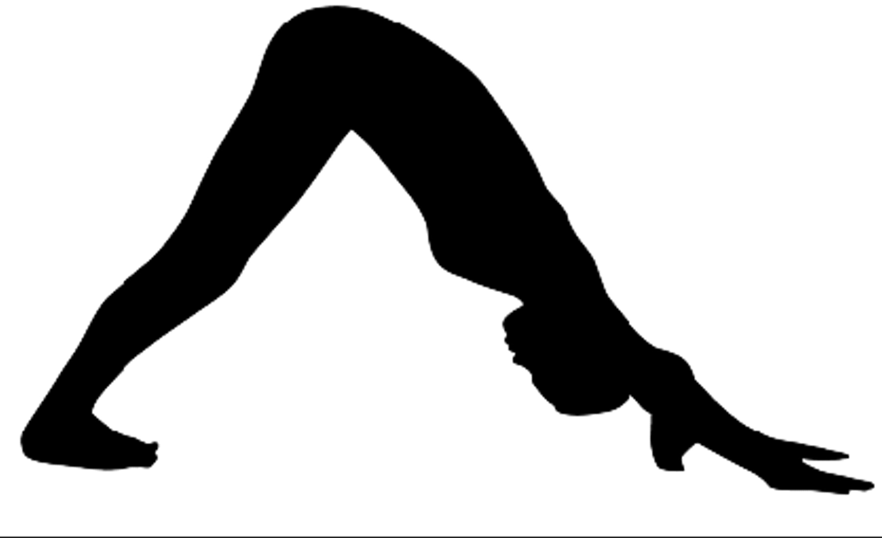
A 51-year-old female distance runner presented with pain along the right posterior lateral heel. The pain was described as a sharp burning, tingling sensation that was most evident while doing the downward dog position in yoga class. In this position the pelvis is flexed while the knees and ankles are in full extension. The individual is on “all fours” (
Fig. 1) and as flexibility improves over time, the hands and feet can be moved in closer proximity.
Objectively, there was no swelling or erythema noted on the lateral posterior right heel. A mild Tinel’s sign was elicited with percussion of the lateral calcaneal branch of the sural nerve. The palpable enlargement of the nerve can be subtlety mobilized under the skin and feels like a lamp cord under a rug (the so-called lamp cord sign) [
7]. Symptoms could also be elicited by this maneuver. The patient had a sharp, lancinating pain emanating along the lateral heel and Achilles area with the knee extended and foot in maximum dorsiflexion. No signs of sciatica or back pain were noted. The peroneal tendons, achilles tendon, and the remainder of the posterior heel were not tender. Biomechanically, ankle dorsiflexion was 2 degrees with knee fully extended and 6 degrees with knee flexed. Hamstring and illiopsoas muscle flexibility was within normal limits. Resting calcaneal stance position was 3 degrees everted bilateral. The right lower extremity was approximately 2 to 3 mm longer than the contralateral limb. Radiographs were unremarkable. No external forces could be implicated since yoga activity was practiced without shoes.
This patient received a conservative protocol consisting of heel lifts under orthotic devices, nonsteroidal anti-inflammatory drugs, and ice. The patient was cautioned to modify her yoga and to omit the downward dog maneuver. Symptoms reduced immediately and she gradually returned to her training program.
Figure 1.
“Down Dog” position in Yoga.
Figure 1.
“Down Dog” position in Yoga.
Discussion
Sural nerve impingement is known to have traumatic and compressional etiologies. The lateral calcaneal branch of the sural nerve can be affected by local trauma, irritation from bony prominences, shoes, or local traction. A fascial restriction component has been proposed, however this affects the sural nerve at its more proximal origins. Local nerve impingement has been linked to fascial inflammation resulting in an increase in compartment pressure. A common example is for Baxter’s neuritis that develops from chronic plantar fasciitis.
The patient presented with localized pain and paresthesia that was aggravated with combined stretching of the calf muscle and ankle dorsiflexion. The authors believe local traction applied to this anatomical branch of the sural nerve both caused and aggravated neuritic symptoms. Symptoms resolved with the use of heel lifts because the traction of the local nerve was relieved. Heel lifts have also been used for Achilles tendonitis, posterior heel pain, and extreme cases of equinus.
However, other biomechanical considerations may have come into play. In our case with a gastrocnemius equinus deformity (whether compensatory or congenital) the sural nerve (and its lateral calcaneal branch) length adapts correspondingly to its “normal physiological length.” In the yoga maneuver “downward dog,” where the participant progressively tries to gain flexibility the lateral sural cutaneous nerve is placed upon a considerable stretch (traction) thereby resulting in nerve injury. To postulate that those with a gastrocnemius equinus deformity may be predisposed to this aforementioned injury while participating in yoga may provide a basis for a future study.