Arthrodesis of the first metatarsophalangeal joint is a commonly performed operation for hallux rigidus, with overall success of 80% to 100%. There are some cadaveric and biomechanical studies [
1–
4] comparing different methods of fixation for achieving metatarsophalangeal joint arthrodesis. However, there is no clinical study, to our knowledge, comparing different methods of fixation for achieving arthrodesis. We sought to compare the use of Memory staples (DePuy Orthopaedics Inc, Warsaw, Indiana) with two other methods of achieving first metatarsophalangeal joint arthrodesis in terms of time to fuse and complications.
Patients and Methods
Patient Selection
We conducted a retrospective study using the medical records and radiographs of consecutive patients who had undergone first metatarsophalangeal joint arthrodesis between April 1, 2006, and April 30, 2008, at three consultants’ practices in the National Health Service. Of the three consultants, two were senior orthopedic surgeons and one was a specialist foot and ankle surgeon. One senior consultant used a single lag screw, and the second consultant used a single lag screw and a circlage wire for all his operations. The foot and ankle surgeon used two Memory staples. Patients with unilateral or bilateral osteoarthritis in the form of hallux rigidus were included in the study. They had to have at least one radiograph postoperatively, and it had to be at least 12 months since having had their surgery. We used a mixture of a computerized coding system, a handwritten theater logbook, and a computerized radiograph archive system to ensure that we included all eligible patients. Radiographs of all eligible patients were anonymized and viewed by an independent observer (a post-fellowship specialist registrar) who was not involved in this study, apart from the first author (K.S.). Patients with inflammatory arthritis, lesser toe surgery, and revision operations were excluded to minimize confounding variables.
Treatment Methods
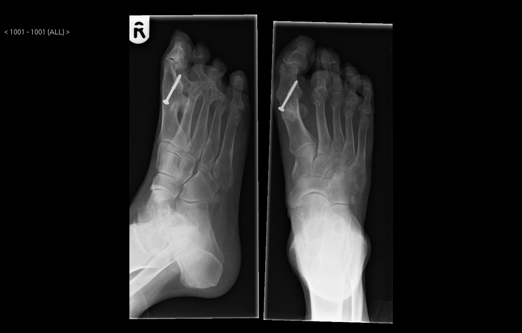
Lag Screw (Group 1). The senior consultant using this method used a dorsomedial incision to prepare the joint similar to a cup-cone, followed by fixation with a single partially threaded 4.0-mm cancellous screw from proximal to distal (
Fig. 1). Postoperatively, patients were given a plaster slipper/walking cast for 6 weeks, followed by physiotherapy for intrinsic muscle exercises.
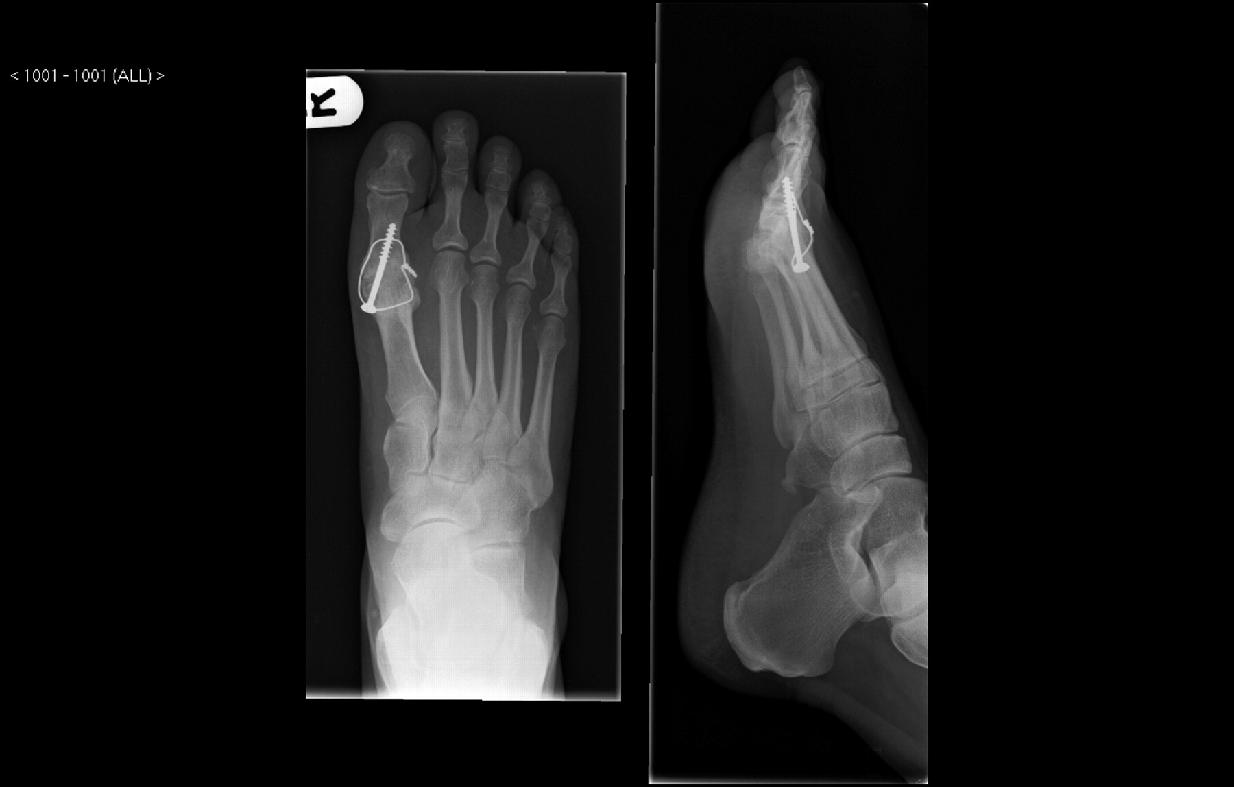
Lag Screw and Circlage Wire (Group 2). The senior consultant using this method used a dorsal incision to prepare the joint similar to a cup-cone, followed by fixation with a single partially threaded 4.0-mm cancellous screw from proximal to distal, along with circlage wire (
Fig. 2). Postoperatively, patients were allowed to bear weight on the heel without a cast.
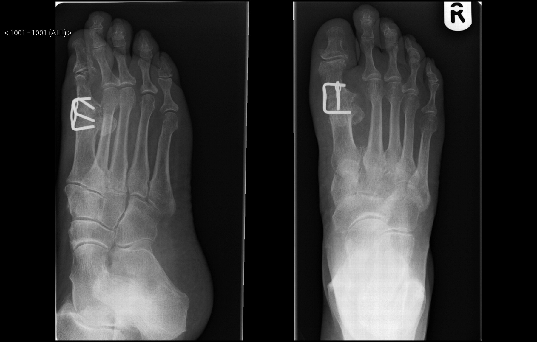
Memory Staples (Group 3). The foot and ankle surgeon using this method used a dorsal incision to make conservative flat-cuts at the joint, followed by fixation with two compression Memory staples at least 60° to 90° to each other (
Fig. 3). Postoperatively, patients were allowed to bear weight on the heel without a cast.
Figure 1.
Lag screw technique.
Figure 1.
Lag screw technique.
Figure 2.
Lag screw and circlage wire technique.
Figure 2.
Lag screw and circlage wire technique.
Postoperative Follow-up
After the operation, the patients were reevaluated at the end of the second week for a postoperative wound check. Further follow-up was performed 6 weeks after surgery and at 4- to 6-week intervals until there was clinical and radiographic evidence of satisfactory arthrodesis. Arthrodesis was considered clinically complete if there was lack of tenderness/movement on examination of the arthrodesis, and radiographic arthrodesis was considered to be present if trabeculae were observed to traverse the arthrodesis interface without loss of the corrected alignment achieved at the time of arthrodesis. The observations between the two clinicians (the first author [K.S.] and an independent observer) were compared for reliability.
Figure 3.
Two compression Memory staples technique.
Figure 3.
Two compression Memory staples technique.
Statistical Analysis
Summary statistics of the two outcome variables, proportion of cases in which fusion took place and time to fusion, are presented. Differences in the proportions were analyzed with the Fisher exact test, and the average healing times were compared with Kruskal-Wallis one-factor nonparametric analysis of variance given the skewed nature of the time variable. All of the tests were performed at the 5% level of significance.
Results
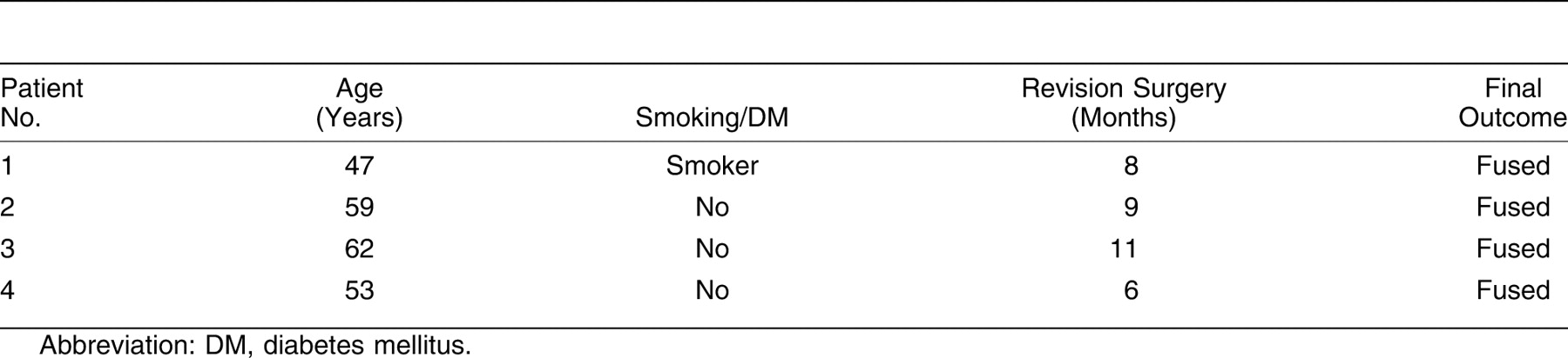
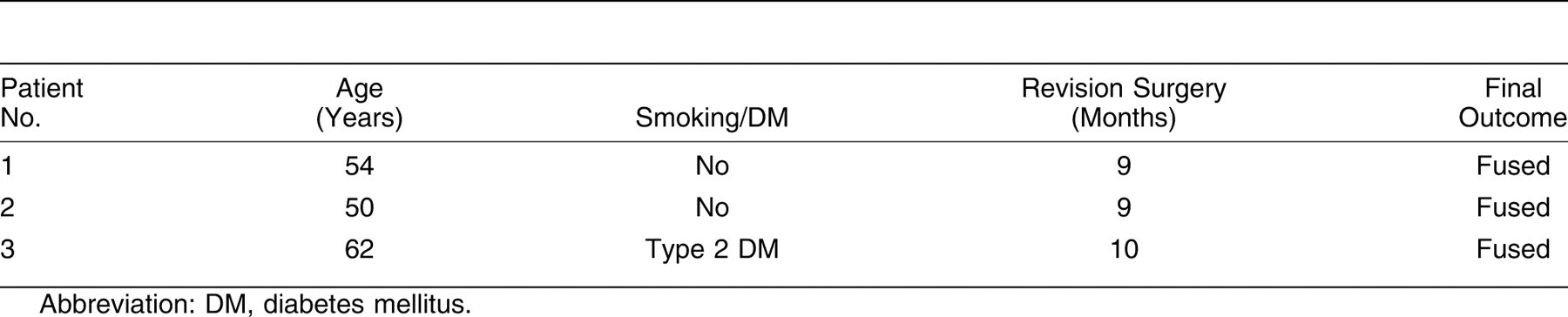
Forty-six patients had metatarsophalangeal joint arthrodesis between April 1, 2006, and April 30, 2008. The numbers in each group were similar (15 patients in groups 1 and 3 and 16 in group 2). The average age of the patients was 53.4 years. There were 28 women and 18 men. The interobserver reliability for radiologic arthrodesis was excellent. The results for group 1 (
Table 1 and
Table 2), group 2 (
Table 3 and
Table 4), and group 3 (
Table 5 and
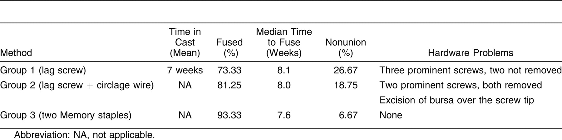
Table 6) are demonstrated. A summary of the results is given in
Table 7.
In group 1, there were three patients with prominent screws, but only one of them was removed. In group 2, there were two patients with prominent screws, and both were removed. One patient had developed a tender bursa over the tip of the screw that was excised. In group 3, there was one patient who had a broken staple that did not require removal because the joint was fused and the broken staple was not prominent.
No significant differences were found among groups in terms of the proportion of cases where fusion took place (P = .300–.685). Similarly, no significant difference was found in the average time until fusion (P = .943).
Table 1.
Group 1: Single Lag Screw Fixation.
Table 1.
Group 1: Single Lag Screw Fixation.
Table 2.
Nonunions in Group 1 (Single Lag Screw Fixation).
Table 2.
Nonunions in Group 1 (Single Lag Screw Fixation).
Table 3.
Group 2: Lag Screw and Circlage Wire Fixation.
Table 3.
Group 2: Lag Screw and Circlage Wire Fixation.
Table 4.
Nonunions in Group 2 (Lag Screw and Circlage Wire Fixation).
Table 4.
Nonunions in Group 2 (Lag Screw and Circlage Wire Fixation).
Table 5.
Group 3: Two Compression Memory Staples Fixation.
Table 5.
Group 3: Two Compression Memory Staples Fixation.
Table 6.
Nonunion in Group 3 (Two Compression Memory Staples Fixation).
Table 6.
Nonunion in Group 3 (Two Compression Memory Staples Fixation).
Discussion
Many different osteosynthesis techniques for first metatarsophalangeal joint arthrodesis have been described, including crossed Kirschner wires, vertical or horizontal intraosseous wire sutures, dorsal plate and screws, interfragmentary screws, and Memory staples. Multiple retrospective studies [
5–
10] have reported success rates for first metatarsophalangeal joint arthrodesis of 80% to 100%. However, contradictory evidence exists in the literature regarding cadaveric biomechanical studies aimed at assessing the strength and rigidity of different fixation methods used to achieve first metatarsophalangeal joint arthrodesis. Curtis et al [
11] and Sykes and Hughes [
12] found the interfragmentary lag screw fixation method to be the most stable fixation method. On the contrary, Neufeld et al [
2] found that combined plate and screw constructs were stronger regarding force to failure and initial stiffness compared with compression Memory staples; however, they also noted that Memory staples have the advantage in their ease of insertion and theoretical continuous compressive force across the arthrodesis site. At Southern General Hospital (Glasgow, UK), we use a lag screw, a lag screw and circlage wire, or two compression Memory staples; the plate and screw construct is reserved for revision procedures. In this study, we compared the use of compression Memory staples with the single lag screw and single lag screw with circlage wire techniques. There are no clinical studies, to our knowledge, that compare three different methods of fixation for first metatarsophalangeal joint arthrodesis. We found that the difference between the median time to fuse when using Memory staples was quicker (although not significant,
P = .943) compared with the other two methods of fixation. The arthrodesis rate in the Memory staples group (93.33%), with one case of nonunion, was comparable with that in a similar study, [
13] although the time to arthrodesis in the present study was much quicker (7.6 vs 8.2 weeks).
Table 7.
Summary of the Results.
Table 7.
Summary of the Results.
Satisfactory arthrodesis requires good joint preparation and fixation. The most important aspect of good joint preparation is denudation of all cartilage. Once all the cartilage is denuded, preparations of the joint surface similar to a cup-cone or matching flat surfaces depends on the preference of the surgeon. There is no evidence to suggest that any one technique of joint preparation (cup-cone or flat surfaces) is better than the other. A method that is easy to perform and reproducible will lead to more predictable outcomes. An ideal fixation should provide compression, resist bending and torsion, and cause no hardware-related problems. Theoretically, the lag screw technique provides compression, the lag screw and circlage wire technique provides compression and resists torsion, and the Memory staples technique offers “dynamic compression,” [
14] and the use of more than one staple resists torsion as well. [
15] Good bone apposition in arthrodesis is important to optimize staple fixation, and we believe that making flat surfaces gives better compression. Fixation effectiveness is known to be increased by a greater number of staples. To decrease rotational forces, more than one staple is recommended. Our experience corroborates the advantages of staples as described in the literature, including good approximation of bone fragments and technically easy application with fewer steps than an AO-applied screw.
One limitation of this study is that it was not performed in a randomized controlled manner. A prospective randomized controlled study would be of value to minimize bias and to further evaluate this method of internal fixation for the first metatarsophalangeal joint arthrodesis. The other limitation is the relatively short follow-up of 1 year. Degeneration of the adjacent joints may develop or increase with time, but this has not been reported to be a factor in other studies. [
16–
18]
Conclusions
Each fixation technique can produce satisfactory results, although a median arthrodesis time of 7.6 weeks, a shorter recovery time, no hardware-related problems, and a high rate of union (93.33%) seem to suggest that compression Memory staples have an advantage over the other techniques. Despite the small number in each group to draw sufficient conclusions, we advocate Memory staples for first metatarsophalangeal joint arthrodesis.