


Abstract
Extra-abdominal desmoid tumors account for 0.03% of all neoplasms and rarely present in the foot. They are benign but locally aggressive, and wide local surgical excision is the treatment of choice owing to the high rate of recurrence in the lower extremities. Invasiveness into the surrounding soft-tissue structures often makes wide excision difficult without compromise of function. We describe a 34-year-old woman with a large, pedal, extra-abdominal desmoid fibroma treated by wide local surgical excision with minimal complications postoperatively and no clinical evidence of recurrence at 28 months.
Case Report
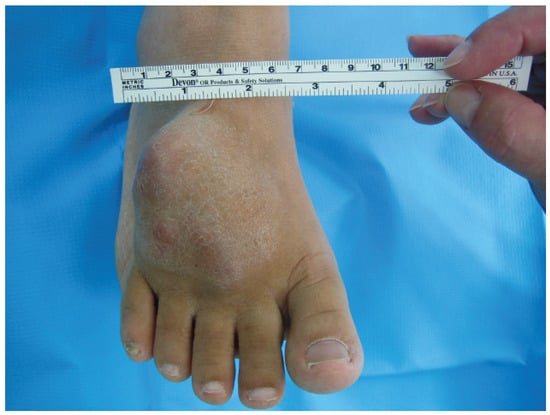
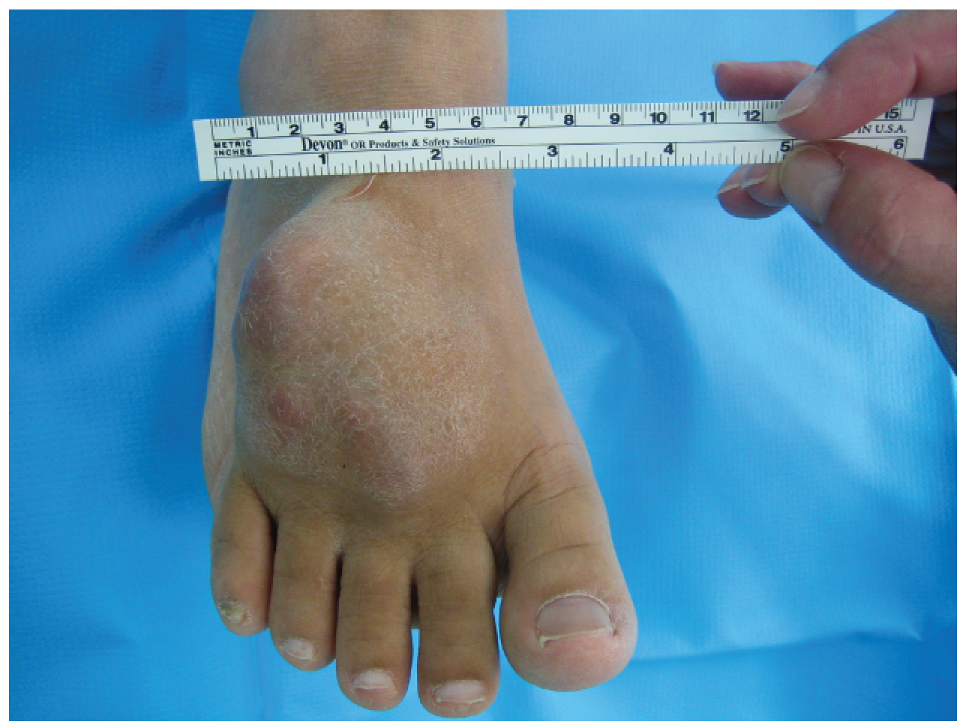
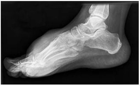
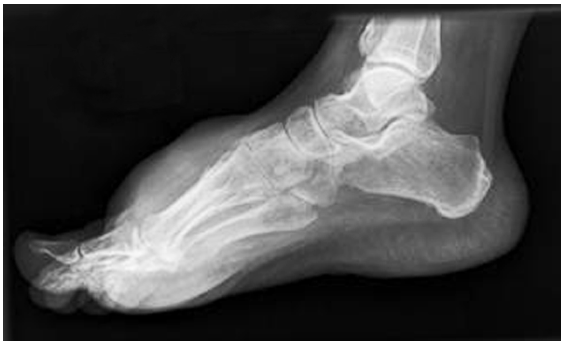
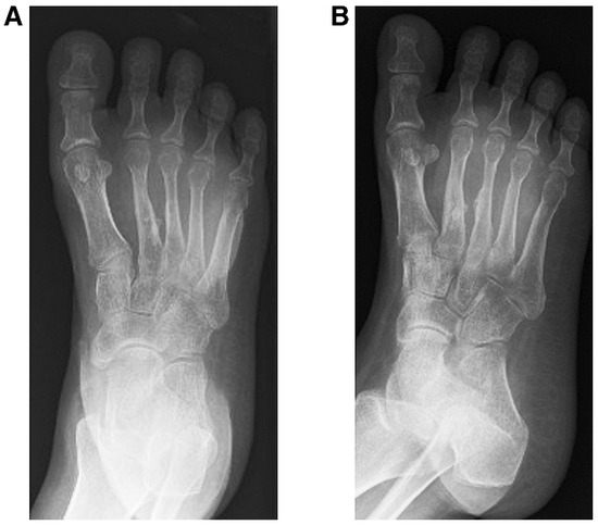
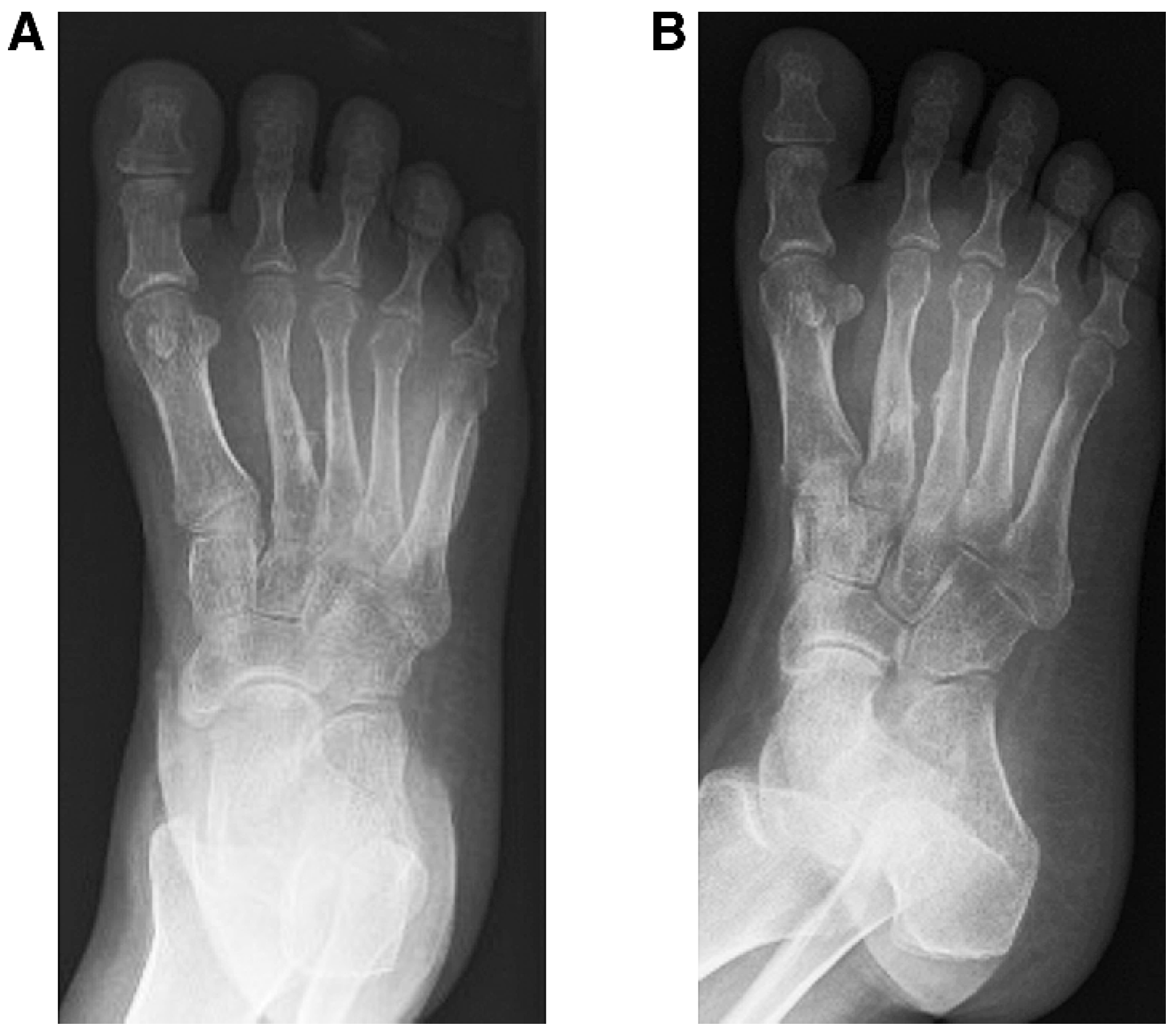
A 34-year-old woman presented in September 2005 with a large, painful mass on the dorsum of her foot that had been present for 1 year. The mass had continued to increase in size and had limited her ability to wear most footwear. Physical examination demonstrated a large, firm, nonmobile, lobulated mass overlying the dorsal tarsometatarsal region (Figure 1). Standard radiography revealed a large softtissue mass with mild calcification adjacent to the second and third metatarsals suspicious for periosteal reaction. Mild bowing of the metatarsals was noted secondary to prolonged pressure of the mass, but no evidence of gross bony destruction was identified (Figure 2 and Figure 3).
Figure 1.
Clinical photograph of the large, firm, nonmobile dorsal mass.
Figure 2.
Lateral radiograph showing increased softtissue contour and density at the dorsal forefoot.
Figure 3.
Dorsoplantar (A) and oblique (B) radiographs showing increased soft-tissue density and calcification near the shafts of the second and third metatarsals suspicious for periosteal reaction.
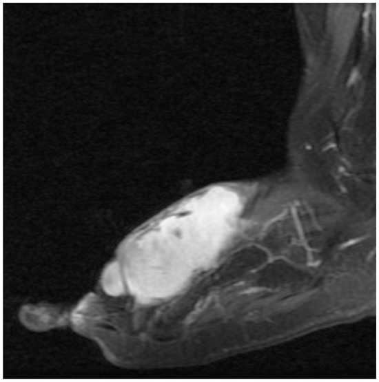
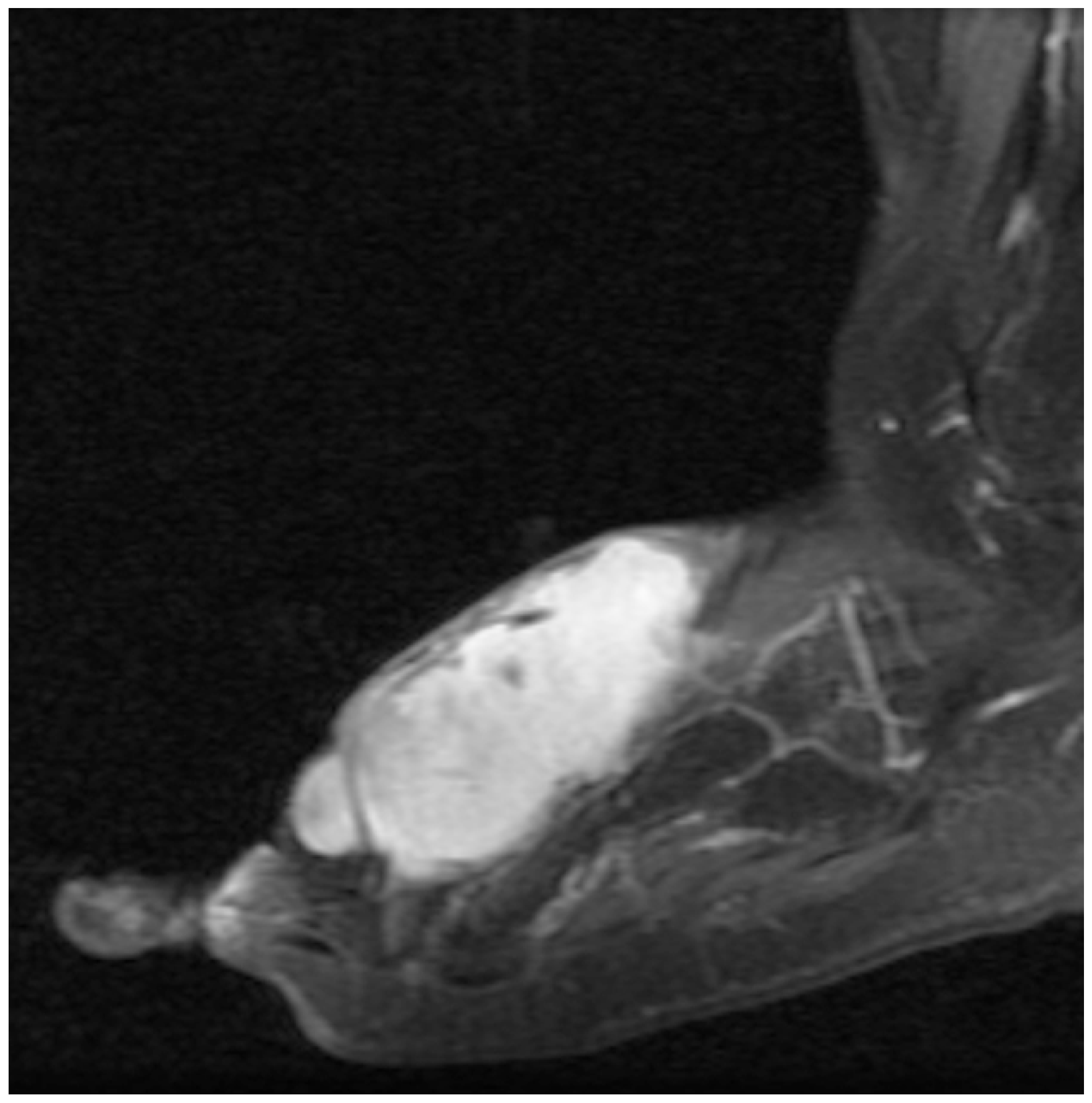
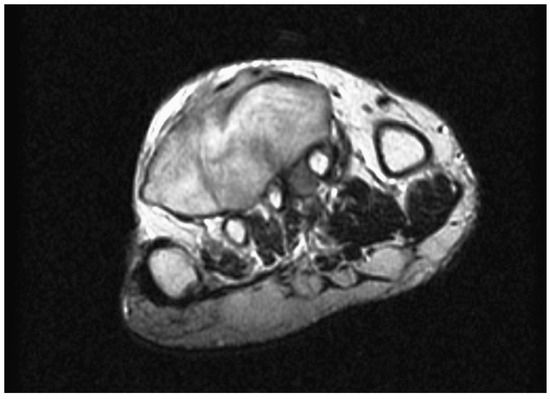
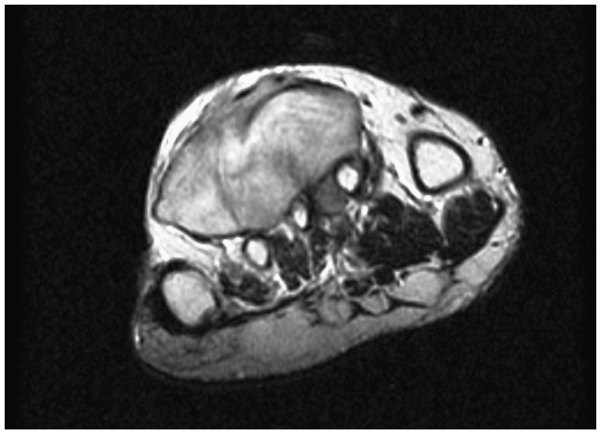
She was admitted to the hospital 2 weeks later for further imaging and evaluation. Total-body magnetic resonance imaging was performed to rule out metastatic lesions, and none were identified. Pedal magnetic resonance images with and without contrast demonstrated an avidly enhancing, welldefined mass located in the dorsum of the forefoot that extended into the second and third intermetatarsal spaces and encompassed two vessels. The mass was immediately adjacent to, but did not breach, the cortex of the second, third, and fourth metatarsals (Figure 4 and Figure 5). Radiologic differential diagnoses included schwannoma, neurofibroma, giant cell tumor of the tendon sheath, and malignant etiologies that could not be ruled out such as clear cell sarcoma, synovial cell sarcoma, and malignant fibrous histiocytoma.
Figure 4.
Sagittal spin-echo T1 with contrast magnetic resonance image showing the avidly enhancing, large, lobulated neoplasm dorsal to the lesser metatarsals.
Figure 5.
Axial fast spin-echo T2 magnetic resonance image showing the nonfibrous portion of the mass having a lesser signal intensity than that of fat.
The patient was brought to the operating room for incisional biopsy of the large soft-tissue mass. The biopsy sample was sent to the pathology department for fresh frozen section diagnosis. Results reported back intraoperatively described a spindle cell neoplasm with myxoid features with a low risk of metastasis although locally invasive.
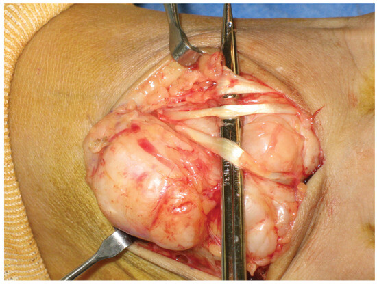
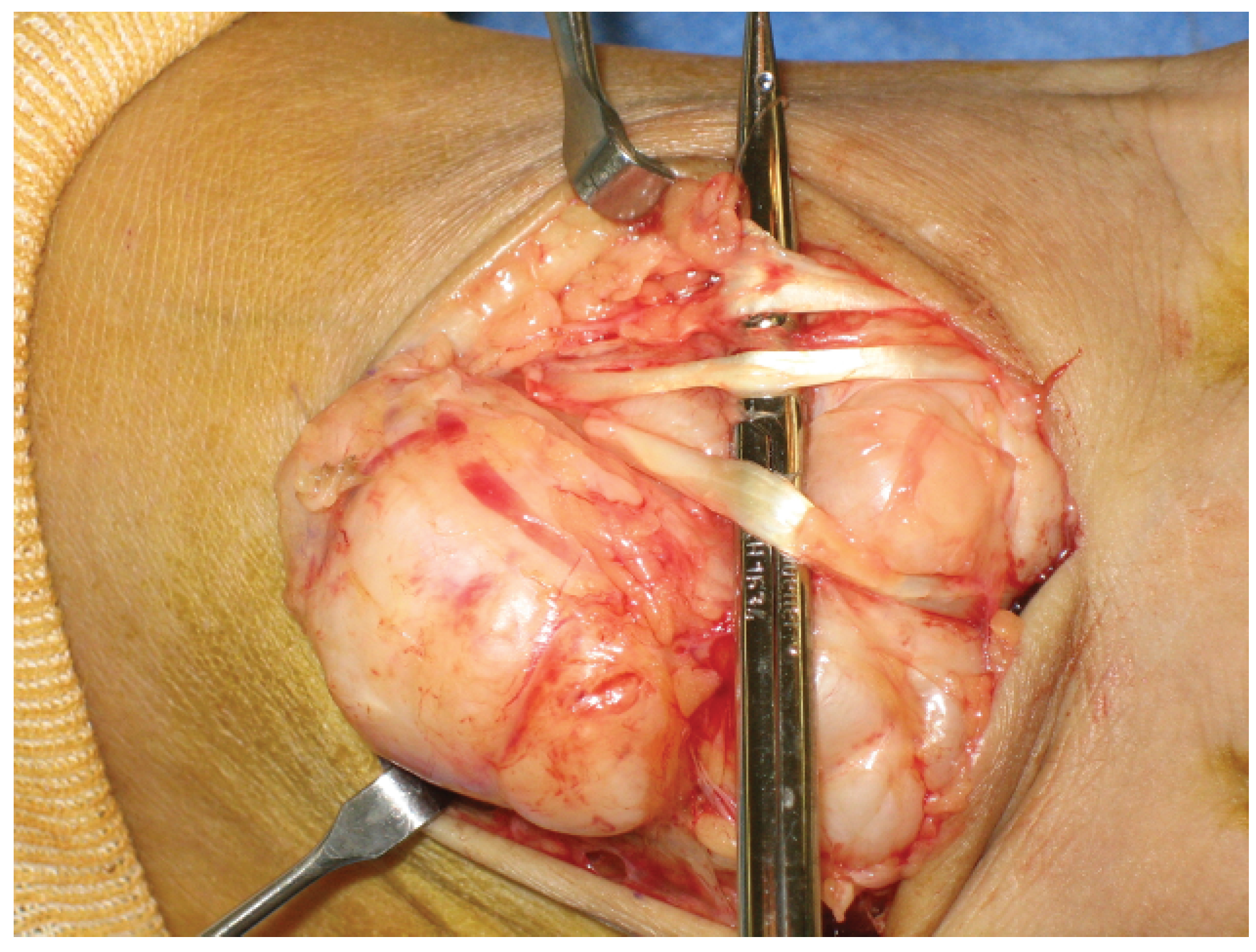
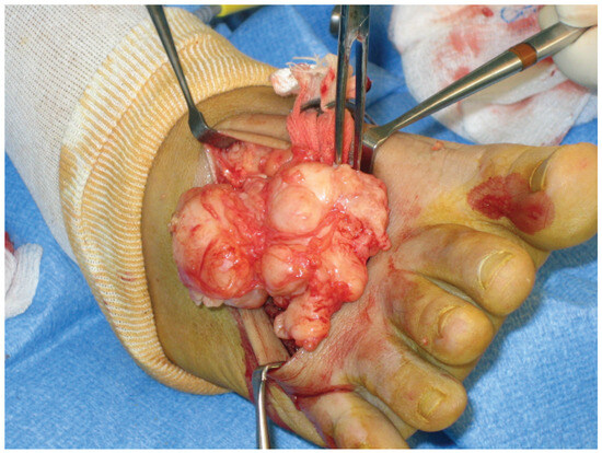
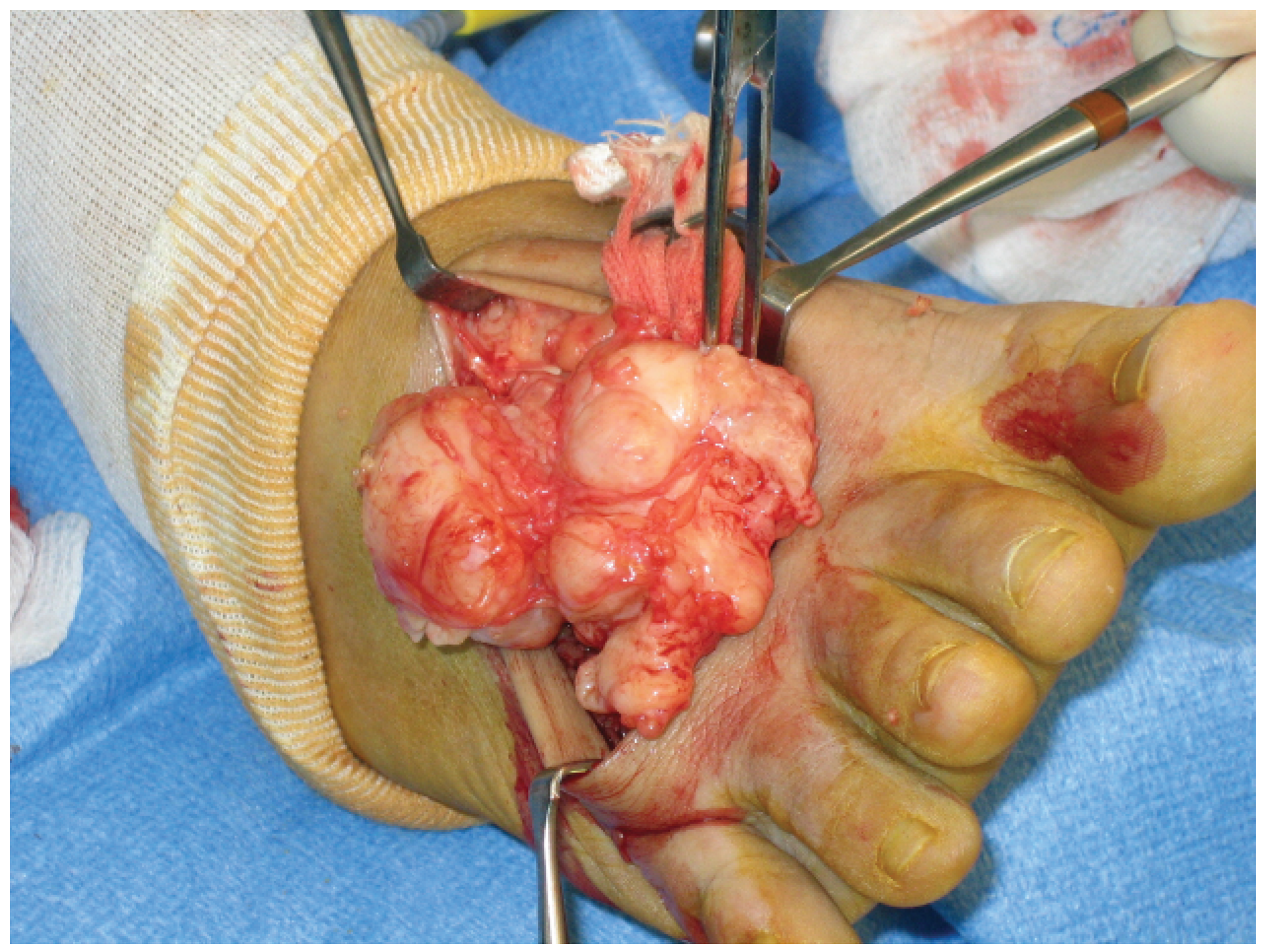
Complete excision of the mass immediately followed. The superficial neurovascular structures were easily retracted and preserved, and no evidence of deep neurovascular entrapment was identified. Although easily separated from the surrounding tissues at the dorsal, medial, and lateral aspects, the mass entrapped the long extensor tendons to the second, third, and fourth digits and had multiple adhesions near the base of the second, third, and fourth metatarsals (Figure 6 and Figure 7). The extensor tendons were freed from the mass, and dissection of the tumor was completed by removing a portion of the involved periosteum overlying the lesser metatarsal bases. The mass was removed in toto without any visible remains in the foot. A TLS drain (Porex Technologies Corp, Fairburn, Georgia) was inserted, and the wound was closed in layers. A compressive dressing was applied, and the patient was kept nonweightbearing. The drain was removed the next day, and the patient was discharged 2 days after surgery.
Figure 6.
Entrapped tendons of the extensor digitorum longus muscle in the dense, fibrous tumor.
Figure 7.
The long extensor tendons are retracted along with the neurovascular supply, and the tumor is carefully dissected free.
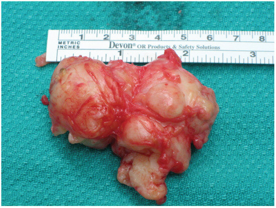
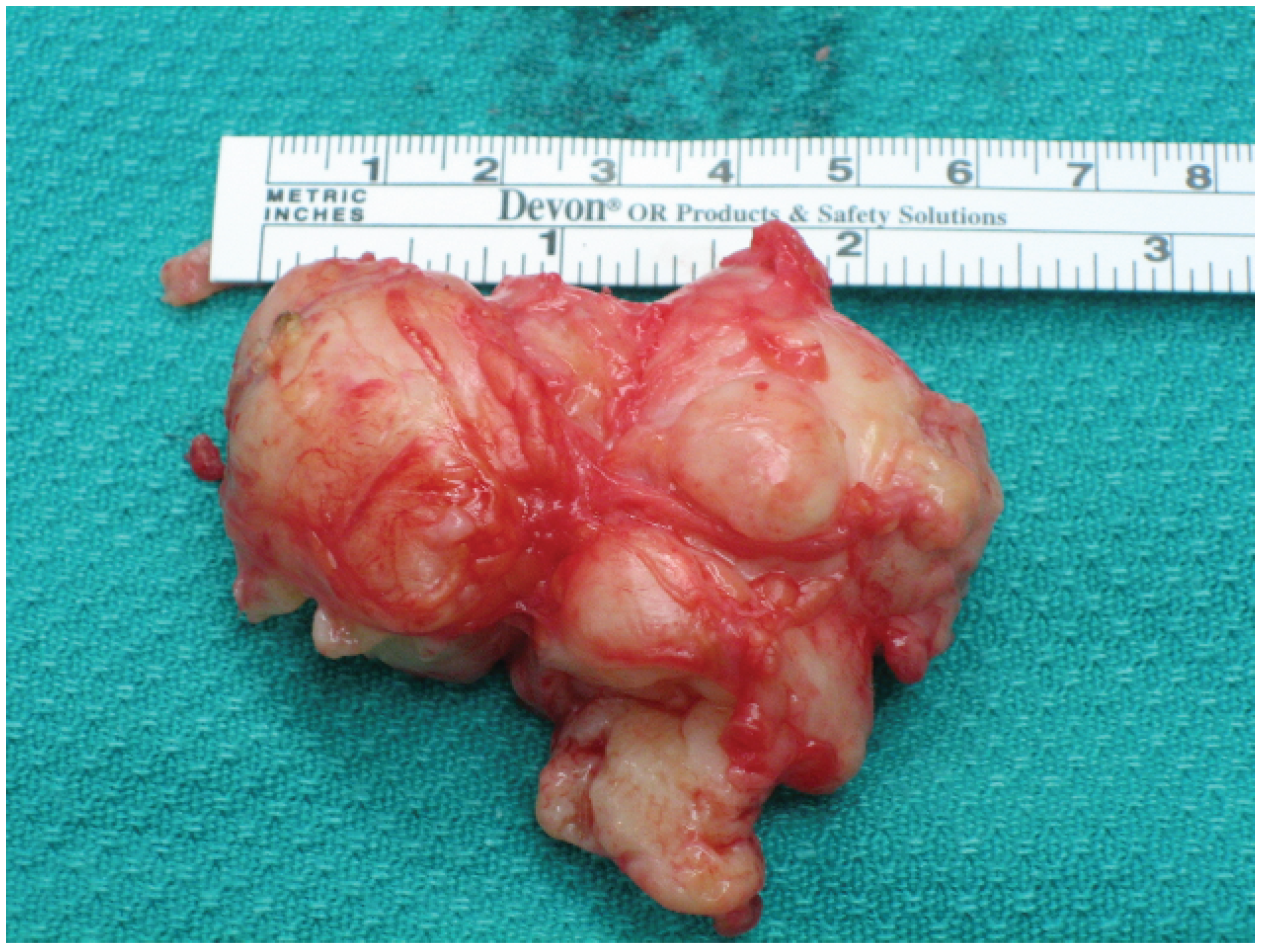
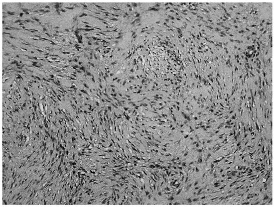
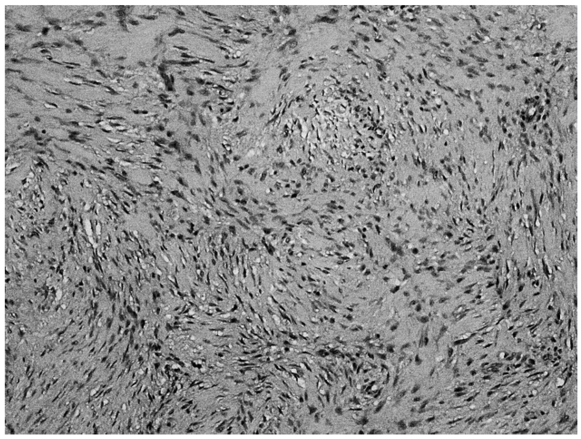
The tumor had a mass of 38 g, measured 6.5 × 5.0 × 2.5 cm, and appeared as encapsulated, firm, pinktan tissue (Figure 8). Confirmed pathologic diagnosis was delayed owing to intradepartmental and interdepartmental consultations. Tissue samples were sent to the Harvard Medical School Department of Pathology for review. The pathology report noted that the neoplasm exhibited focally myxoid spindle cells with a predominant fascicular growth pattern, an unusual finding in low-grade fibromyxoid sarcoma. Furthermore, many of the nuclei in the fascicular areas showed multifocal striking keloidal hyalinization (Figure 9). Immunostains showed focal positivity for smooth muscle actin and rare neo-plastic cells positive for S-100 protein. Staining for β-catenin was positive in cell cytoplasm but not in nuclei, as is usually expected in desmoid fibromatoses. Most important, the pathology report described positive surgical margins of the tissue, indicating incomplete resection of the mass from the foot.
Figure 8.
The tumor had a mass of 38 g and measured 6.5 × 5.0 × 2.5 cm.
Figure 9.
Photomicrograph showing focally myxoid spindle cells with a predominant fascicular growth pattern (3200).
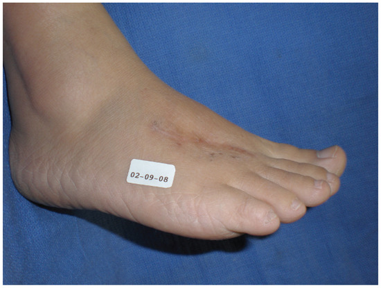
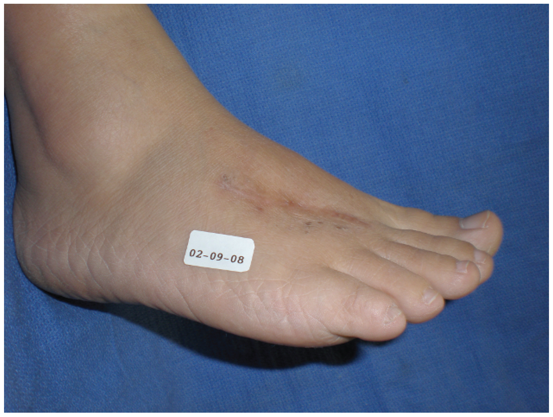
The patient had an unremarkable postoperative course. No osseous complications were noted; however, she had generalized numbness over the dorsal distal forefoot and mild extensor weakness to the second, third, and fourth digits. She was pain free and fully functional in shoes 4 weeks after surgery. No clinical or radiographic evidence of recurrence was noted up to 28 months after excision of the lesion (Figure 10).
Figure 10.
Postoperative clinical photograph showing no evidence of recurrence. The patient maintained function of the extremity and noted only numbness over the incision site.
Discussion
Extra-abdominal desmoid tumors, or aggressive fibromatosis, account for only 0.03% of all neoplasms and for less than 3% of soft-tissue neoplasms.[1,2] The estimated incidence in the general population is 2 to 4 per million population per year, and women aged 25 to 35 are most commonly affected.[3] They are classified as rare, slow-growing, histologically benign tumors of fibroblastic origin.[1,2,4,5] Although benign, they exhibit aggressive local invasion into the surrounding structures and have often been classified as low-grade fibrosarcomas. Transformation of an extra-abdominal desmoid tumor into a fibrosarcoma is rare but has been reported in the literature.[6]
Magnetic resonance imaging is the modality of choice for the assessment of soft-tissue tumors. Although it does not provide histologic specificity, some magnetic resonance imaging features can help differentiate desmoids from similarly appearing malignant lesions.[7] Specifically, the nonfibrous portion of a desmoid tumor has lesser signal intensity than does that of fat on a T2 image compared with a malignant lesion.[8]
Immunohistochemical analysis of extra-abdominal desmoid fibromas has shown cells to be positive for vimentin, α-smooth muscle actin, and desmin.[9] Nuclear β-catenin staining can discriminate desmoid tumors from other proliferations of fibroblastic or myofibroblastic origin.[10] This tumor exhibited positive immunostaining for α-smooth muscle actin and β-catenin.
Bouffier advocated wide excision in 1899, and it has remained the treatment of choice.[2] However, complete wide excision of a lower-extremity aggressive fibroma may prove to be a challenging endeavor when also attempting to preserve function. Rates of recurrence vary greatly and have been reported as low as 19% to as high as 77%, with an average of approximately 40%.[1,11,12] In one study [12] of 132 patients, 68% experienced a recurrence at an average of 1.4 years after initial treatment. Other studies [4,12] have noted an increased probability of recurrence in the foot and calf compared with in other anatomical locations.
Adjunctive chemotherapy and endocrine therapy have been studied, but the results are unclear.[2,12] Radiotherapy in addition to surgical excision has been shown to be helpful in the management of these tumors.[1,2,3,5,12,13] Some studies [4,12] have suggested that the histologic margin of resection accurately reflects the true extent of resection and, therefore, subsequent risk of local recurrence. In the case of positive surgical margins and specific anatomical areas of high risk of recurrence, adjunctive radiotherapy may need to be considered. However, one study [2] noted associated pain of the weightbearing area developing 2 to 3 years later and avoided radiotherapy to the foot altogether. Careful consideration must be given to the use of adjunctive radiotherapy in the presence of positive surgical margins after resection of an aggressive fibroma from the foot.
Although the histologic surgical margins of this tumor were positive, radiotherapy was not used adjunctively on the patient. A ‘‘wait-and-see’’ approach after surgery had been recommended by the Harvard Medical School Department of Pathology if further radical resection of tissue was not possible. Given the size and location of the mass, further resection was not attempted so as not to compromise the function of the extremity. The patient was kept under periodic observation postoperatively, and this approach has proved successful, with no clinical or radiographic evidence of recurrence 28 months after surgery.
Financial Disclosure
None reported.
Conflicts of Interest
None reported.
References
- NUYTTENS, J.J.; RUST, P.F.; THOMAS, C.R., JR. ET AL: Surgery versus radiation therapy for patients with aggressive fibromatosis or desmoid tumors: a comparative review of 22 articles. Cancer 2000, 88, 1517. [Google Scholar] [CrossRef]
- KARAKOUSIS, C.P.; MAYORDOMO, J.; ZOGRAFOS, G.C. ET AL: Desmoid tumors of the trunk and extremity. Cancer 1993, 72, 1637. [Google Scholar] [CrossRef] [Scilit] [PubMed]
- DUGGAL, A.; DICKINSON, I.C.; SOMMERVILLE, S. ET AL: The management of extra-abdominal desmoid tumors. Int Orthop 2004, 28, 252. [Google Scholar] [CrossRef] [Scilit] [PubMed]
- PRITCHARD, D.J.; NASCIMENTO, A.G.; PETERSEN, I.A. Local control of extra-abdominal desmoid tumors. J Bone Joint Surg Am 1996, 78, 848. [Google Scholar] [CrossRef] [Scilit] [PubMed]
- PAPAGELOPOULOS, P.J.; MAVROGENIS, A.F.; MITSIOKAPA, E.A. ET AL: Current trends in the management of extra-abdominal desmoid tumours. World J Surg Oncol 2006, 4, 21. [Google Scholar] [CrossRef] [Scilit] [PubMed]
- SCHWICKERATH, J.; KUNZIG, H.J. Spontaneous malignant transformation of extra-abdominal fibromatosis to fibrosarcoma. Geburtshilfe Frauenheilk 1995, 55, 173. [Google Scholar] [CrossRef] [Scilit] [PubMed]
- WALDT, S.; RECHL, H.; RUMMENY, E.J. ET AL: Imaging of benign and malignant soft tissue masses of the foot. Eur Radiol 2003, 13, 1125. [Google Scholar] [CrossRef] [Scilit] [PubMed]
- HARTMAN, T.E.; BERQUIST, T.H.; FETSCH, J.F. MR imaging of extraabdominal desmoids: differentiation from other neoplasms. AJR Am J Roentgenol 1992, 158, 581. [Google Scholar] [CrossRef] [Scilit] [PubMed]
- WEVER, I.D.; CIN, P.D.; FLETCHER, C.D.M. ET AL: Cytogenetic, clinical, and morphological correlations in 78 cases of fibromatoses: report from the CHAMP study group. Mod Pathol 2000, 13, 1080. [Google Scholar] [CrossRef] [Scilit] [PubMed]
- OWENS, C.L.; SHARMA, R.; ALI, S.Z. Deep fibromatosis (desmoid tumor): cytopathologic characteristics, clinicoradiologic features, and immunohistochemical findings on fine-needle aspiration. Cancer Cytopathol 2007, 111, 166. [Google Scholar] [CrossRef] [Scilit] [PubMed]
- MEHROTRA, A.K.; SHEIKH, S.; AARON, A.D. ET AL: Fibromatoses of the extremities: clinicopathologic study of 36 cases. J Surg Oncol 2000, 74, 291. [Google Scholar] [CrossRef] [Scilit] [PubMed]
- ROCK, M.G.; PRITCHARD, D.J.; REIMAN, H.M. ET AL: Extraabdominal desmoid tumors. J Bone Joint Surg Am 1984, 66, 1369. [Google Scholar] [CrossRef] [Scilit] [PubMed]
- BALLO, M.T.; ZAGARS, G.K.; POLLACK, A. ET AL: Desmoid tumor: prognostic factors and outcome after surgery, radiation therapy or combined surgery and radiation therapy. J Clin Oncol 1999, 17, 158. [Google Scholar] [CrossRef] [Scilit] [PubMed]
© 2011 American Podiatric Medical Association