


Abstract
Ankle dorsiflexion measurement is important for clinical and research use. With so much evidence on the unreliability of goniometric measurements, a systematic review was performed to investigate various alternative techniques for measuring ankle dorsiflexion in the nonneurologic patient. All of the major databases were queried electronically to identify studies that used any method of ankle dorsiflexion measurement in the nonneurologic subject. Keywords included ankle dorsiflexion NOT cerebral palsy NOT stroke, the latter to exclude neurologic conditions. In 755 studies that used some form of ankle joint dorsiflexion measurement, ten different techniques were identified that included various apparatuses designed specifically for this purpose. Reliability testing of these techniques involved test-retest trials with small student populations as subjects, which returned high intraclass correlation coefficient scores. However, their methodological quality would have benefitted from the use of an actual patient population and comparison with a reference standard. When validating ankle dorsiflexion measurement techniques, actual patient populations should be used, otherwise papers would score poorly on methodological quality assessment. Standardizing patient position, foot posture, amount of moment applied, and reference landmarks will ensure that various trial results can be compared directly.
Ankle equinus, or limited ankle dorsiflexion, is a common condition that has been implicated in chronic plantar heel pain,[1] forefoot nerve entrapment,[2] a variety of other foot and ankle conditions,[3] and diabetic ulceration.[4,5] Hill[6] indicated that 96% of patients assessed for musculoskeletal complaints were diagnosed as having ankle equinus. Further-more, decreased ankle mobility has also been associated with delayed healing of venous ulcers.[7] In a study of 1,666 consecutive people with diabetes, Lavery et al4 concluded that those with equinus, defined as 0° of dorsiflexion or less, had significantly higher peak plantar pressures than did those without.
Previous publications suggest that in toe walking[8] and cerebral palsy,[9] equinus can be seen as a fixed plantarflexed foot deformity, with the patient walking on the metatarsophalangeal joint area. In the nonneurologic patient, equinus has been known under a variety of terms, including limited ankle dorsiflexion, gastrocnemius equinus, equinus contracture, functional equinus, and ankle equinus. The determining factor is the amount of dorsiflexion when either a total lack of dorsiflexion or a reduction in ankle dorsiflexion is evident. There is no universal consensus as to the exact definition of this condition. Definitions of 0° of dorsiflexion, ie, with the foot at 90° to the lower leg,[4] less than 5°,[10] less than 10°,[11] or even less than 20°[12] can be found in literature. As opposed to the orthopedic definition of ankle equinus, which is a plantarflexed foot, the podiatric medical definition is less than 10° of dorsiflexion when the subtalar joint is in the neutral position.[13]
Traditionally, it is thought that foot dorsiflexion occurs mainly at the ankle joint. However, because with subtalar joint pronation, dorsiflexion of the calcaneus occurs relative to the talus, calcaneal dorsiflexion relative to the tibia may include ankle joint and subtalar joint motions. Hence, for dorsi-flexion of the whole foot, the term ankle joint complex dorsiflexion[1] has been coined. Recent studies,[14] however, have revealed that the upward movement of the foot toward the tibia may actually be more complex than this. Studying in vivo kinematics, Lundgren et al[14] concluded that the combined motion at the three joints of the medial arch (ie, the motion of the first metatarsal relative to the talus) was 17.6° in the sagittal plane, which is greater than that at the tibiotalar joint (15.3°).
Ankle dorsiflexion measurement is of utmost importance[15] because it determines the diagnosis of equinus and its eventual treatment. In the technique described by Root et al,[13] one arm of a goniometer is held along a lateral bisection of the lower leg and the other arm is held parallel to the lateral plantar border of the foot, from the heel to the fifth metatarsophalangeal joint. The foot is then held at subtalar joint neutral, and a dorsiflexing force, which also locks the midtarsal joint, is applied to the forefoot. The 1924 Silfverskiold[16] technique of assessing this with the knees flexed and extended confirms whether the condition arises from tightness of the gastrocnemius or soleus muscle because the former inserts above and the latter below the knee joint.
This type of measurement has been reported to be highly unreliable[14] and to have poor reproducibility.[17] Although reliability seems to increase with training and experience,[18] apparently no amount of training can make this measurement accurate enough for Van Gheluwe et al[19] to recommend it for use in clinical practice. In fact, they concluded that the interrater reliability may be clinically unacceptable. The responsiveness of ankle range-of-motion measurement is uncertain, and further studies using actual patient populations are required.[20]
In view of the overwhelming evidence[14,17,19,20,21,22] regarding goniometric unreliability, two fundamental questions arise: What is being done in research? What alternative options are available for the researcher/clinician? The main objectives of this structured literature review were to investigate whether alternative techniques to goniometric measurements exist for routine clinical measurement of ankle joint dorsiflexion and to determine how valid and reliable they are, concentrating mainly on nonneurologically induced limited ankle joint dorsiflexion.
Methods
Search Strategy
The databases of PubMed and EBSCO HOST, which includes CINAHL, SPORTSdiscus, DynaMed, and Academic Search Complete, were queried electronically to identify studies that used any method of ankle dorsiflexion measurement in the nonneurologic subject. Keywords included ankle dorsiflexion NOT cerebral palsy NOT stroke, the latter to exclude neurologic conditions.
Study Identification
A total of 755 trials were identified, including 23 papers from hand-searched articles. All of the abstracts were scrutinized to determine whether the studies were appropriate for the review. Studies were excluded if they dealt with neurologically induced equinus such as in cerebral palsy and stroke. Kinematics was also excluded because this method of assessment was believed to be beyond the reach of most clinicians. Although editorials and reviews were also included in the search, these were used only as a source of further references and were not included in the final article selection.
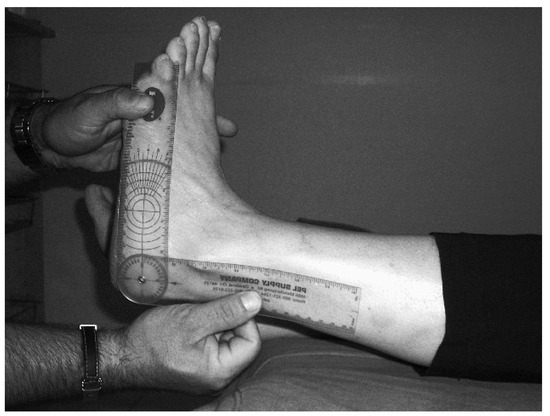
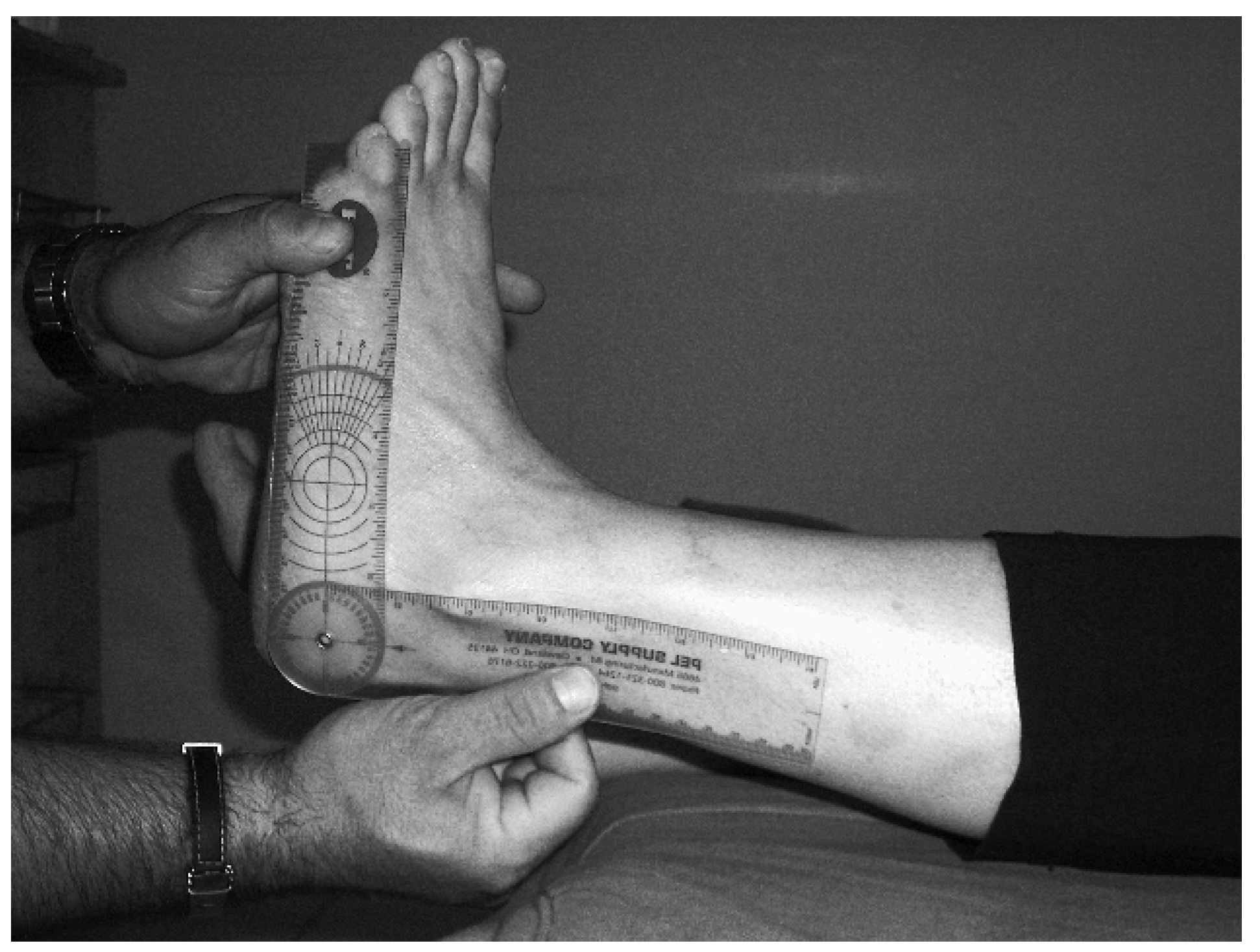
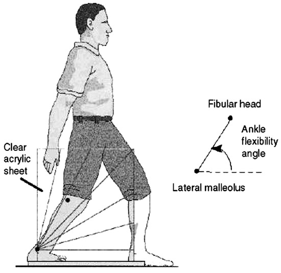
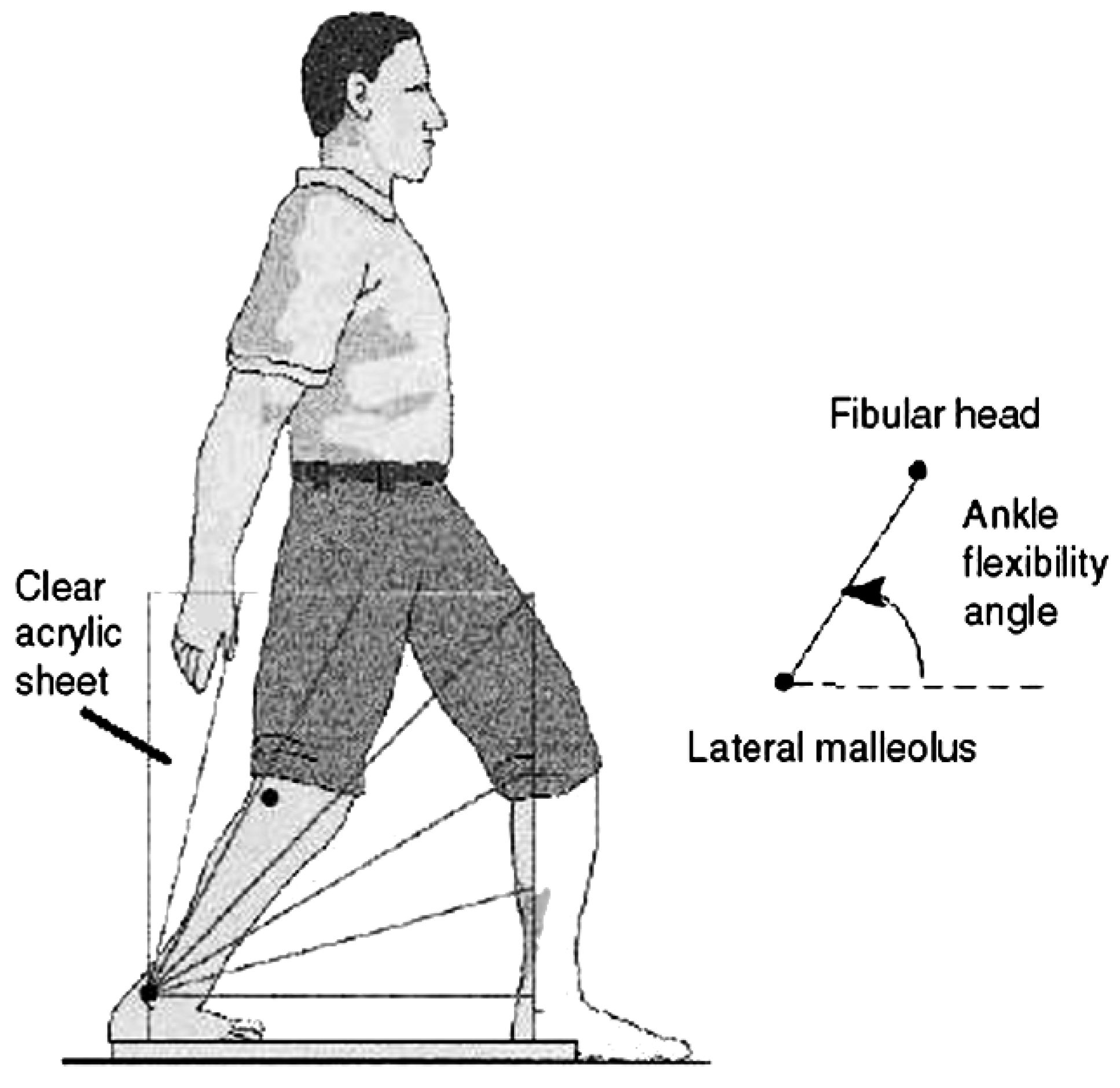
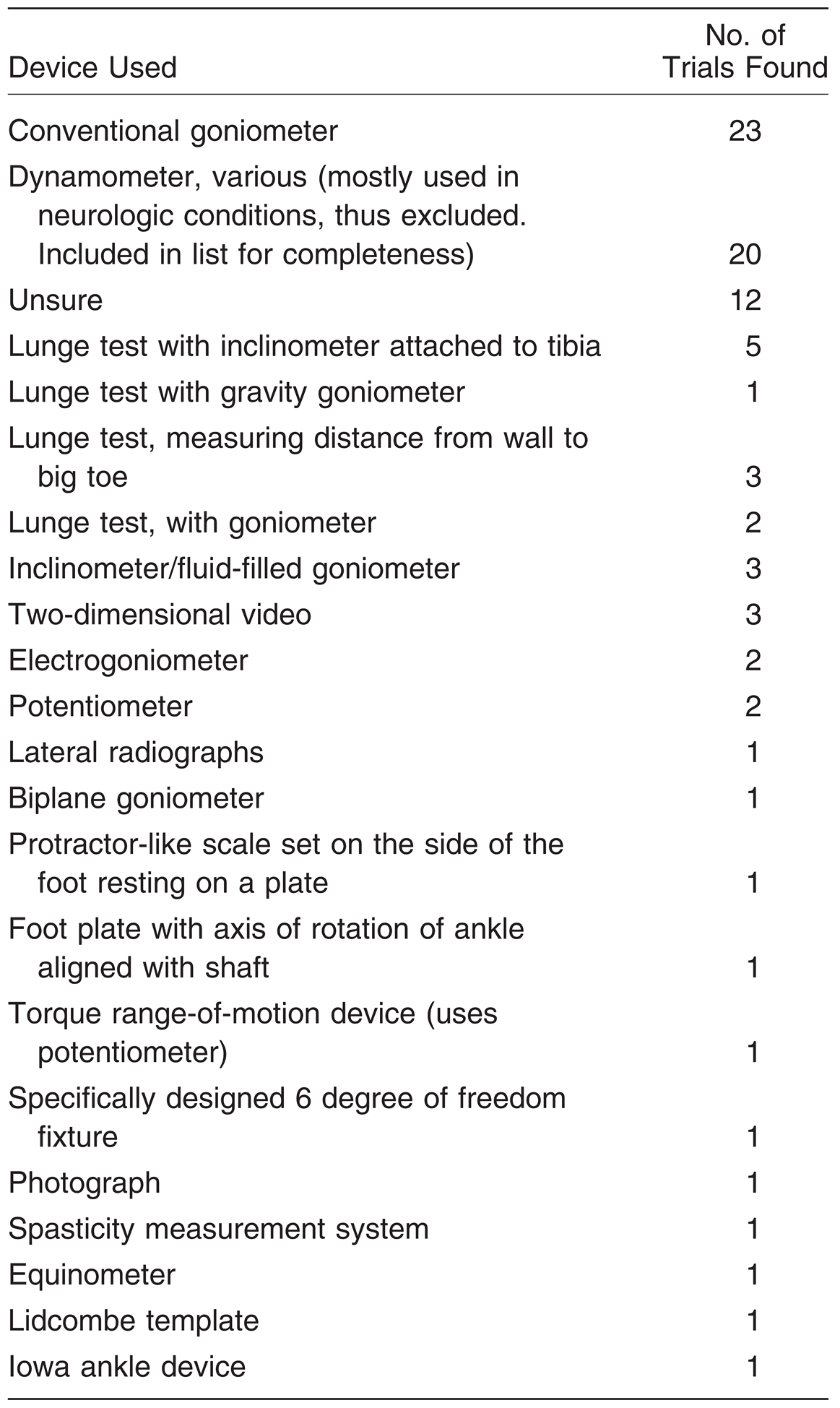
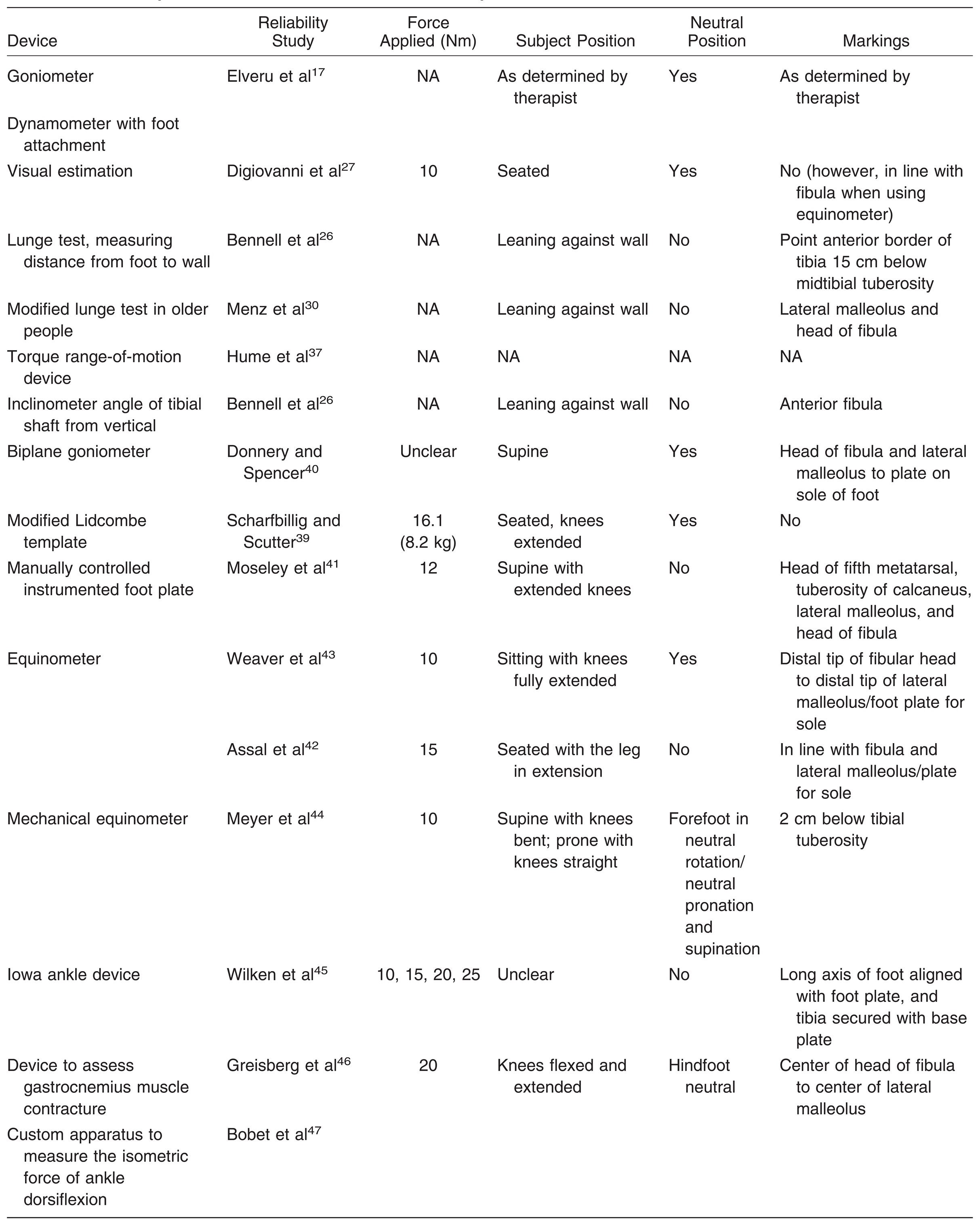
Various methods of measuring ankle dorsiflexion emerged from 87 articles that used some form of ankle joint dorsiflexion measurement (Table 1). From these methods, techniques specific to ankle measurement were identified (Table 2). For completeness in procedure, goniometric and visual estimation were included together with dynamometer and a custom apparatus to measure the isometric force of ankle dorsiflexion (Figure 1 and Figure 2 indicate some of these methods).
Table 1.
The Various Techniques Used to Measure Ankle Joint Complex Dorsiflexion.
Table 2.
Various Specific Ankle Dorsiflexion Tools/Techniques Identified.
Figure 1.
Clinician performing goniometric ankle dorsiflexion measurement.
Figure 2.
Modified lunge test. There are five variations of the original lunge test. (Reprinted with permission from Menz et al[30] and the Australian Journal of Physiotherapy.).
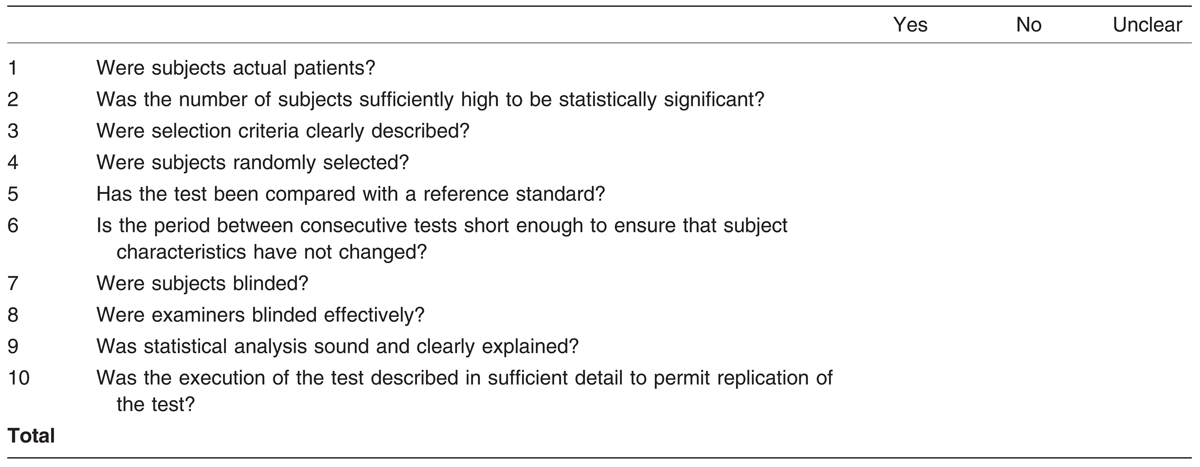
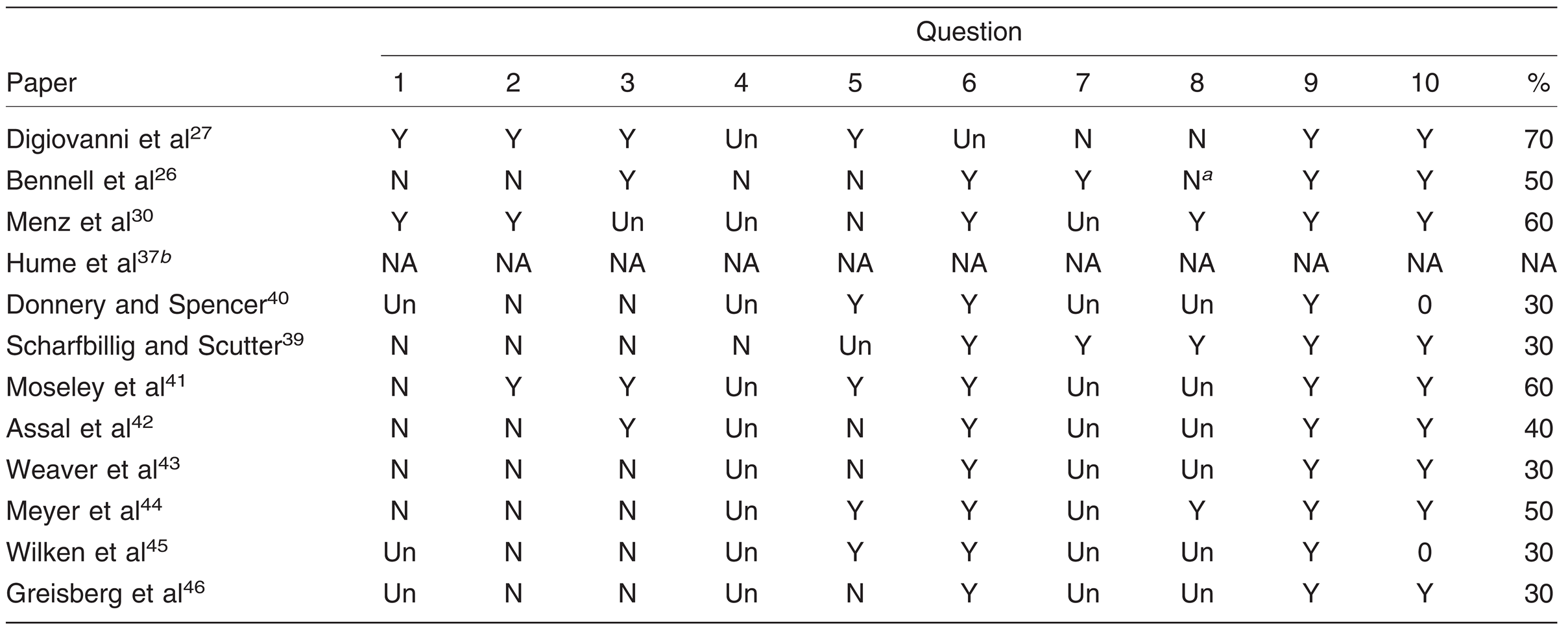
Although appraisal of the methodological quality of primary studies is essential in systematic reviews, no consensus exists on the ideal checklist and scale for assessing methodological quality.[23] Quality assessment of diagnostic tests differs from that of randomized clinical trials. A ten-item quality assessment tool was derived from QUADAS[24] and Jull[25] (Table 3). Two experienced clinicians checked the studies on their own to ensure blinding, and the presented results are the agreed-on scores (Table 4). Data extracted from these studies included assessment technique, amount of force (moment) applied, patient position, and whether the foot was held at subtalar joint neutral.
Table 3.
Quality Assessment Tool.
Table 4.
Quality Assessment Scores.
Results
The 87 articles produced a number of different ankle dorsiflexion measurement techniques, which are summarized in Table 2. From these studies, it was also possible to identify the published papers that validated each technique.
Measurement Techniques
Techniques for measuring ankle joint complex dorsiflexion have been categorized into three broad categories: conventional methods, more sophisticated methods using readily-available equipment, and specifically designed apparatuses and methods. Conventional methods include the goniometer,[14] the lunge test (Figure 2),[26] and visual estimation.[27] These methods do not require any specialized instruments designed specifically for this purpose. The lunge test has five variations: with inclinometer attached to tibia,[26] with gravity goniometer,[28] measuring the distance from the wall to the big toe,[26] with goniometer,[29] and with a transparent scale on the lateral side of the leg.[30] More sophisticated methods using readily-available equipment include the electrogoniometer and potentiometer,[31] inclinometer/gravity goniometer,[32] lateral radiographs,[33] two-dimensional video,[34] and photography.[35]
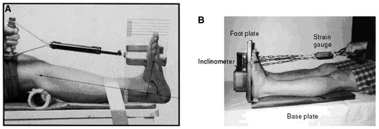
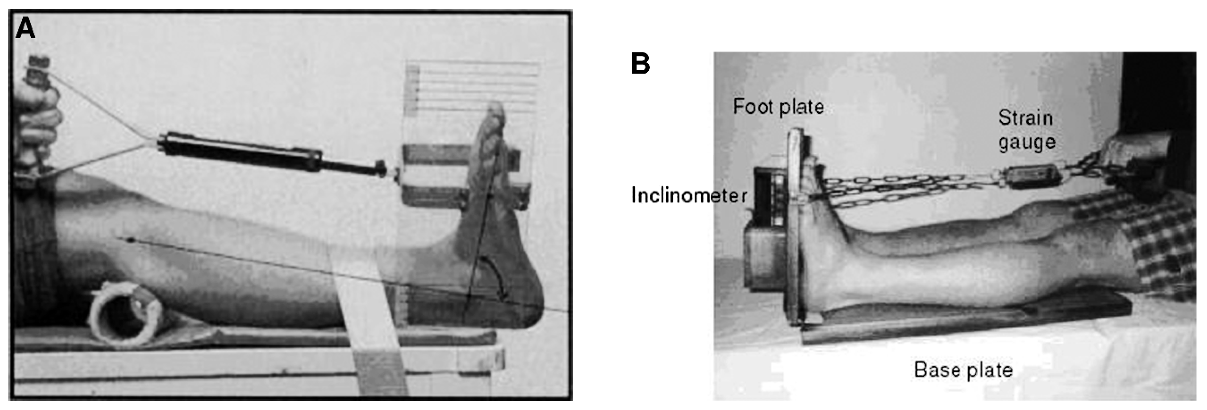
The many apparatuses and methods designed specifically for measuring ankle joint complex dorsiflexion include dynamometer with foot attachment,[36] torque range-of-motion device,[37] Lidcombe template38/modified Lidcombe template[39] (Figure 3), biplane goniometer,[40] manually controlled instrumented foot plate,[41] equinometer,[42,43] mechanical equinometer,[44] Iowa ankle device,[45] device to assess gastrocnemius muscle contracture,[46] and custom apparatus fixed to the leg and foot for measuring the isometric force of ankle dorsiflexion at ankle angles.[47] Several common features are found in these designs. 1) They are composed of a vertical arm and a plate for the sole of the foot or a variation thereof.[39,40,41,43,45,46] 2) The head of the fibula and the midline of the lateral malleolus seem to be accepted markings for lateral alignment.[30,41,42,43,45] Exceptions are the mechanical equinometer, which uses the anterior aspect of the tibia for alignment purposes[42]; the Lidcombe template, which uses the posterior aspect of the lower leg for this purpose[39]; and the device to assess gastrocnemius muscle contracture, which uses the axis of the second ray instead of the plate.[46] 3) The axis of the device is oriented to the axis of the ankle joint in the transverse plane.[41,45] 4) The axis may be in line with the ankle axis.[41,42,43,45] However, the axis below the lateral malleolus as per the Rootian model was not used in any of these specially designed apparatuses except the biplane goniometer.[40] The Lidcombe template axis approximates this by being placed at the rearmost part of the base board and foot plate ‘‘to simulate attachment of the Achilles tendon to the calcaneum and to provide a constant axis from which the force of the foot was applied.’’[39](p574) 5) An average reading is taken from several measurements.[27,46]
Figure 3.
A, The original Lidcombe template had a metatarsal bar. (Reprinted with permission from Moseley and the Australian Journal of Physiotherapy.[38]) B, Scharfbillig and Scutter[39] modified this with a plate, hence the name modified Lidcombe template. (Reprinted with permission from JAPMA.[39]).
Although it is recognized that routine radiographic assessment of ankle dorsiflexion is not normal clinical practice,[33] this type of modality may be indicated when important therapeutic decisions have to be made. Hence, this modality was considered as a part of this review.
Data Extraction
Subject Positioning. Because ankle dorsiflexion may be assessed in a variety of different clinical situations, patient position may affect the resultant ankle dorsiflexion angle. A patient may be assessed lying down in a supine[41] or prone position or in the more normal podiatric clinical position of sitting.[27] Patients are also assessed with knees extended and flexed. Subject position is known to affect the resultant amount of ankle joint dorsiflexion. Thoms and Rome[48] confirmed that although a prone or supine patient position does not affect this result, sitting does influence the result.
Foot Position. Whether foot position is held at subtalar joint neutral seems to be an important issue; however, it is not addressed by many retrieved trials. Podiatric medical theory suggests that a pronated foot allows increased sagittal plane motion, making this an unwanted situation if only ankle joint complex dorsiflexion measurement is desired. In fact, Tiberio et al[49] showed a variation of 10° between pronated and nonpronated feet. This is in accord with the finding of Woodburn,[34] who demonstrated a marked difference among the pronated, neutral, and supinated foot types. This, consequently, implies that to standardize measurements, it must be decided which foot position to use: pronated or nonpronated. A 10° error is unacceptable. It is hypothesized that holding the subtalar joint at neutral prevents dorsiflexion through the midfoot.[34,50] A pronated foot may hide a true diagnosis of limited ankle dorsiflexion, whereas a supinated foot may produce equinus, with its resultant important clinical consequences.[34]
Naturally, issues of subtalar joint neutral position reliability would arise at this time.[43] It is well-known that untrained therapists do not consistently find subtalar joint neutral[17,51]; however, training increases reliability,[18] although there is room for improvement.[51] Trials that performed the measurement at subtalar joint neutral include those by Digiovanni et al,[27] Scharfbillig and Scutter,[39] Donnery and Spencer,[40] Assal et al,[42] and Weaver et al.[43]
Force Applied. When the amount of dorsiflexing force applied by the examiner varies, the resultant angle also varies.[42] The product of the applied force and the distance between the axis of movement, ie, ankle axis, and the point of application of the force gives the moment, which is synonymous with torque. Using moment calculations and angular displacement, passive versus angular displacement curves can be produced as opposed to simpler systems, where only a single point is recorded.[41] The moment applied to the ankle joint complex might fluctuate between one examiner and another or between one examination and another, thus exerting unequal forces on the gastrocnemius muscle. The stretching characteristics of the triceps surae themselves may vary depending on other intrinsic or extrinsic factors, such as temperature.[52,53] The amount of force, or moment, applied to the ankle joint, normally to the metatarsophalangeal joint area, was reported in 75% of assessed studies. In two studies[26,30] that assessed a type of lunge test, this measure was not applicable. The moment applied varied: 10,[27,43,44] 12,[41] 15,[42] 16.1,[39] and 20 Nm.[46] Wilken et al[45] assessed angular displacement over a range of forces at 10, 15, 20, and 25 Nm. The different amounts of force seem to have been chosen arbitrarily, except for that in the study by Digiovanni et al,[27] who justified their 10-Nm moment as having been chosen based on previous work.
Applying the same amount of force to all subjects reduces the variability of subsequent readings between the same patients, making the test seem more reliable than if the force were not controlled. This is because the magnitude of force applied determines the applied torque, directly influencing the ankle dorsiflexion angle.[42] If a known torque is applied to dorsiflex the ankle, reliability is, thus, increased.[38] This may be one way of standardizing one of the variables,[43] thus increasing the possibility of comparing results between trials. Additional apparatuses may be required to do this, although a simple spring balance has been used with good effect to attain this goal.[38]
Methodological Quality Assessment
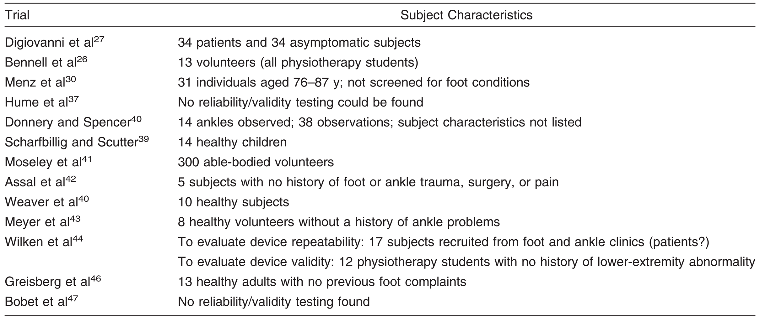
Thirteen papers that reported reliability were identified for quality assessment (Table 5). However, the full text of two papers could not be obtained,[37,47] which reduced the list to eleven.
Table 5.
Summary of Trials Assessed for Quality and Their Subject Characteristics.
Subjects Used in Ankle Dorsiflexion Reliability Assessment
Most assessed studies used small convenience samples of healthy volunteers (Table 5). Only three of the trials used real patients.[27,30,45] ‘‘Evaluations of new tests often omit the essential developmental stage of evaluation in a real clinical population.’’[25](p69) The use of a suitable spectrum of patients is required when evaluating the validity and reliability of new diagnostic tests, otherwise spectrum bias may occur. This is because differences in clinical features between patients may produce considerable variations in results.[24] Regarding sample size, only Digiovanni et al,[27] Menz et al,[30] and Moseley et al[41] had a significant number of subjects. The ability to infer to larger populations in all of the other cases is consequently debatable.
Randomization
Selection bias can be present when subject selection is not random.[54] Although only three trials selected actual patients,[27,30,45] the reporting of whether they were randomly selected is not clear.
Comparison with a Reference Standard
‘‘Was there an independent blind comparison with a reference (gold) standard of diagnosis?’’ is an important question that should be asked to assess diagnostic tests.[25] This mostly refers to the validity of the instrument. In simple words, are the reported results actual results or is there the possibility of confounding elements being present? If a measure is reporting 0° of ankle joint complex dorsiflexion, is it actually 0°? Is there only ankle joint complex dorsiflexion, or does the result incorporate midtarsal joint dorsiflexion as well? This can only be ascertained by cross-checking this result with another measure that is known to produce good results. In this context, this may be difficult to achieve because it is already known that there is a problem with attaining true dorsiflexion angles.
Five studies cross-checked their results with a known measurement technique. Digiovanni et al[27] checked visual estimation of ankle dorsiflexion with an equinometer, although, as the authors themselves point out, there is lack of a gold standard test. Whereas Moseley et al[41] referenced their results with photography, Donnery and Spencer[40] and Meyer et al[44] compared their results with a goniometer, even when this method is known to have high reliability problems. Wilken et al[45] assessed their dorsiflexion results with an Optotrak motion analysis system.
When measuring ankle dorsiflexion, true tibiotalar motion can be measured accurately using radiographic techniques. The vertical midline of the fibula can be compared with a line dividing the talar trochlear.[33] Thus, this type of modality may be indicated as a true reference standard when comparing new techniques/equipment. Notwithstanding this, it is clear that some authors may run into ethical difficulties when proposing this modality because of radiation issues.
Statistical Analysis
Test-retest designs seem to be the preferred method of assessment, with intraclass correlation being the statistical test of choice.[26,30,39,41,43,44,45,46] They follow a trend in reliability trials. ‘‘The consistency or the repeatability of the [range-of-motion] measurement- whether the application of the instrument and the procedures produce the same measurements consistently under the same conditions.’’[15](p1867) Currier,[55] cited by Gajdosik and Bohannon, continues to add that the validity of a measurement ‘‘constitutes the degree to which an instrument measures what it is purported to measure; the extent to which it fulfills its purposes.’’ [15](p1870) Measures of validity tell us whether an item measures what it is supposed to, that is, whether a measurement is true.[56]
Discussion
When embarking on new research projects, it is normal practice to assess the validity and reliability of any outcome measures used. Thus, it is perplexing why, with the current evidence regarding the unreliability of goniometric measurements, so many trials used this type of assessment as their main outcome measure. This not only raises questions as to the reliability and validity of their results but also as to their clinical value.
It is evident that researchers always felt unhappy with goniometric measurements. The number of systematic reviews regarding this issue and the number of different apparatuses that have been developed to improve ankle dorsiflexion measurement techniques attest to this. Most of these specifically designed apparatuses mainly aim to reduce errors of placement by securing the vertical arm to the leg. The accepted method of alignment seems to be from the lateral head of the fibula to the middle of the lateral malleolus for the vertical arm, and a plate to cover the plantar aspect of the foot for the other arm. For the former, several researchers have drawn a vertical line from the lateral head of the fibula to the malleolus to maintain alignment; if this practice is not maintained during clinical goniometric measurements, this may produce a significant error in placement, which can be reduced by having the vertical, stable arm long enough to reach up to the head of the fibula.
It is acceptable that as the forefoot is pushed upward to dorsiflex, kinematic changes occur in all of the joints distal to the ankle joint. Although the study by Lundgren et al[14] was limited to six subjects, who apparently were not controlled for hypermobility (ie, the amount of rotation around the axes) of the midtarsal joint, it confirms that motion of the foot is not solely at the ankle joint complex and that the other foot joints contribute a significant amount of dorsiflexion.
It seems that Root et al[13] may have had a valid point after all in proposing that the ankle dorsiflexion angle be measured at subtalar joint neutral. Woodburn[34] and Tiberio et al[49] observed a significant difference between pronated and nonpronated feet. In view of this, it is surprising that Lundgren et al[14] did not control for subtalar joint position, which could have affected their results.
This means that trials that compared ankle dorsiflexion measurements taken at subtalar joint neutral and those that did not maintain this position cannot be compared directly because in the latter there may be the possibility of having increased angles due to joint motion extraneous to the ankle. In this regard, it follows that subjects with pronated, or flat, feet would exhibit increased ankle dorsiflexion measurements derived from additional midtarsal joint motion. In such cases, the presence, or otherwise, of ankle equinus would be masked by false results because these would include both ankle and midtarsal joint motion.
Whether the subtalar joint neutral position can be consistently established by clinicians is debatable; Diamond et al[18] believe that reliability may be increased by training. However, its use may, in fact, increase measurement error.[14] Currently, there is a paucity of information that compares pronated and nonpronated foot dorsiflexion values with those of supinated feet.[34] Finding a way around the subtalar joint neutral position issue would eliminate one important factor that significantly affects this type of measurement.[44]
With reference to subject positioning, from the trial by Thoms and Rome[48] it can be concluded that this should be either prone or supine. Subject positioning is one of the factors that should be standardized to obtain consistent results in ankle joint measurement trials. However, even here, more research is required because the trial by Thoms and Rome assessed only the prone and supine positions with the knee extended and the sitting position with the knee flexed. This could be because it is known that there is poor correlation between knees extended and knees flexed.[44] It is still puzzling, however, why sitting position with knees extended was not included in this trial.
Although most of the papers reviewed used the term torque,[27,39,41,42,44,45,46] moment may be more appropriate and is the accepted terminology in the biomechanics community. As to the amount of force applied, it emerges that consistency can be attained only by applying the same moment, which directly influences the ankle dorsiflexion angle.[42] This may be one way of standardizing one of the variables, thus increasing the possibility of comparing results between trials. Additional apparatuses may be required to do this, although a simple spring balance has been used with good effect to attain this goal.[38] Also note that the amount of force being quoted[27,39,41,42,43,44,45,46] is generally a small percentage of the actual forces being applied to the foot during locomotion.
Although all of the studies report high intraclass correlation coefficient values for reliability, the methods used to assess the various tools seem to be weak. It has been pointed out that although the number of diagnostic test evaluations is increasing, their methodological quality is, on average, poor.[54] Few studies use actual patient populations. Most lack reference to a reference standard, such as radiographs. Consequently, one cannot discern whether the reported angles are, in fact, the true angles of foot dorsiflexion.
It is also clear that the terms reliability and repeatability are being used interchangeably. According to Gajdosik and Bohannon, ‘‘[r]eliability in goniometry simply means the consistency or the repeatability of the [range-of-motion] measurements, that is, whether the application of the instrument and the procedures produce the same measurements consistently under the same conditions.’’ [15](p1867) This definition, however, does not consider validity, ie, whether the instrument is actually measuring the correct angle. This is one of the main reasons why any such procedure must be compared with the reference standard. In the case of ankle joint complex measurement, following the revelation of Tiberio et al[49] that in a pronated foot there is 10° more dorsiflexion, the instrument under review might actually be measuring both ankle joint complex and midtarsal joint complex dorsiflexion. To complicate this further, it might also be actually measuring the summation of all of the dorsiflexion movements of joints distal to the ankle.[14] Thus, this reference standard may be radiologic, where the movement between the actual bones themselves may be measured more accurately.
Finally, in view of the evidence regarding goniometric unreliability, serious doubt arises as to the value of all of the research performed with this method. A method/technique of assessing pure ankle joint complex dorsiflexion needs to be validated fully and, possibly, most of the research mentioned herein repeated using this method to ensure that all of the results are, in fact, true foot dorsiflexion results.
Conclusions
From this review it can, thus, be deduced that most devised instruments consistently produced the same results in healthy populations. There is still no total agreement as to how the foot dorsiflexion angle can be measured. At the moment, the various techniques are inconsistent, thus causing a deficit in this particular area of research. This is mainly because the lack of standardization makes direct comparisons between trials impossible. Furthermore, it is not clear what is being measured, whether it is tibiotalar joint motion, ankle joint complex motion, or the summation of all of the movements of the joints of the foot. It seems that the latter may be the most likely, thus the term foot dorsiflexion may be more appropriate.
It is being proposed that the following variables be standardized: placement of the vertical arm, use of a plate for the other arm, and the amount of force applied to dorsiflex the foot (currently 10–20 Nm). Further research is required into the following aspects of this field: 1) Subject position: does it influence the final result? 2) Foot position: can subtalar joint neutral be consistently applied? If not, can a way around this problem be found? 3) Placement of the axis, whether in line with the ankle joint axis or below the lateral malleolus, as described by Root et al.[13] 4) The amount of force needed to be applied to the plantar forefoot. Should it try to simulate normal kinetics during gait?
Unless true standardization of measurement is achieved, the accuracy of communication between clinicians, the act of measuring ankle joint dorsiflexion, and the accuracy of the measurement itself, which is important in determining therapeutic options, will be sorely lacking. Furthermore, the research that is going on cannot be responsibly used. It seems that even in this day and age, ankle joint complex dorsiflexion measurement is still a gray area.
Financial Disclosure
None reported.
Conflicts of Interest
None reported.
References
- IRVING, D.B.; COOK, J.L.; MENZ, H.B. Factors associated with chronic plantar heel pain: a systematic review. J Sci Med Sport 2006, 9, 11. [Google Scholar] [CrossRef] [Scilit] [PubMed]
- BARRETT, S.L.; JARVIS, J. Equinus deformity as a factor in forefoot nerve entrapment: treatment with endoscopic gastrocnemius recession. JAPMA 2005, 95, 464. [Google Scholar] [CrossRef] [Scilit] [PubMed]
- DANANBERG, H.J.; SHEARSTONE, J.; GUILLANO, M. Manipulation method for the treatment of ankle equinus. JAPMA 2000, 90, 385. [Google Scholar] [CrossRef] [Scilit]
- LAVERY, L.A.; ARMSTRONG, D.G.; BOULTON, A.J. Ankle equinus deformity and its relationship to high plantar pressure in a large population with diabetes mellitus. JAPMA 2002, 92, 479. [Google Scholar] [CrossRef] [Scilit]
- Armstrong, D.G.; Stacpoole-Shea, S.; Nguyen, H. Lengthening of the Achilles tendon in diabetic patients who are at high risk for ulceration of the foot. J Bone Joint Surg Am 1999, 81, 535. [Google Scholar] [CrossRef] [Scilit]
- HILL, R.S. Ankle equinus: prevalence and linkage to common foot pathology. JAPMA 1995, 85, 295. [Google Scholar] [CrossRef] [Scilit]
- DAVIS, J.A.; BULL, R.H.; FARRELLY, I.H. ET AL: A home-based exercise programme improves ankle range of motion in long-term venous ulcer patients. Phlebology 2007, 22, 86. [Google Scholar] [CrossRef] [Scilit]
- SOBEL, E.; CASELLI, M.A.; VELEZ, Z. Effect of persistent toe walking on ankle equinus: analysis of 60 idiopathic toe walkers. JAPMA 1997, 87, 17. [Google Scholar] [CrossRef] [Scilit]
- ALLINGTON, N.J.; LEROY, N.; DONEUX, C. Ankle joint range of motion measurements in spastic cerebral palsy children: intraobserver and interobserver reliability and reproducibility of goniometry and visual estimation. J Pediatr Orthop B 2002, 11, 236. [Google Scholar]
- JOHNSON, C.H.; CHRISTENSEN, J.C. Biomechanics of the first ray: part V. The effect of equinus deformity: a 3-dimensional kinematic study on a cadaver model. J Foot Ankle Surg 2005, 44, 114. [Google Scholar] [CrossRef] [Scilit] [PubMed]
- ROME K: Ankle joint dorsiflexion measurement studies: a review of the literature. JAPMA 1996, 86, 205. [CrossRef] [Scilit] [PubMed]
- BRANTINGHAM, J.W.; GILBERT, J.L.; SHAIK, J. ET AL: Sagittal plane blockage of the foot, ankle and hallux and foot alignment-prevalence and association with low back pain. J Chirop Med 2006, 5, 123. [Google Scholar] [CrossRef] [Scilit] [PubMed]
- ROOT, M.L.; ORION, W.P.; WEED, J.H. Normal and Abnormal Function of the Foot; Clinical Biomechanics Corp: Los Angeles, 1977. [Google Scholar]
- LUNDGREN, P.; NESTER, C.; LIU, A. ET AL: Invasive in vivo measurement of rear-, mid- and forefoot motion during walking. Gait Posture 2008, 28, 93. [Google Scholar] [CrossRef] [Scilit]
- GAJDOSIK, R.L.; BOHANNON, R.W. Clinical measurement of range of motion: review of goniometry emphasizing reliability and validity. Phys Ther 1987, 67, 1867. [Google Scholar] [CrossRef] [Scilit]
- SILFVERSKIOLD N: Reduction of the uncrossed two-joint muscles of the leg to one-joint muscles in spastic conditions. Acta Chir Scand 1924, 56, 315.
- ELVERU, R.A.; ROTHSTEIN, J.M. LAMB RL: Goniometric reliability in a clinical setting. Phys Ther 1988, 68, 672. [Google Scholar] [CrossRef] [Scilit]
- DIAMOND, J.E.; MUELLER, M.J.; DELITTO, A. ET AL: Reliability of a diabetic foot evaluation. Phys Ther 1989, 69, 797. [Google Scholar] [CrossRef] [Scilit]
- VAN GHELUWE, B.; KIRBY, K.A.; ROOSEN, P. ET AL: Reliability and accuracy of biomechanical measurements of the lower extremities. JAPMA 2002, 92, 317. [Google Scholar] [CrossRef] [Scilit]
- MARTIN, R.L. MCPOIL TG: Reliability of ankle goniometric measurements: a literature review. JAPMA 2005, 95, 564. [Google Scholar] [CrossRef] [Scilit]
- EVANS, A.; SCUTTER, S.D. Sagittal plane range of motion of the pediatric ankle joint: a reliability study. JAPMA 2006, 96, 418. [Google Scholar] [CrossRef] [Scilit]
- WRIGHT, J.G.; FEINSTEIN, A.R. Improving the reliability of orthopaedic measurements. J Bone Joint Surg Br 1992, 74, 287. [Google Scholar] [CrossRef] [Scilit] [PubMed]
- MOJA, L.P.; TELARO, E.; D’AMICO, R.; et al. Assessment of methodological quality primary studies by systematic reviews: results of the metaquality cross sectional study. BMJ 2005, 330, 1053. [Google Scholar] [CrossRef] [Scilit]
- WHITING, P.; RUTJES, A.W.S.; REITSMA, J.B. ET AL: The development of QUADAS: a tool for the quality assessment of studies of diagnostic accuracy included in systematic reviews. BMC Med Res Method 2003, 3, 25. [Google Scholar] [CrossRef] [Scilit] [PubMed]
- JULL A: Evaluation of studies of assessment and screening tools and diagnostic tests. Evid Based Nurs 2002, 5, 68. [CrossRef] [Scilit]
- BENNELL, K.; TALBOT, R.; WAJSWELNER, H.; et al. Intrarater and interrater reliability of a weight-bearing lunge measure of ankle dorsiflexion. Phys Ther 1998, 44, 175. [Google Scholar]
- DIGIOVANNI, C.W.; HOLT, S.; CZERNIECKI, J.M. ET AL: Can the presence of equinus contracture be established by physical exam alone? J Rehabil Res Dev 2001, 38, 335. [Google Scholar]
- NITZ, J.C. Low Choy N. The relationship between ankle dorsiflexion range, falls and activity level in women aged 40 to 80 years. N Z J Physiother 2004, 32, 121. [Google Scholar]
- BAGGETT, B.D.; YOUNG, G. Ankle joint dorsiflexion: establishment of a normal range. JAPMA 1993, 83, 251. [Google Scholar] [CrossRef] [Scilit]
- MENZ, H.B.; TIEDEMANN, A.; KWAN, M.M. ET AL: Reliability of clinical tests of foot and ankle characteristics in older people. JAPMA 2003, 93, 380. [Google Scholar] [CrossRef] [Scilit]
- TESIO, L.; MONZANI, M.; GATTI, R.; et al. Flexible electrogoniometers: kinesiological advantages with respect to potentiometric goniometers. Clin Biomech 1995, 10, 275. [Google Scholar] [CrossRef] [Scilit] [PubMed]
- NYANZI, C.S.; LANGRIDGE, J.; HEYWORTH, J.R.; et al. Randomized controlled study of ultrasound therapy in the management of acute lateral ligament sprains of the ankle joint. Clin Rehabil 1999, 13, 16. [Google Scholar] [CrossRef] [Scilit] [PubMed]
- BACKER, M.; KOFOED, H. Passive ankle mobility: clinical measurement compared with radiography. J Bone Joint Surg Br 1989, 71, 696. [Google Scholar] [CrossRef] [Scilit]
- WOODBURN J: Video joint angle position analysis of the subtalar joint position on maximum ankle joint dorsiflexion. J Br Podiatr Med 1991, 46, 19.
- MOSELEY, A.M. The effect of casting combined with stretching on passive ankle dorsiflexion in adults with traumatic head injuries. Phys Ther 1997, 77, 982. [Google Scholar] [CrossRef] [Scilit] [PubMed]
- MUIR, I.W.; CHESWORTH, B.M.; VANDERVOORT, A.A. Effect of a static calf-stretching exercise on the resistive torque during passive ankle dorsiflexion in healthy subjects. J Orthop Sports Phys Ther 1999, 29, 106. [Google Scholar] [CrossRef] [Scilit]
- HUME, P.; NAKAMURA, T.; BUFORD, W.; et al. Analysis of torquerange-of-motion for the passive viscoelastic properties of the foot/ankle complex. Paper presented at: 18th Annual Conference on Biomedical Engineering Research, Houston, Texas, 2000. [Google Scholar]
- MOSELEY A, ADAMS R: Measure of passive ankle dorsiflexion: procedure and reliability. Aust J Physiother 1991, 37, 175. [CrossRef] [Scilit]
- SCHARFBILLIG, R.; SCUTTER, S.D. Measurement of foot dorsiflexion: a modified Lidcombe template. JAPMA 2004, 94, 573. [Google Scholar] [CrossRef] [Scilit]
- DONNERY, J.; SPENCER, R.B. The biplane goniometer: a new device for measurement of ankle dorsiflexion. JAPMA 1988, 78, 348. [Google Scholar] [CrossRef] [Scilit]
- MOSELEY, A.M.; CROSBIE, J. ADAMS R: Normative data for passive ankle plantarflexion-dorsiflexion flexiblty. Clin Biomech 2001, 16, 514. [Google Scholar] [CrossRef] [Scilit] [PubMed]
- ASSAL, M.; SHOFER, J.B.; ROHR, E.; et al. Assessment of an electronic goniometer designed to measure equinus contracture. J Rehabil Res Dev 2003, 40, 235. [Google Scholar]
- WEAVER, K.; PRICE, R.; CZERNIECKI, J.; et al. Design and validation of an instrument package designed to increase the reliability of ankle range of motion measurements. J Rehabil Res Dev 2001, 38, 471. [Google Scholar]
- MEYER, D.C.; WERNER, C.M.; WYSS, T.; ET, A.L. A mechanical equinometer to measure the range of motion of the ankle joint: interobserver and intraobserver reliability. Foot Ankle Int 2006, 27, 202. [Google Scholar] [CrossRef] [Scilit]
- WILKEN, J.; SALTZMAN, C. YACK H: Reliability and validity of the Iowa ankle range-of-motion device. J Orthop Sports Phys Ther 2004, 34, A17. [Google Scholar]
- GREISBERG, J.; DRAKE, J.; CRISCO, J. ET AL: The reliability of a new device designed to assess gastrocnemius contracture. Foot Ankle Int 2002, 23, 655. [Google Scholar] [CrossRef] [Scilit]
- BOBET, J.; GOSSEN, E.R.; STEIN, R.B. A comparison of models of force production during stimulated isometric ankle dorsiflexion in humans. IEEE Trans Neural Syst Rehabil Eng 2005, 13, 444. [Google Scholar] [CrossRef]
- THOMS V, ROME K: Effect of subject position on the reliability of measurement of active ankle joint dorsiflexion. The Foot 1997, 7, 153. [CrossRef] [Scilit]
- TIBERIO, D.; BOHANNON, R.W.; ZITO, M.A. Effect of subtalar joint position on the measurement of maximum ankle dorsiflexion. Clin Biomech 1989, 4, 189. [Google Scholar] [CrossRef] [Scilit]
- MUELLER, M.J.; DIAMOND, J.E.; DELITTO, A. ET AL: Insensitivity, limited joint mobility, and plantar ulcers in patients with diabetes mellitus. Phys Ther 1989, 69, 453. [Google Scholar] [CrossRef] [Scilit]
- PIERRYNOWSKI, M.R.; SMITH, S.B.; MLYNARCZYK, J.H. Proficiency of foot care specialists to place the rearfoot at subtalar neutral. JAPMA 1996, 86, 217. [Google Scholar] [CrossRef] [Scilit] [PubMed]
- KNIGHT, C.A.; RUTLEDGE, C.R.; COX, M.E.; et al. Effect of superficial heat, deep heat and active exercise warm up on the extensibility of the plantarflexors. Phys Ther 2001, 81. [Google Scholar] [CrossRef] [Scilit]
- ROBERTSON, V.J.; WARD, A.R.; JUNG, P. The effect of heat on tissue extensibility: a comparison of deep and superficial heating. Arch Phys Med Rehabil 2005, 86, 819. [Google Scholar] [CrossRef] [Scilit] [PubMed]
- LIJMER, J.G.; MOL, B.W.; HEISTERKAMP, S. ET AL: Empirical evidence of design-related bias in studies of diagnostic tests. JAMA 1999, 282, 1061. [Google Scholar] [CrossRef] [Scilit] [PubMed]
- CURRIER, D.P. Elements of Research in Physical Therapy, Ed 2, p 166; Williams & Wilkins: Baltimore, 1984. [Google Scholar]
- KIRK, J.; MILLER, M.L. Reliability and Validity in Qualitative Research; Sage Publications: Beverly Hills, 1986. [Google Scholar]

© 2011 American Podiatric Medical Association