Next Article in Journal
Trimethoprim-Sulfamethoxazole–Induced Stevens-Johnson Syndrome. A Case Report
Previous Article in Journal
Mini TightRope System for Hallux Abducto Valgus Deformity. A Discussion and Case Report
 
 
Journal of the American Podiatric Medical Association is published by MDPI from Volume 116 Issue 1 (2026). Previous articles were published by another publisher in Open Access under a CC-BY (or CC-BY-NC-ND) licence, and they are hosted by MDPI on mdpi.com as a courtesy and upon agreement with American Podiatric Medical Association.
Article

Large Subungual Exostosis of the Great Toe. A Case Report

3rd Orthopaedics and Traumatology Clinic, Diskapi Yildirim Beyazit Training and Research Hospital, İrfan Baştuğ Street Diskapi, Ankara, 06110 Turkey
*
Author to whom correspondence should be addressed.
J. Am. Podiatr. Med. Assoc. 2010, 100(4), 296-298; https://doi.org/10.7547/1000296
Published: 1 July 2010

Abstract

A subungual exostosis is a benign lesion described as a variant of an osteochondroma. It mostly affects the great toe, and it can be confused with an osteochondroma and a Nora’s lesion. The curative treatment for a subungual exostosis is complete excision, and the cartilaginous cap must be totally resected to prevent a possible recurrence. In this article, we present findings regarding a large and painful lesion on the dorsomedial aspect of the great toe of a patient.

A subungual exostosis is a rare variant of an osteochondroma that is beneath or adjacent to the nail and mainly arises on distal phalanges. [1] The lesion was first recognized by Dupuytren [2] in 1817. To date, more than 300 cases have been reported. Although the etiology of the condition is unclear, preceding trauma and chronic infection may be precipitating factors for the development of a subungual exostosis. [3] Inflammatory processes or metaplastic changes on the fibrous tissue and cartilage seem to be the pathogenesis of this process. [4]
The risk of malignant degeneration of a solitary osteochondroma is reported to be 1%. []5] Differentiation between a subungual exostosis and a subungal osteochondroma is rather difficult, and the initial appearance of a subungual exostosis resembles an osteochondroma. [6] Another lesion that could be confused with a subungual exostosis is bizarre parosteal osteochondromatous proliferation. This lesion may also be called a Nora’s lesion, and it affects mainly the phalanges of the hands and feet. [7] Both entities are osteochondromatous proliferations with distinct chromosomal rearrangements. Zambrano and Nosé [8] found a balanced X chromosome translocation in their patients with subungual exostoses. They claimed that the subungual exostosis is a neoplastic process rather than a reactive one.
The slow-growing subungual exostosis usually presents in the second and third decades of life. It is often misdiagnosed, thus a detailed physical examination and two-view radiography of the involved digit are important for diagnosis. The curative treatment for this lesion is complete excision, and the cartilaginous cap must be totally resected to prevent a possible recurrence.

Case Report

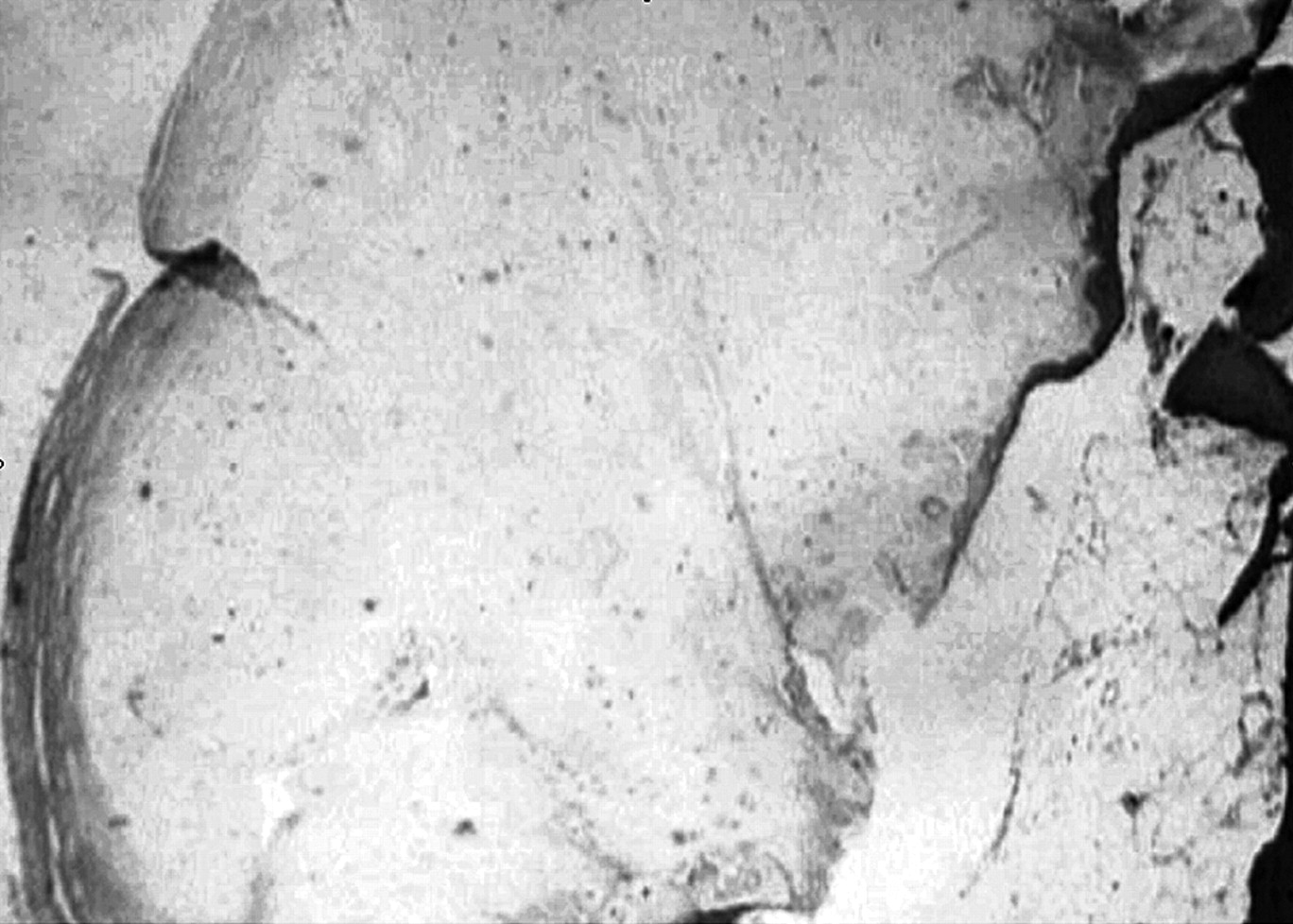
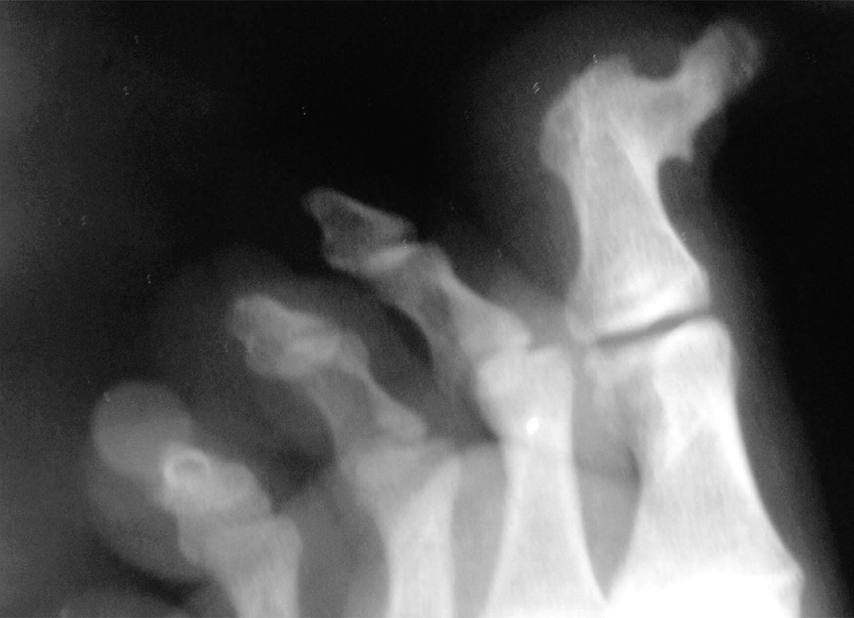
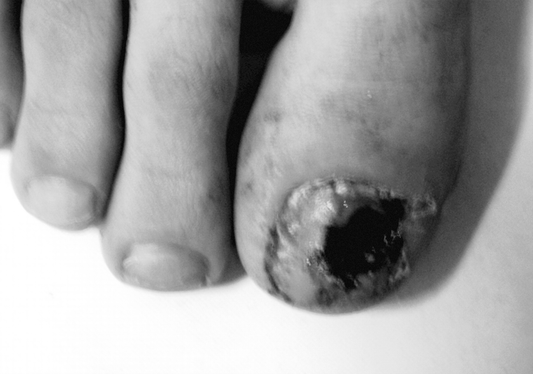
A 15-year-old adolescent was admitted to an out-patient clinic with a 7-month history of a growing painful mass on the dorsal aspect of his right great toe. He had a trauma history. On physical examination, he had a stiff and painful mass on the dorsomedial aspect of the great toe without drainage (Fig. 1). Radiologic examination revealed a bony excrescence arising from the dorsomedial aspect of the great toe (Fig. 2). The nail bed was intact despite the largeness of the lesion. After applying a digital ring, local anesthesia, and a tourniquet, complete resection of the osteocartilaginous tumor was performed using a “fish-mouth”-type incision. The wound was primarily closed (Fig. 3). Pathologic findings were trabecular bone surrounded by a thin cap composed of hyaline cartilage and focally by fibrocartilage or fibrous tissue, which confirms a subungual exostosis (Fig. 4).

Discussion

Subungual exostoses have a painful course, and the pain can be exacerbated by ambulation or physical activity. Onycholysis of the nail plate may also occur if the lesion enlarges and indurates. Although the diagnosis can easily be made using clinical and radiologic features, the differential diagnosis can still include bizarre parosteal osteochondromatous proliferation, florid reactive periostitis, turret exostosis (ossifying hematoma), subungual verruca, pyogenic granuloma, glomus tumor, Koenen tumor, keratoacanthoma, carcinoma of the nail bed, and malignant melanoma. [9] The curative treatment for this lesion is total excision.
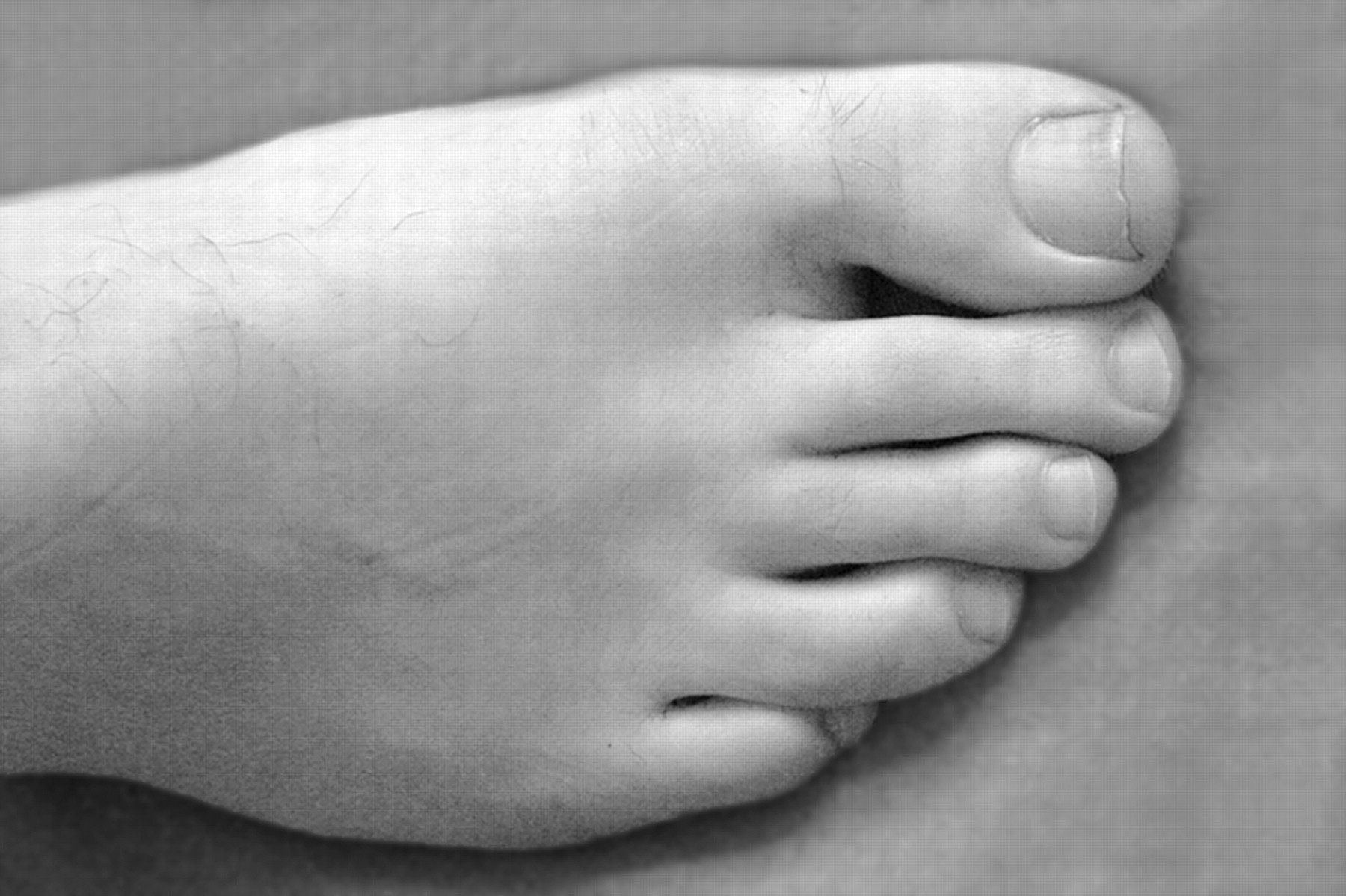
Among the excisional techniques, use of a “fish-mouth”-type incision should be considered to avoid possible disruption of the nail bed. If there is a destroyed nail bed, then an approach to remove the tumor from its top can be applied. [10] The cartilaginous cap must be totally resected to avoid a possible recurrence. The recurrence rate has been reported to vary between 10% and 20%. Another issue in the treatment of subungual exostoses is nail problems. Lemperle et al [11] showed that a simple excision of the destroyed nail matrix could stimulate growth of the residual healthy matrix and regenerate the nail. If there is a remaining large nail bed defect, onycholysis could occur after the operation. Therefore, some authors have used artificial skin to prevent onycholysis. [12] However, they usually do not present functional problems. A complete nail excision yielded highly favorable results in terms of the nail bed. No problems were seen 2 years after the operation (Fig. 5).
In conclusion, a subungual exostosis must be accepted as a tumor, and it can occur in a wide spectrum ranging from a reddish-pink lesion to a large lesion, as in the patient described herein. Although it is a benign lesion, the differential diagnosis of malignant lesions should be kept in mind. The cartilaginous cap must be totally excised to avoid a possible recurrence. Use of a “fish-mouth”-type incision should be considered to avoid disrupting the nail bed.

Financial Disclosure

None reported.

Conflict of Interest

None reported.

References

  1. De Palma L, Gigante A, Specchia N: Subungual exostosis of the foot. Foot Ankle Int17:758,1996.
  2. Dupuytren G: On the Injuries and Diseases of Bones, p 408, Sydenham Society, London, England,1847.
  3. Letts M, Davidson D, Nizalik E: Subungual exostosis: diagnosis and treatment in children.J Trauma44:346,1998.
  4. Wu KK: Subungual exostosis.J Foot Ankle Surg34:96,1995.
  5. Karasick D, Schweitzer ME, Eschelman DJ: Symptomatic osteochondromas: imaging features.AJR Am J Roentgenol168:1507,1997.
  6. Iizuka T, Kinoshita Y, Fukumoto K: Subungual exostosis of the finger.Ann Plast Surg35:330,1995.
  7. Meneses MF, Unni KK, Swee RG: Bizarre parosteal osteochondromatous proliferation of bone (Nora’s lesion).Am J Surg Pathol17:691,1993.
  8. Zambrano E, Nose V: Distinct chromosomal rearrangements in subungual (Dupuytren) exostosis and bizarre parosteal osteochondromatous proliferation (Nora lesion).Am J Surg Pathol28:1033,2004.
  9. Warren KJ, Fairley JA: Stump the experts: subungual exostosis.Dermatol Surg24:287,1998.
  10. Multhopp-Stephens H, Walling AK: Subungual exostosis: a simple technique of excision.Foot Ankle Int16:88,1995.
  11. Lemperle G, Schwarz M, Lemperle SM: Nail regeneration by elongation of the partially destroyed nail bed.Plast Reconstr Surg111:167,2003.
  12. Hsieh SC, Chen SL, Chen TM, et al: Thin split-thickness toenail bed grafts for avulsed nail bed defects.Ann Plast Surg52:375,2004.
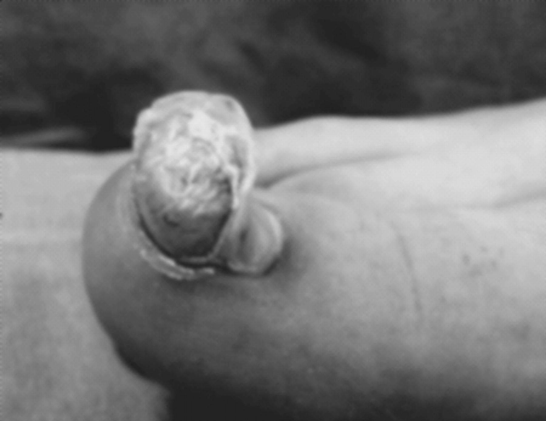
Figure 1. Preoperative view showing the large tumor on the dorsomedial aspect of the great toe.
Japma 100 00296 g001
Figure 2. Oblique preoperative foot radiograph.
Japma 100 00296 g002
Figure 3. Postoperative view of the great toe.
Japma 100 00296 g003
Figure 4. Trabecular bone surrounded by a thin cap composed of hyaline and by fibrocartilage or fibrous tissue, which confirms a subungual exostosis.
Japma 100 00296 g004
Figure 5. Postoperative view of the patient after 2 years.
Japma 100 00296 g005