Article

Establishment of a Protoplasts-Based Transient Expression System in Banana (Musa spp.)

1
Key Laboratory of Ministry of Education for Genetics, Breeding and Multiple Utilization of Crops, Plant Immunity Center, Fujian Agriculture and Forestry University, Fuzhou 350002, China
2
Guangxi Key Laboratory of Biology for Crop Diseases and Insect Pests, Plant Protection Research Institute, Guangxi Academy of Agricultural Sciences, Nanning 530007, China
*
Authors to whom correspondence should be addressed.
Agronomy 2022, 12(11), 2648; https://doi.org/10.3390/agronomy12112648
Submission received: 22 September 2022 / Revised: 19 October 2022 / Accepted: 24 October 2022 / Published: 27 October 2022
(This article belongs to the Special Issue Recent Advances in Horticultural Crops-from Omics to Biotechnology)

Abstract

:
The breeding of triploid banana cultivars with improved traits, such as yield and disease resistance, remains a major challenge for breeders. One reason is that the molecular study and functional gene analysis in bananas fall behind due to the difficulties of its genetic manipulation. The plant protoplast-based transient transformation has been documented and widely used as a versatile and convenient system for functional gene analysis in many plant species. However, an efficient high-quality protoplast isolation and transformation system is still lacking for bananas. Here, we established an efficient protoplast isolation and transformation method for bananas by selecting proper source materials, optimizing conditions for enzymatic hydrolysis and PEG-mediated transfection. We found the best source materials for banana protoplasts’ isolation are young suckers, which give a yield of protoplasts ranging from 2.5 × 10⁶ to 10.1 × 10⁷ g−1 fresh weight after 5 to 6 h of enzymolysis. The yield is sufficient for most assays that have been established in protoplasts-based systems, such as protein subcellular localization and protein interaction assays. Moreover, using the established transient gene expression system in banana protoplasts, we validated the subcellular localization of Arabidopsis VESICLE SORTING RECEPTOR 1 (VSR1) and the protein self-interaction of Arabidopsis CNGC20 on the cell membrane. The results indicated this system works well and could be routinely used for the functional characterization of banana genes.

1. Introduction

Bananas (Musa spp.) are the main food source in many tropical and subtropical countries and one of the most popular fruits in industrialized countries. They are mainly distributed in Africa, Latin America, Oceania, and Asia [1]. Like other crops, bananas are also threatened by a wide range of biotic and abiotic stresses which affect fruit productivity and quality [2]. In recent years, bananas have been seriously damaged worldwide by Fusarium wilt (Fusarium oxysporum f. sp. cubense) due to the lack of disease-resistant varieties [3]. Bananas are also vulnerable to wind damage and cold damage [4]. Due to the polyploidy and high sterility of most banana cultivars, their genetic diversity is lost [5]. Moreover, banana plants are propagated through nutritional reproduction rather than seeds. Traditional breeding methods are difficult to implement on bananas. Therefore, to ensure the healthy and sustainable development of the banana industry, it is urgent to develop a new technical system to breed new varieties of bananas with enhanced disease resistance, high yield, and high quality.
The genome sequence of bananas (Musa accuminata) was sequenced and released, providing an opportunity to conduct functional genomic studies in bananas [6]. Although the genetic transformation system was established in bananas by several methods, including agrobacterium-mediated transformation, particle bombardment, and electroporation, these methods have some disadvantages, such as low efficiency in callus induction, a long period of tissue culture, a low transformation rate, and difficulty of regeneration [7,8]. The shortages make them inefficient and time-consuming for the functional gene analysis in bananas.
The use of transient gene expression in protoplasts, a cell-based system, facilitated the rapid discoveries of gene functions and signaling pathways in several plant species, including Arabidopsis, rice, maize, and tobacco [9,10,11]. The freshly prepared Arabidopsis protoplasts respond to a broad spectrum of signals, including hormones, sugar, hydrogen peroxide, elicitors, and stresses, similar to intact tissues [9]. The protoplasts could be efficiently transfected with plasmids containing interested genes through PEG-mediated transformation. Thus, transient expression in protoplasts was developed into a versatile and convenient system for functional gene analysis, such as protein subcellular localization, protein activity, protein-protein interaction, protein degradation, kinase activation, protein modification, gene regulation, and promoter activity [10]. So far, a protoplast-based transient expression system was established in more and more plant species, including wheat [12], pineapples [13], soybeans [14], Ma bamboo [15], and sugarcane [16]. Although protoplasts’ isolation has also been reported in bananas, using embryogenic cell suspension (ECS), young leaves, and corm, preparing and keeping the ECS is time consuming; somaclonal variation through cell cultures can occur; the yields of reported methods are quite low; and the enzyme digestion time is long [17,18], making them not suitable for practical application in a functional gene study. Hence, an efficient protoplast-based transformation system in bananas is needed to be established.
The key for efficiently isolating protoplasts lies in the use of suitable plant tissue and the enzyme solution for tissue digestion. Here, we established a method for efficiently isolating plenty of banana protoplasts in a relative short time by systemically testing different banana tissues and enzymatic hydrolysis conditions. We found that banana suckers are the best material for protoplasts’ isolation. The yield of protoplasts could achieve 1.0 × 10⁷ g−1 fresh weight after 6 h digestion in an optimized enzyme solution. We also optimized conditions of PEG-mediated plasmids’ transformation. The transfection efficiency could reach above 60%. Furthermore, the subcellular localization of VESICLE SORTING RECEPTOR 1 (VSR1), which has been reported in Arabidopsis [19], was confirmed in banana protoplasts. The protein self-association of CYCLIC NUCLEOTIDE-GATED CHANNEL 20 (CNGC20) was also verified by a bimolecular fluorescence complementation (BiFC) assay in this system [20]. Thus, our method provides a convenient protoplast-based technique for the functional study of genes in bananas.

2. Materials and Methods

2.1. Plant Materials

The banana shoots growing on a Murashige Skoog (MS) medium in the propagation stage (~2-week-old), plantlets growing on a regeneration medium in the rooting stage (~4-week-old), and suckers growing in the field (~1-month-old) (Musa acuminata L., AAA group, Cavendish subgroup, cv. Williams) were used for protoplasts’ isolation in this study. Banana shoots and plantlets were incubated at 28 °C with a 10 h: 14 h, light (2000 lx): dark photoperiod. The harvested banana suckers were saved in a 4 °C refrigerator no more than 48 h before use.

2.2. Protoplast Isolation

The whole shoots and plantlets were cut into pieces (0.5–1 mm) using a sharp razor, while the sheath of the sucker was peeled off layer by layer to obtain the inside tender tissue as the starting material for protoplasts’ isolation. The processed suckers were then cut into 0.5 mm strips. The strips were immediately transferred into a beaker with 10 mL 0.6 M Mannitol (Sigma, 69-65-8) solution to equilibrium osmotic pressure for 10 min. Then, the Mannitol solution was replaced by 10 mL enzyme solutions (ESs) (Table 1) (The differences among ES1 to ES10 are the concentrations of Cellulase, Macerozyme, and Pectinase, e.g., ES9: 9.1 mM MES (Sigma,4432-31-9, pH 5.7), 2.5% (w/v) Cellulase R-10, 1% (w/v) Macerozyme R-10, 3.4 mM CaCl2, 0.1% Bovine Serum Albumin (9048-46-8) (w/v), 0.6 M Mannitol, and 0.035% β-Mercaptoethanol), and the tissue was placed under vacuum for 15 min. Then, the enzymatic digestion was carried out in the dark for 1 h to 20 h with gentle shaking (50 r/min) at room temperature (25 °C). The digestion was stopped by adding a 15 mL W5 buffer (154 mM NaCl, 155.4 mM CaCl2, 5 mM KCl, 1.4 mM MES pH 5.7, 5 mM Glucose) and filtered through a stainless 200 μm mesh sieve. The protoplasts were collected in 50 mL Falcon tubes by centrifuge for 3 min at 400 G. After removing the supernatant, the protoplasts were washed twice by being gently resuspended in a 15 mL W5 buffer. The protoplasts were then resuspended in a 20 mL W5 buffer and stabilized on ice for 30 min. Then, the protoplasts were resuspended in a 1 mL MMG solution (0.6 M Mannitol, 15 mM MgCl2, 3.7 mM MES pH 5.7) for subsequent PEG-mediated transfection.
Cellulase ‘Onozuka’ R-10 (181005-02) and MacerozymeR-10 (171208-02) were obtained from Yakult Honsha, Japan. PEG-4000 (25322-68-3), CaCl2·2H2O (10035-04-8), NaCl (7647-14-5), and MgCl2·6H2O (7791-18-6) were obtained from Sigma-Aldrich, St. Louis, MO, USA.

2.3. Counting of Protoplasts

The protoplasts’ number was counted using a hemocytometer (XB.K.25, QiuJing, Shanghai, China). Eight microliters of protoplast solution were added on the surface of the hemocytometer, and then the cover slide was laid carefully to avoid bubbles’ generation. The number of intact protoplasts in the four corners and center of the grid was counted under the microscope. The protoplast density was calculated: The protoplast number/mL = the average number of the intact cells in the four corners and center of the grid × 104.

2.4. Protoplast Transfection

The protoplast transfection was performed following the procedure below:
(1)
Add 15 μg plasmids into 100 μL of freshly isolated protoplasts in the MMG solution and mix well.
(2)
Add 110 μL of 45% (w/v) PEG solution (PEG 4000, 0.2 mmol/L Mannitol and 0.1 mol/L CaCl2) into the tube and mix well by gently inverting the tubes.
(3)
After a 15 min incubation in the dark, the protoplasts are gently washed by adding a 1 mL W5 buffer and harvested by being centrifuged for 3 min at 300 G and then resuspended in a 1 mL W5 buffer.
(4)
Repeat the above wash step two times.
(5)
The transfected protoplasts in the W5 solution are incubated under dim light at 25 °C for 12–16 h.
The protoplasts were transfected with 35S::GFP to calculate the transformation efficiency using this formula: the protoplast transformation efficiency (%) = (number of protoplasts emitting green fluorescence/(number of all protoplasts under bright field-number of protoplasts emitting red fluorescence after Propidium Iodide staining)) ×100.

2.5. Bimolecular Fluorescent Complimentary (BiFC) Assay

For the BiFC assay, 35S::CNGC20-nYFP and 35S::CNGC20-cYFP plasmids were co-transformed into the banana protoplasts. After incubation for 12–16 h, the fluorescence signal is observed under a laser confocal microscope.

2.6. Microscopy

The counting of the total and the viable protoplasts was performed with the ordinary optical microscope and fluorescent microscope (Carl Zeiss CZ Microscopy GmbH, Jena, Germany), respectively. To observe the GFP and YFP fluorescence in the transformed protoplasts using a confocal microscope (Carl Zeiss LSM880), the excitation wavelengths and emission filters sets were as follows: GFP, 488 nm (Ex)/BP505 to 530 nm (Em); YFP, 514 nm (Ex)/BP525 to 550 nm (Em). Image analysis was conducted with Carl Zeiss ZEN 2 software.

3. Result

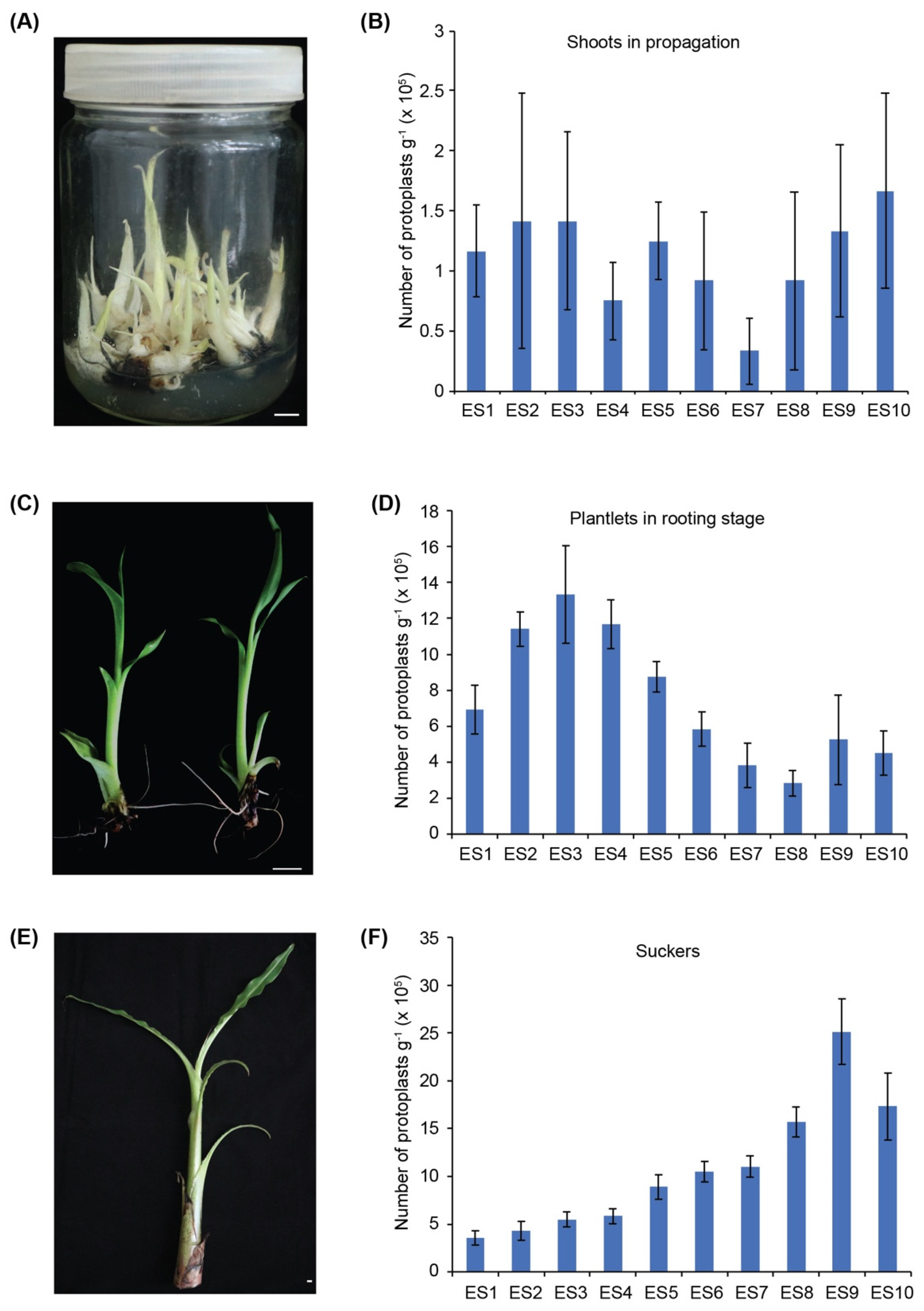
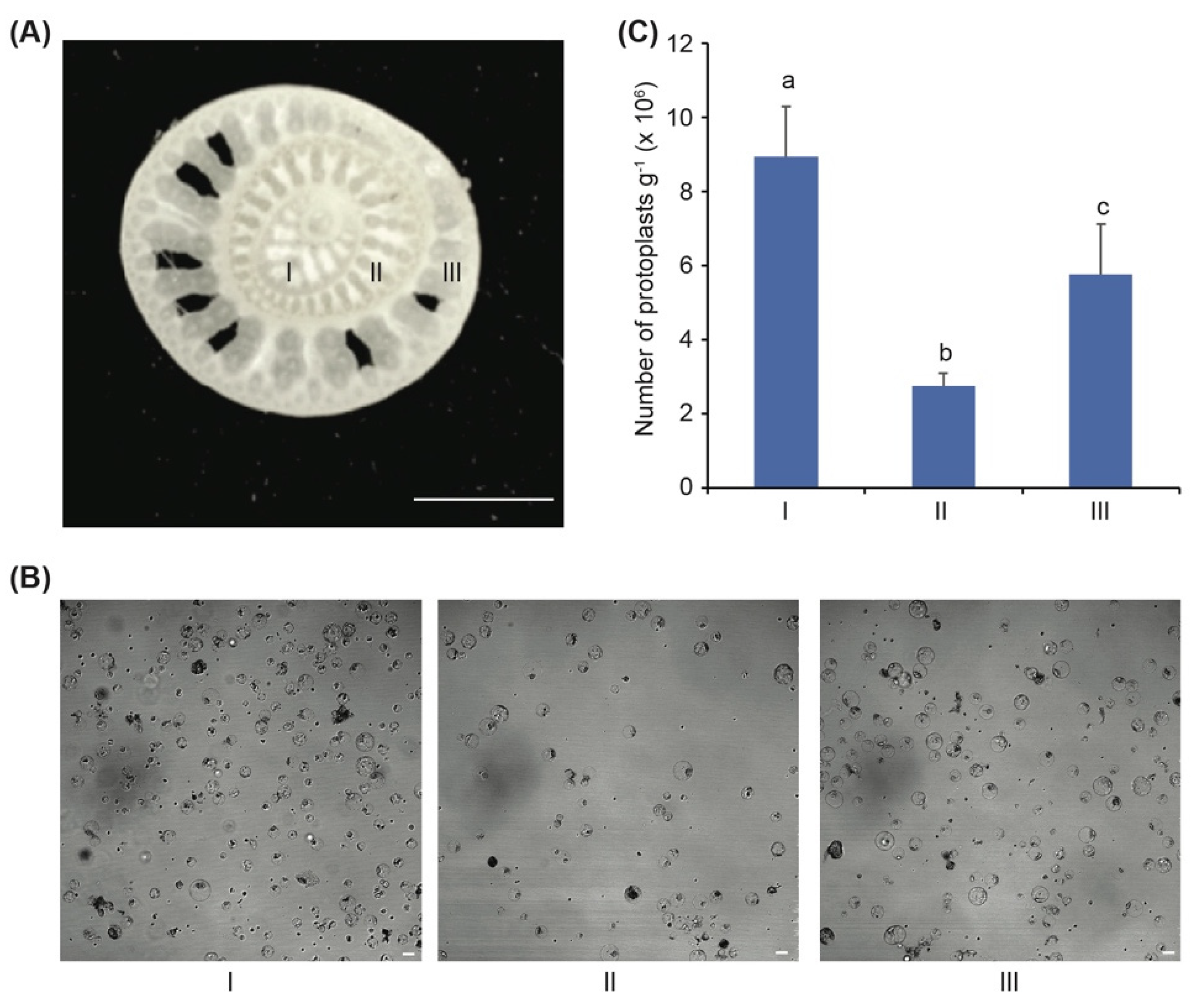
3.1. The Banana Sucker Is the Best Source Material for Isolation of Protoplasts

To establish an efficient transient expression system in bananas for functional gene analysis, obtaining a high yield of protoplasts is necessary. It is critical to use proper source materials for obtaining a high protoplast yield. In order to develop a method for efficient isolation of protoplasts from bananas, we screened banana tender tissues at different developmental stages in combination with different enzyme solutions. In total, 10 enzyme solutions (ESs) with different concentrations of Cellulase R10, Macerozyme R10, and Pectinase Y-23 were tested (Table 1). Firstly, we tried to isolate protoplasts from ~2-week-old young shoots in propagation (Figure 1A). Only a small number of protoplasts (~1 × 10⁵ g−1) was obtained over 20 h of digestion despite the ESs used (Figure 1B), indicating that the shoot in the propagation stage is not suitable for protoplast isolation. Then, we tried ~4-week-old young plantlets in the rooting stage (Figure 1C). Inspiringly, quite a number of protoplasts, from 3 × 10⁵ g−1 to 1.4 × 10⁶ g−1, were obtained after 6 h digestion with different Ess (Figure 1D). The presence of 0.2% to 0.5% pectinase in ES2, ES3, and ES4 could obviously improve the yield of protoplasts. Thus, banana young shoots in propagation are suitable for the protoplast isolation.
The other tender tissue of bananas is the new generated sucker in the field (Figure 1E). As banana suckers could be easily collected during all the growing period, we tested whether they are suitable for the protoplast isolation. Young suckers have only leaf sheaths without a midrib or lamina. The outer 5–6 sheaths were discarded, and the inner three layers of sheaths were subject to enzymatic hydrolysis. Interestingly, lots of protoplasts were isolated after 6 h of digestion under relatively low enzymatic hydrolysis conditions without pectinase (Figure 1F). Enzyme solution ES9 with 2.5% cellulase and 1% macerozyme achieved the highest number (2.5 × 10⁶ g−1) of protoplasts (Figure 1F). Thus, ES9 was considered as the best enzyme solution and used in the later protoplast isolation.
To find the most suitable sections of the banana sucker for isolating protoplasts, the most inner three sucker sheaths Figure 2A(I–III) were prepared separately for enzymatic digestion. After 6 h of digestion, all three sections produced quite a number of spherical protoplasts (Figure 2B). However, the yield of protoplasts from section Ⅰ was 8.4 × 10⁶ g−1, which was significantly higher than those from section II and III (Figure 2C). Thus, we concluded that the most inner sheath of the sucker is the best source material for protoplasts’ isolation in bananas.

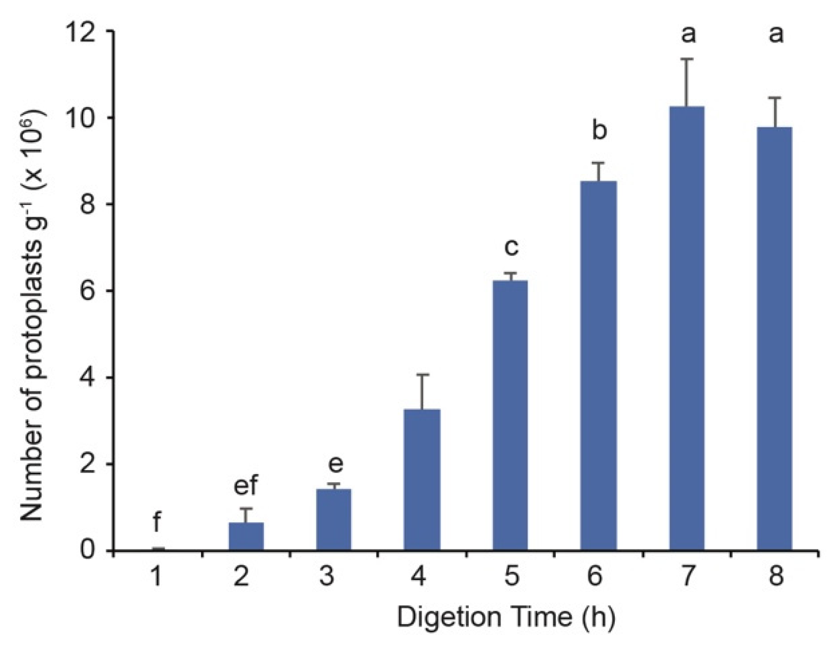
3.2. Five Hours of Enzymatic Hydrolysis Is Sufficient for the Protoplast Isolation from Banana Suckers

We then quantified the yield of protoplasts from banana suckers with 1 to 8 h of enzymatic hydrolysis in enzyme solution ES9 to determine an appropriate digestion time for a good yield. The yields of protoplasts increased significantly along with the digestion time and peaked to 1.01 × 10⁷ g−1 at 7 h (Figure 3). Notably, a good yield, 6.1 × 10⁶ protoplasts g−1, was already achieved with 5 h of enzymatic incubation. In our experience, such a yield is sufficient for transient expression experiments in protoplasts. As a long time of enzymatic incubation could cause damage to the plasma membrane, resulting in the bursting of protoplasts, shortening the digestion time ensures the high quality of protoplasts. To sum up, 2.5% Cellulase and 1% Macerozyme in ES9 is the suitable combination of enzymes in the solution, and a 5 h digestion is sufficient for the protoplast isolation from banana suckers.

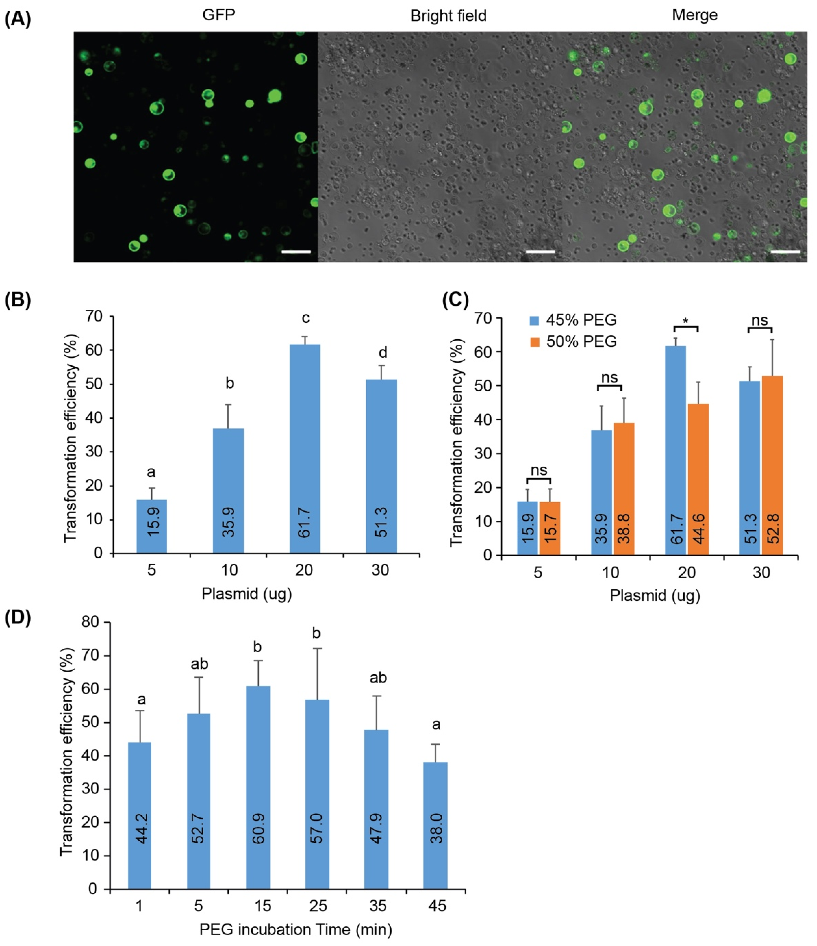
3.3. Transient Expression of GFP in Banana Protoplasts

As we established a method to isolate banana protoplasts efficiently, we then tested whether we could conduct transient expression in this system. PEG-mediated plasmids’ transfection is widely used in the protoplast transformation. We then tried to express the GFP gene in banana protoplasts using PEG-mediated transformation. According to the PEG transfection used for Arabidopsis protoplasts, we transformed 100 µL of banana protoplasts (4 × 10⁴ protoplasts) with 10 µg of plasmid containing GFP under the constitutive 35S promoter. The transfected protoplasts were incubated overnight (12–16 h) under dim light at 25 °C to express GFP proteins. Under a confocal microscope, we observed bright green fluorescence in a lot of banana protoplasts (Figure 4A), indicating that the protoplasts-based transient expression system worked well in bananas.

3.4. Effects of DNA Amount on the Protoplast Transformation Efficiency

We noticed that in Figure 4A, the efficiency of the protoplast transformation was not great (30.4% percent of protoplasts expressing GFP). The amount of plasmid could greatly affect transformation efficiency; therefore, we tested whether the banana protoplast transformation efficiency could be improved by increasing the DNA amount. We transformed 5, 10, 20, and 30 μg of the GFP plasmid into banana protoplasts per the reaction (4 × 10⁴ protoplasts), respectively. The results showed that the transformation efficiency increased along with an increased DNA amount from 5 to 20 μg, but it decreased when the plasmid increased to 30 μg (Figure 4B). The decreased transformation efficiency might be due to the big volume of 30 μg plasmid (more than 30 μL), or too many impurities from the plasmid preparation affected transfection. The data indicate that the optimal amount of plasmid for the banana transformation is 20 μg, which gives a transformation efficiency of 61.7%.

3.5. Effects of PEG Concentration and Incubation Time on the Protoplast Transformation Efficiency

Besides the amount of plasmid, the PEG concentration and incubation time might affect the protoplast transformation efficiency. The transformation efficiency had no difference between 45% and 50% PEG when using 5, 10, or 30 μg plasmids (Figure 4C). However, 50% PEG gave lower transformation efficiency than 45% PEG when using 20 μg plasmids (Figure 4C). Thus, the optimal PEG concentration for banana protoplasts’ transformation is 45%.
To optimize the transient expression system further, we next tested the influence of different PEG incubation times on the protoplast transformation efficiency. We found that 5 min of PEG incubation resulted in the transformation efficiency of 52.7% (Figure 4D). The PEG incubation time was prolonged to 15 min slightly to 60.9% (Figure 4D), while a further extension of the incubation time more than 25 min led to a slightly decreased transformation efficiency (Figure 4D). Therefore, the optimal PEG incubation time for the banana protoplast transformation is 15 min.

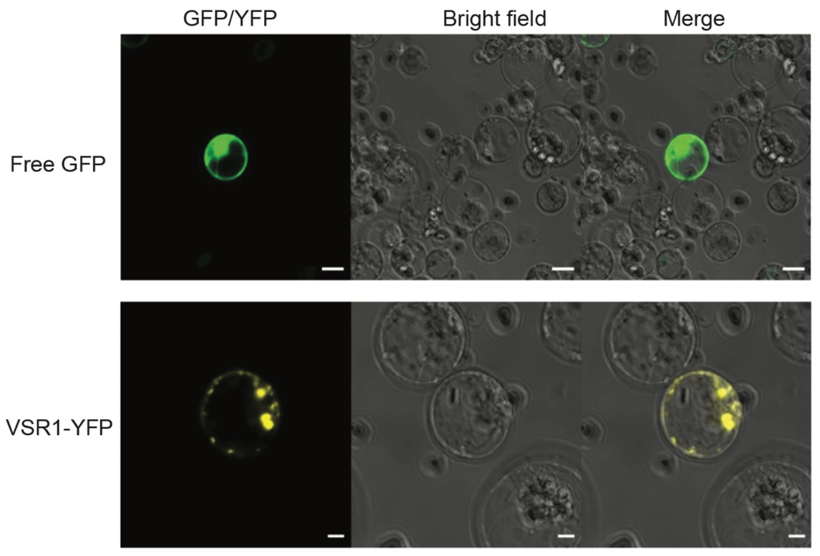
3.6. Protein Localization Assay Using Transient Expression System in Banana Protoplasts

To test the reliability of the banana protoplasts-based transient expression system in protein subcellular localization and functional gene analysis, we verified the subcellular localization of Arabidopsis VACUOLAR SORTING RECEPTOR 1 (VSR1). VSRs are type I membrane proteins involved in the sorting and packaging of soluble vacuolar proteins into transport vesicles. The free GFP localized in both the cytoplasm and nucleus in the banana protoplasts transfected with 35S::GFP (Figure 5). VSR1-YFP was observed mainly in small particles of protoplasts transfected with 35S::VSR1-YFP (Figure 5), indicating its vesicle localization.

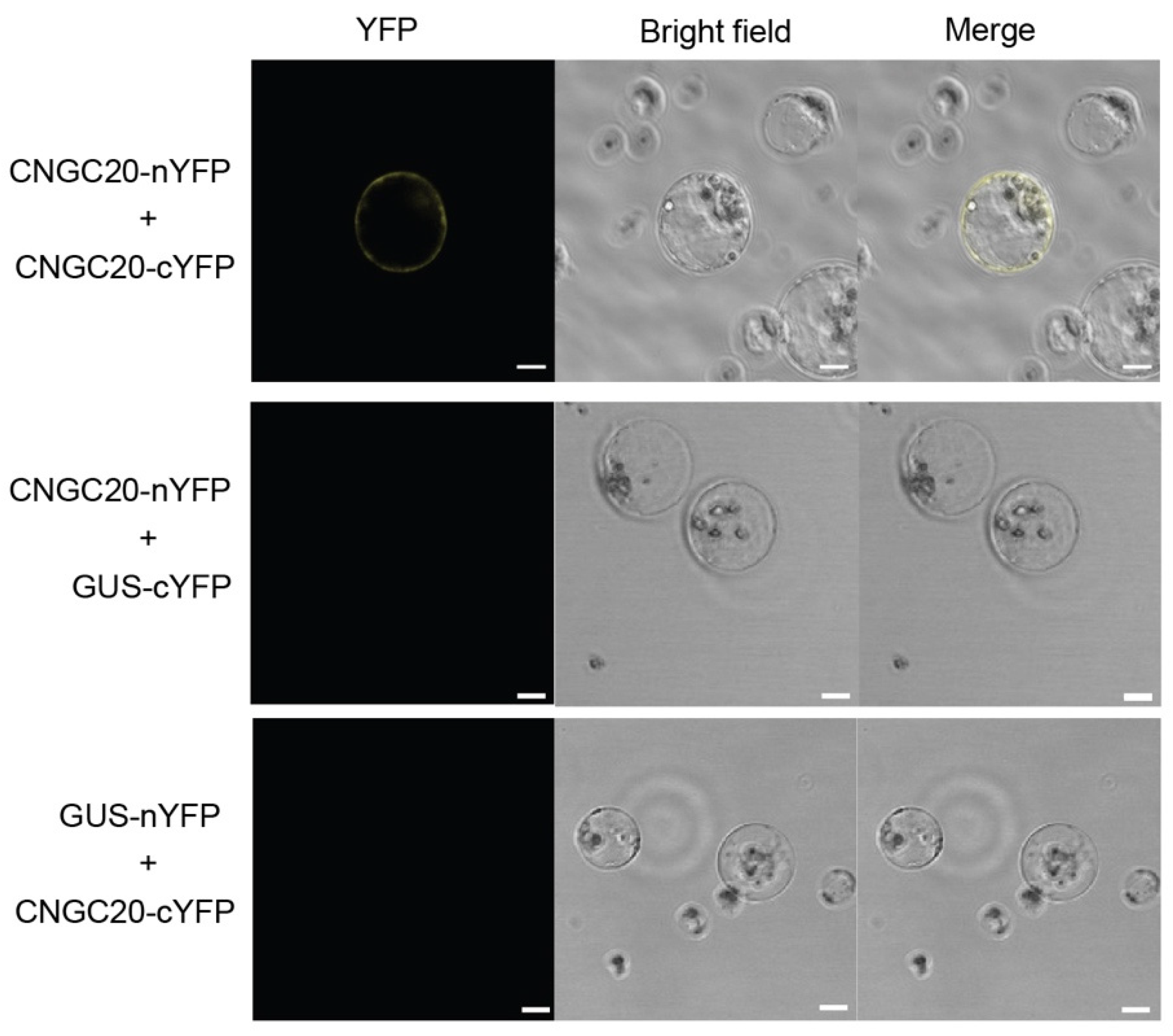
3.7. BiFC Assay Validates Protein-Protein Interactions in Banana Protoplasts

BiFC is widely used in the investigation and direct visualization of protein-protein interactions in live cells. It monitors the interaction and subcellular compartmentalization of protein complexes. To test whether the banana protoplast transient transfection system could also be used for the BiFC assay, we transiently co-expressed Arabidopsis CNGC20-nYFP and CNGC20-cYFP in banana protoplasts. CNGC20 has been shown to form homomeric complexes on the cell membrane [20]. As expected, the yellow fluorescent signal was observed on the cell membrane of banana protoplasts transfected with CNGC20-nYFP and CNGC20-cYFP, but not in the protoplasts transfected with the negative control plasmids (CNGC20-nYFP and GUS-cYFP, or GUS-nYFP and CNGC20-cYFP) (Figure 6), validating the self-association of CNGC20. Therefore, the BiFC assay could be successfully performed in the banana protoplast transient expression system.

4. Discussion

Transient gene expression in plant protoplasts is widely used as a convenient and reliable system to characterize genes, proteins, and study signaling networks rapidly [9,10,21]. To obtain the protoplast system works, it is critical to obtain a large amount and high quality of protoplasts, which depends mainly on the proper source of plant materials and enzyme solutions [22]. Here, we showed that banana plantlets in the rooting stage and suckers are good source materials for protoplasts’ isolation, but different enzyme solutions are needed to obtain the best yield (Figure 1). Especially, the highest yield of 1.01 × 10⁷ protoplasts g−1 fresh weight was achieved with 7 h of enzymatic digestion using the most inner sheaths of banana suckers, which is a great improvement than the previously reported method, which used banana corm as the material and produced 8.7 × 10⁵ protoplasts g−1 after 24 h of enzymatic digestion [18]. As both the young banana plantlet and sucker could be more easily collected than rhizome, the protoplasts’ isolation with our method could be routinely performed.
The enzymolysis solution is very important to ensure the high yield and integrity of protoplasts [23]. The main components of enzymatic hydrolysate are enzymes, enzyme solvents, and osmotic pressure stabilizers. Cellulase, macerozyme, and pectinase are commonly used in the preparation of protoplasts. The combination and concentration of the enzyme were different with different banana varieties, genotypes, and tissues [17,24]. Studies show that the enzyme solution containing 3% cellulase, 1% macerozyme, and 0.2% pectinase produced the maximum yield of banana protoplasts from successive suspension cells [17]. Here, the same formula of three enzymes results in a good yield of protoplasts from banana plantlets in the rooting stage (Figure 1D and Table 1). However, for isolating protoplasts from banana suckers, the pectinase seems to have negative effects. The highest yield of protoplasts was obtained with 2.5% cellulase and 1% macerozyme without pectinase (Figure 1F). The optimal enzymolysis time also varies for different plant species. For example, the optimal enzymolysis time for the pineapple and wheat is 3 h and 4 h, respectively [12,13]. In the process of enzymolysis, the activity of the enzyme may change [25], and a short time is not enough to enzymolize the cell wall fully, but a long time may also break the isolated protoplasts [25]. Our study showed that the optimal enzymolysis time was 5 to 6 h for banana suckers to obtain a relatively high yield of protoplasts (Figure 3). In practice, 4 h of enzymolysis which gives around 3.6 × 10⁶ protoplasts g−1 fresh weight is sufficient for a small scale of experiments, such as the protein subcellular localization assay, BiFC, and reporter assays.
After establishment of the protocol for banana protoplasts’ isolation, we optimized the conditions for PEG-mediated plasmid transfection of banana protoplasts. The PEG-mediated transformation has been successfully applied in a variety of plant protoplasts or microorganisms, most of which have high transformation efficiency at a PEG concentration range of 20–40% [23,26]. In this study, the highest transformation efficiency could be achieved with 45% of PEG for the transfection of banana protoplasts. In addition, the optimal PEG incubation time for transfections is 10–15 min. Another important factor affecting the transformation efficiency in protoplasts is the amount of plasmid. It was reported that within a certain range amount of DNA, the transformation efficiency of protoplasts increases along with the increased amount of DNA and tends to be stable after reaching a peak [26]. It may be because of the increase in the amount of plasmid along with the increased reaction volume which affects the transformation efficiency [27]. In the protoplast transformation system of different plants, the optimal DNA amount varies [28]. Our results showed that the optimal amount of plasmid for the banana protoplast transfection is 20 μg.
Given the difficulty and time-consuming nature of making stable transgenic lines in bananas, the transient gene expression in the protoplasts system is a useful tool for functional gene characterization. In this study, the subcellular localization of AtVSR1 was confirmed in the banana protoplast (Figure 5). Furthermore, the BiFC assay was also successfully performed to validate and achieve visualization protein interactions in the cell in our banana protoplast-based transformation system (Figure 6). Therefore, the banana protoplast isolation and transformation system developed in this study can be easily used for protein subcellular localization and protein-protein interaction assays and shall be a versatile and convenient system for functional gene analysis in bananas.

5. Conclusions

In this study, we established a method for efficiently preparing banana protoplasts from banana suckers or plantlets in the rooting stage. Our method could obtain a great yield of protoplasts within 5 to 6 h of enzymolysis, which is sufficient and convenient for most molecular assays that have been established in the protoplasts-based system. We optimized the conditions for PEG-mediated protoplasts’ transformation. We showed that functional gene analysis such as protein subcellular localization and protein interaction assays could be performed in the established transient expression system using banana protoplasts. As banana suckers and plantlets could be easily collected, such a protoplasts-based system can be routinely used for functional gene study in bananas. Regeneration of the plant from the protoplast is documented for many species [29]. We are working on the regeneration of bananas from protoplasts now. If it works out, it will provide a new strategy for transgene and gene editing in the breeding of new varieties of bananas.

Author Contributions

Conceptualization, H.C. and G.F.; methodology, C.Z., S.L., C.D., H.G. and D.Y.; writing—original draft preparation, C.Z.; writing—review and editing, H.C. and G.F.; supervision, H.C. All authors have read and agreed to the published version of the manuscript.

Funding

This work was funded by the Central Government-Guided Local Science and Technology Development Fund (ZY21195015) and the National Natural Science Foundation of China (31960520).

Conflicts of Interest

The authors declare that they have no conflict of interest.

References

  1. Perrier, X.; De Langhe, E.; Donohue, M.; Lentfer, C.; Vrydaghs, L.; Bakry, F.; Carreel, F.; Hippolyte, I.; Horry, J.P.; Jenny, C.; et al. Multidisciplinary perspectives on banana (Musa spp.) domestication. Proc. Natl. Acad. Sci. USA 2011, 108, 11311–11318. [Google Scholar] [CrossRef] [Green Version]
  2. Butler, D. Fungus threatens top banana. Nature 2013, 504, 195–196. [Google Scholar] [CrossRef] [Green Version]
  3. Ploetz, R.C. Fusarium Wilt of Banana. Phytopathology 2015, 105, 1512–1521. [Google Scholar] [CrossRef] [Green Version]
  4. Pegg, K.G.; Coates, L.M.; O’Neill, W.T.; Turner, D.W. The Epidemiology of Fusarium Wilt of Banana. Front. Plant Sci. 2019, 10, 1395. [Google Scholar] [CrossRef] [PubMed] [Green Version]
  5. Ghag, S.B.; Ganapathi, T.R. Genetically modified bananas: To mitigate food security concerns. Sci. Hortic. 2017, 214, 91–98. [Google Scholar] [CrossRef]
  6. D’Hont, A.; Denoeud, F.; Aury, J.M.; Baurens, F.-C.; Carreel, F.; Garsmeur, O.; Noel, B.; Bocs, S.; Droc, G.; Rouard, M.; et al. The banana (Musa acuminata) genome and the evolution of monocotyledonous plants. Nature 2012, 488, 213–217. [Google Scholar] [CrossRef] [Green Version]
  7. Assani, A.; Haïcour, R.; Wenzel, G.; Foroughi-Wehr, B.; Bakry, F.; Côte, F.-X.; Ducreux, G.; Ambroise, A.; Grapin, A. Influence of donor material and genotype on protoplast regeneration in banana and plantain cultivars (Musa spp.). Plant Sci. 2002, 162, 355–362. [Google Scholar] [CrossRef]
  8. Shivani; Kaur, N.; Awasthi, P.; Tiwari, S. Identification and expression analysis of genes involved in somatic embryogenesis of banana. Acta Physiol. Plant. 2018, 40, 139. [Google Scholar] [CrossRef]
  9. Sheen, J. Signal transduction in maize and Arabidopsis mesophyll protoplasts. Plant Physiol. 2001, 127, 1466–1475. [Google Scholar] [CrossRef]
  10. He, P.; Shan, L.; Sheen, J. The Use of Protoplasts to Study Innate Immune Responses. Methods Mol. Biol. 2007, 354, 1–9. [Google Scholar]
  11. Confraria, A.; Baena-González, E. Using Arabidopsis Protoplasts to Study Cellular Responses to Environmental Stress. Methods Mol. Biol. 2016, 1398, 247–269. [Google Scholar]
  12. Jia, X.; Zhang, X.; Qu, J.; Han, R. Optimization Conditions of Wheat Mesophyll Protoplast Isolation. Agric. Sci. 2016, 7, 850–858. [Google Scholar] [CrossRef]
  13. Priyadarshani, S.V.G.N.; Hu, B.; Li, W.; Ali, H.; Jia, H.; Zhao, L.; Ojolo, S.P.; Azam, S.M.; Xiong, J.; Yan, M.; et al. Simple protoplast isolation system for gene expression and protein interaction studies in pineapple (Ananas comosus L.). Plant Methods 2018, 14, 95. [Google Scholar] [CrossRef] [Green Version]
  14. Wu, F.; Hanzawa, Y. A Simple Method for Isolation of Soybean Protoplasts and Application to Transient Gene Expression Analyses. J. Vis. Exp. 2018, 57258. [Google Scholar] [CrossRef]
  15. Ye, S.; Chen, G.; Kohnen, M.V.; Wang, W.; Cai, C.; Ding, W.; Wu, C.; Gu, L.; Zheng, Y.; Ma, X.; et al. Robust CRISPR/Cas9 mediated genome editing and its application in manipulating plant height in the first generation of hexaploid Ma bamboo (Dendrocalamus latiflorus Munro). Plant Biotechnol. J. 2019, 18, 1501–1503. [Google Scholar] [CrossRef] [Green Version]
  16. Wang, Q.; Yu, G.; Chen, Z.; Han, J.; Hu, Y.; Wang, K. Optimization of protoplast isolation, transformation and its application in sugarcane (Saccharum spontaneum L.). Crop J. 2020, 9, 133–142. [Google Scholar] [CrossRef]
  17. Wu, S.; Zhu, H.; Liu, J.; Yang, Q.; Shao, X.; Bi, F.; Hu, C.; Huo, H.; Chen, K.; Yi, G. Establishment of a PEG-mediated protoplast transformation system based on DNA and CRISPR/Cas9 ribonucleoprotein complexes for banana. BMC Plant Biol. 2020, 20, 425. [Google Scholar] [CrossRef]
  18. Khatri, A.; Dahot, M.; Khan, I.; Nizamani, G.S. An efficient method of protoplast isolation in banana (Musa spp.). Pak. J. Bot. 2010, 42, 1267–1271. [Google Scholar]
  19. Miao, Y.; Yan, P.K.; Kim, H.; Hwang, I.; Jiang, L. Localization of Green Fluorescent Protein Fusions with the Seven Arabidopsis Vacuolar Sorting Receptors to Prevacuolar Compartments in Tobacco BY-2 Cells. Plant Physiol. 2006, 142, 945–962. [Google Scholar] [CrossRef] [Green Version]
  20. Zhao, C.; Tang, Y.; Wang, J.; Zeng, Y.; Sun, H.; Zheng, Z.; Su, R.; Schneeberger, K.; Parker, J.E.; Cui, H. A mis-regulated cyclic nucleotide-gated channel mediates cytosolic calcium elevation and activates immunity in Arabidopsis. New Phytol. 2021, 230, 1078–1094. [Google Scholar] [CrossRef]
  21. Yoo, S.-D.; Cho, Y.-H.; Sheen, J. Arabidopsis mesophyll protoplasts: A versatile cell system for transient gene expression analysis. Nat. Protoc. 2007, 2, 1565–1572. [Google Scholar] [CrossRef] [Green Version]
  22. He, F.; Chen, S.; Ning, Y.; Wang, G.L. Rice (Oryza sativa) Protoplast Isolation and Its Application for Transient Expression Analysis: Rice Protoplast Isolation and Transient Expression Analysis. Curr. Protoc. Plant Biol. 2016, 1, 373–383. [Google Scholar] [CrossRef]
  23. Shen, Y.; Meng, D.; McGrouther, K.; Zhang, J.; Cheng, L. Efficient isolation of Magnolia protoplasts and the application to subcellular localization of MdeHSF1. Plant Methods 2017, 13, 44. [Google Scholar] [CrossRef]
  24. Megia, R.; Haïcour, R.; Rossignol, L.; Sihachakr, D. Callus formation from cultured protoplasts of banana (Musa sp.). Plant Sci. 1992, 85, 91–98. [Google Scholar] [CrossRef]
  25. Shabanian, N.; Rahmani, M.S.; Zarrinjoei, F. The effect of enzyme concentrations and digestion time on protoplast isolation from in vitro grown leaves of Oak (Quercus brantii). J. Biotechnol. 2010, 150, 565. [Google Scholar] [CrossRef]
  26. Armstrong, C.; Petersen, W.; Buchholz, W.; Bowen, B.; Sulc, S. Factors affecting PEG-mediated stable transformation of maize protoplasts. Plant Cell Rep. 1990, 9, 335–339. [Google Scholar] [CrossRef]
  27. Ge, C.-Y.; Duan, Y.-B.; Zhou, M.-G.; Chen, C.-J. A Protoplast Transformation System for Gene Deletion and Complementation in Sclerotinia sclerotiorum. J. Phytopathol. 2013, 161, 800–806. [Google Scholar] [CrossRef]
  28. Chang, S.; Cohen, S.N. High frequency transformation of Bacillus subtilis protoplasts by plasmid DNA. Mol. Gen. Genet. 1979, 168, 111–115. [Google Scholar] [CrossRef]
  29. Reed, K.M.; Bargmann, B.O.R. Protoplast Regeneration and Its Use in New Plant Breeding Technologies. Front. Genome Ed. 2021, 3, 734951. [Google Scholar] [CrossRef]
Figure 1. Yields of the protoplasts from different tissues of banana. (A,C,E) Banana shoots in propagation (A), plantlets in rooting stage (C), and suckers (E) used for protoplasts isolation. Bar = 1 cm. (B,D,F) The yields of protoplasts isolated from (A,C,E), respectively, after 6 h digestion in different enzyme solutions (ES1 to ES10, Table 1). Y-axes represent the number of protoplasts per gram fresh weight ± SD (n = 4).
Agronomy 12 02648 g001
Figure 2. Yields of the protoplasts from different sheaths of the banana sucker. (A) After peeling off the outer layers of the banana sucker sheath, the most inner three layers (IIII) were prepared separately for protoplasts’ isolation. Bar = 1 cm. (B) The protoplasts isolated from different sheaths in (A) under bright field of microscope. Bars = 10 μm. (C) The yields of protoplasts isolated from different sheaths in (A), after 6 h digestion in enzyme solution ES9. Y-axis represents the number of protoplasts per gram fresh weight ± SD (n = 4). The different letters indicate statistically significant differences (p < 0.05).
Agronomy 12 02648 g002
Figure 3. Yields of the protoplasts from banana suckers with different digestion times in enzyme solution ES9. The yields of protoplasts isolated from banana suckers with different digestion times, 1 to 8 h, in enzyme solution ES9. Y-axes represent the number of protoplasts per gram fresh weight ± SD (n = 4). The different letters indicate statistically significant differences (p < 0.05).
Agronomy 12 02648 g003
Figure 4. Effect of plasmid amount, PEG concentration, and incubation time on protoplast transfection. (A) Visualization of banana protoplasts transiently expressing 35S::GFP under a confocal microscope. Bars = 50 μm. (B) The transformation efficiency (TE) of banana protoplasts transfected with various amounts of plasmid DNA (35S::GFP) using PEG-mediated transfection (using 45% PEG solution). TE was calculated after 12 h cultivation of protoplasts after transformation. * indicates a significant difference to 45% PEG (p < 0.05 ) in a student’s t-test. (C) The effects of PEG concentration on the TE of banana protoplasts transfected with various amounts of plasmid DNA. (D) The effects of PEG incubation time on the TE of banana protoplasts. The different letters indicate statistically significant differences (p < 0.05). ns: non-significant differences.
Agronomy 12 02648 g004
Figure 5. Subcellular localization of VSP1 in banana protoplasts. Subcellular localization of free GFP and Arabidopsis VSP1-YFP in banana protoplasts. GFP Bars, 10 μm; VSP1-YFP Bars, 5 μm.
Agronomy 12 02648 g005
Figure 6. Bimolecular fluorescence complementation (BiFC) assay of CNGC20 self-association in banana protoplasts. Arabidopsis CNGC20-nYFP and CNGC20-cYFP were transiently co-expressed in the banana protoplasts. The co-expressions of CNGC20-nYFP and GUS-cYFP, and GUS-nYFP and CNGC20-cYFP were included as negative controls. Images were captured using a confocal microscope 12 h after transformation. Bars = 10 μm.
Agronomy 12 02648 g006
Table 1. Enzyme combinations in solutions tested for the banana protoplast isolation.
Enzyme
Solutions
Cellulase
R-10 (%)
Macerozyme
R-10 (%)
Pectinase
Y-23 (%)
ES11.50.30
ES2310.2
ES3310.5
ES4311
ES5321
ES61.50.750
ES720.750
ES82.50.750
ES92.510
ES10210
Protoplasts’ isolation.
Publisher’s Note: MDPI stays neutral with regard to jurisdictional claims in published maps and institutional affiliations.

