Article

Early Rod Dysfunction Influences Cone Development in a Rhodopsin P23H Mouse Model of Retinitis Pigmentosa


1
Lions Eye Institute, 2 Verdun St., Nedlands, WA 6009, Australia
2
Centre for Ophthalmology and Visual Sciences, The University of Western Australia, 35 Stirling Hwy, Crawley, WA 6009, Australia
3
Department of Optometry and Vision Sciences, University of Melbourne, Parkville, VIC 3052, Australia
4
School of Human Sciences, The University of Western Australia, 35 Stirling Hwy, Crawley, WA 6009, Australia
5
Perron Institute for Neurological and Translational Science, 8 Verdun St., Nedlands, WA 6009, Australia
*
Author to whom correspondence should be addressed.
Pathophysiology 2026, 33(1), 7; https://doi.org/10.3390/pathophysiology33010007

Submission received: 7 October 2025 / Revised: 25 November 2025 / Accepted: 2 January 2026 / Published: 14 January 2026
(This article belongs to the Section Neurodegenerative Disorders)

Abstract

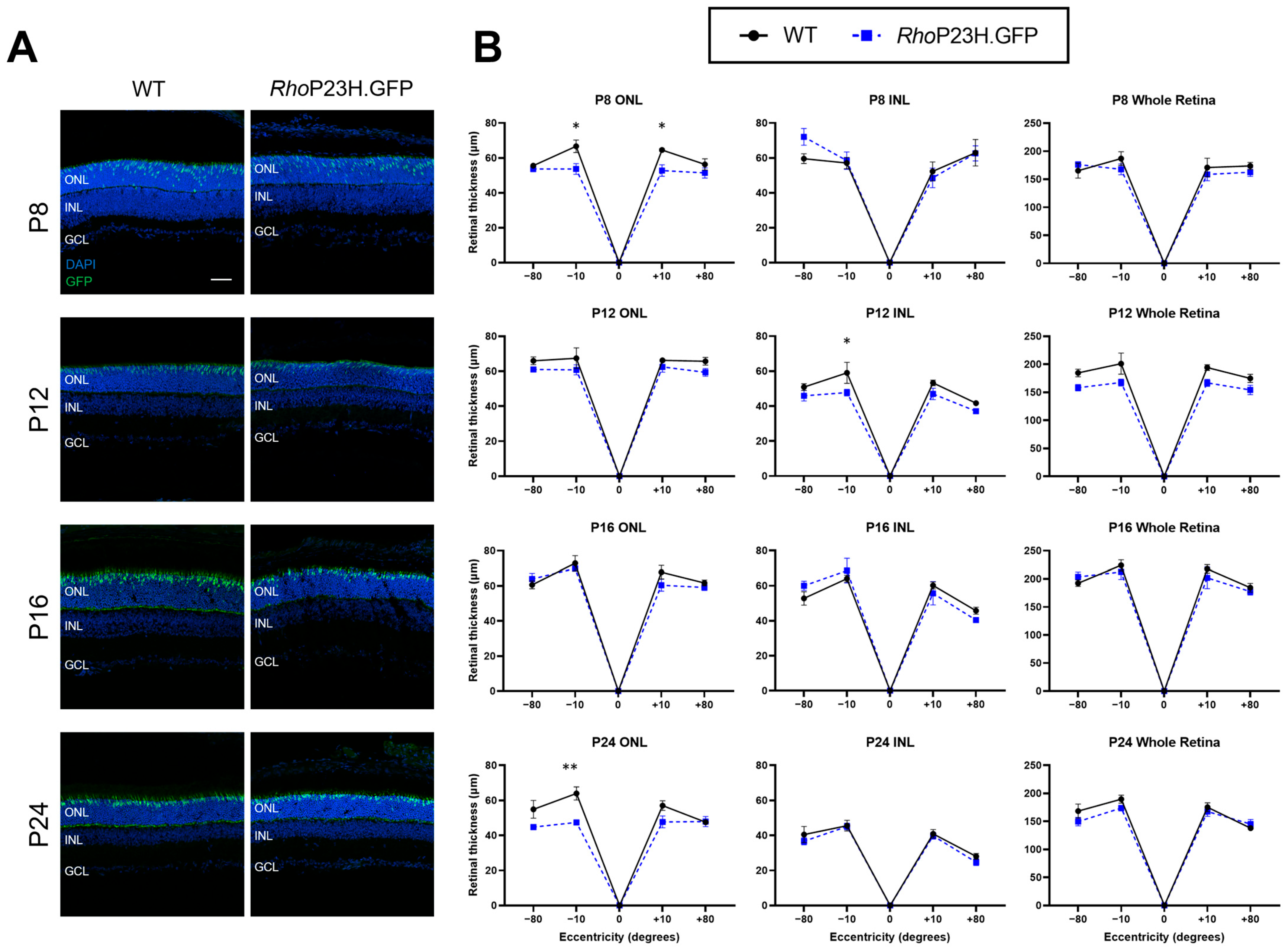
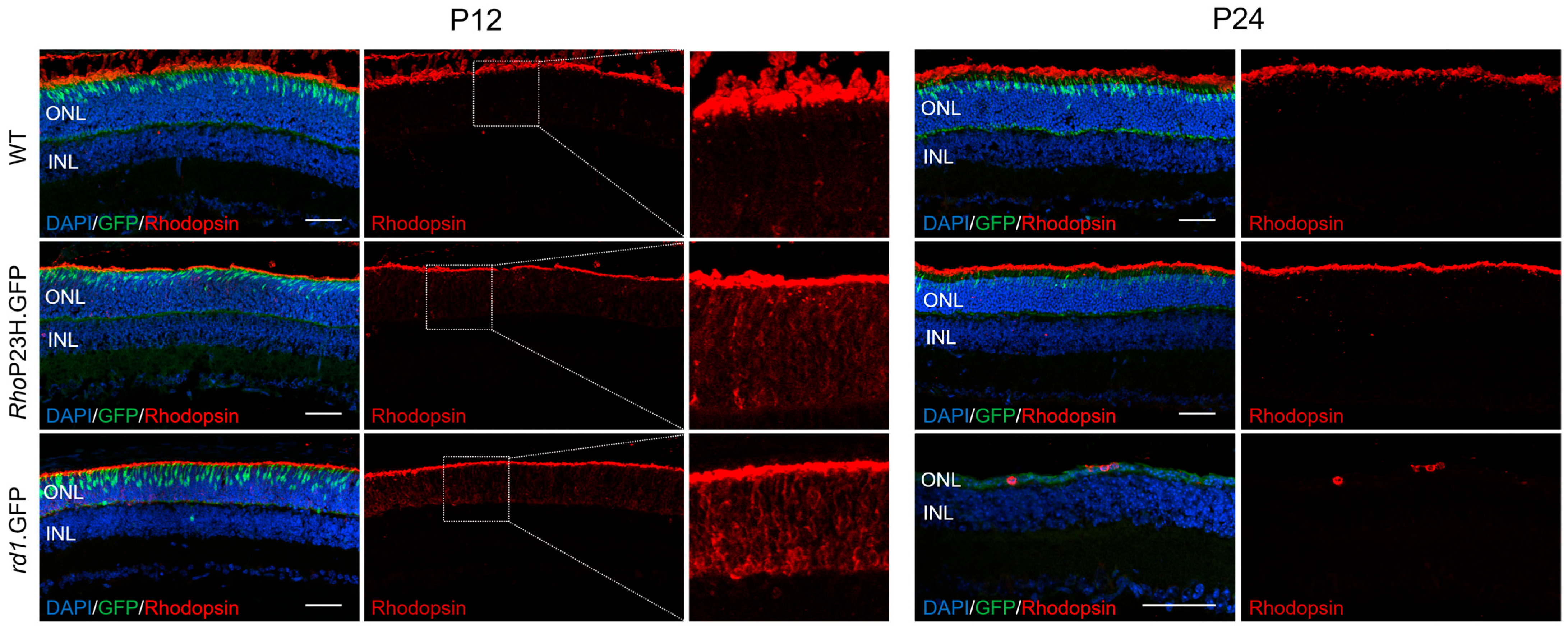
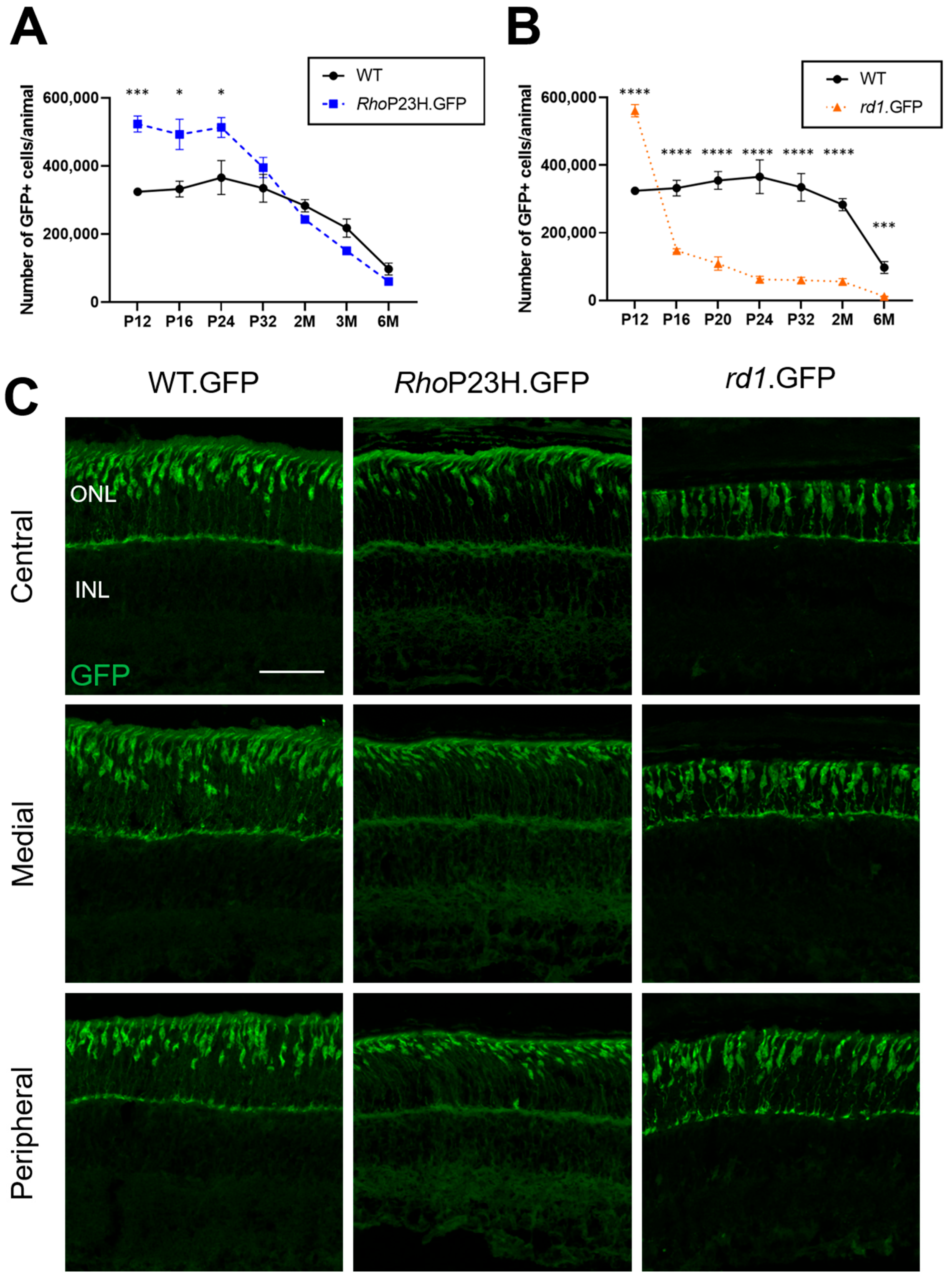
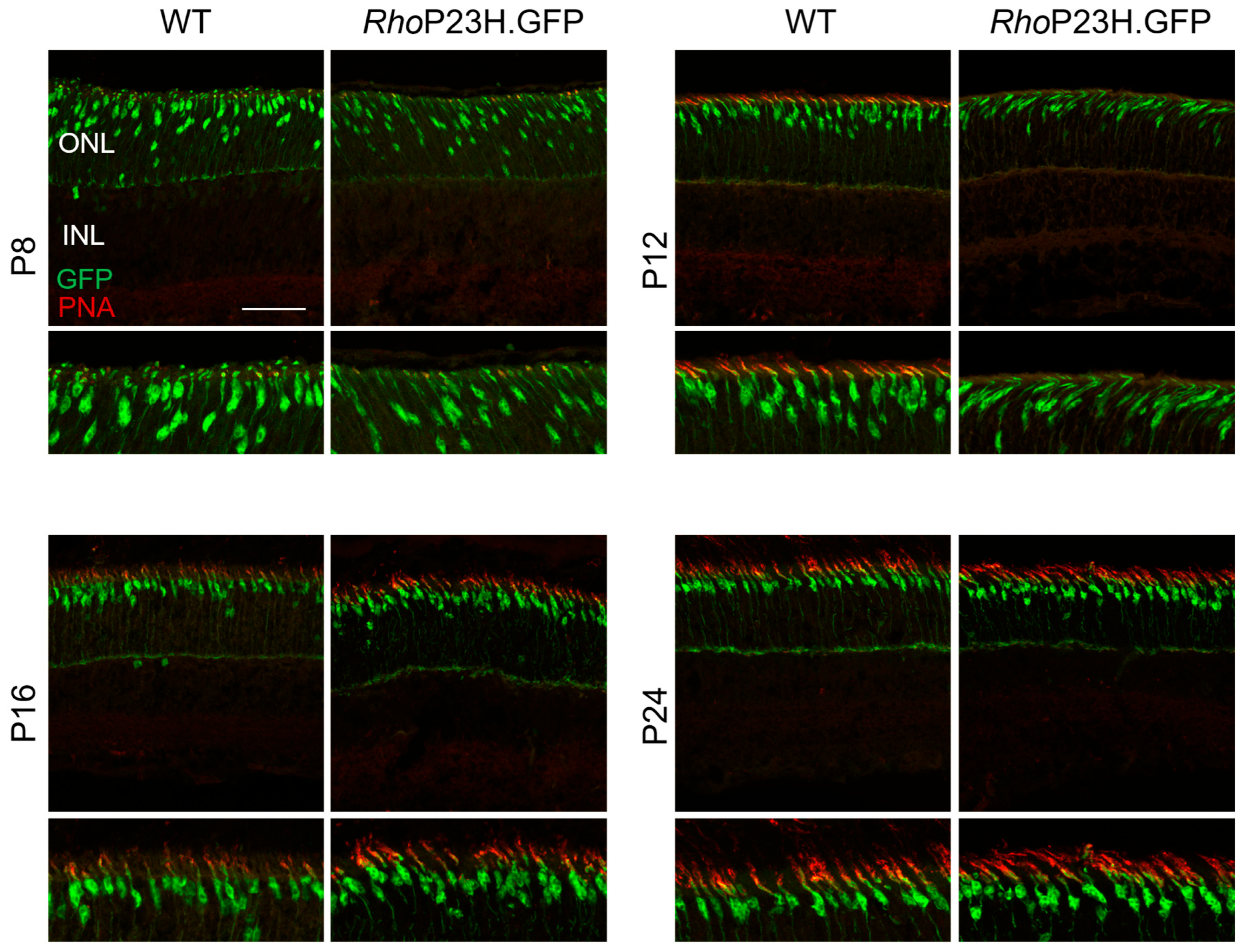
Background/Objectives: The RhoP23H/WT mouse line is a commonly used model to study rhodopsin P23H-associated autosomal dominant retinitis pigmentosa. Previous studies in RhoP23H/WT mice have largely focused on retinal changes occurring at one month of age and later, and have indicated a compensatory thickening of inner retinal layers in response to rod degeneration. However, the effect of disease processes during early postnatal retinal development remains understudied. Methods: In this study, we investigated the retinal response to rod dysfunction during early postnatal developmental ages P8–P24 in our novel RhoP23H/WT reporter line, RhoP23H.GFP, which expresses green fluorescent protein (GFP) exclusively in cone photoreceptors. Results: Histological analysis revealed no significant difference in retinal thickness in RhoP23H.GFP mice compared to healthy controls at the ages investigated. RhoP23H.GFP retinas initially exhibited a greater mislocalization of rhodopsin to the rod cell bodies at P12, though this mislocalization normalized to wildtype by P24. Most notably, flow cytometry revealed significantly increased cone photoreceptor numbers in P12 (61%), P16 (48%), and P24 (40%) RhoP23H.GFP mice compared to wildtype controls, indicating a possible compensatory response of cone photoreceptors to rod dysfunction. Additionally, cone morphology appeared altered in diseased cones. Conclusions: Our results suggest that cones may undergo a developmental compensatory adaptation in response to rod dysfunction, providing new insights into early disease mechanisms of retinitis pigmentosa.
Keywords: retinitis pigmentosa; photoreceptor degeneration; P23H mouse; cone photoreceptor; retinal development; retinal compensation; inherited retinal disease retinitis pigmentosa; photoreceptor degeneration; P23H mouse; cone photoreceptor; retinal development; retinal compensation; inherited retinal disease
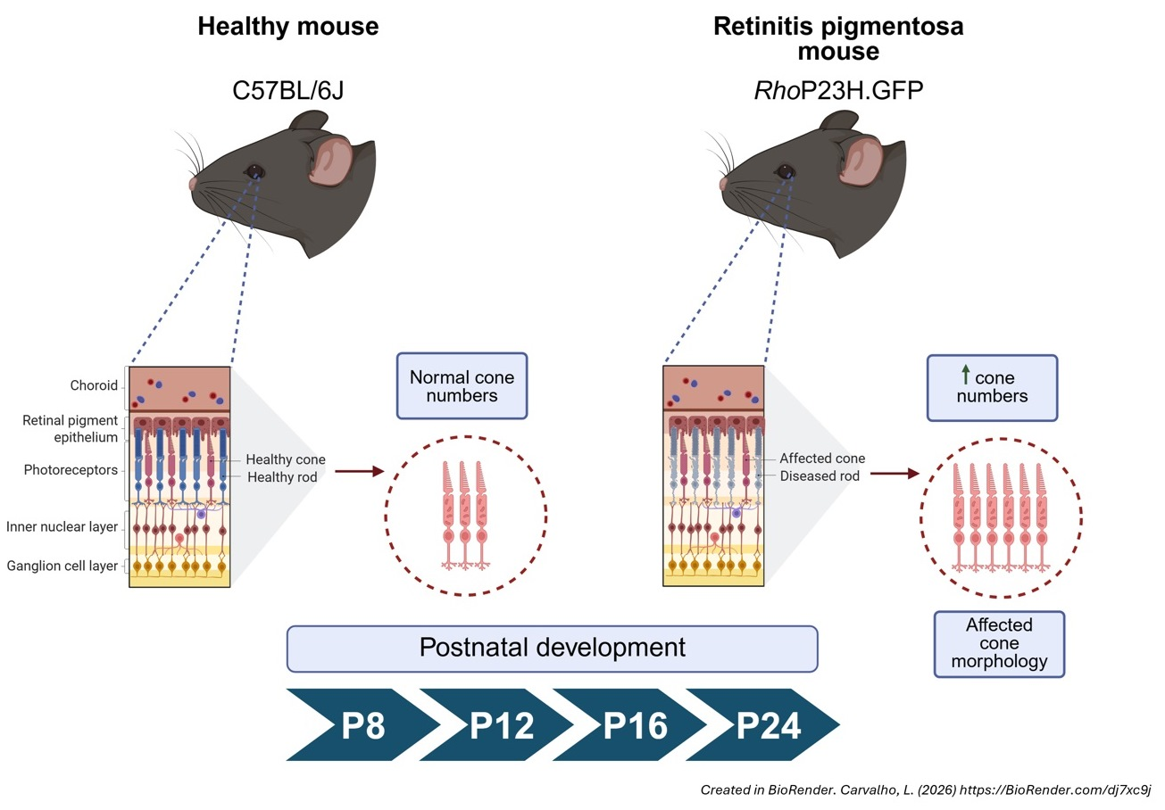
Graphical Abstract