Case Report

ESR1 Y537S Detected in a Brain Metastasis but Not in Plasma ctDNA in HR+/HER2-Low Metastatic Breast Cancer: A Case Report


by
Aemélia Nègre
1,2,*,
Lorène Seguin
1,
Arthur Géraud Cremieux
1,
Frederic Viret
1,
Agnès Tallet
3,
Cornel Popovici
4,
Anthony Gonçalves
1 and
Alexandre de Nonneville
1
1
Department of Medical Oncology, Institute Paoli Calmettes, 13009 Marseille, France
2
Faculty of Medicine, Aix Marseille University, La Timone Campus, 13005 Marseille, France
3
Department of Radiotherapy, Institute Paoli Calmettes, CRCM, 13009 Marseille, France
4
Cancer Biology Department, Institute Paoli Calmettes, 13009 Marseille, France
*
Author to whom correspondence should be addressed.
Curr. Oncol. 2026, 33(8), 454; https://doi.org/10.3390/curroncol33080454
Submission received: 8 June 2026 / Revised: 16 July 2026 / Accepted: 27 July 2026 / Published: 28 July 2026
(This article belongs to the Section Breast Cancer)

Simple Summary

Breast cancer can progress in the brain even when disease elsewhere remains well controlled. We report a woman whose hormone-sensitive breast cancer remained controlled outside the brain during long-term treatment but who developed several brain metastases. Genetic analysis of a surgically removed cerebellar metastasis identified a mutation in the estrogen receptor gene associated with resistance to aromatase inhibitors. The same mutation was not detected in a blood sample using a highly sensitive test. This discrepancy may reflect limited release of tumor DNA from brain lesions into the bloodstream and does not prove that the mutation was present only in the brain. This case shows that a negative blood-based tumor DNA result cannot exclude isolated brain progression and that direct analysis of brain metastasis tissue can provide clinically useful information when surgical material is available.

Abstract

Brain metastases in hormone receptor-positive metastatic breast cancer remain incompletely characterized at the molecular level. We report isolated central nervous system progression in a 60-year-old woman with hormone receptor-positive breast cancer initially diagnosed at the age of 38 and metastatic recurrence diagnosed at the age of 55. After approximately five years of extracranial disease control with letrozole plus palbociclib, she developed multiple brain metastases, including a large cerebellar lesion requiring surgical resection. Molecular profiling of the resected lesion identified ESR1 Y537S and ERBB2 Y772_A775dup, whereas postoperative plasma circulating tumor DNA analyzed using a sensitive next-generation sequencing assay showed no detectable somatic alteration. Extracranial disease remained in complete metabolic response. This tissue–plasma discordance is compatible with spatial genomic heterogeneity but may also reflect a low circulating tumor fraction, postoperative reduction in tumor burden, and limited release of tumor-derived DNA from CNS lesions into the systemic circulation. This case highlights the limitations of plasma circulating tumor DNA for evaluating CNS-limited progression and supports direct molecular profiling of brain metastases when tissue is available.

1. Introduction

Brain metastases represent a major clinical challenge in metastatic breast cancer (MBC), particularly because of their distinct biology and limited sensitivity to systemic therapies due to the differences in drug bioavailability imposed by the blood–brain barrier. Approximately 15–25% of patients with breast cancer will develop brain metastases, with marked differences according to molecular subtype [1].
While HER2-positive and triple-negative tumors have the highest predilection for central nervous system (CNS) spread, hormone receptor-positive (HR+) disease accounts for a non-negligible proportion of cases, mostly late in the natural history of the disease [2].
Metastatic dissemination is known to be accompanied by genomic diversification, with late metastatic sites often diverging from both the primary tumor and other metastatic deposits. This evolutionary phenomenon reflects acquisition of resistance mutations under treatment pressure, and contributes to clinical heterogeneity of therapeutic responses.
Among key resistance mechanisms in HR+ MBC, ESR1 ligand-binding domain mutations (notably Y537S, Y537N, and D538G) are well-established drivers of endocrine resistance. Their prevalence increases with cumulative exposure to aromatase inhibitors. While common in visceral and bone metastases, ESR1 mutations remain exceptionally rare in brain metastases, with only a few documented cases [3].
Importantly, molecular profiling of brain metastases often reveals alterations not detectable in circulating tumor DNA (ctDNA), reflecting both anatomical compartmentalization and biological isolation of CNS lesions [4]. Therefore, matched tissue and liquid biopsy analyses can provide complementary insights.
A previously published case report described a 37-year-old woman with hormone receptor-positive, HER2-negative metastatic breast cancer and isolated leptomeningeal progression during aromatase inhibitor (AI) treatment, in whom an ESR1 activating mutation was detected in free tumor DNA derived from cerebrospinal fluid (CSF), but not in the circulating tumor DNA (ctDNA) derived from matched plasma samples [5].
Here, we report a case of isolated CNS progression in a patient with HR+/HER2-low metastatic breast cancer treated with an aromatase inhibitor plus a CDK4/6 inhibitor. An ESR1 Y537S mutation was detected in the resected cerebellar metastasis but was not identified in postoperative plasma ctDNA analyzed using the Hedera DX HP2 assay.
This case illustrates tissue–plasma molecular discordance during isolated intracranial progression and highlights the complementary diagnostic value of tissue-based sequencing when plasma ctDNA is negative.

2. Method

This case report was conducted in accordance with the CARE (CAse REport) guidelines to ensure transparent and complete reporting of clinical information. Written informed consent for publication, including the use of clinical data and imaging, was obtained from the patient prior to submission. The patient was informed of the purpose of the report and agreed to the anonymous use of her medical information for scientific and educational purposes. A completed CARE checklist has been provided as Supplementary Information. GenAI v5.5 was used only for limited language editing to improve grammar, syntax, and English fluency. It was not used to generate or modify any scientific content, data, analyses, figures, or interpretations.

3. Case Report

A 38-year-old woman was diagnosed in 2003 with a grade II NST invasive carcinoma of the right breast (cT2N0), which was HR-positive and HER2-negative. She underwent surgery followed by adjuvant radiotherapy and tamoxifen, which was discontinued after 1.5 years due to intolerance.
In 2020, 17 years after the initial diagnosis, at the age of 55, she developed a locoregional and pulmonary metastatic recurrence (ER 100%, PR 10%, HER2 IHC 2+/DISH-negative). Letrozole plus palbociclib was initiated, resulting in a complete metabolic response. In June 2021, while extracranial disease remained controlled, she underwent mastectomy with immediate breast reconstruction. The complete extracranial metabolic response was subsequently maintained for approximately four years.
Molecular profiling of tumor tissue was performed using an in-house targeted hybrid-capture 96-gene NGS panel (ISO15189-accredited) [6], enabling detection of single-nucleotide variants, insertions/deletions, and copy number alterations. This analysis identified an ERBB2 Y772_A775dup in-frame duplication with a variant allele frequency (VAF) of 15%.
In October 2022, the patient underwent serial ctDNA monitoring every 3 months using Guardant360 CDx (Guardant Health, Inc., Palo Alto, CA, USA), with no detectable mutations until July 2024.
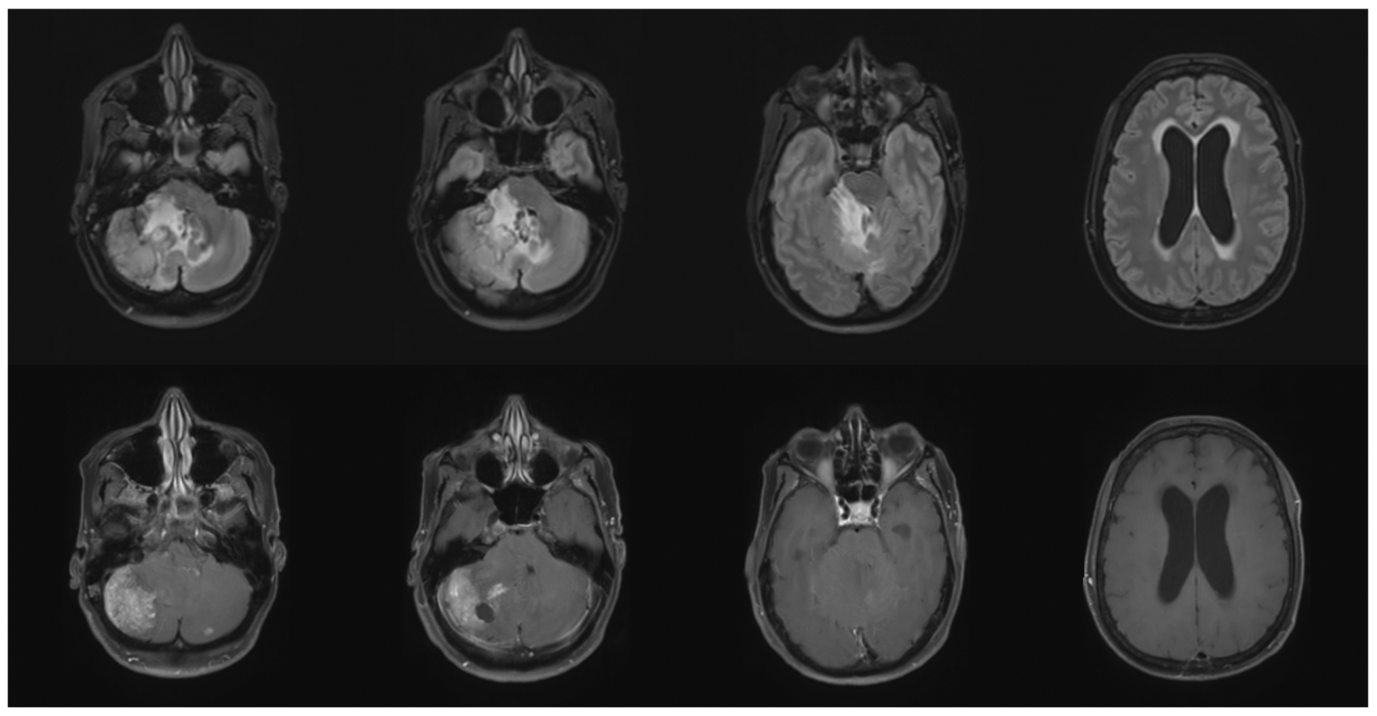
Beginning in September 2024, she developed progressive vertigo and blurred vision, which worsened by April 2025. Neurological examination in May 2025 revealed right-sided dysmetria and a broad-based gait. A brain MRI revealed several infracentimetric supratentorial lesions, along with a large right cerebellar lesion with mass effect and obstructive hydrocephalus (Figure 1). A ventriculoperitoneal shunt was placed emergently, followed by surgical resection of the cerebellar mass. A whole-body PET scan confirmed persistent complete extracranial response, with no visceral or bone activity.
The resected lesion was confirmed as metastatic breast adenocarcinoma, with ER expression of 100%, PR expression of 80%, and a HER2 IHC score of 1+. Although the HR-positive/HER2-negative phenotype was preserved, PR expression was higher than in the 2020 recurrence (80% versus 10%).
A subsequent molecular analysis of the resected tumor tissue, performed on the in-house targeted NGS panel, revealed the following alterations:
  • An already-identified variant in the ERBB2 gene (Y772_A775dup), with a VAF of 59%.
  • A pathogenic variant in the ESR1 gene (Y537S) located in exon 8, with a VAF of 40%, known to be a canonical activating mutation driving complete ligand-independent ER activation and strong AI resistance.
  • A low-level amplification (8 to 10 copies) of the genomic region encompassing the CCND1 locus.
To evaluate whether the ESR1 alteration was detectable in the systemic circulation, postoperative plasma collected on 8 August 2025 was analyzed using the Hedera profiling 2 (HP2) ctDNA assay (Hedera Dx, Epalinges, Switzerland), a hybrid-capture 32-gene NGS panel designed to detect single-nucleotide variants, insertions/deletions, copy number alterations, and selected structural variants [7]. The assay covers clinically relevant ESR1 ligand-binding domain hotspots, including Y537S/N/C, D538G, and E380Q, and has an approximate limit of detection of 0.1–0.5% variant allele frequency for established hotspot mutations.
No somatic alteration was detected in postoperative plasma, including ESR1 Y537S or the previously identified ERBB2 Y772_A775dup in-frame duplication. No copy number alteration was identified. This negative result was compatible with a very low circulating tumor fraction but did not establish that the alterations were confined to the CNS.
Following confirmation of isolated cerebral progression, multidisciplinary treatment included stereotactic radiotherapy to the resection cavity and the remaining intracranial lesions. Endocrine therapy was switched to fulvestrant, based on its expected higher activity against ESR1-mutated hormone-receptor-positive disease compared to letrozole [8]. The presence of an ESR1 Y537S mutation further supported discontinuation of aromatase inhibitor therapy given the strong ligand-independent ER activation associated with this mutation. Palbociclib was replaced by abemaciclib, the latter being selected both for its potential CNS penetration [9] and its documented activity in combination with fulvestrant in the context of disease progression on AI plus CDK4/6 inhibitors [10].
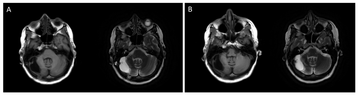
Imaging reassessments performed in November 2025 and February 2026, approximately 3 and 6 months after treatment modification, demonstrated a persistent complete extracranial metabolic response on PET imaging and stable intracranial findings on brain MRI (Figure 2).

4. Discussion

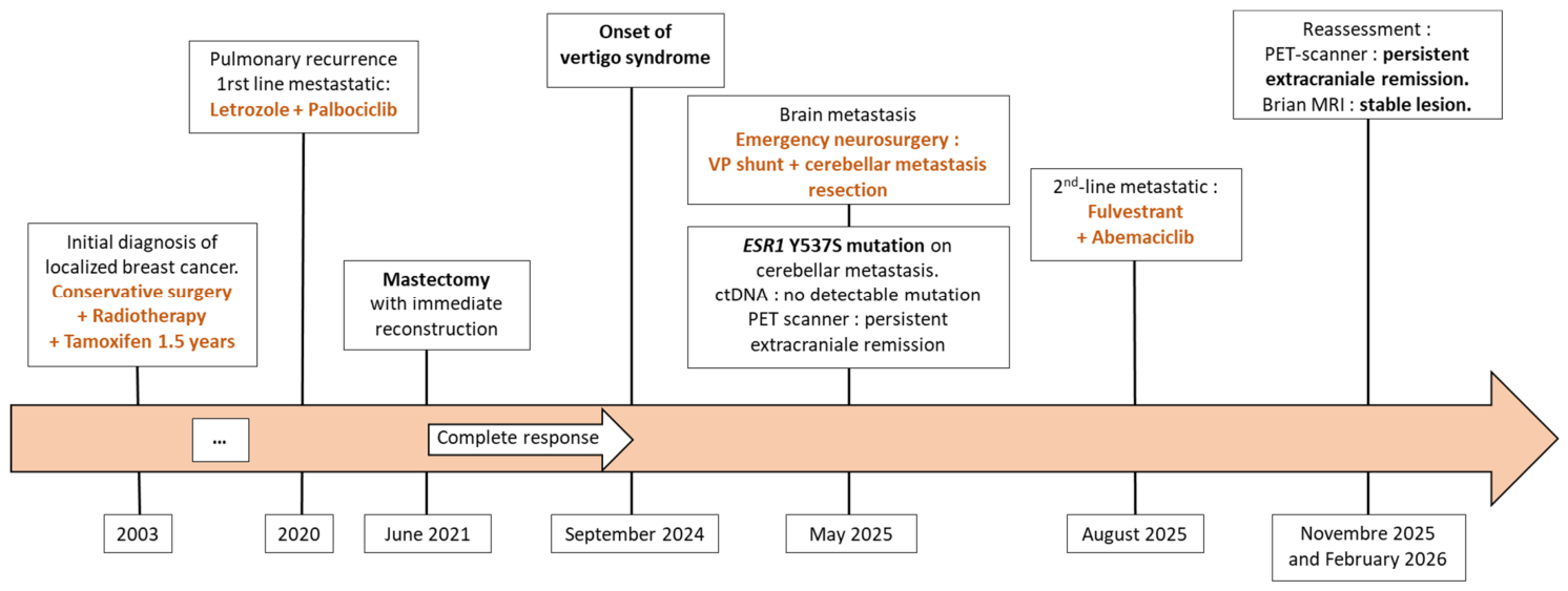
This case illustrates several important biological principles (a summary of the patient’s clinical course and treatments is provided in Figure 3).
First, the detection of ESR1 Y537S in the brain metastasis after prolonged exposure to an aromatase inhibitor is compatible with treatment-driven clonal selection and spatial heterogeneity. However, the available tissue and plasma analyses do not establish that the mutation was confined to the CNS [11].
Second, brain metastases may undergo independent evolutionary trajectories, resulting in molecular divergence from extracranial disease [12,13]. Accordingly, tissue sequencing remains essential when isolated CNS relapse is identified and surgical material is available.
Third, plasma ctDNA may remain negative in patients with CNS-limited progression, emphasizing that a negative ctDNA result does not exclude clinically relevant intracranial disease activity [14,15]. This is of particular importance in the context of emerging ctDNA-guided strategies aiming to adapt treatment intensity and imaging schedules in metastatic breast cancer. In HER2-positive disease, MRD-driven approaches using personalized ctDNA assays are being evaluated to support treatment de-escalation, while in HR-positive disease, trials such as PADA-2 investigate ctDNA dynamics to guide therapeutic modifications. However, this case highlights a key limitation of such approaches, as ctDNA may remain undetectable in the setting of isolated CNS progression. Therefore, reliance on ctDNA alone may be insufficient, and dedicated brain imaging should be maintained when clinically indicated.
The absence of ESR1 Y537S detection in plasma ctDNA does not constitute definitive evidence that the mutation was confined to the CNS. Plasma sampling was performed after resection of the dominant cerebellar lesion, which may have further reduced tumor-derived DNA shedding. The observed discordance may therefore reflect a combination of a low circulating tumor fraction, postoperative reduction in intracranial tumor burden, and limited release of tumor-derived DNA from CNS lesions into the systemic circulation because of biological compartmentalization and the blood–brain barrier.
From a therapeutic perspective, oral selective estrogen receptor degraders represent a potential future options in ESR1-mutated disease, although their role in isolated CNS progression remains undefined [16,17,18,19]. Imlunestrant has shown promising activity and brain penetration in preclinical models [20,21,22], but clinical evidence in this specific setting remains to be established. The relative enrichment of the ERBB2 Y772_A775dup variant in the brain metastasis is hypothesis-generating but does not establish a causal role in CNS progression. Trastuzumab deruxtecan could represent a subsequent treatment option based on the HER2-low phenotype and its reported intracranial activity; however, the predictive significance of this ERBB2 alteration in the present setting remains uncertain.
Beyond endocrine resistance, preclinical data suggest that ESR1 Y537S may influence the tumor microenvironment through YAP1-mediated fibroblast activation and conversion into cancer-associated fibroblasts [23]. Although this mechanism cannot be inferred from the present clinical observation, these findings provide a potential biological framework for future investigation.

5. Conclusions

This case describes isolated CNS progression in a patient with HR+/HER2-low metastatic breast cancer and sustained extracranial disease control. ESR1 Y537S was detected in the resected cerebellar metastasis but not in postoperative plasma ctDNA. Although this discordance is compatible with spatial genomic heterogeneity, plasma negativity cannot establish that the mutation was confined to the CNS and may also reflect low circulating tumor fraction, postoperative reduction in tumor burden, and limited ctDNA shedding from intracranial lesions. This observation highlights the limitations of plasma liquid biopsy in CNS-limited clinical progression and supports an integrated diagnostic approach combining imaging and direct molecular profiling of metastatic tissue when available.

Supplementary Materials

The following supporting information can be downloaded at: https://www.mdpi.com/article/10.3390/curroncol33080454/s1, CARE Checklist of information to include when writing a case report.

Author Contributions

Conceptualization, A.N., A.G. and A.d.N.; methodology, A.N., C.P. and A.d.N.; investigation, A.N., L.S., A.G.C., F.V., A.T., C.P. and A.d.N.; formal analysis, A.N.; data curation, A.N., L.S., A.G.C., F.V., A.T. and C.P.; visualization, A.N. and A.d.N.; writing: original draft preparation, A.N.; writing: review and editing, all authors; supervision, A.G. and A.d.N. All authors have read and agreed to the published version of the manuscript.

Funding

This research received no external funding.

Institutional Review Board Statement

Ethical review and approval were waived for this single-patient case report because it reports retrospective clinical observations obtained exclusively during routine care and involved no research-specific intervention, in accordance with institutional policy.

Informed Consent Statement

Written informed consent was obtained from the patient for publication of the clinical information and images included in this case report.

Data Availability Statement

All relevant data supporting the findings of this case report are included within article. Additional clinical data, including original MRI images, are not publicly available due to patient privacy and ethical restrictions.

Conflicts of Interest

AdN: Gilead (consulting fees, lecture fees, congress invitations), Daiichi Sankyo (consulting fees, lecture fees, congress invitations), Astra Zeneca (consulting fees, lecture fees, congress invitations), Seagen (consulting fees), Lilly (consulting fees, lecture fees, congress invitations), Novartis (consulting fees, lecture fees, congress invitations), MSD (consulting fees, congress invitations, lecture fees), Pfizer (research grants paid to institution, lecture fees, congress invitations), Promise Proteomics (consulting fees). AG: Novartis (consulting or advisory role), MSD (consulting or advisory role, research funding), AstraZeneca (consulting or advisory role, research funding), Gilead Sciences (consulting or advisory role), Daiichi Sankyo/Astra Zeneca (consulting or advisory role, research funding), Roche/Genentech (research funding), Sanofi/Aventis (research funding), Novartis (research funding). AGC: Astra Zeneca (congress invitations), Daiichi Sankyo (congress invitations), Eisai (congres invitations), Lilly (lecture fees, congress invitations), Novartis (congress invitations), Pfizer (congress invitations), Promise Proteomics (consulting fees), Seagen (consulting fees, congress invitations). LS: AstraZeneca (lecture fees), Sivan (lecture fees), MSD (lecture fees), Astellas (lecture fees), IPSEN (meeting registration), Lilly (meeting registration, lecture fees), Pfizer (meeting registrations), Novartis (meeting registration), Gilead (meeting registration). CP: Astra Zeneca (consulting fees, lecture fees, congress invitations), AN, AT, FV: no conflicts of interest to declare. The other authors declare that they have no known competing financial interests or personal relationships that could have appeared to influence the work reported in this paper.

References

  1. Weil, R.J.; Palmieri, D.C.; Bronder, J.L.; Stark, A.M.; Steeg, P.S. Breast Cancer Metastasis to the Central Nervous System. Am. J. Pathol. 2005, 167, 913–920. [Google Scholar] [CrossRef] [PubMed]
  2. Lopes, S.; Vieira, I.; Abreu, M.; Pousa, I.; Ferreira, A.; Sousa, S.; Pereira, D. Prognostic Factors and Survival According to Tumor Subtype in Women With Breast Cancer Brain Metastases. Curr. Probl. Cancer 2022, 46, 100866. [Google Scholar] [CrossRef] [PubMed]
  3. Merenbakh-Lamin, K.; Ben-Baruch, N.; Yeheskel, A.; Dvir, A.; Soussan-Gutman, L.; Jeselsohn, R.; Yelensky, R.; Brown, M.; Miller, V.A.; Sarid, D.; et al. D538G Mutation in Estrogen Receptor-α: A Novel Mechanism for Acquired Endocrine Resistance in Breast Cancer. Cancer Res. 2013, 73, 6856–6864. [Google Scholar] [CrossRef] [PubMed]
  4. Grote, I.; Poppe, A.; Lehmann, U.; Christgen, M.; Kreipe, H.; Bartels, S. Frequency of Genetic Alterations Differs in Advanced Breast Cancer between Metastatic Sites. Genes Chromosomes Cancer 2024, 63, e23199. [Google Scholar] [CrossRef] [PubMed]
  5. Carausu, M.; Melaabi, S.; Pierga, J.-Y.; Bidard, F.-C.; Cabel, L. ESR1 Mutation Detection and Dynamics in Meningeal Carcinomatosis in Breast Cancer. J. Breast Cancer 2019, 23, 218–223. [Google Scholar] [CrossRef] [PubMed]
  6. Frampton, G.M.; Fichtenholtz, A.; Otto, G.A.; Wang, K.; Downing, S.R.; He, J.; Schnall-Levin, M.; White, J.; Sanford, E.M.; An, P.; et al. Development and Validation of a Clinical Cancer Genomic Profiling Test Based on Massively Parallel DNA Sequencing. Nat. Biotechnol. 2013, 31, 1023–1031. [Google Scholar] [CrossRef] [PubMed]
  7. Lescuyer, G.; Harlé, A.; Kumar, H.S.; Constantoulakis, P.; Pfarr, N.; Heitzer, E.; Michon, C.; Russo, G.; Speel, E.-J.M.; Piecyk, M.; et al. Analytical Validation of a Pan-Cancer Next-Generation Sequencing Assay for In-House Liquid Biopsy Testing: An International Multicenter Study. J. Mol. Diagn. 2025, 27, 1174–1188. [Google Scholar] [CrossRef] [PubMed]
  8. Bidard, F.-C.; Hardy-Bessard, A.-C.; Dalenc, F.; Bachelot, T.; Pierga, J.-Y.; de la Motte Rouge, T.; Sabatier, R.; Dubot, C.; Frenel, J.-S.; Ferrero, J.M.; et al. Switch to Fulvestrant and Palbociclib versus No Switch in Advanced Breast Cancer with Rising ESR1 Mutation during Aromatase Inhibitor and Palbociclib Therapy (PADA-1): A Randomised, Open-Label, Multicentre, Phase 3 Trial. Lancet Oncol. 2022, 23, 1367–1377. [Google Scholar] [CrossRef] [PubMed]
  9. Li, J.; Jiang, J.; Wu, J.; Bao, X.; Sanai, N. Physiologically Based Pharmacokinetic Modeling of Central Nervous System Pharmacokinetics of CDK4/6 Inhibitors to Guide Selection of Drug and Dosing Regimen for Brain Cancer Treatment. Clin. Pharmacol. Ther. 2021, 109, 494–506. [Google Scholar] [CrossRef] [PubMed]
  10. Kalinsky, K.; Bianchini, G.; Hamilton, E.; Graff, S.L.; Park, K.H.; Jeselsohn, R.; Demirci, U.; Martin, M.; Layman, R.M.; Hurvitz, S.A.; et al. Abemaciclib Plus Fulvestrant in Advanced Breast Cancer After Progression on CDK4/6 Inhibition: Results from the Phase III postMONARCH Trial. J. Clin. Oncol. 2025, 43, 1101–1112. [Google Scholar] [CrossRef] [PubMed]
  11. Spoerke, J.M.; Gendreau, S.; Walter, K.; Qiu, J.; Wilson, T.R.; Savage, H.; Aimi, J.; Derynck, M.K.; Chen, M.; Chan, I.T.; et al. Heterogeneity and Clinical Significance of ESR1 Mutations in ER-Positive Metastatic Breast Cancer Patients Receiving Fulvestrant. Nat. Commun. 2016, 7, 11579. [Google Scholar] [CrossRef] [PubMed]
  12. Brastianos, P.K.; Carter, S.L.; Santagata, S.; Cahill, D.P.; Taylor-Weiner, A.; Jones, R.T.; Van Allen, E.M.; Lawrence, M.S.; Horowitz, P.M.; Cibulskis, K.; et al. Genomic Characterization of Brain Metastases Reveals Branched Evolution and Potential Therapeutic Targets. Cancer Discov. 2015, 5, 1164–1177. [Google Scholar] [CrossRef] [PubMed]
  13. Saunus, J.M.; McCart Reed, A.E.; Lim, Z.L.; Lakhani, S.R. Breast Cancer Brain Metastases: Clonal Evolution in Clinical Context. Int. J. Mol. Sci. 2017, 18, 152. [Google Scholar] [CrossRef] [PubMed]
  14. De Mattos-Arruda, L.; Caldas, C. Cell-free Circulating Tumour DNA as a Liquid Biopsy in Breast Cancer. Mol. Oncol. 2016, 10, 464–474. [Google Scholar] [CrossRef] [PubMed]
  15. Morganti, S.; Parsons, H.A.; Lin, N.U.; Grinshpun, A. Liquid Biopsy for Brain Metastases and Leptomeningeal Disease in Patients with Breast Cancer. npj Breast Cancer 2023, 9, 43. [Google Scholar] [CrossRef] [PubMed]
  16. Oliveira, M.; Pominchuk, D.; Nowecki, Z.; Hamilton, E.; Kulyaba, Y.; Andabekov, T.; Hotko, Y.; Melkadze, T.; Nemsadze, G.; Neven, P.; et al. Camizestrant, a next-Generation Oral SERD, versus Fulvestrant in Post-Menopausal Women with Oestrogen Receptor-Positive, HER2-Negative Advanced Breast Cancer (SERENA-2): A Multi-Dose, Open-Label, Randomised, Phase 2 Trial. Lancet Oncol. 2024, 25, 1424–1439. [Google Scholar] [CrossRef] [PubMed]
  17. Bidard, F.-C.; Kaklamani, V.G.; Neven, P.; Streich, G.; Montero, A.J.; Forget, F.; Mouret-Reynier, M.-A.; Sohn, J.H.; Taylor, D.; Harnden, K.K.; et al. Elacestrant (Oral Selective Estrogen Receptor Degrader) Versus Standard Endocrine Therapy for Estrogen Receptor–Positive, Human Epidermal Growth Factor Receptor 2–Negative Advanced Breast Cancer: Results From the Randomized Phase III EMERALD Trial. J. Clin. Oncol. 2022, 40, 3246–3256. [Google Scholar] [CrossRef] [PubMed]
  18. Martín, M.; Lim, E.; Chavez-MacGregor, M.; Bardia, A.; Wu, J.; Zhang, Q.; Nowecki, Z.; Cruz, F.M.; Safin, R.; Kim, S.-B.; et al. Giredestrant for Estrogen Receptor–Positive, HER2-Negative, Previously Treated Advanced Breast Cancer: Results From the Randomized, Phase II acelERA Breast Cancer Study. J. Clin. Oncol. 2024, 42, 2149–2160. [Google Scholar] [CrossRef] [PubMed]
  19. Bidard, F.-C.; Mayer, E.L.; Park, Y.H.; Janni, W.; Ma, C.; Cristofanilli, M.; Bianchini, G.; Kalinsky, K.; Iwata, H.; Chia, S.; et al. First-Line Camizestrant for Emerging ESR1-Mutated Advanced Breast Cancer. N. Engl. J. Med. 2025, 393, 569–580. [Google Scholar] [CrossRef] [PubMed]
  20. Bhagwat, S.V.; Mur, C.; Vandekopple, M.; Zhao, B.; Shen, W.; Marugán, C.; Capen, A.; Kindler, L.; Stephens, J.R.; Huber, L.; et al. Imlunestrant Is an Oral, Brain-Penetrant Selective Estrogen Receptor Degrader with Potent Antitumor Activity in ESR1 Wild-Type and Mutant Breast Cancer. Cancer Res. 2025, 85, 777–790. [Google Scholar] [CrossRef] [PubMed]
  21. Jhaveri, K.L.; Neven, P.; Casalnuovo, M.L.; Kim, S.-B.; Tokunaga, E.; Aftimos, P.; Saura, C.; O’Shaughnessy, J.; Harbeck, N.; Carey, L.A.; et al. Imlunestrant with or without Abemaciclib in Advanced Breast Cancer. N. Engl. J. Med. 2025, 392, 1189–1202. [Google Scholar] [CrossRef] [PubMed]
  22. Jhaveri, K.L.; Neven, P.; Casalnuovo, M.L.; Kim, S.-B.; Tokunaga, E.; Aftimos, P.; Saura, C.; O’Shaughnessy, J.; Harbeck, N.; Carey, L.A.; et al. Imlunestrant with or without Abemaciclib in Advanced Breast Cancer: Updated Efficacy Results from the Phase III EMBER-3 Trial. Ann. Oncol. 2026, 37, 532–543. [Google Scholar] [CrossRef] [PubMed]
  23. Gelsomino, L.; Caruso, A.; Tasan, E.; Leonetti, A.E.; Malivindi, R.; Naimo, G.D.; Giordano, F.; Panza, S.; Gu, G.; Perrone, B.; et al. Evidence That CRISPR-Cas9 Y537S-Mutant Expressing Breast Cancer Cells Activate Yes-Associated Protein 1 to Driving the Conversion of Normal Fibroblasts into Cancer-Associated Fibroblasts. Cell Commun. Signal. 2024, 22, 545. [Google Scholar] [CrossRef] [PubMed]
Figure 1. Brain MRI at diagnosis of neurological symptoms: Upper row: axial T2-FLAIR sequences. Lower row: axial T1-weighted sequences after gadolinium injection.
Curroncol 33 00454 g001
Figure 2. Follow-up brain MRI after local and systemic treatment. (A) November 2025 and (B) February 2026. At each time point, contrast-enhanced axial T1-weighted imaging is shown on the left and axial T2-weighted imaging on the right. Post-treatment findings remained stable without radiographic intracranial progression.
Curroncol 33 00454 g002
Figure 3. Clinical timeline summarizing the patient’s disease course, treatments, and molecular findings. The diagram outlines key events from the initial diagnosis of localized breast cancer in 2003 to the metastatic recurrence in 2020, followed by a durable complete response under letrozole plus palbociclib. The patient subsequently developed isolated neurological symptoms in late 2024, leading to the discovery of cerebellar and supratentorial brain metastases in May 2025. Surgical resection revealed an ESR1 Y537S mutation in the analyzed brain metastasis, whereas postoperative plasma ctDNA showed no detectable somatic alteration and whole-body PET confirmed persistent extracranial metabolic response. Fulvestrant plus abemaciclib was initiated after neurosurgical and radiotherapeutic management, with stable intracranial findings and persistent extracranial metabolic response at 3- and 6-month follow-up.
Curroncol 33 00454 g003
Disclaimer/Publisher’s Note: The statements, opinions and data contained in all publications are solely those of the individual author(s) and contributor(s) and not of MDPI and/or the editor(s). MDPI and/or the editor(s) disclaim responsibility for any injury to people or property resulting from any ideas, methods, instructions or products referred to in the content.