Article

Survival Outcomes of Immune Checkpoint Inhibitors in Conjunction with Cranial Radiation for Older Adults with Non-Small Cell Lung Cancer and Synchronous Brain Metastasis

by
Ruchira V. Mahashabde
1,
Sajjad A. Bhatti
2,
Bradley C. Martin
1,
Jacob T. Painter
1,
Mausam Patel
3,
Analiz Rodriguez
4,
Jun Ying
5,6 and
Chenghui Li
1,*
1
Division of Pharmaceutical Evaluation and Policy, College of Pharmacy, University of Arkansas for Medical Sciences, Little Rock, AR 72205, USA
2
Division of Hematology and Medical Oncology, Department of Internal Medicine, College of Medicine, University of Arkansas for Medical Sciences, Little Rock, AR 72205, USA
3
Department of Radiation Oncology, University of Arkansas for Medical Sciences, Little Rock, AR 72205, USA
4
Department of Neurosurgery, University of Arkansas for Medical Sciences, Little Rock, AR 72205, USA
5
Department of Biostatistics, University of Arkansas for Medical Sciences, Little Rock, AR 72205, USA
6
Department of Family Medicine, School of Medicine, University of Colorado, Aurora, CO 80045, USA
*
Author to whom correspondence should be addressed.
Curr. Oncol. 2025, 32(9), 499; https://doi.org/10.3390/curroncol32090499
Submission received: 11 June 2025 / Revised: 21 August 2025 / Accepted: 25 August 2025 / Published: 5 September 2025
(This article belongs to the Section Thoracic Oncology)

Simple Summary

Brain metastases are a serious complication of non-small cell lung cancer (NSCLC) that often lead to poor survival. Combining immunotherapy with brain radiation could be more effective than either treatment alone, but the optimal timing of this combination is unknown. Using real-world data from older adults who had brain metastases at diagnosis of NSCLC, we compared surival outcomes of the combination therapy that was administered within different timing windows. This population is largely underrepresented in clinical trials. Our findings could help clinicians optimize combination treatment strategies for these patients who are at high risk of poor survival outcomes.

Abstract

Immune checkpoint inhibitors (ICIs) display efficacy in non-small cell lung cancers (NSCLCs) with brain metastases (BMs) and studies suggest potential synergy with cranial radiation (CR). However, population-based evaluations of optimal time between ICI-CR combinations are limited in the US. Using SEER-Medicare database (2010–2019), we analyzed patients aged ≥65 years with NSCLC and BM receiving ICI-CR within 6 months of diagnosis, excluding those receiving targeted therapies. First treatment after diagnosis (ICI or CR) was defined as index treatment; followed by subsequent treatment. Findings were validated using an independent cohort from the TriNetX LIVE™ Platform. Patients were grouped by interval between the end of the index treatment and the start of the subsequent treatment: ≤15 days (n = 117), 16–30 days (n = 42), and >30 days (n = 77). Overall survival (OS) was measured from the start of the subsequent treatment until death, end of insurance coverage, or study end. Kaplan–Meier survival curves and multivariable Cox proportional hazards models estimated differences between groups. Among 236 patients, median OS was 134 days, 92 days, and 209 days, respectively. No significant OS differences were found across intervals. However, a survival benefit emerged approximately 300 days after follow-up when ICI was administered within 15 days of CR. These findings offer insight into treatment sequencing in NSCLC with BM and support further investigation in larger cohorts.

1. Introduction

Lung cancer remains the leading cause of cancer-related death in the United States [1]. There are two main types of lung cancer: small cell lung cancer, which accounts for 10–15% of patients, and non-small cell lung cancer (NSCLC), which accounts for 80–85% [2]. Unlike small cell lung cancer, which exhibits neuroendocrine features, NSCLC is histologically and molecularly heterogeneous, and it includes adenocarcinoma squamous cell carcinoma and other subtypes [3]. A critical challenge for treating NSCLC is brain metastases (BMs), which affect up to 40% of patients and confer a predicted survival of only 3–15 months after diagnosis [4,5]. Approximately 10% of patients with NSCLC have “synchronous BM” (SBM) (i.e., BM at diagnosis) [6], which accounts for up to 80% of all SBM cases [5]. Not only is lung cancer the most common SBM-associated cancer, but it also has the worst prognosis, with a median survival of approximately 3.9 months and only 20% survival rate beyond the first year [5].
While SCLC initially responds better to chemotherapy, NSCLC’s genomic diversity has allowed targeted therapies and immune checkpoint inhibitors (ICIs) to transform treatment of advanced NSCLC [7]. ICIs enhance immune-mediated destruction of cancer cells via T-cell activation [8], improving overall survival (OS) for patients with locally advanced or metastatic NSCLC, either as monotherapy [9,10,11,12] or in combination with chemotherapy [13,14,15]. BM may benefit from ICI treatment because half of the NSCLC BMs were found to display PD-L1 expression [16]. Despite this biomarker association and high prevalence of BMs in solid tumors, clinical trials often exclude patients with active or untreated BM. Ad hoc analyses of trials that included stable or treated BMs reported an OS benefit among these patients [13,17,18], further supporting potential efficacy of ICIs in the central nervous system, which historically was considered to be immune privileged [19,20]. Cranial radiation (CR) is standard care for BM; stereotactic radiosurgery (SRS) is recommended for limited BMs, and whole-brain radiation therapy (WBRT) with neuroprotection is recommended for extensive disease [21,22]. Preclinical studies have shown that radiation upregulates immune checkpoints, such as PD-1 on tumor-infiltrating T cells and PD-L1 on cancer cells [23], peaking approximately 3 days after radiation treatment [24]. These immunomodulatory properties of CR, combined with immune-enhancing effects of ICIs, suggest a promising synergistic potential, as previously reported in preclinical and clinical studies [25,26].
However, clinical trial data on ICI–radiation synergy in NSCLC are limited to extracranial radiotherapy and with mixed findings. The PACIFIC trial showed improved progression-free survival and OS among patients with unresectable stage 3 NSCLC who received durvalumab within 42 days after completion of chemoradiation [27]. A phase 2 trial showed enhanced event-free 4-year survival among patients with early-stage NSCLC who were undergoing stereotactic ablative radiotherapy with nivolumab, compared to stereotactic ablative radiotherapy alone [28]. However, several recent phase III trials that evaluated the addition of ICI to radiation therapy in early and locally advanced NSCLC failed to demonstrate a survival benefit. The S1914 trial [29], which tested neoadjuvant concurrent and adjuvant atezolizumab use, with stereotactic body radiation therapy (SBRT) versus only SBRT in stage I/II NSCLC, showed no survival benefit but more adverse events. The KEYNOTE-867 trial [30,31], which assessed concurrent pembrolizumab with SBRT in early-stage NSCLC, was terminated early due to lack of efficacy and increased toxicity. Together, results of these studies call into question the hypothesized ICI–radiation synergy and highlight the need to further investigate and optimize timing and disease stage. Data from clinical trials that combine radiation and ICI for treating metastatic NSCLC remain scarce and limited. A phase 1 trial (n = 37) showed that treatment with SBRT-ICI administered simultaneously, compared to sequentially (used within one week of each other), upregulated key immune pathways and enhanced local and distant tumor responses, although survival differences were not significant [32]. Notably, 9/37 patients with treated or asymptomatic BMs survived through the study period, which was a striking contrast to the non-BM cohort (n = 28) in which approximately two-thirds died by the follow-up (Bestvina et al., 2022, Figure A1 [32]). This suggests that patients with NSCLC and controlled BM or small asymptomatic central nervous system lesions may not have worse survival outcomes. However, the study was underpowered, and it was unclear whether the patients with BM had received prior radiation. To date, no published trials have assessed CR combined with ICIs specifically for NSCLC patients with BM.
Currently, reports of efficacy and tolerability of combined ICI-CR treatment for BM primarily stem from retrospective series that focused on melanoma patients [33,34,35,36]. Among NSCLC-specific studies or studies that included NSCLC in the mixture, only a few patients received both CR and ICI (n < 90) [37,38,39,40,41,42,43,44,45,46] and usually were from a single institution [47,48] (Supplementary Table S1). Despite these limitations, the studies showed better locoregional disease control with concurrent use of CR and ICI [42] than with SRS [45,47] or WBRT [46] administered alone or with ICI monotherapy [49]; this suggests synergistic effects when treating BM. However, a major difficulty with interpreting these findings is that the definitions of “concurrent use” varied widely—e.g., 2 weeks [47], 4 weeks [39], 30 days [46], 1 month [42], 3 months [45], 5 months [48]; therefore, optimal timing of the therapies is unclear. Moreover, existing reviews and meta-analyses synthesizing findings of these studies did not address the variability in optimal treatment timing [49,50,51]. Some preclinical studies show that administering ICI during the initial SRS treatment, when tumor burden is high, may reduce the effects of ICIs [24] and thus the potential synergistic effects. Conversely, others argue for a closer temporal proximity between ICI and SRS (2–4 weeks) to align with the half-life of ICIs (12–27 days) [52], which may enhance the synergy. This unresolved timing dilemma leaves clinicians without evidence-based guidance for effectively sequencing ICI and CR treatments.
To address these gaps, we used a large population-based dataset to compare survival outcomes across three clinically meaningful time windows for ICI administration relative to CR (2 weeks, 30 days, and 31–180 days) within 6 months after a diagnosis of NSCLC and SBM. Our study represents a significant advancement over prior research. It is the first direct evaluation of timing of the ICI-CR combination, assessing intervals that have previously been shown to be associated with better survival. Also, it establishes the largest cohort, to date, of patients with NSCLC and SBM who were treated with both modalities (all previous studies included N < 90). Finally, it focuses on two critically understudied populations—patients with untreated SBM, who typically are excluded from clinical trials and are older adults (≥65 years; mean: 73 years) who comprise >70% of NSCLC cases [53], yet frequently are underrepresented in clinical trials [54].

2. Materials and Methods

2.1. Data Source

This study used the Surveillance, Epidemiology, and End Result (SEER) cancer registry data by National Cancer Institute (NCI), which captures data from 34.6% of patients with cancer in the US and contains information on demographic, tumor, and treatment-related covariates at the diagnosis of the primary malignancy. The methodology, structure, and data collection methods were described elsewhere [55,56]. NCI biannually links SEER data to Medicare enrollment, fee-for-service claims, and Medicare Advantage encounter data (SEER-Medicare), which facilitates research on treatment patterns and outcomes [57]. The SEER-Medicare linked data used in this study included SEER data from January 2010 to December 2017 and Medicare enrollment and fee-for-service claims data from January 2009 to December 2019.

2.2. Study Sample

The study cohort included patients who received a diagnosis of NSCLC and SBM between 2010 and 2017 and who were treated with both ICIs and CR (SRS and non-SRS) within 6 months of diagnosis, a window that captures initial treatments and minimizes the possibility of treatment for disease progression. Patients with NSCLC and SBM were identified by ICD-O-3 codes for primary malignant neoplasm of bronchia and lung with NSCLC histology [58] and with a binary indicator variable for the presence of BM at diagnosis, which was available in SEER from 2010 onward.
For our analyses, we selected data from records of patients who met the following criteria: 1) NSCLC diagnosis at age ≥65 years, (2) NSCLC identified as sole primary cancer, (3) evidence of BM at NSCLC diagnosis, (4) uninterrupted enrollment in Medicare Part A (inpatient) and Part B (outpatient and physician services) but not Medicare Part C (Medicare Advantage) for a at least minimum of 6 months before diagnosis (for assessment of baseline comorbidities and performance status) and continuing until “follow-up start date” (to ensure observation of complete treatment). Patient records were excluded on the basis of the following criteria: (1) diagnosis of small cell lung cancer or other histology of lung cancer, (2) diagnoses of multiple primary cancers during the study period, (3) diagnosed at autopsy or died within 30 days of diagnosis, (4) received any oral targeted therapies during the study period (afatinib dimaleate, alectinib HCl, ceritinib, crizotinib, brigatinib, erlotinib HCl, ceritinib, crizotinib, dabrafenib mesylate, erlotinib HCl, gefitinib, osimertinib mesylate, trametinib dimethyl) to ensure patient cohort with similar tumor characteristics, (5) not treated with ICI throughout the study period, and (6) did not receive both ICI and CR within 6 months after diagnosis. Treatment with ICIs approved for lung cancer by the U.S. Food and Drug Administration was documented through Medicare claim records by using the corresponding Healthcare Common Procedural Coding System (HCPCS) codes [58]. The specific ICI agents included pembrolizumab, nivolumab, ipilimumab, avelumab, atezolizumab, and durvalumab. CR (SRS or non-SRS) was identified by linking Common Procedural Terminology/HCPCS [58] codes for radiation procedures with diagnosis codes for secondary malignant neoplasm of brain (ICD-9 198.3x or ICD-10 C79.31) within the same claim, thereby confirming radiation targeted to brain. Patients receiving dual PD-1 and CTLA-4 blockades were excluded from analysis due to potential survival outcome differences compared to ICI monotherapy or ICI-chemotherapy regimens [59] and later approval (26 May 2020) of ICI combination for lung cancer [60] relative to our study period. Because SEER provides only the month and year of the primary cancer diagnosis, we established the precise date of diagnosis by identifying the first diagnosis from Medicare claims and matching it with the SEER month and year of diagnosis. Further information is provided in the Appendix A section.

2.3. Treatment Groups

We analyzed data from patients who received ICI and CR within 6 months after receiving a diagnosis of NSCLC and SBM. Focusing on ICI and CR, the first treatment with either ICI or CR after diagnosis was designated as the index treatment. If a patient switched to the other treatment (e.g., from ICI to CR or vice versa), then the new treatment was classified as subsequent treatment. The switch was marked by the last claim for index treatment and the first claim for subsequent treatment. The time interval between these two dates was calculated in days. Based on this interval, patients were classified into three study groups: ≤15 days, 16–30 days, and >30 days. Patients were followed from the start of the subsequent treatment (i.e., follow-up start date). For example, if a patient initially received CR and then received ICI, then the date of ICI initiation was designated as the follow-up start date. An illustrative example is provided in Figure 1.

2.4. Survival Outcome

The primary study outcome was OS time, defined as number of days from start of follow-up until death. For patients who remained alive, survival times were censored at first occurrence of (1) loss of enrollment in Medicare Parts A and B, (2) enrollment in Medicare Advantage (Part C), or (3) end of data availability (31 December 2019).

2.5. Covariates

Covariates (available in SEER-Medicare database) were selected for their relevance to survival and/or receipt of treatment, according to our review of the published literature [61,62,63] and based on recommendations from our clinical collaborators. Patient characteristics at diagnosis included age, sex (male/female), race/ethnicity (non-Hispanic White/other), marital status (married/not married), census tract level poverty of the patient’s residence (0–<5%, 5–<10%, 10–<20%, 20–100%, or unknown), and rurality of the patient’s county of residence (rural/urban). Variables related to primary tumors included other metastasis (bone, liver, or lung) and histology of NSCLC (adenocarcinoma, squamous cell carcinoma, other). Variables related to cancer treatment included type of CR (SRS or non-SRS), line of ICI treatment (first-line, ≥second-line), and receipt of chemotherapy and neurosurgical resection within the first 6 months after diagnosis. To account for differences in patients’ baseline health status, we used Medicare claims data to calculate two key measures: (1) Modified Charlson Comorbidity Index (excluding cancer) [64] and (2) a performance status (PS) scale that uses a validated claims-based algorithm, which is a proxy of the Eastern Cooperative Oncology Group (ECOG) [65,66]. These measures were calculated with data from the 6 months preceding diagnosis. Detailed methodology for calculation of the performance status scale is available in the Appendix A section.

2.6. Statistical Analysis

The patient characteristics of the three study groups were compared with standard t-tests for continuous variables and Chi-square tests for categorical variables whenever appropriate. Outcomes were evaluated with survival analysis techniques. Survival curves were calculated using the Kaplan–Meier (KM) method and between-group comparisons of curves were performed using a log rank test. Adjusted multivariable Cox proportional hazards (CPH) regression was used to determine hazard ratios while controlling for potential confounders.
We found that the KM curves overlapped twice with delayed separation, indicating a violation of the proportional hazards (PH) assumption. To account for this violation, we conducted a sensitivity analysis by classifying our sample as ICI-CR administered within 15 days and >15 days. We then estimated a multivariable CPH model with two “change points” [67] at 90 days and 300 days, which is around the time point where the KM curves crossed each other, to estimate separate hazard ratios before and after the crossing points. The “change points” were chosen based on AIC and BIC by comparing models with various change points around the time when the curves crossed; p-values for multiple comparisons were adjusted using Bonferroni correction.
Since previous studies focused only on SRS types of CR, we conducted a subgroup analysis of data from patients who received ICI with SRS by using an adjusted multivariable CPH model. Given the distinct clinical profiles of patients who received SRS vs. non-SRS radiation (e.g., recipients of whole-brain radiotherapy typically present with greater metastatic burden), we performed an exploratory subgroup analysis of non-SRS population by using similarly adjusted models. A p < 0.05 was considered statistically significant. All analyses were performed using SAS statistical software (version 9.4; SAS Institute Inc., Cary, NC, USA).

2.7. Validation Study

To externally validate our findings, we also performed additional analyses using TriNetX LIVE™, a web-based platform which provides access to de-identified electronic health records from healthcare organizations worldwide. This platform allows for real-time cohort building and preliminary outcome analysis across diverse patient populations. We detailed the methods and results for the TriNetX analysis are available in the Supplementary Materials.

3. Results

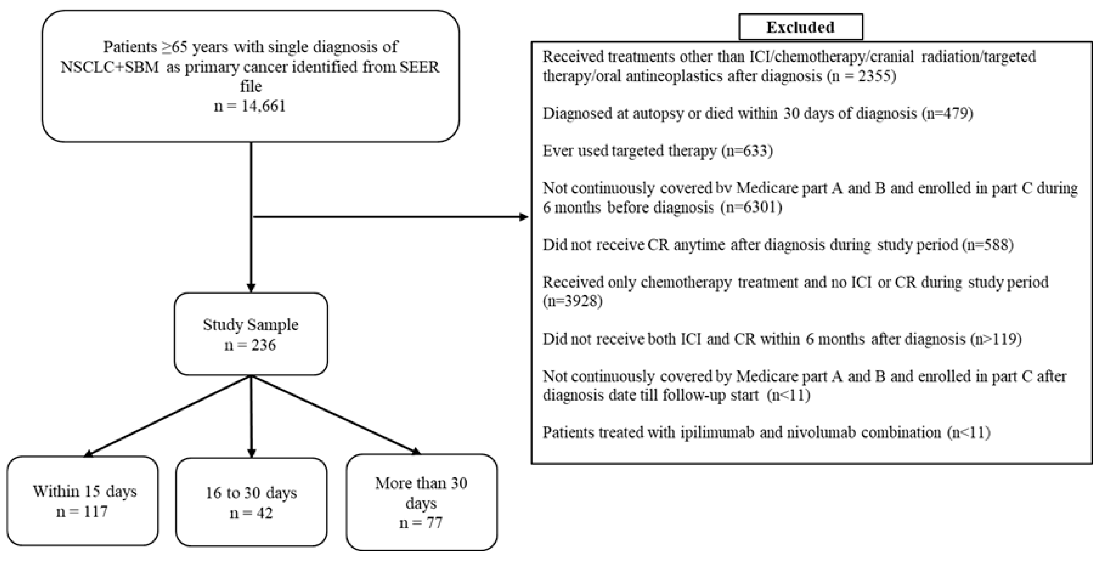
After we applied the inclusion and exclusion criteria, a final study sample of 236 patients remained for the primary analysis (subsequent ICI/CR treatment received ≤15 days: n = 117; 16–30 days: n = 42; >30 days: n = 77). Figure 2 displays the sample selection flow chart. The median age (range) of the sample was 72 years (range: 65–90 years). The median time between ICI and CR was 16 days (range: 0–160 days) for the entire sample. Patients in the >30 days group were significantly more likely (80.52%; p < 0.001) to receive chemotherapy within 6 months of diagnosis compared to the ≤15 days group (26.5%) or the 16–30 days group (30.95%). The study groups were found to be similar with respect to most other covariates. Median follow-up time was 145 days (range: 3–1561) for the entire study sample. The time from diagnosis to index date (mean [SD]: 68.65 [35.17] vs. 90.76 [30.59] vs. 123.7 [31.79]; p ≤ 0.0001) was significantly shorter for patients in the ≤15 days group, compared to the other study groups (Table 1).
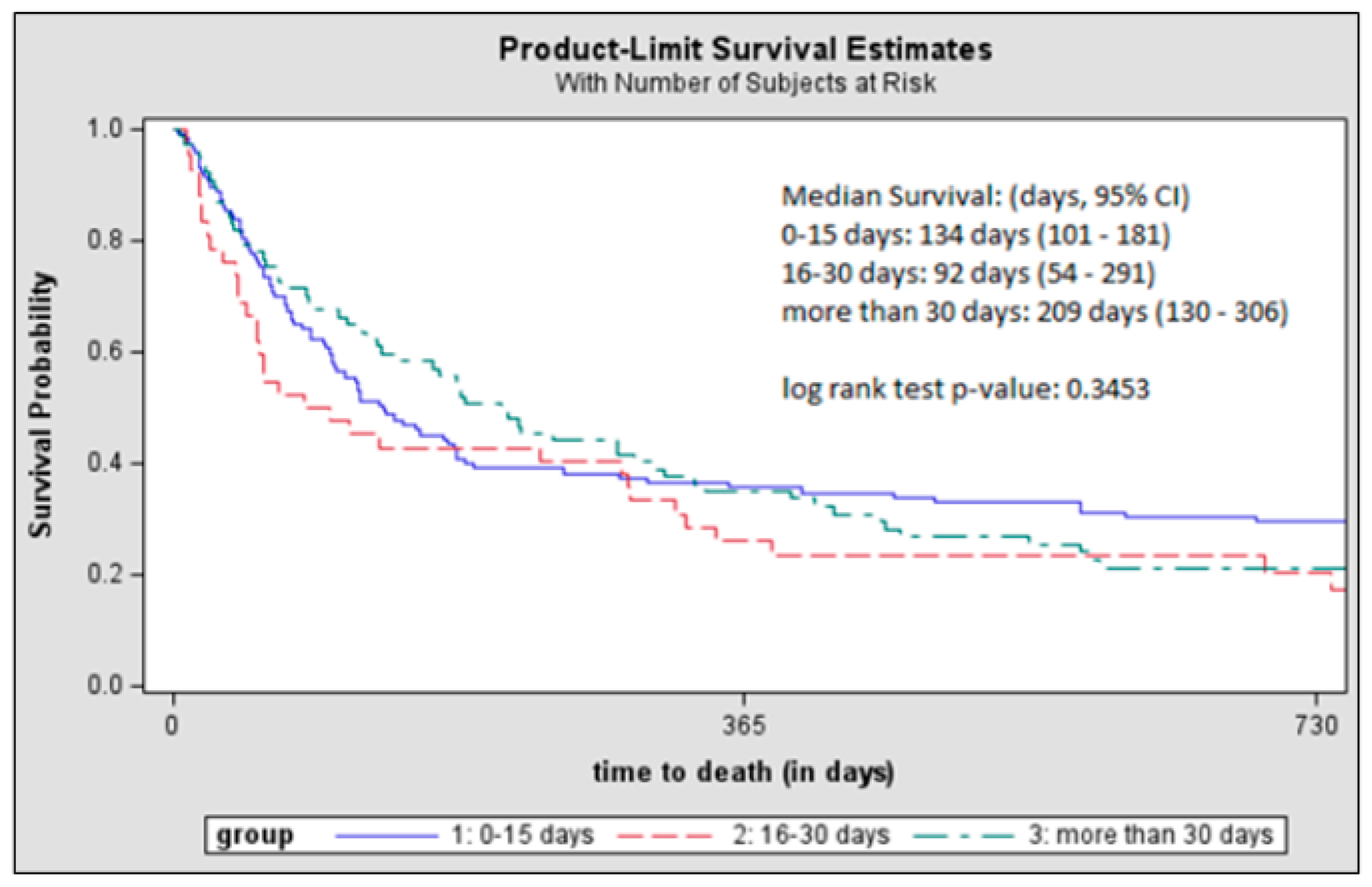
Median survival was shortest for the 16–30 days group (92 days; 95% CI: 54–291), followed by the ≤15 days group (134 days; 95% CI: 101–181), and was longest for the >30 days group (209 days; 95% CI: 130–306); however, the log rank test showed no significant differences (Figure 3). After adjusting for covariates, the hazard of death between the study groups was not statistically significantly different (Table 2).
Under the “change point” CPH model, the hazard of death for the ≤15 days group was significantly lower than the >15 days group only after 300 days from the follow-up start date (Supplementary Table S2).
Similar to results of the primary analysis, hazard of death between the study groups was not significantly different after using KM curves, log rank test (Supplementary Figure S1a,b), and multivariable CPH models (Supplementary Tables S3 and S4) for subgroup analyses of ICI and SRS cohorts and of ICI and non-SRS cohorts.
Findings from the TriNetX validation cohort were consistent with those from the SEER-Medicare, showing no significant overall survival differences across ICI–CR timing intervals using KM curves and log rank test (Supplementary Figure S2a–c).

4. Discussion

In this population-based retrospective study, we compared survival outcomes of US patients with NSCLC and SBM, ages 65 years or older, after receiving ICIs and CR 15 days, 16–30 days, and >30 days apart, with both treatments administered within 6 months after diagnosis. Our analyses showed no significant differences in OS after ICI and CR were administered at these different time intervals. Subgroups analyses of patients who received either SRS or non-SRS with ICI produced similar results. However, for the group that received ICI within 15 days of CR, a survival benefit was observed 300 days after the start of the follow-up. Importantly, we validated our findings using the TriNetX LIVE™ Platform, which provided a complementary real-world cohort. The consistency of results across two independent datasets strengthens the robustness and generalizability of our conclusions.
Our findings on OS in patients with NSCLC and BM who were treated with SRS and ICI align with results of one previous study but contrast with those of other studies, underscoring the impact of the variable definitions for concurrent therapy. Consistent with our results, Ahmed et al. (2017) [37] examined 17 patients with NSCLC and BM who received SRS within 6 months of ICI, and the adjusted multivariate analysis revealed no significant difference in OS. Additionally, their median interval between ICI and SRS for non-concurrent use (median: 1.6 months; range: 0.2–4.7) was comparable to that of our study’s groups with treatment intervals greater than 15 days (median: 1.4 months; range: 0–5.6), reinforcing that broader timing windows may not confer an advantage for OS. In contrast, other studies suggest that survival improves with stricter concurrent timing. Schapira et al. (2018) [42] examined 37 patients with NSCLC and BM who were treated at a single cancer institution, and their results showed that 1-year OS increased when SRS was delivered within 1 month of ICI; however, Chen et al. [47] linked improved OS to a 2-week concurrent treatment window, compared to sequential treatment. Unlike our study, these two studies included patients with mixed histology, and the sample size for NSCLC was limited.
This lack of consensus likely stems from discrepancies in optimal timing and study methodology. The aforementioned studies included less uniform populations—patients who had NSCLC together with SBM (BM at diagnosis) or metachronous BM (developed after diagnosis); our study included only patients who had NSCLC with SBM, a group that is reported to have shorter survival than those with NSCLC and metachronous BM [68]. Moreover, our population includes exclusively patients who were at least 65 years of age at diagnosis. Thus, patients in our study may have succumbed to their systemic disease or to competing risk factors for death, which may partially explain why our results did not show OS benefits with ICI and CR across intervals. Additionally, chemotherapy exposure was higher in our >30-day group (62% vs. 48% for ≤15 days), which studies suggest could change the tumor immune microenvironment [69]. However, when we adjusted for chemotherapy use in our multivariable analysis, the results were not affected.
While no statistically significant differences were found across the intervals, a delayed survival benefit, approximately 300 days after the combination treatment, emerged among patients who received both treatments within 15 days of each other, which warrants further investigation. Preclinical evidence suggests that this delayed effect may stem from the time required for radiation-induced cell death to activate systemic T-cell responses [24,52], as well as the overlap between the pharmacokinetics of ICIs, which have a half-life of 12–27 days [52], and the window of radiation-induced PD-L1 upregulation, which peaks 3–7 days after radiation therapy and lasts up to 30 days [23,24]. However, caution should be exercised when interpreting the effect due to the smaller sample sizes after 300 days (Figure 3) and so larger studies are needed to investigate the validity of this finding.
Our study has several strengths. First, it addresses the underrepresentation of real-world NSCLC populations in the literature; most previous retrospective studies were limited to single-center cohorts. Secondly, existing evidence on the combination of ICI and CR primarily focuses on SRS, even though focal radiation, compared to WBRT, may increase the risk of distant brain failure in patients with NSCLC [70]. Our study included patients who received both SRS and non-SRS CR, which better captures real-world treatment patterns for NSCLC and SBM populations. Lastly, we evaluated the synergy of ICI and CR across three different time intervals, offering valuable insights into the impact of varied treatment timing on patient outcomes.
However, some limitations must be considered. First, although we examined various time windows for ICI and CR combination treatment, there may still be uncertainty about the effects of radiation on T-cell activation. Second, important prognostic factors such as smoking, EGFR/ALK mutations, and performance status were not available in our data. Nonetheless, we excluded patients who received NSCLC-specific targeted therapies to mitigate potential mutation-related confounding, and we used a validated proxy measure for performance status [65,66]. Third, limited sample size prevented separate analyses of the sequencing of CR before and after ICI and of subgroups with comorbidities that may influence treatment choices and outcomes. Accurate sequencing may be an inherent limitation because patients may receive ICI after CR, likely due to systemic progression, or receive CR after ICI, likely due to intracranial progression. Also, the effectiveness of treatment can be impacted by comorbid conditions, which are common in this elderly population (mean age 73) with NSCLC. For instance, COPD may limit the use of aggressive local therapy due to pneumonitis risk [71], and cardiovascular disease could delay ICI initiation due to concerns of myocarditis [72]. Further, non-cancer deaths from conditions like diabetes or renal disease may obscure true cancer-specific survival benefits, especially over longer follow-up. While we have adjusted for the overall comorbidity burden by using the modified Charlson Comorbidity Index, which includes these conditions, we could not separately assess whether optimal timing may differ according to these important comorbid conditions. Fourth, to define our cohort of SBM patients and indicators for other metastases at diagnosis, we used the SEER variable for metastasis sites at diagnosis; however, these variables have not been fully vetted [73]. Lastly, the inherent observational framework of this study may introduce immortal time bias, where patients with longer gaps between ICI and CR may appear to live longer simply because they survived long enough to receive both treatments. To reduce this risk, we required all patients to have received both treatments within 6 months of diagnosis, and their survival was followed from the start of the second treatment. Nonetheless, residual bias may remain if patients with longer time between receiving the first and the second treatment (e.g., 31–180 days) differ clinically from those who received them closer together (≤15 days), despite adjustment for key demographic and clinical prognostic factors. For instance, confounding by indication remains a concern because gaps between ICI and CR may be shorter for younger, healthier patients, whereas older, sicker patients might experience treatment delays due to recovery needs. Still, restrictions on patients receiving both treatments within 6 months, along with adjustment for age, comorbidities, and a proxy frailty score, should mitigate these biases and limit their impact on our comparative findings.

5. Conclusions

This population-based study of older patients with NSCLC and SBM showed no significant difference in OS when ICI and CR were administered within ≤15 days, 16–30 days, or 31–180 days after diagnosis. Notably, we observed a potential delayed survival benefit 300 days after the combination therapy among patients who received ICI ≤15 days of CR, suggesting that early combination therapy may be advantageous. However, the number of patients remaining in each group after 300 days resulted in small samples for this analysis, so large prospective studies are needed to validate this finding and to investigate potential heterogeneity among patients with comorbidities or with newer immunotherapy regimens.

Supplementary Materials

The following supporting information can be downloaded at https://www.mdpi.com/article/10.3390/curroncol32090499/s1, Figure S1: Kaplan-Meier survival curves of subgroup analyses of patients receiving A: ICI and SRS and B: ICI and non-SRS given within 15 days, 16 to 30 days, and more than 30 days apart; Figure S2: Kaplan-Meier survival curves from TriNetX analyses of patients receiving ICI-CR A: within 15 days vs 16 to 30 days, B: within 15 days vs more than 30 days, and C: 16 to 30 days vs more than 30 days; Table S1: Review of retrospective studies involving patients with NSCLC and BM treated with "concurrent" ICI and CR [37,38,39,40,41,42,43,44,45,46,47,48]; Table S2 Adjusted hazard ratios of overall survival from multivariate cox proportional hazard regression analysis with change point of 90 and 300 days. Comparing within 15 days and more than 15 days intervals; Table S3: Adjusted hazard ratios of overall survival of subgroup analysis of patients with ICI and SRS from multivariate cox proportional hazard regression analysis; Table S4: Adjusted hazard ratios of overall survival of subgroup analysis of patients with ICI and non-SRS from multivariate cox proportional hazard regression analysis. File S1: The methods and results for the TriNetX analysis.

Author Contributions

Conceptualization, all authors; methodology, all authors; software, R.V.M.; validation, R.V.M., C.L. and J.Y.; formal analysis, R.V.M. and C.L.; investigation, R.V.M.; resources, R.V.M. and C.L.; data curation, R.V.M. and C.L.; writing—original draft preparation, R.V.M. and C.L.; writing—review and editing, all authors; visualization, R.V.M.; supervision, C.L.; project administration, all authors; funding acquisition, C.L. All authors have read and agreed to the published version of the manuscript.

Funding

C.L. received research support for unrelated projects sponsored by the University of Utah/AstraZeneca. C.L. was supported by the National Center for Advancing Translational Sciences of the National Institutes of Health under Award Number UL1 TR003107-02S2. C.L. is funded by the American Cancer Society (RSGI-23-1039245-01-HOPS). Support of data acquisition has been provided in part by the Arkansas Biosciences Institute, the major research component of the Arkansas Tobacco Settlement Proceeds Act of 2000.

Institutional Review Board Statement

The study was conducted in accordance with the Declaration of Helsinki, and approved by the Institutional Review Board (or Ethics Committee) of University of Arkansas for Medical Sciences (protocol number 260869, approved on 17 March 2021).

Informed Consent Statement

This retrospective study is exempt from informed consent. The data reviewed is a secondary analysis of existing data, does not involve intervention or interaction with human subjects, and is de-identified per the de-identification standard defined in Section §164.514(a) of the HIPAA Privacy Rule. The process by which the data is de-identified is attested to through a formal determination by a qualified expert as defined in Section §164.514(b)(1) of the HIPAA Privacy Rule. This formal determination by a qualified expert refreshed on December 2020.

Data Availability Statement

This study used the linked SEER-Medicare database. The interpreta-tion and reporting of these data are the sole responsibility of the authors. The authors acknowledge the efforts of the National Cancer Institute; the Information Management Services (IMS), Inc.; and the Surveillance, Epidemiology, and End Results (SEERs) Program tumor registries in the creation of the SEER-Medicare database. The datasets used to conduct this study are available on approval of a research protocol from the National Cancer Institute. Instructions for obtaining these data are available at Process for Obtaining SEER-Medicare Data https://healthcaredelivery.cancer.gov/seermedicare/obtain/ (accessed on 28 August 2025).

Acknowledgments

This study used the linked SEER-Medicare database. The interpretation and reporting of these data are the sole responsibility of the authors. The authors acknowledge the efforts of the National Cancer Institute; the Information Management Services (IMS), Inc.; and the Surveillance, Epidemiology, and End Results Program tumor registries in the creation of the SEER-Medicare database. The collection of cancer incidence data used in this study was supported by the California Department of Public Health, pursuant to California Health and Safety Code Section 103885; Centers for Disease Control and Prevention’s National Program of Cancer Registries, under cooperative agreement 1NU58DP007156; the National Cancer Institute’s Surveillance, Epidemiology and End Results Program under contract HHSN261201800032I awarded to the University of California, San Francisco, contract HHSN261201800015I awarded to the University of Southern California, and contract HHSN261201800009I awarded to the Public Health Institute. The ideas and opinions expressed herein are those of the author(s) and do not necessarily reflect the opinions of the State of California, Department of Public Health, the National Cancer Institute, and the Centers for Disease Control and Prevention or their contractors and subcontractors.

Conflicts of Interest

Bradley C. Martin: Honoraria: Institute for Clinical and Economic Review; patents, royalties, other intellectual property: royalties received from TrestleTree, LLC, for the commercialization of an opioid risk prediction tool. Analiz Rodriguez: Honoraria: Nico Corporation; consulting or advisory role: Medexus Pharmaceuticals, Nico Corporation; research funding: Bristol Myers Squibb Foundation; travel, accommodations, and expenses: Nico Corporation, BrainLAB. Chenghui Li: Research Support: AstraZeneca (Inst); no other potential conflicts of interest were reported. The funder had no role in the design of the study; in the collection, analyses, or interpretation of data; in the writing of the manuscript; or in the decision to publish the results.

Abbreviations

The following abbreviations are used in this manuscript:
ICIImmune checkpoint inhibitors
NSCLCNon-small cell lung cancer
BMBrain metastasis
CRCranial radiation
OSOverall survival
SBMSynchronous brain metastasis
SRSStereotactic radiosurgery
WBRTWhole brain radiation therapy
SBRTStereotactic body radiotherapy
NCINational Cancer Institute
SEERSurveillance, Epidemiology, and End Result
HCPCSHealthcare Common Procedural Coding System
ICD-9/10-CMInternational Classification of Diseases Ninth/Tenth Revision Clinical Modification
ECOGEastern Cooperative Oncology Group
KMKaplan–Meier
CPHCox proportional hazards

Appendix A

Imputation of date of lung cancer diagnosis
Because only the month and year of diagnosis are provided in SEER, to determine the day of lung cancer diagnosis, we identified the date of the first medical claim with lung cancer diagnosis (ICD-9/10-CM code: 162.x/C34.x) and compared it to the month and year provided in SEER. Concordance between the month and year in the two datasets was found to be 78.8%, which was similar to 72.2% reported on the SEER-Medicare website [74]. In cases with concordant month and year, the date of the first claim with lung cancer diagnosis was used as the diagnosis date. For patients with discordant months and years of diagnosis, the first day of the month reported in SEER was used to impute the diagnosis date.
Performance Status (PS) Score
The proxy PS was calculated by identifying healthcare services that were significantly associated with a poor disability status. A predicted probability of PS was then calculated by using the beta coefficients from regression models published by Davidoff et al. (2014) [65]. According to a cut-off value of 0.115 recommended in the study, patients who had a predicted probability below the cut-off were classified as having good PS (ECOG PS 0-2) and those who had a predicted probability greater than or equal to the cut-off were classified as having poor PS (ECOG PS ≥ 3) [65,66].

References

  1. Siegel, R.L.; Kratzer, T.B.; Giaquinto, A.N.; Sung, H.; Jemal, A. Cancer statistics, 2025. CA Cancer J. Clin. 2025, 75, 10–45. [Google Scholar] [CrossRef]
  2. Inamura, K. Lung cancer: Understanding its molecular pathology and the 2015 WHO classification. Front. Oncol. 2017, 7, 193. [Google Scholar] [CrossRef]
  3. Huang, D.; Wang, J.; Chen, L.; Jiang, W.; Inuzuka, H.; Simon, D.K.; Wei, W. Molecular Subtypes and Targeted Therapeutic Strategies in Small Cell Lung Cancer: Advances, Challenges, and Future Perspectives. Molecules 2025, 30, 1731. [Google Scholar] [CrossRef]
  4. Schnurman, Z.; Mashiach, E.; Link, K.E.; Donahue, B.; Sulman, E.; Silverman, J.; Golfinos, J.; Oermann, E.; Kondziolka, D. Causes of Death in Patients With Brain Metastases. Neurosurgery 2023, 93, 986–993. [Google Scholar] [CrossRef]
  5. Nayak, L.; Lee, E.Q.; Wen, P.Y. Epidemiology of Brain Metastases. Curr. Oncol. Rep. 2011, 14, 48–54. [Google Scholar] [CrossRef]
  6. Ali, A.; Goffin, J.R.; Arnold, A.; Ellis, P.M. Survival of patients with non-small-cell lung cancer after a diagnosis of brain metastases. Curr. Oncol. 2013, 20, 300–306. [Google Scholar] [CrossRef] [PubMed]
  7. Liu, L.; Soler, J.; Reckamp, K.L.; Sankar, K. Emerging Targets in Non-Small Cell Lung Cancer. Int. J. Mol. Sci. 2024, 25, 10046. [Google Scholar] [CrossRef]
  8. Johnson, D.B.; Rioth, M.J.; Horn, L. Immune Checkpoint Inhibitors in NSCLC. Curr. Treat. Options Oncol. 2014, 15, 658–669. [Google Scholar] [CrossRef]
  9. Reck, M.; Rodríguez-Abreu, D.; Robinson, A.G.; Hui, R.; Csoszi, T.; Fülöp, A.; Gottfried, M.; Peled, N.; Tafreshi, A.; Cuffe, S.; et al. Updated analysis of KEYNOTE-024: Pembrolizumab versus platinum-based chemotherapy for advanced non–small-cell lung cancer with PD-L1 tumor proportion score of 50% or greater. J. Clin. Oncol. 2019, 37, 537–546. [Google Scholar] [CrossRef] [PubMed]
  10. Mok, T.S.K.; Wu, Y.L.; Kudaba, I.; Kowalski, D.M.; Cho, B.C.; Turna, H.Z.; Castro, G.; Srimuninnimit, V.; Laktionov, K.K.; Bondarenko, I.; et al. Pembrolizumab versus chemotherapy for previously untreated, PD-L1-expressing, locally advanced or metastatic non-small-cell lung cancer (KEYNOTE-042): A randomised, open-label, controlled, phase 3 trial. Lancet 2019, 393, 1819–1830. [Google Scholar] [CrossRef] [PubMed]
  11. Sezer, A.; Kilickap, S.; Gümüş, M.; Bondarenko, I.; Özgüroğlu, M.; Gogishvili, M.; Turk, H.; Cicin, I.; Bentsion, D.; Gladkov, O.; et al. Cemiplimab monotherapy for first-line treatment of advanced non-small-cell lung cancer with PD-L1 of at least 50%: A multicentre, open-label, global, phase 3, randomised, controlled trial. Lancet 2021, 397, 592–604. [Google Scholar] [CrossRef]
  12. Herbst, R.S.; Giaccone, G.; de Marinis, F.; Reinmuth, N.; Vergnenegre, A.; Barrios, C.H.; Morise, M.; Felip, E.; Andric, Z.; Geater, S.; et al. Atezolizumab for First-Line Treatment of PD-L1–Selected Patients with NSCLC. N. Engl. J. Med. 2020, 383, 1328–1339. [Google Scholar] [CrossRef]
  13. Gandhi, L.; Rodríguez-Abreu, D.; Gadgeel, S.; Esteban, E.; Felip, E.; De Angelis, F.; Domine, M.; Clingan, P.; Hochmair, M.; Powell, S.; et al. Pembrolizuchmab plus Chemotherapy in Metastatic Non–Small-Cell Lung Cancer. N. Engl. J. Med. 2018, 378, 2078–2092. [Google Scholar] [CrossRef]
  14. Paz-Ares, L.; Luft, A.; Vicente, D.; Tafreshi, A.; Gümüş, M.; Mazières, J.; Hermes, B.; Çay, Ş.; Csőszi, T.; Fülöp, A.; et al. Pembrolizumab plus Chemotherapy for Squamous Non–Small-Cell Lung Cancer. N. Engl. J. Med. 2018, 379, 2040–2051. [Google Scholar] [CrossRef] [PubMed]
  15. West, H.; McCleod, M.; Hussein, M.; Morabito, A.; Rittmeyer, A.; Conter, H.J.; Kopp, H.G.; Daniel, D.; McCune, S.; Mekhail, T.; et al. Atezolizumab in combination with carboplatin plus nab-paclitaxel chemotherapy compared with chemotherapy alone as first-line treatment for metastatic non-squamous non-small-cell lung cancer (IMpower130): A multicentre, randomised, open-label, phase 3 trial. Lancet Oncol. 2019, 20, 924–937. [Google Scholar] [CrossRef] [PubMed]
  16. Berghoff, A.S.; Inan, C.; Ricken, G.; Widhalm, G.; Dieckmann, K.; Birner, P.; Oberndorfer, F.; Dome, B.; Bartsch, R.; Zielinski, C.; et al. Tumor-Infiltrating Lymphocytes (Tils) and Pd-L1 Expression in Non- Small Cell Lung Cancer Brain Metastases (Bm) and Matched Primary Tumors (Pt). Ann. Oncol. 2014, 25, iv465. [Google Scholar] [CrossRef]
  17. Reck, M.; Rodríguez-Abreu, D.; Robinson, A.G.; Hui, R.; Csőszi, T.; Fülöp, A.; Gottfried, M.; Peled, N.; Tafreshi, A.; Cuffe, S.; et al. Pembrolizumab versus Chemotherapy for PD-L1–Positive Non–Small-Cell Lung Cancer. N. Engl. J. Med. 2016, 375, 1823–1833. [Google Scholar] [CrossRef]
  18. Brown, L.J.; Yeo, N.; Gee, H.; Kong, B.Y.; Hau, E.; da Silva, I.P.; Nagrial, A. Immune Checkpoint Inhibitors +/− Chemotherapy for Patients With NSCLC and Brain Metastases: A Systematic Review and Network Meta-Analysis. Thorac. Cancer 2025, 16, e15510. [Google Scholar] [CrossRef]
  19. Berghoff, A.S.; Venur, V.A.; Preusser, M.; Ahluwalia, M.S. Immune Checkpoint Inhibitors in Brain Metastases: From Biology to Treatment. Am. Soc. Clin. Oncol. Educ. Book 2016, 36, e116-22. [Google Scholar] [CrossRef]
  20. Xiao, G.; Liu, Z.; Gao, X.; Wang, H.; Peng, H.; Li, J.; Yang, L.; Duan, H.; Zhou, R. Immune checkpoint inhibitors for brain metastases in non-small-cell lung cancer: From rationale to clinical application. Immunotherapy 2021, 13, 1031–1051. [Google Scholar] [CrossRef]
  21. Referenced with Permission from the NCCN Clinical Practice Guidelines in Oncology (NCCN Guidelines®) for Guideline Name V.2.2021. © National Comprehensive Cancer Network, Inc. 2021. All Rights Reserved. To View the Most Recent and Complete Version of the Guideline, go Online to NCCN.org. Available online: https://www.nccn.org/ (accessed on 8 December 2021).
  22. Referenced with Permission from the NCCN Clinical Practice Guidelines in Oncology (NCCN Guidelines®) for Guideline Name V.4.2025. © National Comprehensive Cancer Network, Inc. 2025. All Rights Reserved. To View the Most Recent and Complete Version of the Guideline, Go Online to NCCN.org. Available online: https://www.nccn.org/ (accessed on 6 June 2025).
  23. Chang, E.L.; Wefel, J.S.; Hess, K.R.; Allen, P.K.; Lang, F.F.; Kornguth, D.G.; Arbuckle, R.B.; Swint, J.M.; Shiu, A.S.; Maor, M.H.; et al. Neurocognition in patients with brain metastases treated with radiosurgery or radiosurgery plus whole-brain irradiation: A randomised controlled trial. Lancet Oncol. 2009, 10, 1037–1044. [Google Scholar] [CrossRef] [PubMed]
  24. Rajeev-Kumar, G.; Pitroda, S.P. Synergizing radiotherapy and immunotherapy: Current challenges and strategies for optimization. Neoplasia 2023, 36, 100867. [Google Scholar] [CrossRef]
  25. Kumari, S.; Mukherjee, S.; Sinha, D.; Abdisalaam, S.; Krishnan, S.; Asaithamby, A. Immunomodulatory Effects of Radiotherapy. Int. J. Mol. Sci. 2020, 21, 8151. [Google Scholar] [CrossRef]
  26. Williamson, C.W.; Sherer, M.V.; Zamarin, D.; Sharabi, A.B.; Dyer, B.A.; Mell, L.K.; Mayadev, J.S. Immunotherapy and radiation therapy sequencing: State of the data on timing, efficacy, and safety. Cancer 2021, 127, 1553–1567. [Google Scholar] [CrossRef] [PubMed]
  27. Antonia, S.J.; Villegas, A.; Daniel, D.; Vicente, D.; Murakami, S.; Hui, R.; Kurata, T.; Chiappori, A.; Lee, K.; de Wit, M.; et al. Overall Survival with Durvalumab after Chemoradiotherapy in Stage III NSCLC. N. Engl. J. Med. 2018, 379, 2342–2350. [Google Scholar] [CrossRef] [PubMed]
  28. Chang, J.Y.; Lin, S.H.; Dong, W.; Liao, Z.; Gandhi, S.J.; Gay, C.M.; Zhang, J.; Chun, S.G.; Elamin, Y.Y.; Fossella, F.V.; et al. Stereotactic ablative radiotherapy with or without immunotherapy for early-stage or isolated lung parenchymal recurrent node-negative non-small-cell lung cancer: An open-label, randomised, phase 2 trial. Lancet 2023, 402, 871–881. [Google Scholar] [CrossRef]
  29. Simone, C.B.; Daly, M.E.; Redman, M.W.; Hsieh, M.-H.; Gray, J.E.; Hesketh, P.J.; Hu, C.; Monjazeb, A.M.; Steuer, C.E.; Ganti, A.K.; et al. SWOG/NRG S1914: Randomized phase III trial of induction/consolidation atezolizumab + SBRT versus SBRT alone in high risk, early-stage NSCLC. J. Clin. Oncol. 2025, 43, 8003. [Google Scholar] [CrossRef]
  30. Jabbour, S.K.; Houghton, B.; Robinson, A.G.; Quantin, X.; Wehler, T.; Kowalski, D.; Ahn, M.J.; Erman, M.; Giaccone, G.; Borghaei, H.; et al. KEYNOTE-867: Phase 3, Randomized, Placebo-Controlled Study of Stereotactic Body Radiotherapy (SBRT) with or without Pembrolizumab in Patients with Unresected Stage I or II Non–Small-Cell Lung Cancer (NSCLC). Int. J. Radiat. Oncol. Biol. Phys. 2022, 114, e376–e377. [Google Scholar] [CrossRef]
  31. Merck Provides Update on Phase 3 KEYNOTE-867 and KEYNOTE-630 Trials—Merck.com. Available online: https://www.merck.com/news/merck-provides-update-on-phase-3-keynote-867-and-keynote-630-trials/ (accessed on 5 June 2025).
  32. Bestvina, C.M.; Pointer, K.B.; Karrison, T.; Al-Hallaq, H.; Hoffman, P.C.; Jelinek, M.J.; Juloori, A.; Melotek, J.M.; Murgu, S.; Partouche, J.; et al. A Phase 1 Trial of Concurrent or Sequential Ipilimumab, Nivolumab, and Stereotactic Body Radiotherapy in Patients With Stage IV NSCLC Study. J. Thorac. Oncol. 2022, 17, 130–140. [Google Scholar] [CrossRef]
  33. An, Y.; Jiang, W.; Kim, B.Y.S.; Qian, J.M.; Tang, C.; Fang, P.; Logan, J.; D’Souza, N.M.; Haydu, L.E.; Wang, X.A.; et al. Stereotactic radiosurgery of early melanoma brain metastases after initiation of anti-CTLA-4 treatment is associated with improved intracranial control. Radiother. Oncol. 2017, 125, 80–88. [Google Scholar] [CrossRef]
  34. Cohen-Inbar, O.; Shih, H.H.; Xu, Z.; Schlesinger, D.; Sheehan, J.P. The effect of timing of stereotactic radiosurgery treatment of melanoma brain metastases treated with ipilimumab. J. Neurosurg. 2017, 127, 1007–1014. [Google Scholar] [CrossRef] [PubMed]
  35. Patel, K.R.; Shoukat, S.; Oliver, D.E.; Chowdhary, M.; Rizzo, M.; Lawson, D.H.; Khosa, F.; Liu, Y.; Khan, M.K. Ipilimumab and Stereotactic Radiosurgery Versus Stereotactic Radiosurgery Alone for Newly Diagnosed Melanoma Brain Metastases. Am. J. Clin. Oncol. 2017, 40, 444–450. [Google Scholar] [CrossRef] [PubMed]
  36. Kiess, A.P.; Wolchok, J.D.; Barker, C.A.; Postow, M.A.; Tabar, V.; Huse, J.T.; Chan, T.A.; Yamada, Y.; Beal, K. Stereotactic radiosurgery for melanoma brain metastases in patients receiving ipilimumab: Safety profile and efficacy of combined treatment. Int. J. Radiat. Oncol. Biol. Phys. 2015, 92, 368–375. [Google Scholar] [CrossRef] [PubMed]
  37. Ahmed, K.A.; Kim, S.; Arrington, J.; Naghavi, A.O.; Dilling, T.J.; Creelan, B.C.; Antonia, S.J.; Caudell, J.J.; Harrison, L.B.; Sahebjam, S.; et al. Outcomes targeting the PD-1/PD-L1 axis in conjunction with stereotactic radiation for patients with non-small cell lung cancer brain metastases. J. Neurooncol. 2017, 133, 331–338. [Google Scholar] [CrossRef]
  38. Enright, T.L.; Witt, J.S.; Burr, A.R.; Yadav, P.; Leal, T.; Baschnagel, A.M. Combined Immunotherapy and Stereotactic Radiotherapy Improves Neurologic Outcomes in Patients with Non–small-cell Lung Cancer Brain Metastases. Clin. Lung Cancer 2021, 22, 110–119. [Google Scholar] [CrossRef]
  39. Koenig, J.L.; Shi, S.; Sborov, K.; Gensheimer, M.F.; Li, G.; Nagpal, S.; Chang, S.D.; Gibbs, I.C.; Soltys, S.G.; Pollom, E.L. Adverse Radiation Effect and Disease Control in Patients Undergoing Stereotactic Radiosurgery and Immune Checkpoint Inhibitor Therapy for Brain Metastases. World Neurosurg. 2019, 126, e1399–e1411. [Google Scholar] [CrossRef]
  40. Lee, M.H.; Cho, K.R.; Choi, J.W.; Kong, D.S.; Seol, H.J.; Nam, D.H.; Jung, H.A.; Sun, J.M.; Lee, S.H.; Ahn, J.S.; et al. Immune checkpoint inhibitors for non-small-cell lung cancer with brain metastasis: The role of gamma knife radiosurgery. J. Korean Neurosurg. Soc. 2021, 64, 271–281. [Google Scholar] [CrossRef]
  41. Qian, J.M.; Martin, A.M.; Martin, K.; Hammoudeh, L.; Catalano, P.J.; Hodi, F.S.; Cagney, D.N.; Haas-Kogan, D.A.; Schoenfeld, J.D.; Aizer, A.A. Response rate and local recurrence after concurrent immune checkpoint therapy and radiotherapy for non–small cell lung cancer and melanoma brain metastases. Cancer 2020, 126, 5274–5282. [Google Scholar] [CrossRef]
  42. Schapira, E.; Hubbeling, H.; Yeap, B.; Mehan, W.A.; Shaw, A.T.; Oh, K.S.; Gainor, J.; Shih, H.A. Improved Overall Survival and Locoregional Disease Control With Concurrent PD-1 Pathway Inhibitors and Stereotactic Radiosurgery for Lung Cancer Patients With Brain Metastases. Int. J. Radiat. Oncol. 2018, 101, 624–629. [Google Scholar] [CrossRef]
  43. Shepard, M.J.; Xu, Z.; Donahue, J.; Eluvathingal Muttikkal, T.J.; Cordeiro, D.; Hansen, L.; Mohammed, N.; Gentzler, R.D.; Larner, J.; Fadul, C.E.; et al. Stereotactic radiosurgery with and without checkpoint inhibition for patients with metastatic non-small cell lung cancer to the brain: A matched cohort study. J. Neurosurg. 2019, 133, 685–692. [Google Scholar] [CrossRef]
  44. Singh, C.; Qian, J.M.; Yu, J.B.; Chiang, V.L. Local tumor response and survival outcomes after combined stereotactic radiosurgery and immunotherapy in non–small cell lung cancer with brain metastases. J. Neurosurg. 2019, 132, 512–517. [Google Scholar] [CrossRef]
  45. Yomo, S.; Oda, K.; Oguchi, K. Synergistic effects of immune checkpoint inhibitors in combination with stereotactic radiosurgery for patients with lung cancer and brain metastases: A propensity score–matched analysis. J. Neurosurg. 2023, 139, 1628–1637. [Google Scholar] [CrossRef]
  46. Khan, M.; Zhao, Z.; Li, X.; Liao, G. Anti-pd1 therapy plus whole-brain radiation therapy may prolong pfs in selected non–small cell lung cancer patients with brain metastases: A retrospective study. Int. J. Gen. Med. 2021, 14, 8903–8918. [Google Scholar] [CrossRef]
  47. Chen, L.; Douglass, J.; Kleinberg, L.; Ye, X.; Marciscano, A.E.; Forde, P.M.; Brahmer, J.; Lipson, E.; Sharfman, W.; Hammers, H.; et al. Concurrent Immune Checkpoint Inhibitors and Stereotactic Radiosurgery for Brain Metastases in Non-Small Cell Lung Cancer, Melanoma, and Renal Cell Carcinoma. Int. J. Radiat. Oncol. Biol. Phys. 2018, 100, 916–925. [Google Scholar] [CrossRef] [PubMed]
  48. Kotecha, R.; Kim, J.M.; Miller, J.A.; Juloori, A.; Chao, S.T.; Murphy, E.S.; Peereboom, D.M.; Mohammadi, A.M.; Barnett, G.H.; Vogelbaum, M.A.; et al. The impact of sequencing PD-1/PD-L1 inhibitors and stereotactic radiosurgery for patients with brain metastasis. Neuro. Oncol. 2019, 21, 1060–1068. [Google Scholar] [CrossRef] [PubMed]
  49. Kim, D.Y.; Kim, P.H.; Suh, C.H.; Kim, K.W.; Kim, H.S. Immune Checkpoint Inhibitors with or without Radiotherapy in Non-Small Cell Lung Cancer Patients with Brain Metastases: A Systematic Review and Meta-Analysis. Diagnostics 2020, 10, 1098. [Google Scholar] [CrossRef]
  50. Badrigilan, S.; Meola, A.; Chang, S.D.; Rezaeian, S.; Nemati, H.; Almasi, T.; Rostampour, N. Stereotactic radiosurgery with immune checkpoint inhibitors for brain metastases: A meta-analysis study. Br. J. Neurosurg. 2023, 37, 1533–1543. [Google Scholar] [CrossRef] [PubMed]
  51. Lu, V.M.; Goyal, A.; Rovin, R.A.; Lee, A.; Mcdonald, K.L. Concurrent versus non-concurrent immune checkpoint inhibition with stereotactic radiosurgery for metastatic brain disease: A systematic review and meta-analysis. J. Neurooncol. 2019, 141, 1–12. [Google Scholar] [CrossRef]
  52. Maritaz, C.; Broutin, S.; Chaput, N.; Marabelle, A.; Paci, A. Immune checkpoint-targeted antibodies: A room for dose and schedule optimization? J. Hematol. Oncol. 2022, 15, 6. [Google Scholar] [CrossRef]
  53. Lung and Bronchus Cancer—Cancer Stat Facts. Available online: https://seer.cancer.gov/statfacts/html/lungb.html (accessed on 4 May 2025).
  54. Montrone, M.; Rosati, G.; Longo, V.; Catino, A.; Massafra, R.; Nardone, A.; Pesola, F.; Montagna, E.S.; Marech, I.; Pizzutilo, P.; et al. Immunotherapy in Elderly Patients Affected by Non-Small Cell Lung Cancer: A Narrative Review. J. Clin. Med. 2023, 12, 1833. [Google Scholar] [CrossRef]
  55. Ambs, A.; Warren, J.L.; Bellizzi, K.M.; Topor, M.; Haffer, S.C.; Clauser, S.B. Overview of the SEER—Medicare Health Outcomes Survey Linked Dataset. Health Care Financ. Rev. 2008, 29, 5. [Google Scholar]
  56. Warren, J.L.; Klabunde, C.N.; Schrag, D.; Bach, P.B.; Riley, G.F. Overview of the SEER-Medicare data: Content, research applications, and generalizability to the United States elderly population. Med. Care 2002, 40, IV-3–IV-18. [Google Scholar] [CrossRef]
  57. Overview of the SEER Program. Available online: https://seer.cancer.gov/about/overview.html (accessed on 4 May 2022).
  58. Mahashabde, R.; Bhatti, S.A.; Martin, B.C.; Painter, J.T.; Rodriguez, A.; Ying, J.; Li, C. Real-World Survival of First-Line Immune Checkpoint Inhibitor Treatment Versus Chemotherapy in Older Patients With Non–Small-Cell Lung Cancer and Synchronous Brain Metastases. JCO Oncol. Pract. 2023, 19, 1009–1019. [Google Scholar] [CrossRef]
  59. Yang, K.; Li, J.; Bai, C.; Sun, Z.; Zhao, L. Efficacy of Immune Checkpoint Inhibitors in Non-small-cell Lung Cancer Patients With Different Metastatic Sites: A Systematic Review and Meta-Analysis. Front. Oncol. 2020, 10, 1098. [Google Scholar]
  60. FDA Approves Nivolumab Plus Ipilimumab and Chemotherapy for First-Line Treatment of Metastatic NSCLC|FDA. Available online: https://www.fda.gov/drugs/resources-information-approved-drugs/fda-approves-nivolumab-plus-ipilimumab-and-chemotherapy-first-line-treatment-metastatic-nsclc (accessed on 4 May 2022).
  61. Lamba, N.; Kearney, R.B.; Catalano, P.J.; Hassett, M.J.; Wen, P.Y.; Haas-Kogan, D.A.; Aizer, A.A. Population-based estimates of survival among elderly patients with brain metastases. Neuro-Oncol. 2021, 23, 661–676. [Google Scholar] [CrossRef] [PubMed]
  62. Sadetsky, N.; Hernandez, A.; Wallick, C.J.; McKenna, E.F.; Surinach, A.; Colburn, D.E. Survival outcomes in an older US population with advanced melanoma and central nervous system metastases: SEER-Medicare analysis. Cancer Med. 2020, 9, 6216–6224. [Google Scholar] [CrossRef]
  63. Youn, B.; Trikalinos, N.A.; Mor, V.; Wilson, I.B.; Dahabreh, I.J. Real-world use and survival outcomes of immune checkpoint inhibitors in older adults with non–small cell lung cancer. Cancer 2020, 126, 978–985. [Google Scholar] [CrossRef]
  64. SEER-Medicare: Comorbidity SAS Macros. Available online: https://healthcaredelivery.cancer.gov/seermedicare/considerations/calculation.html (accessed on 2 May 2022).
  65. Davidoff, A.J.; Gardner, L.D.; Zuckerman, I.H.; Hendrick, F.; Ke, X.; Edelman, M.J. Validation of Disability Status, a Claims-Based Measure of Functional Status for Cancer Treatment and Outcomes Studies. Med. Care 2014, 52, 500. [Google Scholar] [CrossRef]
  66. Davidoff, A.J.; Zuckerman, I.H.; Pandya, N.; Hendrick, F.; Ke, X.; Hurria, A.; Lichtman, S.M.; Hussain, A.; Weiner, J.P.; Edelman, M.J. A Novel Approach to Improve Health Status Measurement in Observational Claims-based Studies of Cancer Treatment and Outcomes. J. Geriatr. Oncol. 2013, 4, 157. [Google Scholar] [CrossRef] [PubMed]
  67. Liang, K.-Y.; Self, G.S.; Liu, X. The Cox Proportional Hazards Model with Change Point: An Epidemiologic Application. Int. Biom. Soc. 1990, 46, 783–793. [Google Scholar] [CrossRef]
  68. Jiang, K.; Parker, M.; Materi, J.; Azad, T.D.; Kamson, D.O.; Kleinberg, L.; Ye, X.; Rincon-Torroella, J.; Bettegowda, C. Epidemiology and survival outcomes of synchronous and metachronous brain metastases: A retrospective population-based study. Neurosurg. Focus 2023, 55, E3. [Google Scholar] [CrossRef]
  69. Merlano, M.C.; Denaro, N.; Galizia, D.; Ruatta, F.; Occelli, M.; Minei, S.; Abbona, A.; Paccagnella, M.; Ghidini, M.; Garrone, O. How Chemotherapy Affects the Tumor Immune Microenvironment: A Narrative Review. Biomedicines 2022, 10, 1822. [Google Scholar] [CrossRef] [PubMed]
  70. Ma, L.H.; Li, G.; Zhang, H.W.; Wang, Z.Y.; Dang, J.; Zhang, S.; Yao, L.; Zhang, X.M. Hypofractionated stereotactic radiotherapy with or without whole-brain radiotherapy for patients with newly diagnosed brain metastases from non-small cell lung cancer. J. Neurosurg. 2012, 117 (Suppl.), 49–56. [Google Scholar] [CrossRef] [PubMed]
  71. Lycan, T.W.; Norton, D.L.; Ohar, J.A. COPD and Immune Checkpoint Inhibitors for Cancer: A Literature Review. Int. J. Chron. Obstruct. Pulmon. Dis. 2024, 19, 2689. [Google Scholar] [CrossRef] [PubMed]
  72. Muğlu, H.; Sünger, E.; Köylü, B.; Tunalı, D.; Erol, C.; Selcukbiricik, F.; Bilici, A.; Olmez, O.F. Late-Onset Myocarditis Following Immune Checkpoint Inhibitors Therapy: A Case Series with Literature Review. Medicina (B Aires) 2025, 61, 302. [Google Scholar] [CrossRef]
  73. Measures That Are Limited or Not Available in the Data. Available online: https://healthcaredelivery.cancer.gov/seermedicare/considerations/measures.html#12 (accessed on 29 June 2025).
  74. Defining Date of Diagnosis & Treatment—SEER-Medicare. Available online: https://healthcaredelivery.cancer.gov/seermedicare/considerations/date.html (accessed on 1 January 2022).
Figure 1. Illustrative example of study design. Abbreviations: Dx: diagnosis; ICI: immune checkpoint inhibitors; CR: cranial radiation.
Curroncol 32 00499 g001
Figure 2. Sample selection flow chart. Abbreviations: SEER: Surveillance Epidemiology and End Results, SBM: synchronous brain metastases, NSCLC: non-small cell lung cancer, ICI: immune checkpoint inhibitors, CR: cranial radiation.
Curroncol 32 00499 g002
Figure 3. Kaplan–MeierKaplan-Meier survival curves of patients who received ICI and CR ≤ 15 days, 16 to 30 days, and >30 days apart. Kaplan–MeierKaplan-Meier curves are truncated at 730 days due to small sample sizes (all groups N < 11 after this point). Numbers at risk are reported where N ≥ 11; smaller values are suppressed as “<11” per SEER-Medicare data-use agreements. p-values are significant at α ≤ 0.05. Abbreviations: ICI, immune checkpoint inhibitors; CR, cranial radiation.
Curroncol 32 00499 g003
Table 1. Patient demographic and clinical characteristics.
Subsequent ICI-CR Treatment Received
Within 15 Days16 to 30
Days
More than 30 Days
n = 117n = 42n = 77
Mean (SD)Mean (SD)Mean (SD)p-value
Age at diagnosis (years) 73.6 (5.8)73.3 (5.7)72.1 (5.7)0.2914
Time from diagnosis to first ICI (days) 64.6 (33.1)84.9 (29.4)123.7 (31.7)<0.0001
Time from diagnosis to first radiation (days) 52.8 (32.5)64.3 (35.0)41.0 (21.3)0.0100
Time from diagnosis to start of follow-up (days) 68.6 (35.1)90.7 (30.5)123.7 (31.7)<0.0001
N%N%N%
Sex
Male 6051.31740.53342.90.3505
Female 5748.72559.54457.1
Race/Ethnicity
Non-Hispanic White 8976.1>31_a>66_a0.0944
Other 2823.9<11_a<11_a
Marital status at diagnosis
Non-married 4639.32047.62836.40.4814
Married 7160.72252.44963.6
Census Tract Poverty Indicator
0–<5% poverty 2622.2>11_a>20_a0.4676
5–<10% 3328.2<11_a>19_a
10–<20% 3328.2<11_a>20_a
20–100% or unknown 2521.4<11_a<11_a
Rurality of patient’s county of residence
Metropolitan 10287.2>31_a>66_a0.9196
Non-metropolitan 1512.8<11_a<11_a
Lung metastases at diagnosis
No 9379.5>31_a<66_a0.6670
Yes 2420.5<11_a>11_a
Bone metastases at diagnosis
No 7967.52661.95571.40.5664
Yes 3832.51638.12228.6
Liver metastases at diagnosis
No 9883.8>31_a<66_a0.7481
Yes 1916.2<11_a>11_a
Baseline Charlson Comorbidity
0 5950.4<18_a<48_a0.1690
1 3126.5>14_a>11_a
≥2 2723.1<11_a<18_a
Baseline ECOG performance status proxy
ECOG 0–2 >106_a>31_a>66_a0.2423
ECOG 3–4 <11_a<11_a<11_a
First treatment after diagnosis
ICI 2823.9<11_a<11_a<0.0001
Radiation 8976.1>31_a>66_a
SRS within 6 months after diagnosis
No 5446.12559.54153.30.2903
Yes 6353.91740.53646.7
Neurosurgical resection within 6 months after diagnosis
No 9480.3>31_a<66_a0.7259
Yes 2319.7<11_a>11_a
Type of treatment
First-line ICI 10589.7>31_a<66_a<0.0001
Second or greater line ICI 1210.3<11_a>11_a
Chemotherapy within 6 months after diagnosis
No 8673.52969.11519.5<0.0001
Yes 3126.51330.96280.5
Year of diagnosis
2015 <11_a<11_a>11_aNA
2016 >25_a<18_a>18_a
2017 >18_a>14_a<48_a
NSCLC histology
Adenocarcinoma 8370.9>14_a<48_a0.0528
Squamous cell carcinoma 1613.7<11_a>11_a
Other 1815.4<18_a<18_a
Primary tumor grade
Grade I/Grade II (well differentiated/moderately differentiated) <11_a<11_a<11_a0.7529
Grade III/Grade IV (poorly differentiated/undifferentiated; anaplastic) >25_a<18_a>18_a
cell type not determined >18_a>14_a<48_a
Primary tumor laterality
Bilateral involvement/midline/one side/lateral origin unknown <11_a<11_a<11_a0.2750
Right: origin of primary>25_a<18_a>18_a
Left: origin of primary>18_a>14_a<48_a
Abbreviations: ECOG, Eastern Cooperative Oncology Group; ICI, immune checkpoint inhibitor; CR, cranial radiation; NSCLC, non-small cell lung cancer; SRS: stereotactic radiosurgery. All p-values significant at α ≤ 0.05. _a: Per the SEER-Medicare data-use agreement, counts and percentages are masked/concealed due to a case count of <11.
Table 2. Adjusted hazard ratios of overall survival from multivariate cox proportional hazard regression analysis.
VariablesHR (95% CI)p-Value
Timing of ICI-CR treatment
  ≤15 vs. >30 days1.09 (0.71–1.66)0.6724
  16–30 vs. >30 days1.51 (0.90–2.53)0.1145
  ≤15 vs. 16–30 days0.72 (0.45–1.14)0.1629
Age at diagnosis (years)1.03 (1.00–1.06)0.0128
Marital status at diagnosis
  Not married1-
  Married1.07 (0.77–1.49)0.6510
Sex
  Female1-
  Male0.73 (0.53–1.01)0.0644
Race/Ethnicity
  Non-Hispanic White1-
  Other0.81 (0.50–1.32)0.4153
Census tract poverty indicator
  0–<5%1-
  5–<101.08 (0.69–1.68)0.7156
  10–<200.82 (0.53–1.28)0.3981
  20–1001.63 (0.95–2.80)0.0757
  unknown1.14 (0.55–2.34)0.7146
Rurality of patient’s county of residence
  Metropolitan area1-
  Non-metropolitan area0.82 (0.50–1.34)0.4333
Metastases at diagnosis
  Bone metastasis1.45 (1.04–2.02)0.0270
  Liver metastasis0.96 (0.64–1.44)0.8616
  Lung metastasis1.43 (0.99–2.07)0.0542
Charlson comorbidity index score
  01-
  10.89 (0.60–1.32)0.5887
  ≥20.87 (0.58–1.32)0.5361
Baseline ECOG performance status proxy
  0–21-
  3–40.83 (0.30–2.28)0.7309
Histology
  Adenocarcinoma1-
  Squamous cell0.81 (0.53–1.24)0.0092
  Other type1.81 (1.15–2.83)0.3408
Tumor grade
  1–21-
  3–40.90 (0.49–1.63)0.7323
  undetermined1.24 (0.70–2.21)0.4516
Treatments within 6 months from diagnosis
  SRS0.67 (0.49–0.93)0.0170
  Chemotherapy1.32 (0.89–1.95)0.1578
  Neurosurgical resection0.58 (0.37–0.91)0.0180
Abbreviations: HR, hazard ratio; CI, confidence intervals; ICI, immune checkpoint inhibitors; CR, cranial radiation; ECOG, Eastern Cooperative Oncology Group; SRS, stereotactic radiosurgery. All p-values significant at α ≤ 0.05.
Disclaimer/Publisher’s Note: The statements, opinions and data contained in all publications are solely those of the individual author(s) and contributor(s) and not of MDPI and/or the editor(s). MDPI and/or the editor(s) disclaim responsibility for any injury to people or property resulting from any ideas, methods, instructions or products referred to in the content.