Case Report

Immunologic Alteration After Total En-Bloc Spondylectomy with Anterior Spinal Column Reconstruction with Frozen Tumor-Containing Bone Autologous Grafts: A Case Report in a Prospective Study

1
Department of Orthopedic Surgery, Graduate School of Medical Sciences, Nagoya City University, Nagoya 467-8601, Japan
2
Department of Orthopaedics, Graduate School of Medical Science, Kyoto Prefectural University of Medicine, 465 Kajii-cho, Kawaramachi-Hirokoji, Kamigyo-ku, Kyoto 602-8566, Japan
3
Department of Orthopaedics, Toyohashi Medical Center, 50 Hamamichiue, Iimura-cho, Toyohashi 444-0836, Japan
4
Clinica Ortopedica e Traumatologica III a Prevalente Indirizzo Oncologico, IRCCS Istituto Ortopedico Rizzoli, Via Pupilli 1, 40136 Bologna, Italy
*
Author to whom correspondence should be addressed.
Curr. Oncol. 2025, 32(8), 432; https://doi.org/10.3390/curroncol32080432 (registering DOI)
Submission received: 14 June 2025 / Revised: 23 July 2025 / Accepted: 30 July 2025 / Published: 31 July 2025
(This article belongs to the Special Issue 2nd Edition: Treatment of Bone Metastasis)

Simple Summary

Cryotherapy could stimulate immune responses, including abscopal effects. This study explored a novel approach for treating spinal bone tumors, utilizing frozen tumor-containing autologous bone grafts for anterior spinal reconstruction after total en-bloc spondylectomy. T-cell receptor (TCR) repertoire changes were analyzed pre- and post-surgery to assess immune activation, focusing on T-cell diversity. Blood samples were collected pre- and post-surgery, followed by RNA extraction and immunosequencing. Results showed a decrease in the diversity and abundance of CDR3 regions in TCR α and β chains postoperatively, indicating the emergence of selective T-cell clones that may influence immune responses. This suggests that transplanting frozen tumor-containing autologous bone grafts impacts the immune system, potentially enhancing antitumor immunity. This approach offers promising insights for developing treatments aimed at activating immune responses and improving outcomes for spinal bone tumors.

Abstract

Cryotherapy could stimulate immune responses and induce abscopal effects. A novel technique was developed for treating spinal bone tumors involving the use of frozen tumor-containing autologous bone grafts for anterior spinal reconstruction following total en-bloc spondylectomy, with the aim of activating cryoimmunity. This study focused on analyzing changes in the T-cell receptor (TCR) repertoire after surgery to evaluate T-cell diversity. Blood samples were collected pre- and post-operatively, with subsequent RNA extraction and immunosequencing. Compared to pre-surgery samples, the diversity and abundance of the Complementarity-Determining Region 3, regions of the TCR α and β chains decreased, suggesting that more selective clones may have emerged and influenced immune responses. Through TCR repertoire analysis, this study demonstrated that transplantation of frozen tumor-containing autologous bone impacted the immune system. This study is expected to provide a foundation for developing treatments that may enhance immune activation.

1. Introduction

Activation of the immune response following cryotherapy was first described by Shulman et al. in the 1960s; they observed that the transplantation of autologous frozen tumors resulted in the release of large amounts of antigens, triggering an immune reaction [1]. Moreover, Soanes et al. reported the regression of distant metastases in patients with prostate cancer treated with cryoablation at the primary site [2]. These phenomena are related to structural changes in proteins after freezing, resulting in the exposure of more immunogenic epitopes and enhancement of their immunogenicity [3]. Although immune tolerance, which can be exploited by tumor cells to escape from the host immune system, is essential for preventing hyper-reactions to self-antigens, cryotherapy may provide a means to elicit a more robust immune response against tumors [3].
The abscopal effect is a phenomenon in which radiotherapy causes regression of the targeted tumor and shrinks tumors at metastatic sites [4]. This effect occurs through the activation of systemic antitumor immune responses triggered by radiation [4]. The abscopal effect has been reported in several malignant tumors, such as malignant melanoma and non-small cell lung cancer [5,6,7], with various types of local treatment, including cryotherapy [8,9].
Cryotherapy for bone tumors is initiated as a reconstruction method for bone tumors of the extremities [10,11]. It has been experimentally proved that tumor cells in the tumor-bearing bone are completely killed by immersion in liquid nitrogen for 20 min [12]. Currently, autografts of frozen bone are considered safe and effective and are widely utilized [10,11]. This technique was utilized for spinal metastatic or primary bone tumors; after total en-bloc spondylectomy (TES), anterior spinal column reconstruction is performed with frozen tumor-containing bone autografts (second-generation TES) with the intention to activate cryo-immunity [12,13] (Figure 1). A case series analysis of patients who received second-generation TES reported that among 51 patients, 24 (47.1%) had no growth or emergence of metastatic lesions [14]. Additionally, four patients, two each with breast cancer and thyroid cancer, experienced spontaneous regression of metastatic lesions or a decrease in tumor markers without chemotherapy [14].
This prospective study investigated immune activation following second-generation TES, focusing on changes in the T-cell receptor (TCR) repertoire after surgery. In this case report, the methodology was described, and a representative case was presented as a preliminary result of an ongoing prospective study.

2. Materials and Methods
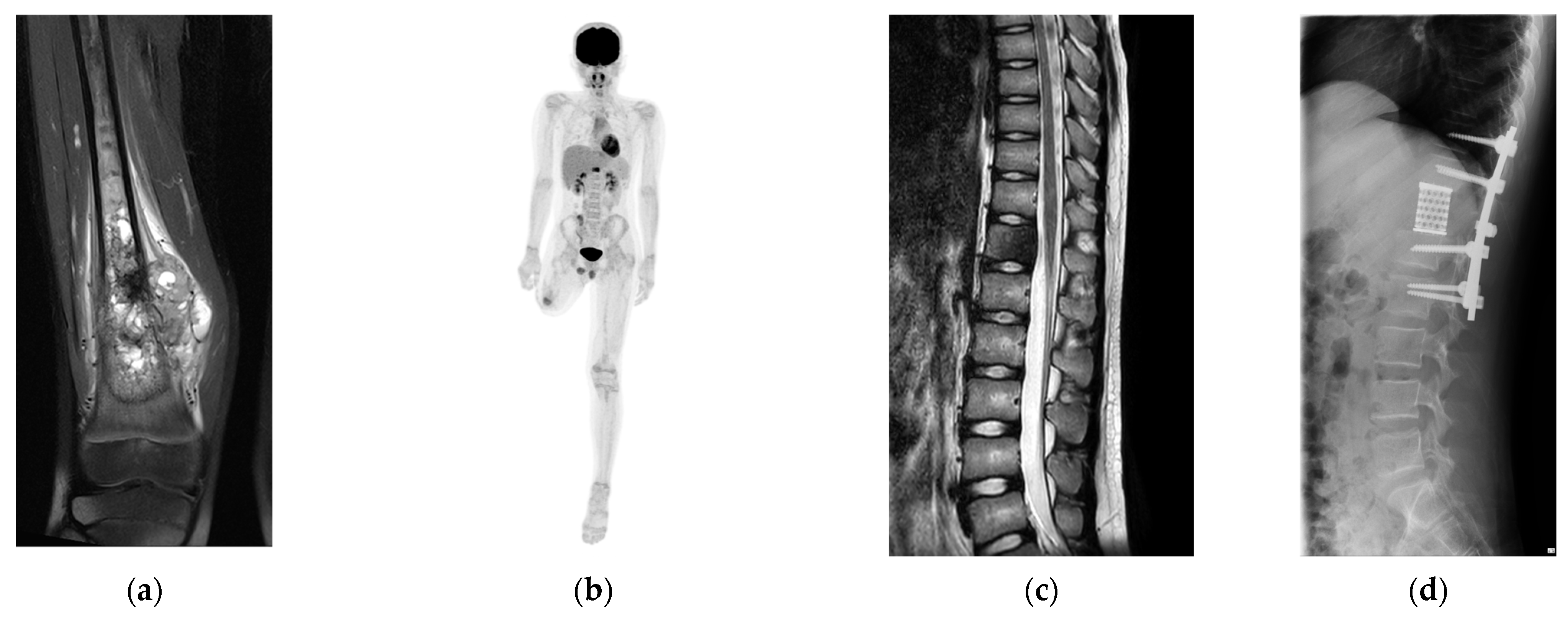
2.1. Patient

A 10-year-old boy was diagnosed with conventional osteosarcoma of the right distal femur. The patient underwent perioperative chemotherapy with tumor resection. Three years later, the patient underwent above-knee amputation due to recurrence at the surgical site. Seven months later, a solitary metastasis in the 12th thoracic spine was pointed at 14 years old. To achieve a disease-free status and prevent possible invasion of the spinal canal, second-generation TES was planned (Figure 2).

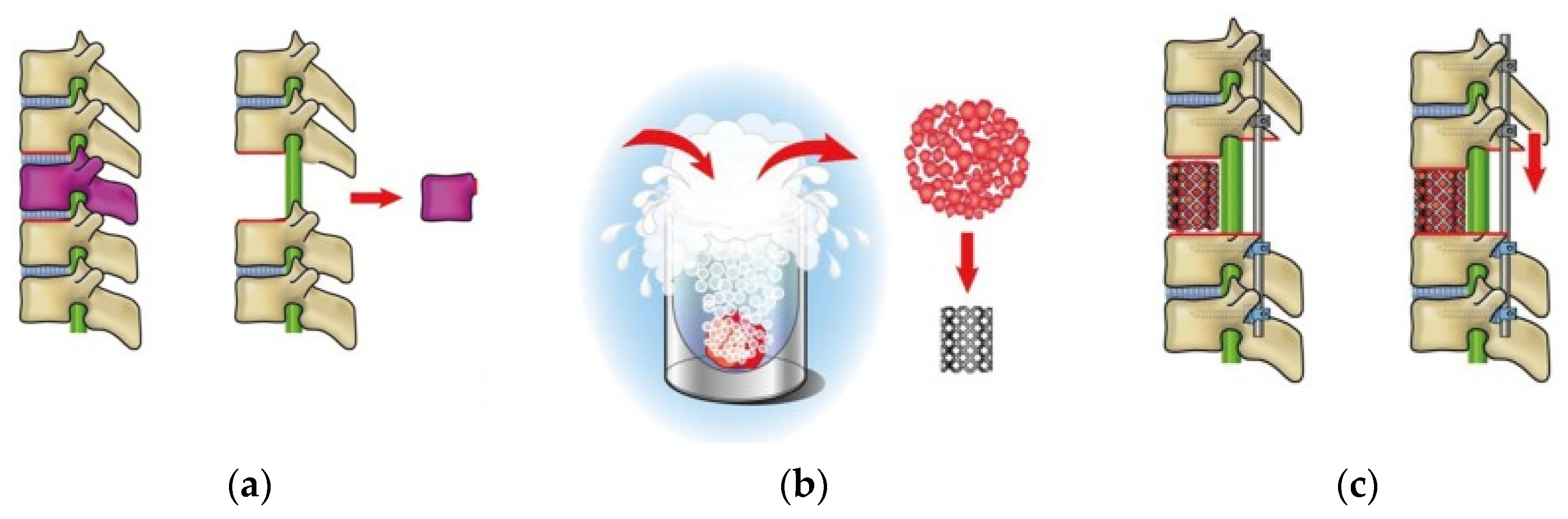
2.2. Tumor Resection and Reconstruction

Second-generation TES was performed with the patient in the prone position under general anesthesia. A midline incision was made to expose the posterior surface of the thoracic vertebrae. The bases of the 12th ribs were cut bilaterally. The spinous process and inferior articular facet of the 11th thoracic vertebra were resected. Surgical thread wire saws were inserted from both sides of the 12th thoracic vertebral foramen, and the vertebral arch and transverse processes of the 12th thoracic vertebra were extracted. The 12th thoracic nerve roots were resected bilaterally. The anterior surface of the 12th thoracic vertebra was gently separated from the surrounding tissues. After inserting pedicle screws and setting-rods to stabilize the vertebral column (covering the 10–11th thoracic and 1–2nd lumbar vertebrae), the 12th thoracic vertebra was resected en bloc. The tumor-containing 12th vertebral body was crushed into small pieces, frozen with liquid nitrogen (−196 °C for 20 min), then packed into a cage with autologous, unfrozen, crashed bones (harvested from the bases of the 12th ribs, as well as the spinous process and facet of the 11th thoracic vertebra) for reconstruction of the 12th vertebral body. The total operative time was 322 min, and the total blood loss was 311 mL. The bony fusion of the anterior column was confirmed three months postoperatively.

2.3. RNA Extraction and the Assessment of Integrity for the Sequence

RNA was extracted from whole blood using the PAXgene Blood RNA System (Becton Dickinson, Franklin Lakes, NJ, USA) just before second-generation TES and 3 months postoperatively. The concentration and quality were tested using a NanoDrop 2000 spectrophotometer (Thermo Fisher Scientific, Waltham, MA, USA). Agilent 2100 (Agilent Technologies, Santa Clara, CA, USA) was used for the precise evaluation of RNA integrity, which was assessed based on the RNA Integrity Number value and flatness of the electropherogram baseline.

2.4. Immunome Sequencing Method

A 5′ rapid amplification of cDNA ends was performed for reverse transcription. The reverse transcription products were amplified using PCR and purified using DNA magnetic beads. The terminal repair was performed using End Prep Enzyme Mix (QIAGEN, Venro, The Netherlands) with sequencing adapters at both ends. Subsequently, clean DNA beads were purified and amplified using the P5 and P7 primers. Bead-based methods were used to purify the PCR products, which were then evaluated for quality with the Qsep100 system (Bioptic, Taiwan, China). Quantification was performed using the Qubit 3.0 Fluorometer (Invitrogen, Carlsbad, CA, USA). Multiple libraries with unique indices were combined (multiplexed) and subsequently loaded onto an Illumina MiSeq or NovaSeq device following the instructions provided by the manufacturer (Illumina, San Diego, CA, USA). Sequencing was conducted using a 2 × 250 or 2 × 300 paired-end configuration, and image analysis and base calling were conducted using the MiSeq Control Software (Version 4.0)/Novaseq Control Software (Version 1.8) on the MiSeq/Novaseq instrument (Illumina).

2.5. Data Analysis

The raw FASTQ files underwent an initial quality evaluation (Table A1). Poor-quality bases and adapter sequences (Q-value < 20) were trimmed using Cutadapt (version 1.9.1) to produce cleaned, high-quality data. Paired-end reads were then merged using FLASH (version 2.2.00). The resulting merged sequences were blasted against the IMGT reference database to determine the best correspondence between germline V(D)J gene segments and the sequences within the Complementarity-Determining Region 1 (CDR1), CDR2, and CDR3. V(D)J recombination is a distinctive genetic rearrangement process that occurs exclusively in developing lymphocytes during early T- and B-cell development [15,16]. This mechanism results in many V(D)J combinations, engineering a highly diverse repertoire of immunoglobulins and T-cell receptor combinations. After obtaining the clone and CDR sequence information, customized scripts were written to perform the following statistical analyses: For diversity analysis, CDR3, an important region for antigen recognition [17], resulting from V(D)J recombination, was analyzed using rank–abundance curves. Additionally, the richness [18] of CDR3 types was analyzed using the abundance-based coverage estimator (ACE) [19], Chao1 [19], Shannon [20], and Simpson indices [21].

3. Results

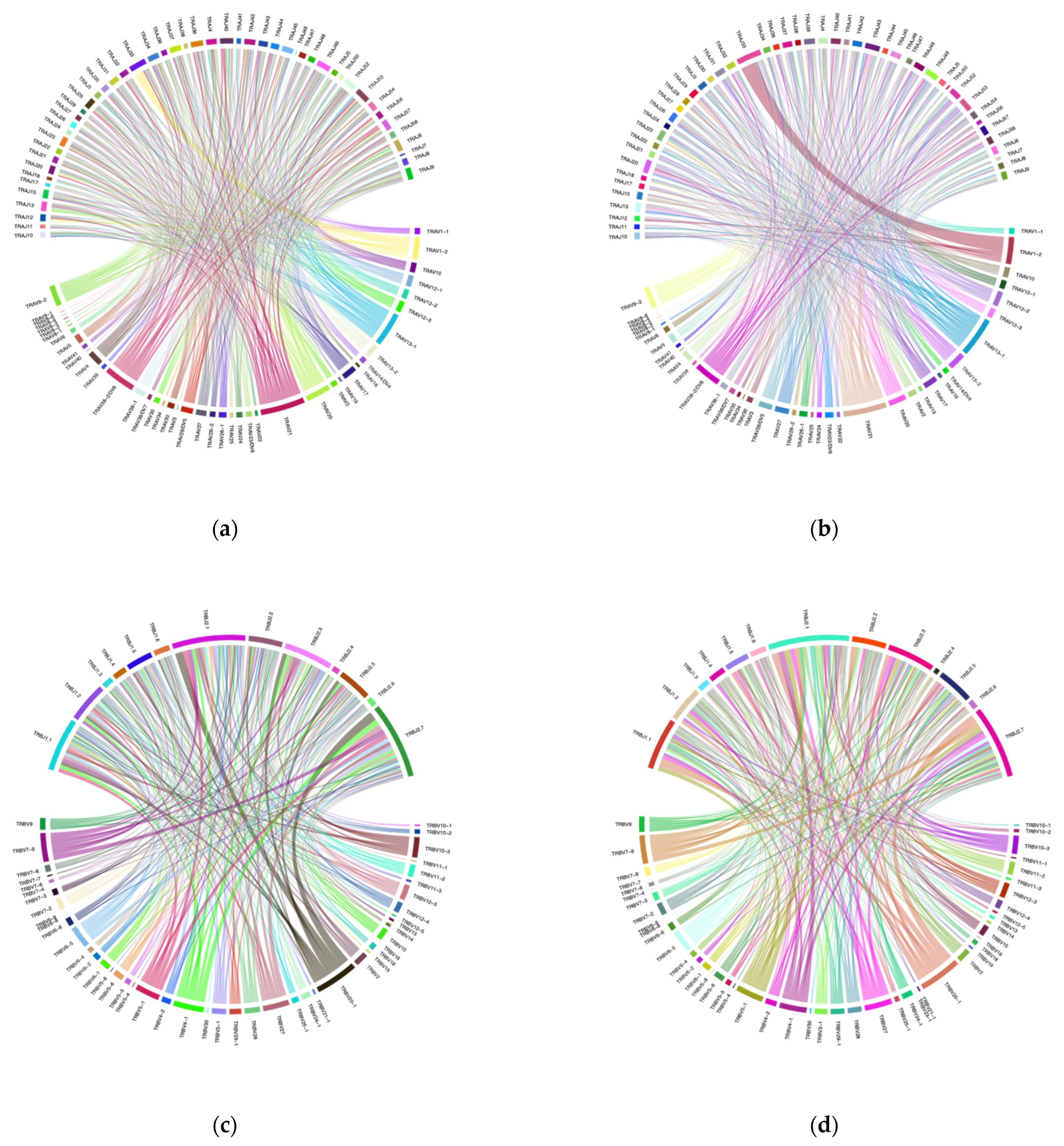
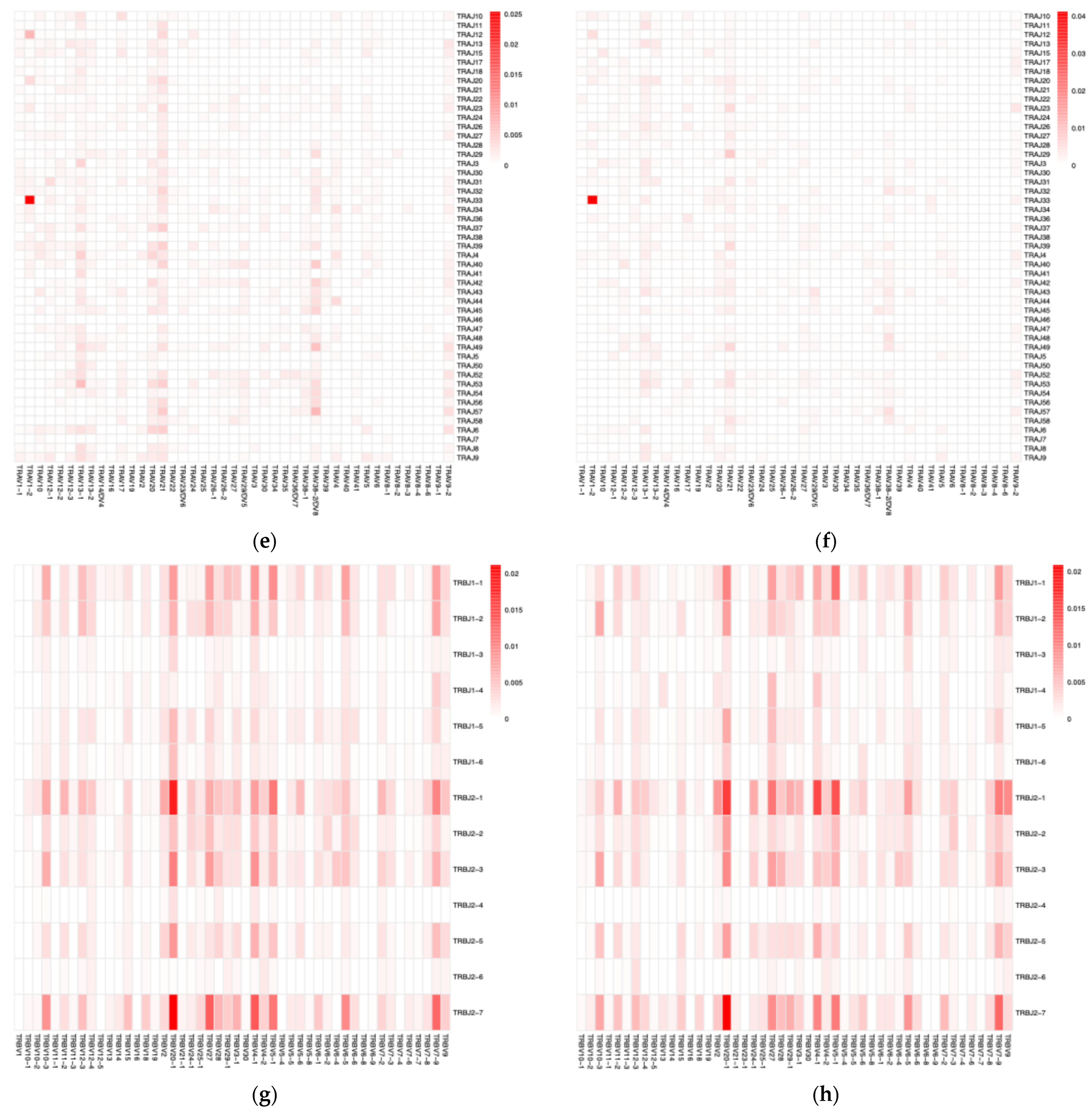
3.1. VJ Gene Recombinations

The merged sequences were BLASTed against the IMGT reference database to identify the best match for the germline VJ genes. The abundance of clones after mapping and clustering was calculated for each chain type at various time points. The Circo plots illustrate the interaction between TRAV–TRAJ or TRBV–TRBJ gene segments, where the connecting lines represent the pairing frequency of specific segments, with thicker lines indicating more common combinations (Figure 3a–d). The heatmaps revealed the dominant pairing genes between TRAV–TRAJ or TRBV–TRBJ, indicating clonal expansion and a favored V-J gene combination in the dataset (Figure 3e–h). Analysis of the full-length sequence and abundance of the identified VJ region in each sample is shown in Supplemental Table S1.

3.2. Diversity Analysis

The rank–abundance curves showed that the diversity of the CDR3 α chain in the preoperative period exhibited a higher level of repertoire diversity and richness compared to that during the postoperative period (Figure 4a). Specifically, the preoperative CDR3 α chain had approximately 2967 unique clonotypes, whereas the postoperative sample had approximately 1865. The ACE and Chao1 indices, which estimate total unseen diversity, were also higher in the preoperative CDR3 α chain. The Shannon index was 11.085 for the preoperative CDR3 α chain, slightly exceeding the postoperative value, indicating a more balanced distribution of clones. Both samples demonstrated high Simpson indices (close to 1), implying that overall diversity remained high. Likewise, the diversity of the CDR3 β chain in the preoperative period exhibited a higher level of immune repertoire diversity compared to that during the postoperative period (Figure 4b). The preoperative CDR3 β chain had approximately 5070 unique clonotypes, higher than the 4050 clonotypes in the postoperative sample. The ACE and Chao1 indices were also higher in the preoperative CDR3 β chain, suggesting that the preoperative repertoire was more extensive and included many rare clones. The Shannon index for the preoperative period (11.974) was slightly higher than that of the postoperative period (11.630), indicating a more even distribution of clones in the preoperative period. Both samples showed high Simpson indices (close to 1), indicating that diversity was high and no single clone dominated either repertoire (Table 1).

3.3. Patient’s Outcome

The patient remained disease-free for 1 year following second-generation TES; however, a solitary metastasis was noted in the right lower lobe. The patient received a lobectomy and is currently disease-free (2 years after second-generation TES).

4. Discussion

This case report presents preliminary results from a prospective study of immunologic alterations following second-generation TES, focusing on the repertoire of TCRs. The findings demonstrated that the repertoires of the CDR3 regions of the TCR α and β chains decreased after surgery, suggesting that more selective clones may have emerged and influenced immune responses.
TCRs are transmembrane glycoprotein heterodimers mainly formed by the combination of α and β chains [22]. The genes encoding the α (TRA) and β (TRB) chains consist of multiple non-continuous segments, including variable (V), diversity (D) segments, and joining (J) segments [22], and TCRs acquire their diversity through V(D)J recombination [22]. Diversity in humans is approximately 2 × 10⁷ different clonotypes [23]. With exposure to various antigens, the diversity of the TCR repertoire dynamically changes throughout its life but it generally decreases with aging [24].
The tumor microenvironment (TME) contains numerous immune-related cells, and cytotoxic T cells play a central role [25,26]. In the TME, tumor antigens are presented by antigen-presenting cells, resulting in the clonal expansion of T cells and restriction of the TCR repertoire [27]. The relationship between TCR repertoire and prognosis has been studied in various tumors [28,29]. Campana et al. analyzed TCR repertoires based on colorectal cancer datasets from the Cancer Genome Atlas and found that patients with numerous TCR repertoires tended to have longer overall survival compared to those with fewer TCR repertoires [30]. Moreover, based on an analysis of patients with metastatic breast cancer, lymphopenia before chemotherapy and lower TCR diversity were independent predictors of prognosis [31]. Thus, the loss of TCR diversity is believed to result from the aggressiveness of tumors and may lead to immune system failure [22].
Tumor mutation burden (TMB) influences the TCR repertoire, as higher values often lead to increased tumor clonality [22]. Tumors with higher TMB levels produce more tumor neoantigens with high affinity for the human leukocyte antigen, thereby increasing their chances of being recognized by T cells [22]. Notably, a study suggested that patients with higher TMB and clonality respond better to immunotherapies, especially in tumors with mismatch repair deficiency (MMR-d) or high microsatellite instability (MSI), due to increased tumor neoantigen production, thereby promoting tumor-specific lymphocyte infiltration and activation [32]. Additionally, in colorectal cancer, MSI-high/MMR-d tumors show higher T-cell infiltration and more clonally expanded TCR repertoires that proliferate further after immunotherapy [33].
Matzinger et al. reported that when tumor tissue is frozen, necrosis primarily occurs in the center of the tumor. In contrast, apoptosis mainly occurs in the periphery of the tumor, depending on the freezing temperature [34]. In necrotic tissue, various molecules, including DNA, RNA, and damage-associated molecular patterns (DAMPs) are released from ruptured cells as danger signals [34]. High mobility group box protein 1 (HMGB1), heat shock proteins, calreticulin, and GP96 have been identified as DAMPs, and HMGB1 is considered a key factor in activating CD8+ T cells through the innate immune system via Toll-like receptor-4 [35,36]. Furthermore, necrotic tissue creates a cytokine-rich environment—such as interleukin (IL)-2, interferon (IFN)-γ, tumor necrosis factor-α, and IL-12—that induces CD8+ T-cell responses, and is presumably in a state conducive to immune reactions against the antigens released from frozen tissue [37]. The TME surrounding the transplanted frozen tissue enhances T-cell activation by tumor neoantigens, with epitopes altered by freezing, potentially resulting in the clonal proliferation of T cells. The results of the current case report indicate a decrease in repertoire diversity after second-generation TES, which may have been caused by reactions to frozen tissue transplantation.
Immune checkpoint inhibitors (ICIs) suggestively enhance the abscopal effect [38]. According to a case report on patients with melanoma treated with cytotoxic T-lymphocyte antigen-4 inhibitors, the radiotherapy of thoracic spinal lesions reduced other metastatic tumors and increased NY-ESO-1 antibodies, a key factor in mediating the abscopal effect, was observed during radiotherapy [38]. Analysis of the TCR repertoire is critical to predicting the efficacy of ICIs [39]. Analysis of blood samples from patients with melanoma who received nivolumab revealed increased TCR clonality after ICI treatment was observed in both peripheral and tumor tissues [39]. Similarly, a randomized controlled study of neoadjuvant ipilimumab and nivolumab for early-stage colon cancer showed that ICI responders had higher baseline CD8+PD-1+ T-cell infiltration and TCR clonality [40]. Similarly to radiotherapy, cryotherapy destroyed tumor tissue and increased tumor antigens potentially leading to a synergistic immune response, especially when combined with ICI [41]. Thus, frozen tissue transplantation used in second-generation TES may enhance antitumor effects when combined with ICIs.
As the results are based on a single case report, it is difficult to gain a comprehensive understanding of the changes in TCR repertoires. Additionally, control data from patients who did not undergo frozen tissue transplantation are necessary for a meaningful comparison. Furthermore, the effects of cryoimmunization may vary depending on factors such as the type of primary lesion, amount of transplanted tissue, and subsequent treatments. To the best of our knowledge, this study is the first to employ TCR repertoire analysis following second-generation TES. Further validation and verification studies are planned.

5. Conclusions

Through TCR repertoire analysis, this study demonstrated that the transplantation of frozen tumor-containing autologous bone grafts has an impact on the immune system.

Supplementary Materials

The following supporting information can be downloaded at: https://www.mdpi.com/article/10.3390/curroncol32080432/s1, Table S1. Full-length sequence of each sample.

Author Contributions

Conceptualization, H.A. and H.M.; methodology, H.A.; software, H.A.; validation, H.A.; formal analysis, H.A.; investigation, H.A., R.T., K.M. and H.M.; resources, H.A. and H.M.; data curation, H.A., R.T., K.M. and H.M.; writing—original draft preparation, H.A.; writing—review and editing, H.A., H.K., R.T., N.S., K.K., K.Y., M.Y., K.M., S.S., A.A., C.E. and H.M.; visualization, H.A. and R.T.; supervision, C.E. and H.M.; project administration, H.A., T.S. and H.K.; funding acquisition, H.A. and H.K. All authors have read and agreed to the published version of the manuscript.

Funding

This work was supported by JSPS KAKENHI Grant Number 20K09485 and 20K18006.

Institutional Review Board Statement

The study was conducted in accordance with the Declaration of Helsinki and approved by the Institutional Review Board of Nagoya City University Hospital (protocol code 60-21-0032; date of approval: 11 October 2021).

Informed Consent Statement

Written informed consent has been obtained from the patient to publish this paper.

Data Availability Statement

We provide details regarding where data supporting reported results in a supplemental dataset.

Acknowledgments

We gratefully thank Tomoko Koshimae for the management of this study. The immunosequencing was outsourced to Genomics from Azenta Life Sciences (Waltham, MA, USA). The authors have reviewed and edited the output and take full responsibility for the content of this publication.

Conflicts of Interest

The authors declare no conflicts of interest.

Abbreviations

The following abbreviations are used in this manuscript:
ACEAbundance-based Coverage Estimator
CDRComplementarity-Determining Region
DAMPsDamage-Associated Molecular Patterns
HMGBHigh Mobility Group Box Protein
ICIImmune Checkpoint Inhibitor
IFNInterferon
ILInterleukin
IMGTInternational ImMunoGeneTics
MMR-dMismatch Repair-deficient
MSIMicrosatellite Instability
TCRT-cell Receptor
TESTotal En-Bloc Spondylectomy
TMBTumor Mutation Burden
TMETumor Microenvironment
TRAT-cell Receptor α Chain
TRBT-cell Receptor β Chain

Appendix A

Table A1. Sequence quality.
SampleTotal ReadsTotal BasesAverage LengthQ20Q30GC%N%
TCR α chain
(preoperative)
3157530938871105297.3496.8488.2348.110.08
TCR β chain
(preoperative)
3261872967060609296.4796.9288.6653.890.08
TCR α chain
(postoperative)
3120218928251874297.596.8488.1748.080.08
TCR β chain
(postoperative)
41820781235268425295.3797.0689.2254.420.08
Q20, Q30: Percentage of bases with quality scores (Qphred) higher than 20 or 30; GC%: Percentage of G+C in the read; N%: Number of bases N per million bases; TCR: T-cell receptor.

References

  1. Shulman, S.; Yantorno, C.; Bronson, P. Cryo-Immunology: A Method of Immunization to Autologous Tissue. Proc. Soc. Exp. Biol. Med. 1967, 124, 658–661. [Google Scholar] [CrossRef] [PubMed]
  2. Soanes, W.A.; Ablin, R.J.; Gonder, M.J. Remission of Metastatic Lesions Following Cryosurgery in Prostatic Cancer: Immunologic Considerations. J. Urol. 1970, 104, 154–159. [Google Scholar] [CrossRef] [PubMed]
  3. Gulley, J.L.; Madan, R.A.; Pachynski, R.; Mulders, P.; Sheikh, N.A.; Trager, J.; Drake, C.G. Role of Antigen Spread and Distinctive Characteristics of Immunotherapy in Cancer Treatment. J. Natl. Cancer Inst. 2017, 109, djw261. [Google Scholar] [CrossRef]
  4. Mole, R.H. Whole Body Irradiation; Radiobiology or Medicine? Br. J. Radiol. 1953, 26, 234–241. [Google Scholar] [CrossRef] [PubMed]
  5. Golden, E.B.; Demaria, S.; Schiff, P.B.; Chachoua, A.; Formenti, S.C. An Abscopal Response to Radiation and Ipilimumab in a Patient with Metastatic Non-Small Cell Lung Cancer. Cancer Immunol. Res. 2013, 1, 365–372. [Google Scholar] [CrossRef] [PubMed]
  6. Liu, Y.; Dong, Y.; Kong, L.; Shi, F.; Zhu, H.; Yu, J. Abscopal Effect of Radiotherapy Combined with Immune Checkpoint Inhibitors. J. Hematol. Oncol. 2018, 11, 104. [Google Scholar] [CrossRef]
  7. Chandra, R.A.; Wilhite, T.J.; Balboni, T.A.; Alexander, B.M.; Spektor, A.; Ott, P.A.; Ng, A.K.; Hodi, F.S.; Schoenfeld, J.D. A Systematic Evaluation of Abscopal Responses Following Radiotherapy in Patients with Metastatic Melanoma Treated with Ipilimumab. Oncoimmunology 2015, 4, e1046028. [Google Scholar] [CrossRef]
  8. Kodet, O.; Němejcova, K.; Strnadová, K.; Havlínová, A.; Dundr, P.; Krajsová, I.; Štork, J.; Smetana, K., Jr.; Lacina, L. The Abscopal Effect in the Era of Checkpoint Inhibitors. Int. J. Mol. Sci. 2021, 22, 7204. [Google Scholar] [CrossRef]
  9. Mole, R.H. The Effect of X-Irradiation on the Basal Oxygen Consumption of the Rat. Q. J. Exp. Physiol. Cogn. Med. Sci. 1953, 38, 69–74. [Google Scholar] [CrossRef]
  10. Tsuchiya, H.; Wan, S.L.; Sakayama, K.; Yamamoto, N.; Nishida, H.; Tomita, K. Reconstruction Using an Autograft Containing Tumour Treated by Liquid Nitrogen. J. Bone Jt. Surg. Br. 2005, 87, 218–225. [Google Scholar] [CrossRef]
  11. Takeuchi, A.; Tsuchiya, H.; Setsu, N.; Gokita, T.; Tome, Y.; Asano, N.; Minami, Y.; Kawashima, H.; Fukushima, S.; Takenaka, S.; et al. What Are the Complications, Function, and Survival of Tumor-Devitalized Autografts Used in Patients with Limb-sparing Surgery for Bone and Soft Tissue Tumors? A Japanese Musculoskeletal Oncology Group Multi-Institutional Study. Clin. Orthop Relat. Res. 2023, 481, 2110–2124. [Google Scholar] [CrossRef]
  12. Murakami, H.; Demura, S.; Kato, S.; Nishida, H.; Yoshioka, K.; Hayashi, H.; Inoue, K.; Ota, T.; Shinmura, K.; Yokogawa, N.; et al. Increase of Il-12 Following Reconstruction for Total En Bloc Spondylectomy Using Frozen Autografts Treated with Liquid Nitrogen. PLoS ONE 2013, 8, e64818. [Google Scholar] [CrossRef]
  13. Ishii, T.; Murakami, H.; Demura, S.; Kato, S.; Yoshioka, K.; Fujii, M.; Igarashi, T.; Tsuchiya, H. Invasiveness Reduction of Recent Total En Bloc Spondylectomy: Assessment of the Learning Curve. Asian Spine J. 2016, 10, 522–527. [Google Scholar] [CrossRef]
  14. Murakami, H.; Demura, S.; Kato, S.; Yoshioka, K.; Hayashi, H.; Inoue, K.; Ota, T.; Shinmura, K.; Yokogawa, N.; Fang, X.; et al. Systemic Antitumor Immune Response Following Reconstruction Using Frozen Autografts for Total En Bloc Spondylectomy. Spine J. 2014, 14, 1567–1571. [Google Scholar] [CrossRef]
  15. Roth, D.B. V(D)J Recombination: Mechanism, Errors, and Fidelity. Microbiol. Spectr. 2014, 2, 313–324. [Google Scholar] [CrossRef]
  16. Hozumi, N.; Tonegawa, S. Evidence for Somatic Rearrangement of Immunoglobulin Genes Coding for Variable and Constant Regions. Proc. Natl. Acad. Sci. USA 1976, 73, 3628–3632. [Google Scholar] [CrossRef]
  17. Mazzotti, L.; Gaimari, A.; Bravaccini, S.; Maltoni, R.; Cerchione, C.; Juan, M.; Navarro, E.A.-G.; Pasetto, A.; Nascimento Silva, D.; Ancarani, V.; et al. T-Cell Receptor Repertoire Sequencing and Its Applications: Focus on Infectious Diseases and Cancer. Int. J. Mol. Sci. 2022, 23, 8590. [Google Scholar] [CrossRef]
  18. Morris, E.K.; Caruso, T.; Buscot, F.; Fischer, M.; Hancock, C.; Maier, T.S.; Meiners, T.; Müller, C.; Obermaier, E.; Prati, D.; et al. Choosing and Using Diversity Indices: Insights for Ecological Applications from the German Biodiversity Exploratories. Ecol. Evol. 2014, 4, 3514–3524. [Google Scholar] [CrossRef] [PubMed]
  19. Klein-Jöbstl, D.; Schornsteiner, E.; Mann, E.; Wagner, M.; Drillich, M.; Schmitz-Esser, S. Pyrosequencing Reveals Diverse Fecal Microbiota in Simmental Calves During Early Development. Front. Microbiol. 2014, 5, 622. [Google Scholar] [CrossRef] [PubMed]
  20. Spellerberg, I.F.; Fedor, P.J. A Tribute to Claude Shannon (1916–2001) and a Plea for More Rigorous Use of Species Richness, Species Diversity and the ‘Shannon–Wiener’ Index. Glob. Ecol. Biogeogr. 2003, 12, 177–179. [Google Scholar] [CrossRef]
  21. Simpson, E.H. Measurement of Diversity. Nature 1949, 163, 688. [Google Scholar] [CrossRef]
  22. Aran, A.; Garrigós, L.; Curigliano, G.; Cortés, J.; Martí, M. Evaluation of the TCR Repertoire as a Predictive and Prognostic Biomarker in Cancer: Diversity or Clonality? Cancers 2022, 14, 1771. [Google Scholar] [CrossRef]
  23. Arstila, T.P.; Casrouge, A.; Baron, V.; Even, J.; Kanellopoulos, J.; Kourilsky, P. A Direct Estimate of the Human Alphabeta T Cell Receptor Diversity. Science 1999, 286, 958–961. [Google Scholar] [CrossRef]
  24. Attaf, M.; Huseby, E.; Sewell, A.K. Aβ T Cell Receptors as Predictors of Health and Disease. Cell. Mol. Immunol. 2015, 12, 391–399. [Google Scholar] [CrossRef] [PubMed]
  25. Takahashi, H.; Yoshimatsu, G.; Faustman, D.L. The Roles of TNFR2 Signaling in Cancer Cells and the Tumor Microenvironment and the Potency of TNFR2 Targeted Therapy. Cells 2022, 11, 1952. [Google Scholar] [CrossRef] [PubMed]
  26. Chandra, R.; Ehab, J.; Hauptmann, E.; Gunturu, N.S.; Karalis, J.D.; Kent, D.O.; Heid, C.A.; Reznik, S.I.; Sarkaria, I.S.; Huang, H.; et al. The Current State of Tumor Microenvironment-Specific Therapies for Non-Small Cell Lung Cancer. Cancers 2025, 17, 1732. [Google Scholar] [CrossRef] [PubMed]
  27. Song, I.H.; Lee, S.B.; Jeong, B.K.; Park, J.; Kim, H.; Lee, G.; Cha, S.M.; Lee, H.; Gong, G.; Kwon, N.J.; et al. T Cell Receptor Clonotype in Tumor Microenvironment Contributes to Intratumoral Signaling Network in Patients with Colorectal Cancer. Immunol. Res. 2024, 72, 921–937. [Google Scholar] [CrossRef]
  28. Takahashi, H.; Hanaoka, K.; Wada, H.; Kojima, D.; Watanabe, M. The Current Status of T Cell Receptor (TCR) Repertoire Analysis in Colorectal Cancer. Int. J. Mol. Sci. 2025, 26, 2698. [Google Scholar] [CrossRef]
  29. Porciello, N.; Franzese, O.; D’Ambrosio, L.; Palermo, B.; Nisticò, P. T-Cell Repertoire Diversity: Friend or Foe for Protective Antitumor Response? J. Exp. Clin. Cancer Res. 2022, 41, 356. [Google Scholar] [CrossRef]
  30. Campana, L.G.; Mansoor, W.; Hill, J.; Macutkiewicz, C.; Curran, F.; Donnelly, D.; Hornung, B.; Charleston, P.; Bristow, R.; Lord, G.M.; et al. T-Cell Infiltration and Clonality May Identify Distinct Survival Groups in Colorectal Cancer: Development and Validation of a Prognostic Model Based on The Cancer Genome Atlas (TCGA) and Clinical Proteomic Tumor Analysis Consortium (CPTAC). Cancers 2022, 14, 5883. [Google Scholar] [CrossRef]
  31. Manuel, M.; Olivier, T.; Thomas, B.; Gilles, C.; Anais, C.; Gilles, P.; Tioka, R.; Audrey, G.; Solène, P.; Jean-François, M.; et al. Lymphopenia Combined with Low Tcr Diversity (Divpenia) Predicts Poor Overall Survival in Metastatic Breast Cancer Patients. OncoImmunology 2012, 1, 432–440. [Google Scholar] [CrossRef]
  32. Sahin, I.H.; Akce, M.; Alese, O.; Shaib, W.; Lesinski, G.B.; El-Rayes, B.; Wu, C. Immune Checkpoint Inhibitors for the Treatment of Msi-H/Mmr-D Colorectal Cancer and a Perspective on Resistance Mechanisms. Br. J. Cancer 2019, 121, 809–818. [Google Scholar] [CrossRef] [PubMed]
  33. Akiyama, Y.; Kondou, R.; Iizuka, A.; Miyata, H.; Maeda, C.; Kanematsu, A.; Ashizawa, T.; Nagashima, T.; Urakami, K.; Shimoda, Y.; et al. Characterization of the Immunological Status of Hypermutated Solid Tumors in the Cancer Genome Analysis Project HOPE. Anticancer Res. 2022, 42, 3537–3549. [Google Scholar] [CrossRef] [PubMed]
  34. Matzinger, P. Tolerance, Danger, and the Extended Family. Annu. Rev. Immunol. 1994, 12, 991–1045. [Google Scholar] [CrossRef]
  35. Janopaul-Naylor, J.R.; Shen, Y.; Qian, D.C.; Buchwald, Z.S. The Abscopal Effect: A Review of Pre-Clinical and Clinical Advances. Int. J. Mol. Sci. 2021, 22, 11061. [Google Scholar] [CrossRef]
  36. Shabalkina, A.V.; Izosimova, A.V.; Ryzhichenko, E.O.; Shurganova, E.V.; Myalik, D.S.; Maryanchik, S.V.; Ruppel, V.K.; Knyazev, D.I.; Khilal, N.R.; Barsova, E.V.; et al. Nodal Expansion, Tumor Infiltration and Exhaustion of Neoepitope-Specific Th Cells After Prophylactic Peptide Vaccination and Anti-CTLA4 Therapy in Mouse Melanoma B16. Int. J. Mol. Sci. 2025, 26, 6453. [Google Scholar] [CrossRef] [PubMed]
  37. Abdo, J.; Cornell, D.L.; Mittal, S.K.; Agrawal, D.K. Immunotherapy Plus Cryotherapy: Potential Augmented Abscopal Effect for Advanced Cancers. Front. Oncol. 2018, 8, 85. [Google Scholar] [CrossRef]
  38. Postow, M.A.; Callahan, M.K.; Barker, C.A.; Yamada, Y.; Yuan, J.; Kitano, S.; Mu, Z.; Rasalan, T.; Adamow, M.; Ritter, E.; et al. Immunologic Correlates of the Abscopal Effect in a Patient with Melanoma. N. Engl. J. Med. 2012, 366, 925–931. [Google Scholar] [CrossRef]
  39. Inoue, H.; Park, J.H.; Kiyotani, K.; Zewde, M.; Miyashita, A.; Jinnin, M.; Kiniwa, Y.; Okuyama, R.; Tanaka, R.; Fujisawa, Y.; et al. Intratumoral Expression Levels of Pd-L1, Gzma, and Hla-a Along with Oligoclonal T Cell Expansion Associate with Response to Nivolumab in Metastatic Melanoma. Oncoimmunology 2016, 5, e1204507. [Google Scholar] [CrossRef]
  40. Chalabi, M.; Fanchi, L.F.; Dijkstra, K.K.; Van den Berg, J.G.; Aalbers, A.G.; Sikorska, K.; Lopez-Yurda, M.; Grootscholten, C.; Beets, G.L.; Snaebjornsson, P.; et al. Neoadjuvant Immunotherapy Leads to Pathological Responses in Mmr-Proficient and Mmr-Deficient Early-Stage Colon Cancers. Nat. Med. 2020, 26, 566–576. [Google Scholar] [CrossRef]
  41. Yonezawa, N.; Murakami, H.; Demura, S.; Kato, S.; Miwa, S.; Yoshioka, K.; Shinmura, K.; Yokogawa, N.; Shimizu, T.; Oku, N.; et al. Abscopal Effect of Frozen Autograft Reconstruction Combined with an Immune Checkpoint Inhibitor Analyzed Using a Metastatic Bone Tumor Model. Int. J. Mol. Sci. 2021, 22, 1973. [Google Scholar] [CrossRef] [PubMed]
Figure 1. Schema of second-generation TES. (a) en-bloc excision of the vertebra (or vertebrae); (b) the resected lamina and vertebral body, containing tumors, are used as bone grafts for anterior spinal reconstruction. These are placed in liquid nitrogen (−196 °C) for 20 min before being crushed and packed into a titanium cage with a sufficient amount of autologous, unfrozen grafts harvested during the resection procedure); (c) the cage is placed between the adjacent healthy vertebral bodies, and spinal shortening is performed to stabilize the cage [12].
Curroncol 32 00432 g001
Figure 2. Clinical course of a 10-year-old male with conventional osteosarcoma. (a) Image of the primary lesion (right proximal femur, short τ inversion recovery). (b) Positron emission tomography shows a single metastasis in the 12th thoracic spine. (c) Sagittal image of the spine (T2-weighted image) and (d) after surgical resection of the 12th thoracic spine and reconstruction.
Curroncol 32 00432 g002
Figure 3. VJ genes recombination. (a) Circo plots for the combination of TRAV–TRAJ genes before surgery; (b) Circo plots for the combination of TRAV–TRAJ genes 3 months after surgery; (c) Circo plots for the combination of TRBV–TRBJ genes before surgery; (d) Circo plots for the combination of TRBV–TRBJ genes 3-month after surgery; (e) heatmap for the combination of TRAV–TRAJ genes before surgery; (f) heatmap for the combination of TRAV–TRAJ genes 3-month after surgery; (g) heatmap for the combination of TRBV–TRBJ genes before surgery; and (h) heatmap for the combination of TRBV–TRBJ genes 3-month after surgery.
Curroncol 32 00432 g003aCurroncol 32 00432 g003b
Figure 4. Rank–abundance curve plot. The horizontal axis represents CDR3 abundance ranking. The vertical axis represents the CDR3 abundance frequency corresponding to the number, and the shaded area indicates the 0.95 confidence interval obtained using the bootstrap method. The rank–abundance curves indicated a distribution pattern characterized by a few highly abundant clones at the top ranks and a long tail of numerous rare sequences. (a) CDR3 α chain; (b) CDR3 β chain. The blue tones indicate “preoperative” and red tones indicate “postoperative”.
Curroncol 32 00432 g004
Table 1. Diversity in the CDR3 chains. ACE, abundance-based coverage estimator; CDR, complementarity-determining region.
Analyzed PointsRichnessACEChao1ShannonSimpson
CDR3 α chain (preoperative)29673017300611.090.9993
CDR3 α chain (postoperative)18651921194010.460.9990
CDR3 β chain (preoperative)50705154517411.970.9997
CDR3 β chain (postoperative)40504094410111.630.9996
Disclaimer/Publisher’s Note: The statements, opinions and data contained in all publications are solely those of the individual author(s) and contributor(s) and not of MDPI and/or the editor(s). MDPI and/or the editor(s) disclaim responsibility for any injury to people or property resulting from any ideas, methods, instructions or products referred to in the content.

