Article

Concordance of Abundance for Mutational EGFR and Co-Mutational TP53 with Efficacy of EGFR-TKI Treatment in Metastatic Patients with Non-Small-Cell Lung Cancer

1
Department of Medical Oncology, The Second Affiliated Hospital, School of Medicine, Zhejiang University, Hangzhou 310009, China
2
State Key Laboratory of Oncology in South China, Collaborative Innovation Center for Cancer Medicine, Department of Medical Oncology, Sun Yat-sen University Cancer Center, Guangzhou 510060, China
3
Department of Pathology, The Second Affiliated Hospital, School of Medicine, Zhejiang University, Hangzhou 310009, China
*
Author to whom correspondence should be addressed.
These authors contributed equally to this work.
Curr. Oncol. 2023, 30(9), 8464-8476; https://doi.org/10.3390/curroncol30090616
Submission received: 11 August 2023 / Revised: 6 September 2023 / Accepted: 13 September 2023 / Published: 15 September 2023
(This article belongs to the Section Thoracic Oncology)

Abstract

:
The present study aimed to investigate the influence of the mutation abundance of the epidermal growth factor receptor (EGFR) and its co-mutation with TP53 on the therapeutic efficacy of tyrosine kinase inhibitor (TKI) treatment in patients with metastatic lung adenocarcinoma (LUAD). In total, 130 patients (January 2018-September 2022) with metastatic LUAD from the Second Affiliated Hospital of Zhejiang University were included. Kaplan–Meier analysis was performed to measure the duration of drug application (DDA) and the log-rank test was used to compare differences. Univariate and multivariate analyses of Cox proportional hazard regression models were used to evaluate the association between the relevant clinicopathological factors and DDA. Hazard ratios with 95% confidence intervals were also calculated. Among the 130 patients who were treated with first-generation EGFR-TKIs, 86 showed high-EGFR mutation abundance (>22.0%) and 44 showed low-EGFR mutation abundance (≤22.0%). Patients in the high-EGFR group had a greater DDA than those in the low-EGFR group (p < 0.05). The results of the subgroup analysis were consistent with those of the total mutation population (exon19: >18.5% vs. ≤18.5%, 14 months vs. 10 months, p = 0.049; exon21: >22.0% vs. ≤22.0%, 15 months vs. 9 months, p = 0.005). In addition, the mutation abundance of TP53 was negatively correlated with the DDA (p < 0.05). Patients in the combination group had a better DDA than those in the monotherapy group (p < 0.05). Subgroup analysis showed that, among the low mutation abundance of the EGFR exon 21 or 19 cohort, the combination group had a better DDA than the monotherapy group (p < 0.05). An EGFR mutation abundance greater than 22.0% was a positive predictor of DDA in patients with metastatic LUAD. However, a TP53 mutation abundance higher than 32.5% could reverse this situation. Finally, first-line treatment with EGFR-TKIs plus chemotherapy is a potential treatment strategy for patients with low-abundance EGFR mutations.

1. Introduction

Lung cancer is a common and fatal malignant tumor found worldwide [1]. Non-small-cell lung cancer (NSCLC) is the most common histological type of lung cancer, accounting for more than 85% of lung cancer cases [2]. Epidermal growth factor receptor (EGFR) is a common driver gene in NSCLC, and approximately 20% of NSCLC patients have activating mutations in EGFR, particularly those who have an adenocarcinoma histology and have never smoked or have a history of light smoking [3]. EGFR-mutated NSCLC is a genetically heterogeneous disease with more than 200 distinct mutations. Deletion of exon 19 and L858R point mutation in exon 21 are dominant [4,5]. In the field of NSCLC, progress in genetic testing technology and the discovery of new tumor driver genes have promoted the development and clinical application of NSCLC-targeted therapeutic drugs. EGFR tyrosine kinase inhibitors (TKIs) can remarkably improve progression-free survival (PFS) and are recommended as the first-line treatment for EGFR-mutated metastatic NSCLC according to the NCCN guidelines [6,7]. To date, several TKIs have been approved for the treatment of EGFR-mutated NSCLC, including first-generation (Erlotinib, Gefitinib), second-generation (Afatinib), and third-generation (Osimertinib) TKIs [8,9]. Each of these TKIs works by targeting specific mutations in the EGFR gene, inhibiting the tyrosine kinase activity, and blocking downstream signaling pathways that promote cancer growth. First-generation TKIs have shown efficacy in the initial treatment of EGFR-mutated NSCLC, while second-generation TKIs offer broader coverage against EGFR mutations. Osimertinib, a third-generation TKI, specifically targets the T790M resistance mutation, which is common in patients who develop acquired resistance to first-generation TKIs. It has demonstrated remarkable effectiveness in both first- and second-line treatment settings.
However, there are significant differences in the clinical efficacy of TKIs among individuals. Few studies have been conducted on the primary drug resistance of EGFR-TKIs, and the related mechanisms are unclear. Previous studies have shown that the efficacy of EGFR-TKIs is highly heterogeneous [10]. Co-mutations play an important role in the response and resistance to EGFR-TKIs in EGFR-mutant NSCLC, partially explaining the effect of heterogeneity [11,12]. The majority of metastatic EGFR-mutant lung adenocarcinomas (LUAD) harbor one or more co-mutations, and TP53 is a common co-mutated gene in EGFR-mutant NSCLC, accounting for 55–65% of cases [13,14]. Tumors bearing co-mutations in TP53 exhibit high genomic instability and somatic mutation burden [15]. Multiple clinical studies have identified TP53 co-alteration as a negative prognostic marker in LUAD with EGFR mutations and as a predictor of poor clinical outcomes of EGFR-TKI treatment [16].
However, intratumoral heterogeneity is another factor limiting the efficacy of EGFR-TKIs [17]. Several reports have focused on the quantitative assessment of genetic mutations instead of qualitative tests [18]. The abundance of EGFR mutations differs among tumors, or in different samples obtained from the same tumor, because mutant and wild-type (WT) EGFR can exist concurrently in the same primary NSCLC [17]. Wu et al. first proposed individual differences in the abundance of EGFR mutations in NSCLC by direct sequencing and using the amplification refractory mutation system (ARMS), which indicated that patients with high mutation abundance treated with EGFR-TKIs had significantly longer PFS than those with low mutation abundance [18]. However, determining the mutation abundance in tumor tissues to investigate the difference in sensitivity between ARMS and PCR is difficult. The application of ARMS is primarily limited by false positive results caused by non-specific primers that may be produced by WT DNA, as well as the missed detection of low-frequency mutations [19,20]. Lung cancer cells have a high incidence of EGFR amplification, which can affect the abundance of EGFR mutations, as determined by the ARMS assay. Therefore, a quantitative and highly sensitive method is necessary to detect the abundance of EGFR mutants.
Compared to traditional genetic testing methods, second-generation gene sequencing (NGS) has several advantages. It can simultaneously detect point mutations, insertion/deletion, copy number variation, and gene rearrangement of four variant forms, from several genes to hundreds of genes in whole exomes or whole genomes, and it can also provide comprehensive gene mutation data [21]. In patients previously diagnosed with metastatic NSCLC who were found to be negative for EGFR and anaplastic lymphoma kinase (ALK) variants using traditional single-gene testing, 17.4% carried at least one EGFR or ALK variant, as outlined by NGS [22]. Investigators performed NGS analysis in patients with LUAD who showed negative genetic variation (EGFR/KRAS) using traditional genetic testing methods, and 31–65% of the samples showed genetic variation; some patients received approved drugs in accordance with the NCCN guidelines and achieved clinical benefit [23,24]. Therefore, in the case of false negative results from a non-NGS technology, NGS retesting is recommended for patients with negative traditional testing methods [25].
Currently, no reports have been found on whether the co-mutation abundance of EGFR and TP53 affects the efficacy of EGFR-TKIs. Thus, this study aimed to identify the role of EGFR mutation abundance using NGS in the prognosis of metastatic LUAD and the landscape and influence of its co-mutations, as well as to explore therapeutic modalities for low-EGFR mutation abundance. The detection of mutation abundance is of great significance for individualized treatment of patients. Therefore, the population carrying EGFR mutations can be subdivided according to the mutation abundance, thereby accurately distinguishing the targeted therapy population.

2. Materials and Methods

2.1. Patients

Data on metastatic LUAD patients with EGFR mutations were retrospectively extracted from the Second Affiliated Hospital of Zhejiang University School of Medicine (SAHZU) between January 2018 and September 2022. Detailed inclusion criteria were as follows: (a) patients with stage IV LUAD; (b) histopathological examination showing lung adenocarcinoma; (c) all tissue samples for NGS detection evaluated with HE staining by well-trained, experienced pathologists to ensure that the tumor cell content was greater than 50%; (d) NGS analysis showing EGFR exon 19/21 mutation, with conclusive mutation abundance data; (e) oral EGFR-TKI therapy used as first-line treatment for more than 1 month; (f) patients who underwent chest and abdominal CT, brain MRI, and other imaging examinations every 2–3 months to evaluate the potential therapeutic effects on target or non-target lesions during EGFR-TKI administration; and (g) complete clinical information. The exclusion criteria were as follows: (a) NGS test after treatment (n = 4); (b) second- or third-generation EGFR-TKIs as first-line treatment (n = 30); (c) non-EGFR exon19/21 mutation (n = 5); (d) non-tissue samples for NGS (n = 41); and (e) EGFR T790 mutation (n = 5) (Supplementary Figure S1). Finally, 130 metastatic LUAD patients with EGFR mutations who were treated with EGFR-TKIs as the first-line treatment were included. Of these, 55 had TP53 mutations. To determine the cut-off values, the maximum mutation abundance was incorporated into the statistical analysis for patients with more than one mutation type. The study protocol was approved by the Ethical Committee of SAHZU.

2.2. Abundance of EGFR Mutations

Common EGFR mutations, including those in exons 19 and 21, were detected using NGS. NGS was performed using the TruSight One Sequencing Panel Kit (Illumina, San Diego, CA, USA). Briefly, DNA was extracted from tissue samples using a DNA tissue extraction reagent kit. The ILLUMINA NOVASEQ 6000 sequencing platform was used for the enrichment of targeted region hybridization and high-throughput parallel sequencing of disease-related gene mutations. The average sequencing depth was 2000×, with a sensitivity of 0.1%. A total of 641 disease-related gene mutations were identified. The pathogenicity of mutations was assessed based on international databases (ClinVar, Catalog of Somatic Mutations in Cancer (COSMIC), and OncoKB) and then filtered through the company’s self-built database and methods. Using X-tile software (version 3.6.1), the optimal cut-off values of EGFR exon 19 and exon 21 mutation abundance for the duration of drug application (DDA) were 18.5% and 22.0%, respectively. Moreover, the optimal cutoff values of EGFR and TP53 mutation abundance for DDA were 22.0% and 32.5%, respectively.

2.3. Statistical Analysis

Categorical variables are reported as numbers and percentages, and continuous variables are reported as means and standard deviations. DDA was defined as the treatment time from the initial use of one EGFR-TKI to the cessation of the EGFR-TKI or the change to another EGFR-TKI until disease progression or death from any cause. The DDA times were analyzed using Kaplan–Meier analysis and compared among the groups using the log-rank test. Propensity score matching (PSM) analysis was performed to reduce the impact of other clinical characteristics on DDA. PSM analysis is a statistical technique used to reduce bias and confounding in observational studies. It aims to match individuals or groups based on their propensity scores, which estimate the probability of receiving a particular treatment or exposure. The propensity score is calculated using observed characteristics or covariates that are believed to influence both the treatment assignment and outcome. PSM analysis helps balance the distribution of confounding factors across treatment groups, making it a valuable method for drawing causal inferences from observational data. Univariate and multivariate analyses of Cox proportional hazard regression models were used to evaluate the association between the relevant clinicopathological factors and DDA. Hazard ratios (HRs) with 95% confidence intervals (CIs) were calculated. All statistical analyses were performed using R software (version 4.2.1). Statistical significance was defined as a two-sided p value < 0.05.

3. Results

3.1. Characteristics of the Included Patients

Among 130 patients with metastatic LUAD who were treated with first-generation EGFR-TKIs as a first-line treatment, 86 showed high-EGFR mutation abundance (high-EGFR group, >22.0%), and 44 showed low-EGFR mutation abundance (low-EGFR group, ≤22.0%). Patient characteristics are shown in Table 1. Icotinib was the preferred first-generation TKI for first-line treatment, accounting for 70% of the high-EGFR group and 61% of the low-EGFR group, followed by gefitinib and erlotinib. Most patients were treated with EGFR-TKI monotherapy, regardless of the EGFR mutation abundance, particularly in the high-EGFR group (88%). Of the 130 patients, 43 received third-generation EGFR-TKIs as a second-line treatment following first-line first-generation EGFR-TKIs. Most patients achieved a complete/partial response after first-line treatment, accounting for 77% of the high-EGFR group and 77% of the low-EGFR group. More than 80% of patients had one EGFR mutation. Exons 19 and 21 were the most common mutation types of EGFR, and the number of patients with only exon 19 mutations and exon 21 mutations was 55 and 63, respectively; 12 of these cases were exon 19/21 combined with other non-T790 mutations. More than half of the patients (51%) had a combined TP53 mutation in the high-EGFR group.
A total of 26 patients underwent combination therapy (EGFR-TKIs plus chemotherapy or anti-angiogenesis (the combination group)) and 104 patients underwent EGFR-TKI monotherapy (the single group) as first-line treatment. The abundance of EGFR mutations was lower in the combination group than in the single group (p < 0.05, Figure 1A), and patients in the combination group had better DDA than those in the single group (23 months vs. 12 months, p < 0.05, Figure 1B). No difference in EGFR mutation abundance or DDA was observed between EGFR mutation numbers and types (Supplementary Figure S2).

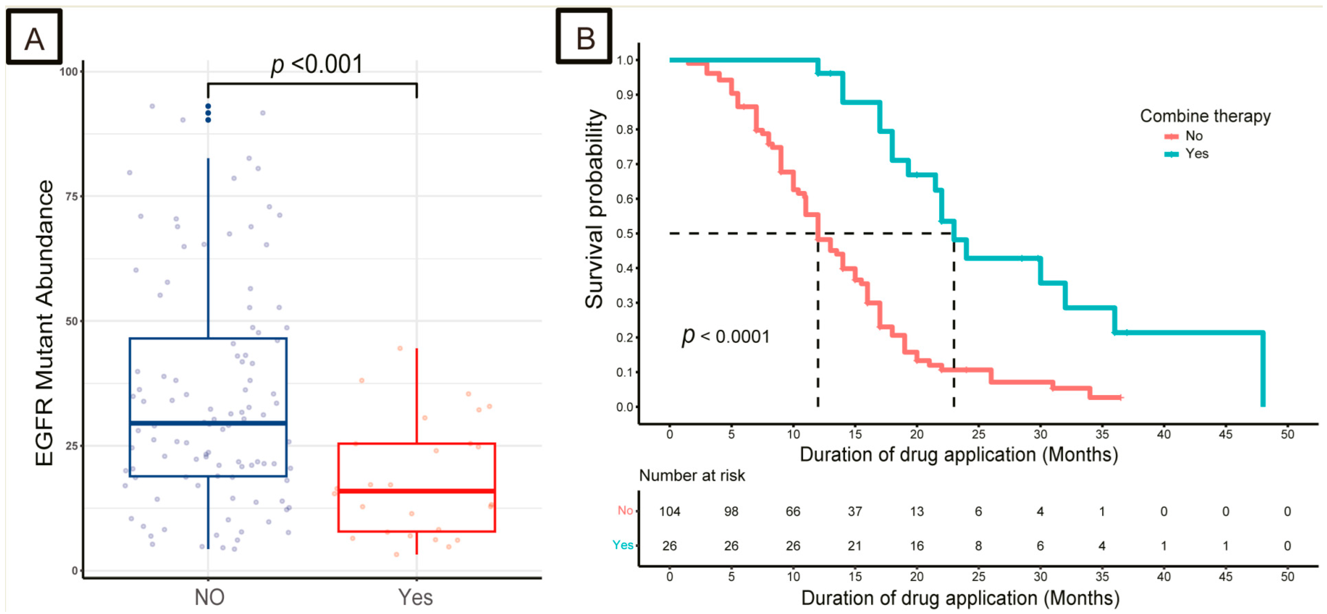
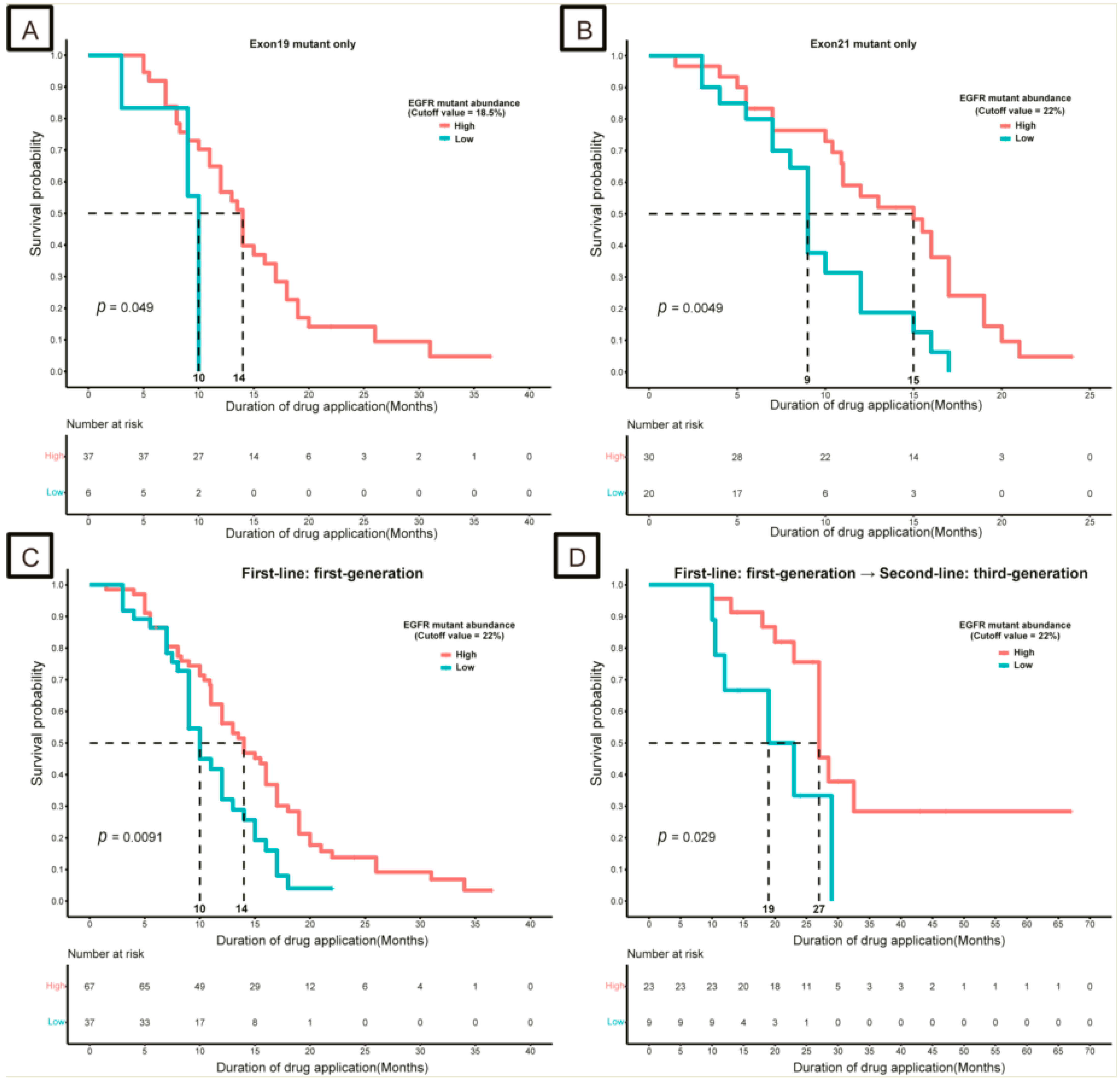
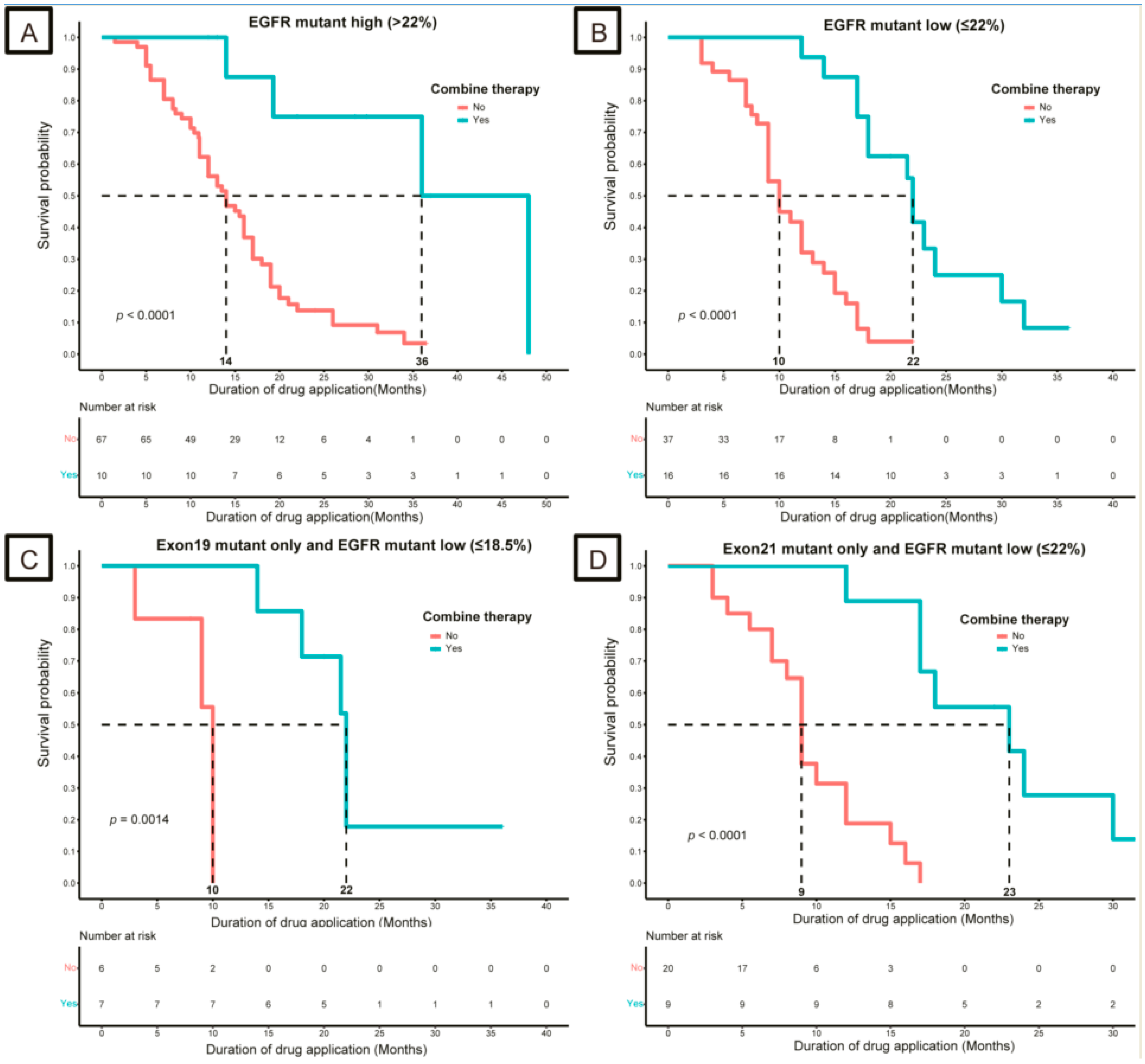
3.2. Impact of EGFR and TP53 Mutation Abundance on Response to TKIs

Based on X-tile software, the optimal cut-off values of EGFR exon 19 and exon 21 mutation abundance for DDA were 18.5% and 22.0%, respectively.
Patients with high-EGFR exon 19 mutations (>18.5%) had a greater DDA than those with low abundance (≤18.5%) (14 months vs. 10 months, p = 0.049; Figure 2A). Consistently, high -EGFR exon 21 mutation abundance (>22.0%) was also associated with good DDA (15 months vs. 9 months, p = 0.005; Figure 2B).
Based on X-tile software, the optimal cutoff values of EGFR and TP53 mutation abundance for DDA were 22.0% and 32.5%, respectively.
A total of 104 patients who received first-generation TKIs as first-line treatment provided tissue samples for NGS, and high-EGFR mutation abundance (>22.0%) was associated with good DDA (14 months vs. 10 months, p = 0.009; Figure 2C). In addition, 32 patients received first-generation EGFR-TKIs as the first-line treatment, followed by third-generation EGFR-TKIs as the second-line treatment. Similarly, in these patients, a high abundance (>22.0%) of EGFR mutations was associated with good DDA, compared with a low abundance (≤22.0%; 27 months vs. 19 months, p = 0.029; Figure 2D).
Among these patients, 23 had a high mutation abundance of TP53 (>32.5%, the high-TP53 group), 20 had a low mutation abundance of TP53 (≤32.5%, the low-TP53 group), and 27 had no TP53 mutation (WT group). The high-TP53 group had worse DDA (8 months) than those in the low-TP53 group (13.5 months, Cox regression analysis, p < 0.05) and WT group (17 months, Cox regression analysis, p < 0.05; Figure 3A). In addition, patients in the high-TP53 group had a higher EGFR mutation abundance of EGFR compared with those in the low-TP53 group and WT groups (p < 0.05, Figure 3B).
PSM was performed to reduce the impact of other clinical characteristics on the DDA. After PSM analysis, 39 patients were included in the high-EGFR and low-EGFR groups and 14 patients were included in the high-TP53 group and low-TP53 groups, respectively. The characteristics of the two groups were matched (p > 0.05) and are presented in Supplementary Tables S1 and S2. Similar results showed that the high mutation abundance of EGFR (>22.0%) was associated with a greater DDA (17 months vs. 12 months, p = 0.011), and a high mutation abundance of TP53 (>32.5%) was associated with poor DDA (13.5 months vs. 8.3 months, p = 0.004) (Supplementary Figure S3).
To explore the association between EGFR and TP53 mutations, univariate and multivariate Cox regression analyses were performed in the tissue sample cohort. Univariate Cox regression analysis showed that the EGFR mutation abundance (low vs. high, HR [95% CI], 1.407 [0.797, 2.482], p > 0.05) was not an independent predictor of DDA compared to TP53 mutation abundance (low vs. WT, HR [95% CI], 2.621 [1.307, 5.256], p = 0.007; high vs. WT, HR [95% CI], 7.300 [3.475, 15.338], p < 0.001). However, based on multivariate Cox regression analysis, EGFR mutation abundance (low vs. high, HR [95% CI], 2.074 [1.108, 3.882], p = 0.023) and TP53 mutation abundance (low vs. WT, HR [95% CI], 2.354 [1.163, 4.763], p = 0.017; high vs. WT, HR [95% CI], 8.836 [4.091, 19.084], p < 0.001) were independent predictors of DDA (Figure 3C).

3.3. Strategies for Patients with Low-Abundance Mutations

Patients in the combination group were associated with good DDA compared with those in the single group, regardless of whether they were high or low (p < 0.05, Figure 4A,B). Among the EGFR exon 19 mutations and low-EGFR mutation abundance (≤18.5%) cohort, the combination group had a better DDA than the monotherapy group in tissue samples (22 months vs. 10 months, p < 0.05, Figure 4C). Similar results were observed only in the EGFR exon 21 mutation and low-EGFR mutation abundance (≤22.0%) cohorts (p < 0.05, Figure 4D).

4. Discussion

For metastatic NSCLC with EGFR mutations, the first-line use of TKI greatly improves the survival time and quality of life of patients [26,27]. However, some patients have a poor response to TKIs, and approximately 20% of patients develop primary resistance to EGFR-TKIs [6], indicating unknown mechanisms restricting the efficacy of TKIs. Intratumoral heterogeneity is an important cause of treatment failure and refers to the presence of heterogeneous targeted molecules (such as EGFR) in tumor tissues; that is, EGFR-mutant and WT clones in the same tumor [28,29]. Therefore, different levels of mutant molecules may be part of the variable response to TKIs. In this study, the EGFR mutation abundance was different in different samples from the same patient and at different sites in the same sample. Previous studies have demonstrated that the abundance of EGFR-activating mutations is significantly associated with an objective response to EGFR-TKIs, and the PFS of patients with high mutation abundance is significantly better than that of patients with low mutation abundance [30,31]. In addition, the emergence of the T790M mutation in the kinase domain of EGFR is another common and known mechanism of acquired resistance, occurring in approximately 60% of tumors resistant to first-generation EGFR-TKIs [32]. Therefore, in this study, we excluded patients with the EGFR T790M mutation.
However, factors affecting the mutation abundance and efficacy of EGFR-TKIs have not been explored in previous studies. Therefore, in this study, we aimed to investigate the influence of sensitive mutation abundance and EGFR co-mutations on the therapeutic efficacy of TKIs. Consistent with previous reports, we found that a high EGFR-mutation abundance determined by NGS was positively associated with the efficacy of EGFR-TKIs. The cut-off values calculated by ROC analysis were used to divide EGFR-mutant NSCLC into high (≥22.0%) and low (<22.0%) groups to minimize the number of false positives. Subgroup analysis showed that the cutoff values for the EGFR exon 19 and 21 mutation cohorts were 18.5% and 22.0%, respectively. Patients in the high-EGFR mutation group had greater DDA than those in the low-EGFR mutation group. Further analysis showed that high-EGFR mutation abundance was also positively associated with DDA in the EGFR exon 21 and exon 19 mutation cohorts. In contrast to Zhou’s reports [18], no difference in EGFR mutation abundance was observed between the different EGFR mutation types (exons 19 and 21). This may be due to the small number of patients in the cohort.
In EGFR-mutant lung cancers, several reports have suggested that concurrent TP53 alterations are associated with a lower likelihood of response to EGFR-TKIs and shorter overall survival [33,34]. Several studies have shown that TP53 mutations are important factors in primary EGFR-TKI resistance [35,36]. We analyzed the effect of TP53 mutations on the response to first-line TKIs in patients with EGFR-mutated LUAD. The results showed that TP53 mutations reduced the TKI responsiveness. Patients in the high-TP53 group (>32.5%) had a worse DDA (8 months) than those in the low-TP53 group (≤32.5%; 13.5 months) and WT group (17 months). A statistically significant difference was observed between low TP53 mutation abundance and WT TP53 on DDA (p = 0.002). PSM analysis also verified these results. Using univariate Cox regression analyses, we also explored the association between EGFR and TP53 in the TP53 mutation cohort and confirmed that TP53 mutation abundance was an independent predictor of DDA. The abundance of EGFR and TP53 mutations was associated with DDA, according to multivariable Cox regression analyses. In addition, patients in the high-EGFR group had poor DDA, which was accompanied by high TP53 mutation abundance, indicating that high TP53 abundance was associated with poor prognosis. Consistent with our research, Xu et al. [37] reported that patients with a short-term response to EGFR-TKIs (PFS < 6 months) had a high incidence of TP53 co-mutation using NGS (88% vs. 13%, p < 0.001) in metastatic NSCLC patients with EGFR exon 21 and exon 19 mutations, indicating that TP53 co-mutation is a poor prognostic factor in EGFR-mutant NSCLC. However, TP53 abundance was not analyzed in the present study. Multiple studies have shown that TP53 mutations can be used as a poor prognostic factor in metastatic NSCLC, and mutations in exons at different sites have different effects on clinical prognosis in clinical studies with small samples [38,39,40,41]. Therefore, refined typing of EGFR-TP53 co-mutations has important clinical value for the future clinical prognosis of NSCLC diagnosis and treatment. This method will expand the sample size and promote the analysis of the influence of TP53 mutation types on the prognosis and efficacy of EGFR-TKIs. Apart from EGFR and TP53, other mutation types are also of great importance, but the number is small and does not constitute a statistical sample. Therefore, further research analyzing the relationship between EGFR and other mutation types with a large cohort should be carried out.
Based on the above-mentioned analysis, primary resistance to EGFR-TKIs and poor prognosis in patients with EGFR-sensitive mutations were partly due to the low abundance of EGFR mutations and co-mutations of TP53. Therefore, patients must develop a comprehensive EGFR-TKI-based treatment program. The NEJ009 study [42] compared first-line treatment with gefitinib with or without chemotherapy for metastatic NSCLC with EGFR mutations, and the results showed that the combination group had a higher ORR and better PFS than the gefitinib group (p < 0.001). Furthermore, Yan et al. [43] showed that first-line treatment with EGFR-TKI plus chemotherapy significantly improved PFS and OS because of the low abundance of EGFR mutations; however, the low abundance of the cutoff value (<10%) was defined based on the ARMS detection technique. We also conducted an exploratory analysis of the treatment strategies for patients with low-abundance mutations. We found that the patients in the combination group (chemotherapy or anti-angiogenesis) had better DDA than those in the single-TKI group, regardless of the abundance of EGFR mutations, and the statistical difference in DDA was more significant in the low-abundance EGFR (<22.0%) group (p < 0.0001, Figure 4A,B). Subgroup analysis showed that, for patients with a low abundance of EGFR exon 19 mutations (≤18.5%), the combination group had a better DDA than the monotherapy group in all samples (p < 0.05, Figure 4C). In addition, the same results were observed in the low-abundance EGFR exon 21 cohort (≤22.0%, p < 0.05, Figure 4D). Combination therapy is promising, and positive results are expected to confirm the superiority of this regimen. The OPAL study [44], a phase 2 study of osimertinib in combination with platinum and pemetrexed in patients with previously untreated EGFR-mutated advanced non-squamous non-small-cell lung cancer, consistently showed excellent efficacy, with the ORR, CRR, and DCR being 90.9% (95% confidence interval [CI], 84.0–97.8), 3.0% (0.0–7.2), and 97.0% (92.8–100.0), respectively. Our study provides a therapeutic approach for patients with a low abundance of EGFR-sensitive mutations. However, the study data were limited to DDA and further clinical studies must be conducted to confirm whether combination therapy has a survival benefit.
Detection of mutation abundance is of great importance for the individualized treatment of patients. Therefore, in a population carrying EGFR-sensitive mutations, patients can be further subdivided based on their mutation abundance to accurately determine the treatment population and timing. We selected DDA as the primary endpoint because it is more accurate and consistent with clinical practice than PFS. In addition, the efficacy and toxicity, as well as some cases of slow progression, must be considered. However, the relationship between TP53 abundance and different EGFR mutation sites, as well as the different sites of TP53 mutations and DDA, should be further confirmed by expanding the sample size. Additionally, due to the sample size limitation, we only focused on analyzing the cohort using first-generation TKIs and first-generation TKIs, followed by third-generation TKIs.

5. Conclusions

EGFR-TKIs are the current standard first-line treatment regimen for metastatic EGFR mutations in NSCLC. Primary drug resistance caused by EGFR/TP53 co-mutation is a significant limitation in molecular targeted therapy, particularly for high-abundance TP53 mutations. In this study, we found that an EGFR mutation abundance greater than 22.0% was a positive predictor of DDA in patients with metastatic LUAD. However, a TP53 mutation abundance higher than 32.5% could reverse this situation. Combination therapy (chemotherapy or anti-angiogenesis) may be an effective treatment strategy for patients with low-abundance EGFR mutations or high-abundance TP53 mutations. However, large, prospective, randomized, controlled clinical studies must be conducted to validate the clinical implications of our results.

Supplementary Materials

The following supporting information can be downloaded at: https://www.mdpi.com/article/10.3390/curroncol30090616/s1, Supplementary Figure S1. Flowchart of detailed inclusion and exclusion criteria. Supplementary Figure S2. Distribution of EGFR mutation abundance and Kaplan–Meier survival curves for duration of drug application (DDA). Distribution of EGFR mutation abundance in LUAD patients in different clinical groups: (A) EGFR mutation number and (C) EGFR mutation type. Kaplan–Meier survival curves for the duration of drug application (DDA) in different clinical groups, (B) EGFR mutation number, and (D) EGFR mutation type. EGFR, epidermal growth factor receptor; NS, not significant. Supplementary Figure S3. Kaplan–Meier survival curves for the duration of drug application (DDA) for different EGFR (A) and TP53 (B) mutation abundances after PSM analysis. EGFR, epidermal growth factor receptor; PSM, propensity score matching. Supplementary Table S1. Propensity score matching (PSM) analysis results of EGFR mutation abundance. Supplementary Table S2. Propensity score matching (PSM) analysis results of TP53 mutation abundance.

Author Contributions

Y.W. collected the related data and drafted the manuscript. H.L. conducted the analysis and reviewed the manuscript. N.Y. and X.X. provided the patient information. All authors have read and agreed to the published version of the manuscript.

Funding

This research received no external funding.

Institutional Review Board Statement

This study was approved by the Ethics Committee of the 2nd Affiliated Hospital of Zhejiang University and by the 1964 Declaration of Helsinki.(2023)lsyd(0001).

Informed Consent Statement

Patient consent was waived due to the retrospective nature of the study and the use of de-identified data, ensuring patient privacy and confidentiality.

Data Availability Statement

The datasets used in this study are all publicly available.

Conflicts of Interest

No authors report any conflict of interest.

References

  1. Siegel, R.L.; Miller, K.D.; Wagle, N.S.; Jemal, A. Cancer statistics, 2023. CA Cancer J. Clin. 2023, 73, 17–48. [Google Scholar] [CrossRef] [PubMed]
  2. Testa, U.; Castelli, G.; Pelosi, E. Lung Cancers: Molecular Characterization, Clonal Heterogeneity and Evolution, and Cancer Stem Cells. Cancers 2018, 10, E248. [Google Scholar] [CrossRef] [PubMed]
  3. da Cunha Santos, G.; Shepherd, F.A.; Tsao, M.S. EGFR mutations and lung cancer. Annu. Rev. Pathol. 2011, 6, 49–69. [Google Scholar] [CrossRef] [PubMed]
  4. Roskoski, R. Small molecule inhibitors targeting the EGFR/ErbB family of protein-tyrosine kinases in human cancers. Pharmacol. Res. 2019, 139, 395–411. [Google Scholar] [CrossRef]
  5. Castellanos, E.; Feld, E.; Horn, L. Driven by Mutations: The Predictive Value of Mutation Subtype in EGFR-Mutated Non-Small Cell Lung Cancer. J. Thorac. Oncol. 2017, 12, 612–623. [Google Scholar] [CrossRef]
  6. Maemondo, M.; Inoue, A.; Kobayashi, K.; Sugawara, S.; Oizumi, S.; Isobe, H.; Gemma, A.; Harada, M.; Yoshizawa, H.; Kinoshita, I.; et al. Gefitinib or chemotherapy for non-small-cell lung cancer with mutated EGFR. N. Engl. J. Med. 2010, 362, 2380–2388. [Google Scholar] [CrossRef]
  7. Ettinger, D.S.; Wood, D.E.; Aisner, D.L.; Akerley, W.; Bauman, J.R.; Bharat, A.; Bruno, D.S.; Chang, J.Y.; Chirieac, L.R.; D’Amico, T.A.; et al. Non-Small Cell Lung Cancer, Version 3.2022, NCCN Clinical Practice Guidelines in Oncology. J. Natl. Compr. Canc Netw. 2022, 20, 497–530. [Google Scholar] [CrossRef]
  8. Sun, X.; Xu, S.; Yang, Z.; Zheng, P.; Zhu, W. Epidermal growth factor receptor (EGFR) tyrosine kinase inhibitors for the treatment of non-small cell lung cancer: A patent review (2014–present). Expert Opin. Ther. Pat. 2021, 31, 223–238. [Google Scholar] [CrossRef]
  9. Tan, C.-S.; Kumarakulasinghe, N.B.; Huang, Y.-Q.; Ang, Y.L.E.; Choo, J.R.-E.; Goh, B.-C.; Soo, R.A. Third generation EGFR TKIs: Current data and future directions. Mol. Cancer 2018, 17, 29. [Google Scholar] [CrossRef]
  10. Li, X.; Cai, W.; Yang, G.; Su, C.; Ren, S.; Zhao, C.; Hu, R.; Chen, X.; Gao, G.; Guo, Z.; et al. Comprehensive Analysis of EGFR-Mutant Abundance and Its Effect on Efficacy of EGFR TKIs in Advanced NSCLC with EGFR Mutations. J. Thorac. Oncol. 2017, 12, 1388–1397. [Google Scholar] [CrossRef]
  11. Blakely, C.M.; Watkins, T.B.K.; Wu, W.; Gini, B.; Chabon, J.J.; McCoach, C.E.; McGranahan, N.; A Wilson, G.; Birkbak, N.J.; Olivas, V.R.; et al. Evolution and clinical impact of co-occurring genetic alterations in advanced-stage EGFR-mutant lung cancers. Nat. Genet. 2017, 49, 1693–1704. [Google Scholar] [CrossRef] [PubMed]
  12. Barnet, M.B.; O’Toole, S.; Horvath, L.G.; Selinger, C.; Yu, B.; Ng, C.C.; Boyer, M.; Cooper, W.A.; Kao, S. EGFR-Co-Mutated Advanced NSCLC and Response to EGFR Tyrosine Kinase Inhibitors. J. Thorac. Oncol. 2017, 12, 585–590. [Google Scholar] [CrossRef] [PubMed]
  13. Hong, S.; Gao, F.; Fu, S.; Wang, Y.; Fang, W.; Huang, Y.; Zhang, L. Concomitant Genetic Alterations with Response to Treatment and Epidermal Growth Factor Receptor Tyrosine Kinase Inhibitors in Patients with EGFR-Mutant Advanced Non-Small Cell Lung Cancer. JAMA Oncol. 2018, 4, 739–742. [Google Scholar] [CrossRef] [PubMed]
  14. Kim, Y.; Lee, B.; Shim, J.H.; Lee, S.-H.; Park, W.-Y.; Choi, Y.-L.; Sun, J.-M.; Ahn, J.-S.; Ahn, M.-J.; Park, M. Concurrent Genetic Alterations Predict the Progression to Target Therapy in EGFR-Mutated Advanced NSCLC. J. Thorac. Oncol. 2019, 14, 193–202. [Google Scholar] [CrossRef]
  15. Nahar, R.; Zhai, W.; Zhang, T.; Takano, A.; Khng, A.J.; Lee, Y.Y.; Liu, X.; Lim, C.H.; Koh, T.P.T.; Aung, Z.W.; et al. Elucidating the genomic architecture of Asian EGFR-mutant lung adenocarcinoma through multi-region exome sequencing. Nat. Commun. 2018, 9, 216. [Google Scholar] [CrossRef]
  16. Helena, A.Y.; Suzawa, K.; Jordan, E.J.; Zehir, A.; Ni, A.; Kim, H.R.; Kris, M.G.; Hellmann, M.D.; Li, B.T.; Somwar, R.; et al. Concurrent Alterations in EGFR-Mutant Lung Cancers Associated with Resistance to EGFR Kinase Inhibitors and Characterization of MTOR as a Mediator of Resistance. Clin. Cancer Res. 2018, 24, 3108–3118. [Google Scholar] [CrossRef]
  17. Cai, W.; Lin, D.; Wu, C.; Li, X.; Zhao, C.; Zheng, L.; Chuai, S.; Fei, K.; Zhou, C.; Hirsch, F.R. Intratumoral Heterogeneity of ALK-Rearranged and ALK/EGFR Coaltered Lung Adenocarcinoma. J. Clin. Oncol. 2015, 33, 3701–3709. [Google Scholar] [CrossRef]
  18. Zhou, Q.; Zhang, X.-C.; Chen, Z.-H.; Yin, X.-L.; Yang, J.-J.; Xu, C.-R.; Yan, H.-H.; Chen, H.-J.; Su, J.; Zhong, W.-Z.; et al. Relative abundance of EGFR mutations predicts benefit from gefitinib treatment for advanced non-small-cell lung cancer. J. Clin. Oncol. 2011, 29, 3316–3321. [Google Scholar] [CrossRef]
  19. Schrock, A.B.; Frampton, G.M.; Herndon, D.; Greenbowe, J.R.; Wang, K.; Lipson, D.; Yelensky, R.; Chalmers, Z.R.; Chmielecki, J.; Elvin, J.A.; et al. Comprehensive Genomic Profiling Identifies Frequent Drug-Sensitive EGFR Exon 19 Deletions in NSCLC not Identified by Prior Molecular Testing. Clin. Cancer Res. 2016, 22, 3281–3285. [Google Scholar] [CrossRef]
  20. Frampton, G.M.; Ali, S.M.; Rosenzweig, M.; Chmielecki, J.; Lu, X.; Bauer, T.M.; Akimov, M.; Bufill, J.A.; Lee, C.; Jentz, D.; et al. Activation of MET via diverse exon 14 splicing alterations occurs in multiple tumor types and confers clinical sensitivity to MET inhibitors. Cancer Discov. 2015, 5, 850–859. [Google Scholar] [CrossRef]
  21. Pennell, N.A.; Arcila, M.E.; Gandara, D.R.; West, H. Biomarker Testing for Patients with Advanced Non-Small Cell Lung Cancer: Real-World Issues and Tough Choices. Am. Soc. Clin. Oncol. Educ. Book 2019, 39, 531–542. [Google Scholar] [CrossRef]
  22. Rozenblum, A.B.; Ilouze, M.; Dudnik, E.; Dvir, A.; Soussan-Gutman, L.; Geva, S.; Peled, N. Clinical Impact of Hybrid Capture-Based Next-Generation Sequencing on Changes in Treatment Decisions in Lung Cancer. J. Thorac. Oncol. 2017, 12, 258–268. [Google Scholar] [CrossRef]
  23. Drilon, A.; Wang, L.; Arcila, M.E.; Balasubramanian, S.; Greenbowe, J.R.; Ross, J.S.; Stephens, P.; Lipson, D.; Miller, V.A.; Kris, M.G.; et al. Broad, Hybrid Capture-Based Next-Generation Sequencing Identifies Actionable Genomic Alterations in Lung Adenocarcinomas Otherwise Negative for Such Alterations by Other Genomic Testing Approaches. Clin. Cancer Res. 2015, 21, 3631–3639. [Google Scholar] [CrossRef] [PubMed]
  24. Lim, S.M.; Kim, E.Y.; Kim, H.R.; Ali, S.M.; Greenbowe, J.R.; Shim, H.S.; Chang, H.; Lim, S.; Paik, S.; Cho, B.C. Genomic profiling of lung adenocarcinoma patients reveals therapeutic targets and confers clinical benefit when standard molecular testing is negative. Oncotarget 2016, 7, 24172–24178. [Google Scholar] [CrossRef] [PubMed]
  25. Chinese Society of Clinical Oncology, Expert Committee on Non-Small Cell Lung Cancer. Chinese Expert Consensus on Next Generation Sequencing Diagnosis 
for Non-small Cell Lung Cancer (2020 Edition). Zhongguo Fei Ai Za Zhi 2020, 23, 741–761. [Google Scholar] [CrossRef]
  26. Zhou, C.; Wu, Y.L.; Chen, G.; Feng, J.; Liu, X.-Q.; Wang, C.; Zhang, S.; Wang, J.; Zhou, S.; Ren, S.; et al. Final overall survival results from a randomised, phase III study of erlotinib versus chemotherapy as first-line treatment of EGFR mutation-positive advanced non-small-cell lung cancer (OPTIMAL, CTONG-0802). Ann. Oncol. 2015, 26, 1877–1883. [Google Scholar] [CrossRef] [PubMed]
  27. Hu, X.; Han, B.; Gu, A.; Zhang, Y.; Jiao, S.C.; Wang, C.-L.; He, J.; Jia, X.; Zhang, L.; Peng, J.; et al. A single-arm, multicenter, safety-monitoring, phase IV study of icotinib in treating advanced non-small cell lung cancer (NSCLC). Lung Cancer 2014, 86, 207–212. [Google Scholar] [CrossRef]
  28. Swanton, C. Intratumor heterogeneity: Evolution through space and time. Cancer Res. 2012, 72, 4875–4882. [Google Scholar] [CrossRef]
  29. Turtoi, A.; Blomme, A.; Castronovo, V. Intratumoral heterogeneity and consequences for targeted therapies. Bull. Cancer 2015, 102, 17–23. [Google Scholar] [CrossRef]
  30. Wang, H.; Zhang, M.; Tang, W.; Ma, J.; Wei, B.; Niu, Y.; Zhang, G.; Li, P.; Yan, X.; Ma, Z. Mutation abundance affects the therapeutic efficacy of EGFR-TKI in patients with advanced lung adenocarcinoma: A retrospective analysis. Cancer Biol. Ther. 2018, 19, 687–694. [Google Scholar] [CrossRef]
  31. Wang, Y.; Duan, J.; Chen, H.; Bai, H.; An, T.; Zhao, J.; Wang, Z.; Zhuo, M.; Wang, S.; Wang, J. Analysis of EGFR mutation status in tissue and plasma for predicting response to EGFR-TKIs in advanced non-small-cell lung cancer. Oncol. Lett. 2017, 13, 2425–2431. [Google Scholar] [CrossRef]
  32. Remon, J.; Steuer, C.E.; Ramalingam, S.S.; Felip, E. Osimertinib and other third-generation EGFR TKI in EGFR-mutant NSCLC patients. Ann. Oncol. 2018, 29, i20–i27. [Google Scholar] [CrossRef]
  33. Labbé, C.; Cabanero, M.; Korpanty, G.J.; Tomasini, P.; Doherty, M.K.; Mascaux, C.; Jao, K.; Pitcher, B.; Wang, R.; Pintilie, M.; et al. Prognostic and predictive effects of TP53 co-mutation in patients with EGFR-mutated non-small cell lung cancer (NSCLC). Lung Cancer 2017, 111, 23–29. [Google Scholar] [CrossRef] [PubMed]
  34. Aisner, D.L.; Sholl, L.M.; Berry, L.D.; Rossi, M.R.; Chen, H.; Fujimoto, J.; Moreira, A.L.; Ramalingam, S.S.; Villaruz, L.C.; Otterson, G.A.; et al. The Impact of Smoking and TP53 Mutations in Lung Adenocarcinoma Patients with Targetable Mutations-The Lung Cancer Mutation Consortium (LCMC2). Clin. Cancer Res. 2018, 24, 1038–1047. [Google Scholar] [CrossRef] [PubMed]
  35. Vokes, N.I.; Chambers, E.; Nguyen, T.; Coolidge, A.; Lydon, C.A.; Le, X.; Sholl, L.; Heymach, J.V.; Nishino, M.; Van Allen, E.M.; et al. Concurrent TP53 Mutations Facilitate Resistance Evolution in EGFR-Mutant Lung Adenocarcinoma. J. Thorac. Oncol. 2022, 17, 779–792. [Google Scholar] [CrossRef] [PubMed]
  36. Qin, K.; Hou, H.; Liang, Y.; Zhang, X. Prognostic value of TP53 concurrent mutations for EGFR-TKIs and ALK-TKIs based targeted therapy in advanced non-small cell lung cancer: A meta-analysis. BMC Cancer 2020, 20, 328. [Google Scholar] [CrossRef]
  37. Xu, Y.; Tong, X.; Yan, J.; Wu, X.; Shao, Y.W.; Fan, Y. Short-Term Responders of Non-Small Cell Lung Cancer Patients to EGFR Tyrosine Kinase Inhibitors Display High Prevalence of TP53 Mutations and Primary Resistance Mechanisms. Transl. Oncol. 2018, 11, 1364–1369. [Google Scholar] [CrossRef]
  38. Cheng, Y.; Ma, L.; Liu, Y.; Zhu, J.; Xin, Y.; Liu, X.; Wang, Y.; Zhang, T.; Yang, C.; Wang, S.; et al. Comprehensive characterization and clinical impact of concomitant genomic alterations in EGFR-mutant NSCLCs treated with EGFR kinase inhibitors. Lung Cancer 2020, 145, 63–70. [Google Scholar] [CrossRef]
  39. Li, X.-M.; Li, W.-F.; Lin, J.-T.; Yan, H.-H.; Tu, H.-Y.; Chen, H.-J.; Wang, B.-C.; Wang, Z.; Zhou, Q.; Zhang, X.-C.; et al. Predictive and Prognostic Potential of TP53 in Patients with Advanced Non-Small-Cell Lung Cancer Treated with EGFR-TKI: Analysis of a Phase III Randomized Clinical Trial (CTONG 0901). Clin. Lung Cancer 2021, 22, 100–109. [Google Scholar] [CrossRef]
  40. Canale, M.; Petracci, E.; Delmonte, A.; Bronte, G.; Chiadini, E.; Ludovini, V.; Dubini, A.; Papi, M.; Baglivo, S.; De Luigi, N.; et al. Concomitant TP53 Mutation Confers Worse Prognosis in EGFR-Mutated Non-Small Cell Lung Cancer Patients Treated with TKIs. J. Clin. Med. 2020, 9, 1047. [Google Scholar] [CrossRef]
  41. Liu, Y.; Xu, F.; Wang, Y.; Wu, Q.; Wang, B.; Yao, Y.; Zhang, Y.; Han-Zhang, H.; Ye, J.; Zhang, L.; et al. Mutations in exon 8 of TP53 are associated with shorter survival in patients with advanced lung cancer. Oncol. Lett. 2019, 18, 3159–3169. [Google Scholar] [CrossRef] [PubMed]
  42. Hosomi, Y.; Morita, S.; Sugawara, S.; Kato, T.; Fukuhara, T.; Gemma, A.; Takahashi, K.; Fujita, Y.; Harada, T.; Minato, K.; et al. Gefitinib Alone Versus Gefitinib Plus Chemotherapy for Non-Small-Cell Lung Cancer with Mutated Epidermal Growth Factor Receptor: NEJ009 Study. J. Clin. Oncol. 2020, 38, 115–123. [Google Scholar] [CrossRef] [PubMed]
  43. Yan, X.; Wang, H.; Li, P.; Zhang, G.; Zhang, M.; Yang, J.; Zhang, X.; Zheng, X.; Ma, Z. Efficacy of first-line treatment with epidermal growth factor receptor-tyrosine kinase inhibitor (EGFR-TKI) alone or in combination with chemotherapy for advanced non-small cell lung cancer (NSCLC) with low-abundance mutation. Lung Cancer 2019, 128, 6–12. [Google Scholar] [CrossRef]
  44. Saito, R.; Sugawara, S.; Ko, R.; Azuma, K.; Morita, R.; Maemondo, M.; Oizumi, S.; Takahashi, K.; Kagamu, H.; Tsubata, Y.; et al. Phase 2 study of osimertinib in combination with platinum and pemetrexed in patients with previously untreated EGFR-mutated advanced non-squamous non-small cell lung cancer: The OPAL Study. Eur. J. Cancer 2023, 185, 83–93. [Google Scholar] [CrossRef] [PubMed]
Figure 1. Distribution of EGFR mutation abundance and Kaplan–Meier survival curves for duration of drug application (DDA). (A) The distribution of EGFR mutation abundance of LUAD patients in the single group and the combination group. (B) Kaplan–Meier survival curves for duration of drug application (DDA) in the single group and the combination group. EGFR, epidermal growth factor receptor.
Curroncol 30 00616 g001
Figure 2. Kaplan–Meier survival curves for the duration of drug application (DDA) in different EGFR mutation abundance groups. (A) Only EGFR exon 19 mutation cohort; (B) only EGFR exon 21 mutation cohort; (C) first-generation EGFR-TKIs as first-line treatment cohort; (D) first-generation EGFR-TKIs for first-line treatment, followed by third-generation EGFR-TKIs for second-line treatment cohort. EGFR, epidermal growth factor receptor.
Curroncol 30 00616 g002
Figure 3. Survival analysis results in different EGFR and TP53 mutation abundance groups. (A) Kaplan–Meier survival curves for the duration of drug application (DDA) of different TP53 mutation abundance groups in the first-generation EGFR-TKIs as first-line treatment cohort. (B) The distribution of EGFR mutation abundance of different TP53 mutation abundance groups in the first-generation EGFR-TKIs as first-line treatment cohort. (C) Forest plot for univariable and multivariable Cox regression analysis results. EGFR, epidermal growth factor receptor; NS, not significant; HR, hazard ratio; CI, confidence interval.
Curroncol 30 00616 g003
Figure 4. Kaplan–Meier survival curves for duration of drug application (DDA) based on whether or not combination therapy was received. (A) High-EGFR mutation abundance (>22.0%) cohort; (B) low-EGFR mutation abundance (≤22.0%) cohort; (C) only EGFR exon 19 mutation and low-EGFR mutation abundance (≤18.5%) cohort; (D) only EGFR exon 21 mutation and low-EGFR mutation abundance (≤22.0%) cohort. EGFR, epidermal growth factor receptor.
Curroncol 30 00616 g004
Table 1. Characteristics of the included patients.
CharacteristicsHigh-EGFR Group
(N = 86)
Low-EGFR Group
(N = 44)
Age (years, range)63 (56, 69)67 (56, 73)
Gender
 Male39 (45%)18 (41%)
 Female47 (55%)26 (59%)
First-line therapy
 Icotinib60 (70%)27 (61%)
 Gefitinib26 (30%)16 (36%)
 Erlotinib0 (0%)1 (2.3%)
Combination therapy (first-line)
 No76 (88%)28 (64%)
 Chemotherapy4 (4.7%)8 (18%)
 Target therapy5 (5.8%)7 (16%)
 Chemotherapy plus target therapy1 (1.2%)1 (2.3%)
Second-line therapy
 Third-generation TKIs30 (35%)13 (30%)
    Osimertinib27 (31%)10 (23%)
    Almonertinib2 (2.3%)1 (2.3%)
    Furmonertinib1 (1.2%)2 (4.5%)
 Other or unknown56 (65%)31 (70%)
Best therapy response (first-line)
 CR/PR66 (77%)34 (77%)
 SD19 (22%)7 (16%)
 PD1 (1.2%)3 (6.8%)
EGFR mutant number
 174 (86%)41 (93%)
 ≥212 (14%)3 (6.8%)
EGFR mutant type
 E19 only39 (45%)16 (36%)
 E21 only38 (44%)25 (57%)
 Others9 (10%)3 (6.8%)
TP53 mutant
 Yes44 (51%)11 (25%)
 No24 (28%)8 (18%)
 Undetected or Unknown18 (21%)25 (57%)
Abbreviation: EGFR, epidermal growth factor receptor; TKI, tyrosine kinase inhibitor; CR, complete response; PR, partial response; SD, stable disease; PD, progression disease.
Disclaimer/Publisher’s Note: The statements, opinions and data contained in all publications are solely those of the individual author(s) and contributor(s) and not of MDPI and/or the editor(s). MDPI and/or the editor(s) disclaim responsibility for any injury to people or property resulting from any ideas, methods, instructions or products referred to in the content.

