Severe Fatal Mucormycosis in a Patient with Chronic Lymphocytic Leukaemia Treated with Zanubrutinib: A Case Report and Review of the Literature

 ,
,  ,
,  , ,
, , 
Abstract
1. Introduction
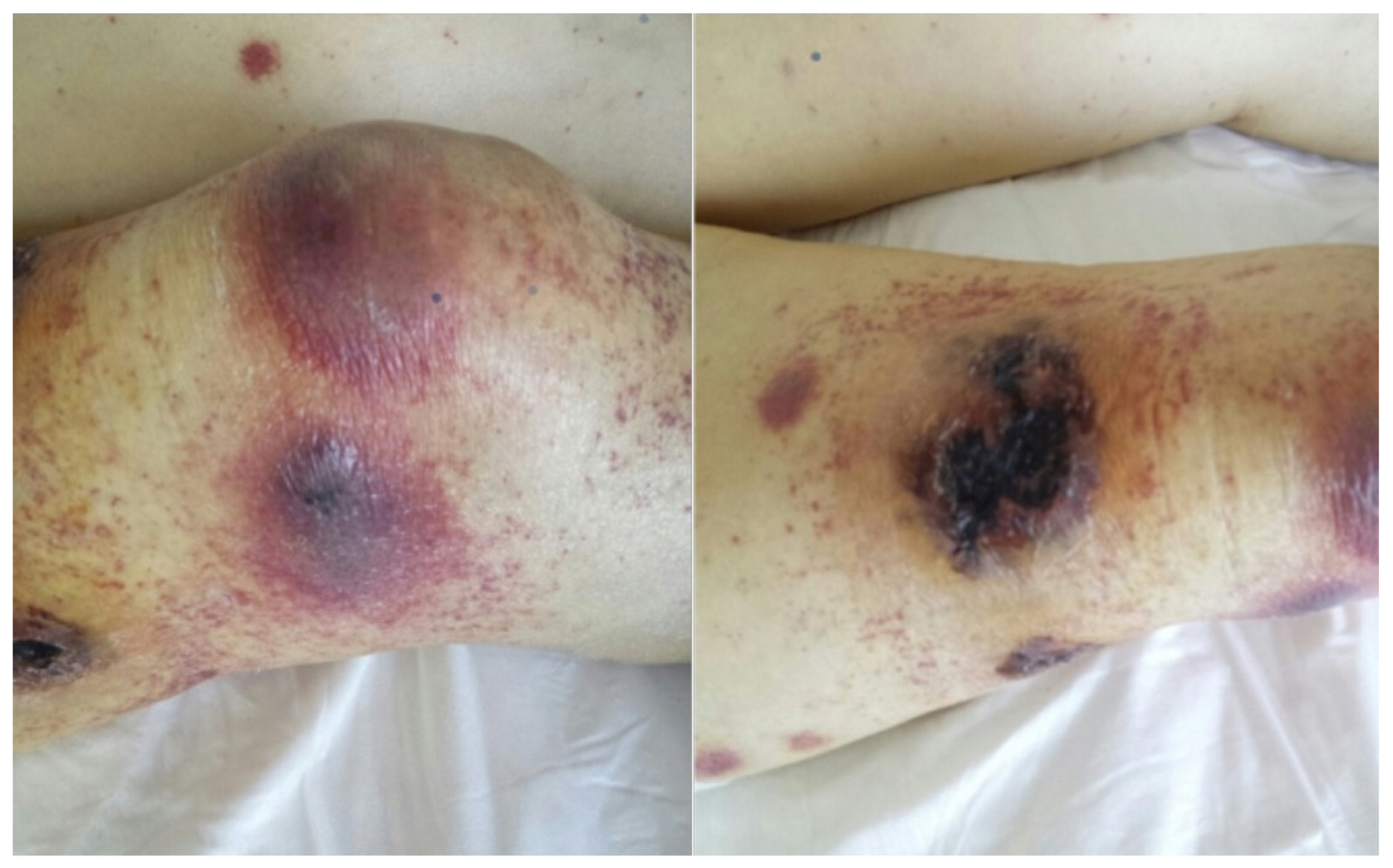
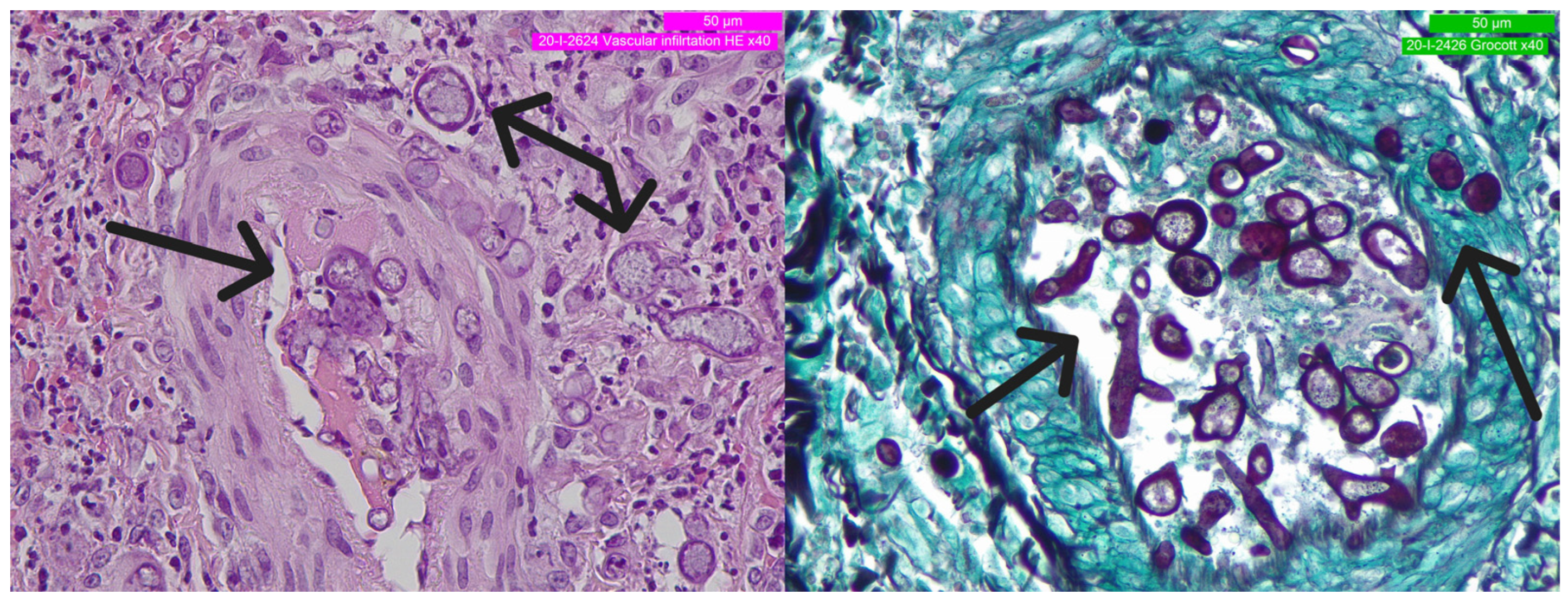
2. Detailed Case Description
3. Discussion
4. Conclusions
Author Contributions
Funding
Institutional Review Board Statement
Informed Consent Statement
Data Availability Statement
Conflicts of Interest
References
- Kipps, T.J.; Stevenson, F.K.; Wu, C.J.; Croce, C.M.; Packham, G.; Wierda, W.G.; O’Brien, S.; Gribben, J.; Rai, K. Chronic lymphocytic leukaemia. Nat. Rev. Dis. Prim. 2017, 3, 16096. [Google Scholar] [CrossRef] [PubMed]
- Forconi, F.; Moss, P. Perturbation of the normal immune system in patients with CLL. Blood 2015, 126, 573–581. [Google Scholar] [CrossRef] [PubMed]
- Fayad, L.; Keating, M.J.; Reuben, J.M.; O’Brien, S.; Lee, B.-N.; Lerner, S.; Kurzrock, R. Interleukin-6 and interleukin-10 levels in chronic lymphocytic leukemia: Correlation with phenotypic characteristics and outcome. Blood 2001, 97, 256–263. [Google Scholar] [CrossRef]
- Ramsay, A.G.; Johnson, A.J.; Lee, A.M.; Gorgün, G.; Le Dieu, R.; Blum, W.; Byrd, J.C.; Gribben, J.G. Chronic lymphocytic leukemia T cells show impaired immunological synapse formation that can be reversed with an immunomodulating drug. J. Clin. Investig. 2008, 118, 2427–2437. [Google Scholar] [CrossRef] [PubMed]
- Herishanu, Y.; Pérez-Galán, P.; Liu, D.; Biancotto, A.; Pittaluga, S.; Vire, B.; Gibellini, F.; Njuguna, N.; Lee, E.; Stennett, L.; et al. The lymph node microenvironment promotes B-cell receptor signaling, NF-κB activation, and tumor proliferation in chronic lymphocytic leukemia. Blood 2011, 117, 563–574. [Google Scholar] [CrossRef]
- Peters, F.S.; Strefford, J.C.; Eldering, E.; Kater, A. T-cell dysfunction in chronic lymphocytic leukemia from an epigenetic perspective. Haematologica 2021, 106, 1234–1243. [Google Scholar] [CrossRef]
- Visentin, A.; Molinari, M.C.; Pravato, S.; Cellini, A.; Angotzi, F.; Cavaretta, C.A.; Ruocco, V.; Imbergamo, S.; Piazza, F.; Proietti, G.; et al. A Retrospective Study on the Efficacy of Subcutaneous Immunoglobulin as Compared to Intravenous Formulation in Patients with Chronic Lymphocytic Leukemia and Secondary Antibody Deficiency. Curr. Oncol. 2023, 30, 274–283. [Google Scholar] [CrossRef]
- Dhalla, F.; Lucas, M.; Schuh, A.; Bhole, M.; Jain, R.; Patel, S.Y.; Misbah, S.; Chapel, H. Antibody Deficiency Secondary to Chronic Lymphocytic Leukemia: Should Patients be Treated with Prophylactic Replacement Immunoglobulin? J. Clin. Immunol. 2014, 34, 277–282. [Google Scholar] [CrossRef]
- Noto, A.; Cassin, R.; Mattiello, V.; Bortolotti, M.; Reda, G.; Barcellini, W. Should treatment of hypogammaglobulinemia with immunoglobulin replacement therapy (IgRT) become standard of care in patients with chronic lymphocytic leukemia? Front. Immunol. 2023, 14, 1062376. [Google Scholar] [CrossRef]
- Mohamed, A.J.; Yu, L.; Bäckesjö, C.-M.; Vargas, L.; Faryal, R.; Aints, A.; Christensson, B.; Berglöf, A.; Vihinen, M.; Nore, B.F.; et al. Bruton’s tyrosine kinase (Btk): Function, regulation, and transformation with special emphasis on the PH domain. Immunol. Rev. 2009, 228, 58–73. [Google Scholar] [CrossRef]
- Tasso, B.; Spallarossa, A.; Russo, E.; Brullo, C. The Development of BTK Inhibitors: A Five-Year Update. Molecules 2021, 26, 7411. [Google Scholar] [CrossRef] [PubMed]
- Maffei, R.; Maccaferri, M.; Arletti, L.; Fiorcari, S.; Benatti, S.; Potenza, L.; Luppi, M.; Marasca, R. Immunomodulatory effect of ibrutinib: Reducing the barrier against fungal infections. Blood Rev. 2020, 40, 100635. [Google Scholar] [CrossRef] [PubMed]
- Dubovsky, J.A.; Beckwith, K.A.; Natarajan, G.; Woyach, J.A.; Jaglowski, S.; Zhong, Y.; Hessler, J.D.; Liu, T.-M.; Chang, B.Y.; Larkin, K.M.; et al. Ibrutinib is an irreversible molecular inhibitor of ITK driving a Th1-selective pressure in T lymphocytes. Blood 2013, 122, 2539–2549. [Google Scholar] [CrossRef] [PubMed]
- Strijbis, K.; Tafesse, F.G.; Fairn, G.D.; Witte, M.D.; Dougan, S.K.; Watson, N.; Spooner, E.; Esteban, A.; Vyas, V.K.; Fink, G.R.; et al. Bruton’s Tyrosine Kinase (BTK) and Vav1 Contribute to Dectin1-Dependent Phagocytosis of Candida albicans in Macrophages. PLoS Pathog. 2013, 9, e1003446. [Google Scholar] [CrossRef]
- Varughese, T.; Taur, Y.; Cohen, N.; Palomba, M.L.; Seo, S.K.; Hohl, T.M.; Redelman-Sidi, G. Serious Infections in Patients Receiving Ibrutinib for Treatment of Lymphoid Cancer. Clin. Infect. Dis. 2018, 67, 687–692. [Google Scholar] [CrossRef]
- Pilmis, B.; Kherabi, Y.; Huriez, P.; Zahar, J.-R.; Mokart, D. Infectious Complications of Targeted Therapies for Solid Cancers or Leukemias/Lymphomas. Cancers 2023, 15, 1989. [Google Scholar] [CrossRef]
- Estupiñán, H.Y.; Berglöf, A.; Zain, R.; Smith, C.I.E. Comparative Analysis of BTK Inhibitors and Mechanisms Underlying Adverse Effects. Front. Cell Dev. Biol. 2021, 9, 630942. [Google Scholar] [CrossRef]
- Little, J.S.; Weiss, Z.F.; Hammond, S. Invasive Fungal Infections and Targeted Therapies in Hematological Malignancies. J. Fungi 2021, 7, 1058. [Google Scholar] [CrossRef]
- Ruiz-Camps, I.; Aguilar-Company, J. Risk of infection associated with targeted therapies for solid organ and hematological malignancies. Ther. Adv. Infect. Dis. 2021, 8, 204993612198954. [Google Scholar] [CrossRef]
- Walther, G.; Wagner, L.; Kurzai, O. Updates on the taxonomy of mucorales with an emphasis on clinically important taxa. J. Fungi 2019, 5, 106. [Google Scholar] [CrossRef]
- Kwon-Chung, K.J. Taxonomy of fungi causing mucormycosis and entomophthoramycosis (zygomycosis) and nomenclature of the disease: Molecular mycologic perspectives. Clin. Infect. Dis. 2012, 54 (Suppl. 1), S8–S15. [Google Scholar] [CrossRef]
- Spatafora, J.W.; Chang, Y.; Benny, G.L.; Lazarus, K.; Smith, M.E.; Berbee, M.L.; Bonito, G.; Corradi, N.; Grigoriev, I.; Gryganskyi, A.; et al. A phylum-level phylogenetic classification of zygomycete fungi based on genome-scale data. Mycologia 2016, 108, 1028–1046. [Google Scholar] [CrossRef] [PubMed]
- Wijayawardene, N.N.; Pawłowska, J.; Letcher, P.M.; Kirk, P.M.; Humber, R.A.; Schüßler, A.; Wrzosek, M.; Muszewska, A.; Okrasińska, A.; Istel, Ł.; et al. Notes for genera: Basal clades of Fungi (including Aphelidiomycota, Basidiobolomycota, Blastocladiomycota, Calcarisporiellomycota, Caulochytriomycota, Chytridiomycota, Entomophthoromycota, Glomeromycota, Kickxellomycota, Monoblepharomycota, Mortierellomyc. Fungal Divers. 2018, 92, 43–129. [Google Scholar] [CrossRef]
- Wagner, L.; de Hoog, S.; Alastruey-Izquierdo, A.; Voigt, K.; Kurzai, O.; Walther, G. A revised species concept for opportunistic Mucor species reveals species-specific antifungal susceptibility profiles. Antimicrob. Agents Chemother. 2019, 63, 1–8. [Google Scholar] [CrossRef] [PubMed]
- Jeong, W.; Keighley, C.; Wolfe, R.; Lee, W.L.; Slavin, M.A.; Kong, D.C.M.; Chen, S.C.-A. The epidemiology and clinical manifestations of mucormycosis: A systematic review and meta-analysis of case reports. Clin. Microbiol. Infect. 2019, 25, 26–34. [Google Scholar] [CrossRef]
- Nicolás, F.E.; Murcia, L.; Navarro, E.; Navarro-Mendoza, M.I.; Pérez-Arques, C.; Garre, V. Mucorales species and macrophages. J. Fungi 2020, 6, 94. [Google Scholar] [CrossRef]
- Petrikkos, G.; Skiada, A.; Lortholary, O.; Roilides, E.; Walsh, T.J.; Kontoyiannis, D. Epidemiology and Clinical Manifestations of Mucormycosis. Clin. Infect. Dis. 2012, 54 (Suppl. 1), S23–S34. [Google Scholar] [CrossRef]
- Hlaing, K.M.; Monday, L.M.; Nucci, M.; Nouér, S.A.; Revankar, S.G. Invasive Fungal Infections Associated with COVID-19. J. Fungi 2023, 9, 667. [Google Scholar] [CrossRef]
- Sharma, B.; Nonzom, S. Mucormycosis and Its Upsurge during COVID-19 Epidemic: An Updated Review. Curr. Microbiol. 2023, 80, 322. [Google Scholar] [CrossRef]
- Walther, G.; Wagner, L.; Kurzai, O. Outbreaks of Mucorales and the Species Involved. Mycopathologia 2020, 185, 765–781. [Google Scholar] [CrossRef]
- Roden, M.M.; Zaoutis, T.E.; Buchanan, W.L.; Knudsen, T.A.; Sarkisova, T.A.; Schaufele, R.L.; Sein, M.; Sein, T.; Chiou, C.C.; Chu, J.H.; et al. Epidemiology and outcome of zygomycosis: A review of 929 reported cases. Clin. Infect. Dis. 2005, 41, 634–653. [Google Scholar] [CrossRef] [PubMed]
- Chayakulkeeree, M.; Ghannoum, M.A.; Perfect, J.R. Zygomycosis: The re-emerging fungal infection. Eur. J. Clin. Microbiol. Infect. Dis. 2006, 25, 215–229. [Google Scholar] [CrossRef] [PubMed]
- Chakrabarti, A.; Chatterjee, S.S.; Das, A.; Panda, N.; Shivaprakash, M.R.; Kaur, A.; Varma, S.C.; Singhi, S.; Bhansali, A.; Sakhuja, V. Invasive zygomycosis in India: Experience in a tertiary care hospital. Postgrad. Med. J. 2009, 85, 573–581. [Google Scholar] [CrossRef] [PubMed]
- Roilides, E.; Zaoutis, T.E.; Katragkou, A.; Benjamin, D.K.; Walsh, T.J. Zygomycosis in neonates: An uncommon but life-threatening infection. Am. J. Perinatol. 2009, 26, 565–573. [Google Scholar] [CrossRef]
- Meis, J.F.; Chakrabarti, A. Changing epidemiology of an emerging infection: Zygomycosis. Clin. Microbiol. Infect. 2009, 15 (Suppl. 5), 10–14. [Google Scholar] [CrossRef]
- Lecointe, K.; Cornu, M.; Leroy, J.; Coulon, P.; Sendid, B. Polysaccharides cell wall architecture of mucorales. Front. Microbiol. 2019, 10, 469. [Google Scholar] [CrossRef]
- Ellis, M.; Al-Ramadi, B.; Finkelman, M.; Hedstrom, U.; Kristensen, J.; Ali-Zadeh, H.; Klingspor, L. Assessment of the clinical utility of serial β-d-glucan concentrations in patients with persistent neutropenic fever. J. Med. Microbiol. 2008, 57, 287–295. [Google Scholar] [CrossRef]
- Odabasi, Z.; Mattiuzzi, G.; Estey, E.; Kantarjian, H.; Saeki, F.; Ridge, R.J.; Ketchum, P.A.; Finkelman, M.A.; Rex, J.H.; Ostrosky-Zeichner, L. -D-Glucan as a Diagnostic Adjunct for Invasive Fungal Infections: Validation, Cutoff Development, and Performance in Patients with Acute Myelogenous Leukemia and Myelodysplastic Syndrome. Clin. Infect. Dis. 2004, 39, 199–205. [Google Scholar] [CrossRef]
- Maertens, J.A.; Klont, R.; Masson, C.; Theunissen, K.; Meersseman, W.; Lagrou, K.; Heinen, C.; Crepin, B.; Eldere, J.V.; Tabouret, M.; et al. Optimization of the cutoff value for the Aspergillus double-sandwich enzyme immunoassay. Clin. Infect. Dis. 2007, 44, 1329–1336. [Google Scholar] [CrossRef]
- Miceli, M.H.; Kauffman, C.A. Aspergillus Galactomannan for Diagnosing Invasive Aspergillosis. JAMA 2017, 318, 1175–1176. [Google Scholar] [CrossRef]
- Millon, L.; Herbrecht, R.; Grenouillet, F.; Morio, F.; Alanio, A.; Letscher-Bru, V.; Cassaing, S.; Chouaki, T.; Kauffmann-Lacroix, C.; Poirier, P.; et al. Early diagnosis and monitoring of mucormycosis by detection of circulating DNA in serum: Retrospective analysis of 44 cases collected through the French Surveillance Network of Invasive Fungal Infections (RESSIF). Clin. Microbiol. Infect. 2016, 22, 810.e1–810.e8. [Google Scholar] [CrossRef]
- Bitar, D.; Van Cauteren, D.; Lanternier, F.; Dannaoui, E.; Che, D.; Dromer, F.; Desenclos, J.-C.; Lortholary, O. Increasing incidence of zygomycosis (mucormycosis), France 1997–2006. Emerg. Infect. Dis. 2009, 15, 1395–1401. [Google Scholar] [CrossRef]
- Alghamdi, A.; Lutynski, A.; Minden, M.; Rotstein, C. Successful Treatment of Gastrointestinal Mucormycosis in an Adult with Acute Leukemia: Case Report and Literature Review. Curr. Oncol. 2017, 24, 61–64. [Google Scholar] [CrossRef][Green Version]
- Prakash, H.; Chakrabarti, A. Global epidemiology of mucormycosis. J. Fungi 2019, 5, 26. [Google Scholar] [CrossRef]
- Skiada, A.; Pagano, L.; Groll, A.; Zimmerli, S.; Dupont, B.; Lagrou, K.; Lass-Florl, C.; Bouza, E.; Klimko, N.; Gaustad, P.; et al. Zygomycosis in Europe: Analysis of 230 cases accrued by the registry of the European Confederation of Medical Mycology (ECMM) Working Group on Zygomycosis between 2005 and 2007. Clin. Microbiol. Infect. 2011, 17, 1859–1867. [Google Scholar] [CrossRef]
- Tilak, R.; Raina, P.; Gupta, S.; Tilak, V.; Prakash, P.; Gulati, A. Cutaneous zygomycosis: A possible postoperative complication in immunocompetent individuals. Indian J. Dermatol. Venereol. Leprol. 2009, 75, 596–599. [Google Scholar] [CrossRef] [PubMed]
- Sridhara, S.R.; Paragache, G.; Panda, N.K.; Chakrabarti, A. Mucormycosis in immunocompetent individuals: An increasing trend. J. Otolaryngol. 2005, 34, 402–406. [Google Scholar] [CrossRef] [PubMed]
- Heimann, S.; Vehreschild, M.; Cornely, O.; Heinz, W.; Grüner, B.; Silling, G.; Kessel, J.; Seidel, D.; Vehreschild, J. Healthcare burden of probable and proven invasive mucormycosis: A multi-centre cost-of-illness analysis of patients treated in tertiary care hospitals between 2003 and 2016. J. Hosp. Infect. 2019, 101, 339–346. [Google Scholar] [CrossRef]
- Kontoyiannis, D.P.; Yang, H.; Song, J.; Kelkar, S.S.; Yang, X.; Azie, N.; Harrington, R.; Fan, A.; Lee, E.; Spalding, J.R. Prevalence, clinical and economic burden of mucormycosis-related hospitalizations in the United States: A retrospective study. BMC Infect. Dis. 2016, 16, 730. [Google Scholar] [CrossRef] [PubMed]
- Visentin, A.; Facco, M.; Gurrieri, C.; Pagnin, E.; Martini, V.; Imbergamo, S.; Frezzato, F.; Trimarco, V.; Severin, F.; Raggi, F.; et al. Prognostic and Predictive Effect of IGHV Mutational Status and Load in Chronic Lymphocytic Leukemia: Focus on FCR and BR Treatments. Clin. Lymphoma Myeloma Leuk. 2019, 19, 678–685.e4. [Google Scholar] [CrossRef]
- Visentin, A.; Bonaldi, L.; Rigolin, G.M.; Mauro, F.R.; Martines, A.; Frezzato, F.; Pravato, S.; Gargarella, L.R.; Bardi, M.A.; Cavallari, M.; et al. The complex karyotype landscape in chronic lymphocytic leukemia allows the refinement of the risk of Richter syndrome transformation. Haematologica 2021, 107, 868–876. [Google Scholar] [CrossRef]
- Chakrabarti, A. Cutaneous zygomycosis: Major concerns. Indian J. Med. Res. 2010, 131, 739–741. [Google Scholar]
- Walsh, T.J.; Gamaletsou, M.N.; McGinnis, M.R.; Hayden, R.T.; Kontoyiannis, D. Early clinical and laboratory diagnosis of invasive pulmonary, extrapulmonary, and disseminated mucormycosis (zygomycosis). Clin. Infect. Dis. 2012, 54 (Suppl. 1), 55–60. [Google Scholar] [CrossRef]
- Marchesini, G.; Nadali, G.; Facchinelli, D.; Candoni, A.; Cattaneo, C.; Laurenti, L.; Fanci, R.; Farina, F.; Lessi, F.; Visentin, A.; et al. Infections in patients with lymphoproliferative diseases treated with targeted agents: SEIFEM multicentric retrospective study. Br. J. Haematol. 2021, 193, 316–324. [Google Scholar] [CrossRef]
- Tisi, M.C.; Hohaus, S.; Cuccaro, A.; Innocenti, I.; De Carolis, E.; Za, T.; D’alò, F.; Laurenti, L.; Fianchi, L.; Sica, S.; et al. Invasive fungal infections in chronic lymphoproliferative disorders: A monocentric retrospective study. Haematologica 2017, 102, e108–e111. [Google Scholar] [CrossRef] [PubMed]
- Visentin, A.; Gurrieri, C.; Imbergamo, S.; Lessi, F.; Di Maggio, S.A.; Frezzato, F.; Adami, F.; Zambello, R.; Piazza, F.; Semenzato, G.; et al. Epidemiology and risk factors of invasive fungal infections in a large cohort of patients with chronic lymphocytic leukemia. Hematol. Oncol. 2017, 35, 925–928. [Google Scholar] [CrossRef]
- Visentin, A.; Mauro, F.R.; Cibien, F.; Vitale, C.; Reda, G.; Fresa, A.; Ciolli, S.; Pietrasanta, D.; Marchetti, M.; Murru, R.; et al. Continuous treatment with Ibrutinib in 100 untreated patients with TP 53 disrupted chronic lymphocytic leukemia: A real-life campus CLL study. Am. J. Hematol. 2022, 97, E95–E99. [Google Scholar] [CrossRef] [PubMed]
- Mauro, F.R.; Giannarelli, D.; Visentin, A.; Reda, G.; Sportoletti, P.; Frustaci, A.M.; Chiarenza, A.; Ciolli, S.; Vitale, C.; Laurenti, L.; et al. Prognostic Impact and Risk Factors of Infections in Patients with Chronic Lymphocytic Leukemia Treated with Ibrutinib. Cancers 2021, 13, 3240. [Google Scholar] [CrossRef] [PubMed]
- Fiorcari, S.; Maffei, R.; Vallerini, D.; Scarfò, L.; Barozzi, P.; Maccaferri, M.; Potenza, L.; Ghia, P.; Luppi, M.; Marasca, R. BTK Inhibition Impairs the Innate Response against Fungal Infection in Patients with Chronic Lymphocytic Leukemia. Front. Immunol. 2020, 11, 2158. [Google Scholar] [CrossRef]
- Krunic, A.L.; Medenica, M.; Busbey, S. Solitary embolic cutaneous aspergillosis in the immunocompromised patient with acute myelogenous leukemia—A propos another case caused by Aspergillus flavus. Int. J. Dermatol. 2003, 42, 946–950. [Google Scholar] [CrossRef]
- Skiada, A.; Pavleas, I.; Drogari-Apiranthitou, M. Epidemiology and Diagnosis of Mucormycosis: An Update. J. Fungi 2020, 6, 265. [Google Scholar] [CrossRef] [PubMed]
- Burnham-Marusich, A.R.; Hubbard, B.; Kvam, A.J.; Gates-Hollingsworth, M.; Green, H.R.; Soukup, E.; Limper, A.H.; Kozel, T.R. Conservation of Mannan Synthesis in Fungi of the Zygomycota and Ascomycota Reveals a Broad Diagnostic Target. mSphere 2018, 3, e00094-18. [Google Scholar] [CrossRef] [PubMed]
- Schwarz, P.; Cornely, O.A.; Dannaoui, E. Antifungal combinations in Mucorales: A microbiological perspective. Mycoses 2019, 62, 746–760. [Google Scholar] [CrossRef] [PubMed]
- Dannaoui, E. Antifungal resistance in mucorales. Int. J. Antimicrob. Agents 2017, 50, 617–621. [Google Scholar] [CrossRef] [PubMed]
- Spellberg, B.; Fu, Y.; Edwards, J.E.; Ibrahim, A.S. Combination therapy with amphotericin B lipid complex and caspofungin acetate of disseminated zygomycosis in diabetic ketoacidotic mice. Antimicrob. Agents Chemother. 2005, 49, 830–832. [Google Scholar] [CrossRef] [PubMed]
- Reed, C.; Bryant, R.; Ibrahim, A.S.; Edwards, J.J.; Filler, S.G.; Goldberg, R.; Spellberg, B. Combination polyene-caspofungin treatment of rhino-orbital-cerebral mucormycosis. Clin. Infect. Dis. 2008, 47, 364–371. [Google Scholar] [CrossRef]
- Kazak, E.; Aslan, E.; Akalın, H.; Saraydaroğlu, Ö.; Hakyemez, B.; Erişen, L.; Yazıcı, B.; Gürcüoğlu, E.; Yılmaz, E.; Ener, B.; et al. A mucormycosis case treated with a combination of caspofungin and amphotericin B. J. Mycol. Med. 2013, 23, 179–184. [Google Scholar] [CrossRef] [PubMed]
- Gargouri, M.; Marrakchi, C.; Feki, W.; Charfi, S.; Maaloul, I.; Lahiani, D.; Elleuch, E.; Koubaa, M.; Mnif, Z.; Ayadi, A.; et al. Combination of amphotericin B and caspofungin in the treatment of mucormycosis. Med. Mycol. Case Rep. 2019, 26, 32–37. [Google Scholar] [CrossRef]
- Kyvernitakis, A.; Torres, H.A.; Jiang, Y.; Chamilos, G.; Lewis, R.E.; Kontoyiannis, D. Initial use of combination treatment does not impact survival of 106 patients with haematologic malignancies and mucormycosis: A propensity score analysis. Clin. Microbiol. Infect. 2016, 22, 811.e1–811.e8. [Google Scholar] [CrossRef]
- Cornely, O.A.; Alastruey-Izquierdo, A.; Arenz, D.; Chen, S.C.A.; Dannaoui, E.; Hochhegger, B.; Hoenigl, M.; Jensen, H.E.; Lagrou, K.; Lewis, R.E.; et al. Global guideline for the diagnosis and management of mucormycosis: An initiative of the European Confederation of Medical Mycology in cooperation with the Mycoses Study Group Education and Research Consortium. Lancet Infect. Dis. 2019, 19, e405–e421. [Google Scholar] [CrossRef] [PubMed]
- Vallejo, C.; Jarque, I.; Fortun, J.; Casado, A.; Peman, J. IFISTRATEGY: Spanish National Survey of Invasive Fungal Infection in Hemato-Oncologic Patients. J. Fungi 2023, 9, 628. [Google Scholar] [CrossRef] [PubMed]
- Lindsay, J.; Teh, B.W.; Micklethwaite, K.; Slavin, M. Azole antifungals and new targeted therapies for hematological malignancy. Curr. Opin. Infect. Dis. 2019, 32, 538–545. [Google Scholar] [CrossRef] [PubMed]


| Non-Immunological Risk Factors | Immunological Risk Factors | Special and Novel Risk Factors |
|---|---|---|
| Decompensated diabetes mellitus and ketoacidosis [31,32,33] | Immunodepression Primitive: solid and/or hematologic malignancies; autoimmunity | Premature neonates [34] |
| Iron overload | ||
| Major trauma | ||
| Prolonged use of corticosteroids | ||
| Intravenous drug abuse [27] | ||
| Iatrogenic/secondary: hematopoietic stem cell (HSCT); solid organ transplant | Preventive or therapeutic antimycotic drugs (voriconazole, itraconazole, or caspofungin) [35] | |
| BTK inhibitor [12,15,16,17,18,19] | ||
| SARS-CoV-2 infecion and treatment [28,29] |
| 2006 | FCR protocol (Fludarabine-Cyclophosphamide-Rituximab) |
| 2010 | FCR |
| 2012 | Rituximab |
| 2013 | Bendamustine |
| 2014–2016 | Ibrutinib (discontinued due to infections) |
| 2017–2020 | Venetoclax |
| 2020 | Idelalisib-Rituximab |
| May 2020 | Zanubrutinib |
Disclaimer/Publisher’s Note: The statements, opinions and data contained in all publications are solely those of the individual author(s) and contributor(s) and not of MDPI and/or the editor(s). MDPI and/or the editor(s) disclaim responsibility for any injury to people or property resulting from any ideas, methods, instructions or products referred to in the content. |
© 2023 by the authors. Licensee MDPI, Basel, Switzerland. This article is an open access article distributed under the terms and conditions of the Creative Commons Attribution (CC BY) license (https://creativecommons.org/licenses/by/4.0/).
Share and Cite
Maggioni, G.; Fedrigo, M.; Visentin, A.; Carturan, E.; Ruocco, V.; Trentin, L.; Alaibac, M.; Angelini, A. Severe Fatal Mucormycosis in a Patient with Chronic Lymphocytic Leukaemia Treated with Zanubrutinib: A Case Report and Review of the Literature. Curr. Oncol. 2023, 30, 8255-8265. https://doi.org/10.3390/curroncol30090599
Maggioni G, Fedrigo M, Visentin A, Carturan E, Ruocco V, Trentin L, Alaibac M, Angelini A. Severe Fatal Mucormycosis in a Patient with Chronic Lymphocytic Leukaemia Treated with Zanubrutinib: A Case Report and Review of the Literature. Current Oncology. 2023; 30(9):8255-8265. https://doi.org/10.3390/curroncol30090599
Chicago/Turabian StyleMaggioni, Giuseppe, Marny Fedrigo, Andrea Visentin, Elisa Carturan, Valeria Ruocco, Livio Trentin, Mauro Alaibac, and Annalisa Angelini. 2023. "Severe Fatal Mucormycosis in a Patient with Chronic Lymphocytic Leukaemia Treated with Zanubrutinib: A Case Report and Review of the Literature" Current Oncology 30, no. 9: 8255-8265. https://doi.org/10.3390/curroncol30090599
APA StyleMaggioni, G., Fedrigo, M., Visentin, A., Carturan, E., Ruocco, V., Trentin, L., Alaibac, M., & Angelini, A. (2023). Severe Fatal Mucormycosis in a Patient with Chronic Lymphocytic Leukaemia Treated with Zanubrutinib: A Case Report and Review of the Literature. Current Oncology, 30(9), 8255-8265. https://doi.org/10.3390/curroncol30090599