Review

Potential Molecular Targeted Therapy for Unresectable Hepatocellular Carcinoma

by
Shashank Kumar
1,* and
Abhay Kumar Pandey
2,*
1
Molecular Signaling & Drug Discovery Laboratory, Department of Biochemistry, Central University of Punjab, Guddha, Bathinda 151401, Punjab, India
2
Department of Biochemistry, University of Allahabad, University Road, Prayagraj 211002, Uttar Pradesh, India
*
Authors to whom correspondence should be addressed.
Curr. Oncol. 2023, 30(2), 1363-1380; https://doi.org/10.3390/curroncol30020105
Submission received: 17 December 2022 / Revised: 16 January 2023 / Accepted: 16 January 2023 / Published: 18 January 2023
(This article belongs to the Special Issue Hepatocellular Carcinoma: Epidemiology, Pathogenesis and Treatment)

Abstract

:
Hepatocellular carcinoma (HCC) is one of the most prevalent and lethal cancers, representing a serious worldwide health concern. The recurrence incidence of hepatocellular carcinoma (HCC) following surgery or ablation is as high as 70%. Thus, the clinical applicability of standard surgery and other locoregional therapy to improve the outcomes of advanced HCC is restricted and far from ideal. The registered trials did not identify a treatment that prolonged recurrence-free survival, the primary outcome of the majority of research. Several investigator-initiated trials have demonstrated that various treatments extend patients’ recurrence-free or overall survival after curative therapies. In the past decade, targeted therapy has made significant strides in the treatment of advanced HCC. These targeted medicines produce antitumour effects via specific signals, such as anti-angiogenesis or advancement of the cell cycle. As a typical systemic treatment option, it significantly improves the prognosis of this fatal disease. In addition, the combination of targeted therapy with an immune checkpoint inhibitor is redefining the paradigm of advanced HCC treatment. In this review, we focused on the role of approved targeted medicines and potential therapeutic targets in unresectable HCC.

1. Introduction

The Global Burden of Disease places liver cancer as the second leading cause years lost due to ill health. In the past 40 years, the incidence of liver cancer has risen drastically with increased mortality. Hepatocellular carcinoma (HCC) is the most common liver cancer subtype, produced from liver cells or intrahepatic biliary epithelial cells [1,2]. About 90% of initial liver tumours are HCC. One of the main curative methods for HCC is liver resection. After surgical resection, the annual recurrence rate of HCC is 10% and increases to 70–80% after 5 years [3]. Identification of patients with a high risk of HCC recurrence following curative surgical resection is crucial from a therapeutic standpoint. The five-year survival rate following curative resection is commonly stated to be between 40 and 70 percent. Up to 50% of individuals with resectable HCC who first have surgery later experience another recurrence [4]. The high risk of recurrence has been ascribed to the lack of efficient adjuvant therapy as well as the presence of occult micrometastasis from the primary tumour at the time of surgery. Tumour size, tumour count, and portal vein invasion are additional factors that are linked to early recurrence following resection. However, a pathological assessment for an early HCC recurrence has not been provided [4,5]. In this review, we look at current developments in targeted therapy to combat HCC recurrence and the underlying mechanism. We also give an outline of the key areas of HCC research, such as cutting-edge clinical trials that promise to enable significant advancement over the course of the following ten years. Various target-specific drugs and their mechanisms of action are described in the subsequent section of the review.

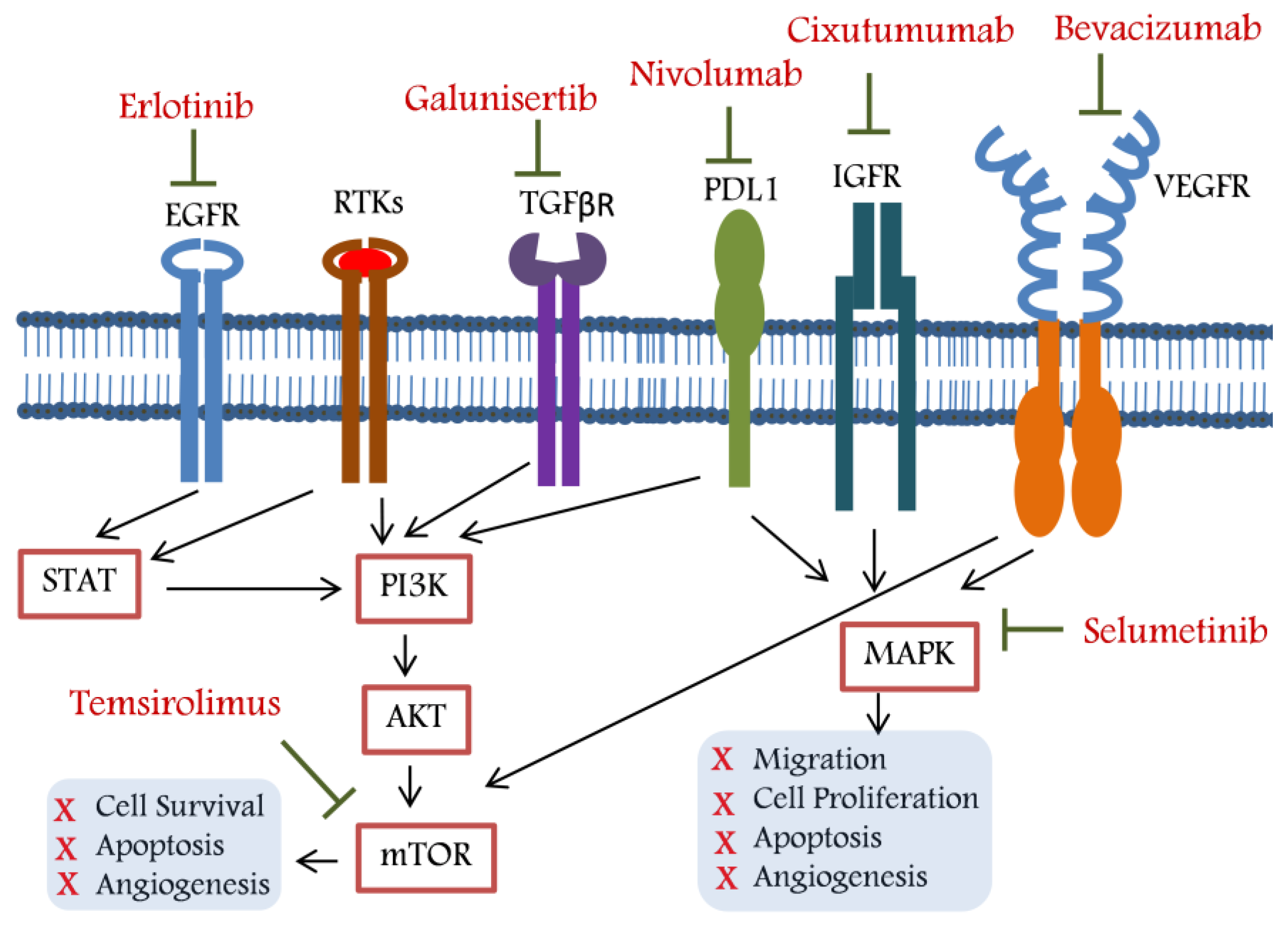
2. Targeted Therapy for Unresectable HCC

2.1. Belinostat: A Histone Deacetylase Inhibitor (HDACi)

Histone deacetylase (HDAC) enzymes are mostly zinc-dependent proteins that compress chromatin and restrict the transcription of related genes by catalysing deacetylation of histone proteins. Increased HDAC expression is associated with cancer initiation/progression and thus, it is an appropriate therapeutic target [6]. HDAC inhibitors (HDACi) are now acknowledged as a distinct and powerful class of anticancer drugs. In HCC cell lines and xenografts, it has been shown that inhibitors of HDAC cause apoptosis and tumour regression in addition to having anti-proliferative, anti-metastatic, and anti-invasive properties [7]. One member of the family, belinostat, has undergone testing for the treatment of hepatocellular carcinoma both alone and in combination with other chemotherapies and biological agents [8]. PXD101 (belinostat), an inhibitor of histone deacetylase, inhibits the development of hepatocarcinoma cells in a dose-dependent manner and causes histone acetylation. PXD101 therapy induces apoptosis without having a large impact on viral gene expression. Twelve cellular genes with tumour suppressor roles experienced varied changes in expression after being exposed to PXD101 for up to 48 h [9]. Phase I and phase II trials consisted of 19 and 36 patients, respectively. In phase II trial, the median progression-free survival (PFS) and OS were 2.83 and 8.89 months, respectively. The study concluded that further in-depth studies are required, as the targeted PFS endpoint was not achieved [10]. Wang et al. (2013) studied pharmacokinetics of belinostat in HCC patients to determine its associated metabolic mode of action. The study identified UGT1A1-mediated glucuronidation as a major mode of metabolic pathway alteration in HCC patients. The clinical significance of this finding remains to be determined [11].

2.2. Bevacizumab: A Vascular Endothelial Growth Factor (VEGF) Inhibitor

The vast majority of HCC cases are diagnosed at an advanced stage, despite the fact that many patients with the condition present at a younger age. With surgery or local ablation, patients who present with early- and intermediate-stage illness may be cured. The interaction between tumour cells and the elements of the tumour microenvironment is evidently suggested to be a significant contributor to the development of HCC. The establishment of an immunosuppressive environment is a crucial stage in the development of tumours, and as HCC is an inflammation-associated tumour, targeting the immune network may be a useful treatment strategy. Previous studies have shown that in unresectable HCC, the simultaneous blockage of vascular endothelial growth factor inhibitor (VEGF) and PD-L1 results in a longer progression-free survival [12]. Philip et al. (2012) studied the effect of erlotinib and bevacizumab as combination therapy in HCC patients in a two-stage phase II clinical trial. The combination therapy resulted in minimal activity with a median survival of 9.5 months. Further studies are required to understand the effects of combination therapy in molecularly categorised patients [13].
Metastatic recurrences after curative hepatic resections are common; thus, therapeutic interventions to produce antiangiogenesis effects have been attempted to aid HCC prognosis. VEGF, a dimeric glycoprotein and an important angiogenic factor, increases blood vessels’ permeability and is a potent mitogenic agent. Thus, by increasing angiogenesis and metastasis, VEGF is directly involved in HCC recurrence. Niu et al. (2000) showed higher serum levels of VEGF in the increasing order of normal, cirrhosis, and HCC patients [14]. In a different study, Guo et al. (2012) found significantly higher serum VEGF levels in HCC patients compared to normal individuals. Different studies correlated higher serum VEGF levels with poor overall survival in HCC patients, indicating VEGF’s prognostic value in the disease [15,16].
Bevacizumab, a humanised monoclonal antibody targeting VEGF-A, has shown anticancer efficacy in different types of cancers. The antibody inhibits the interaction between VEGF and its receptor molecule by directly binding to it, which in turn checks the growth and angiogenesis in tumour cells [17]. Bevacizumab showed better efficacy in HCC patients compared to available standard therapy and has been approved (2020) as an initial therapy for HCC patients with metastasis or those who cannot be treated with surgery [18].

2.3. Bortezomib: A Proteasome Inhibitor (PI)

Proteasome inhibitors (PI) have shown promising potential as chemotherapeutic agents in the treatment of HCC [19,20]. Dawson reviewed the mechanisms of different PIs in HCC [21]. As the first in class, bortezomib (BZB) is recommended for the treatment of mantle cell lymphoma and refractory multiple myeloma. It was given FDA approval in 2003 and is now the only PI with clinical approval [22]. The use of BZB alone or in combination with other treatments for HCC has drawn a lot of attention. The role of BZB in the management of non-surgical and metastatic HCC was expertly examined by Huang et al. (2018) as an alternative or a supplement to the current treatment paradigm [23]. Ciombor et al. (2014) studied the effects and tolerability of BZB and doxorubicin (DOX) in HCC patients in a phase II clinical trial [24]. The proteasome inhibition was correlated with the therapy response and survival in advanced HCC patients. The combination therapy showed suitable tolerance in the patients, but the primary endpoint (objective response rate) was not met. The study suggested the prognostic role of proteasome inhibition markers in advanced HCC patients [24].

2.4. Cixutumumab: An IGF-IR Signalling Inhibitor

The insulin-like growth factor (IGF) pathway includes a family of receptors and ligands. Since the IGF-1R (insulin-like growth factor-1 receptor) route regulates cellular motility, apoptosis, and proliferation, it has been suggested that signalling through this pathway contributes to a variety of cancers [25]. In HCC, IGF-IR expression and activity are both increased. The genesis and progression of HCC tumours are also hypothesised to be accelerated by elevated IGF expression [26]. HCC cell lines have experienced growth suppression and apoptosis as a result of pre-clinical investigations with a range of anti-IGF-1R techniques. In addition, studies indicate that the IGF-IR signalling cascade has an angiogenic component since it can trigger the release of VEGF [27]. As a result, anti-IGFR medication increases the efficacy of anti-VEGF therapy by further reducing tumour-associated VEGF release. Recombinant human IgG1 monoclonal antibody cixutumumab (formerly IMC-A12, ImClone Systems, Inc, Bridgewater, NJ, USA) is aimed at IGF-1R. IGF-1R is internalised and degraded by cixutumumab, which also potentially binds to it (Kd = 0.04 nM). Cixutumumab also inhibits the interaction between IGF and their respective receptors and thereby reduces cell viability/proliferation in HCC cells [28,29,30]. Fully human IgG1 monoclonal antibody cixutumumab binds to IGF-R1 with great specificity. The IFG-R1 tyrosine kinase domain is activated by IGF-1. The Akt survival route and the MAP kinase proliferation pathway are both activated by phosphotyrosine residues. In a human tumour xenograft model, cixutumumab efficiently prevented ligand-induced phosphorylation, which inhibits tumour cell proliferation and induces apoptosis. Through internalisation and degradation of the receptor, cixutumumab regulates the receptor localisation on tumour cell surface [31]. A phase II trial (NCT00639509) studied the efficacy of cixutumumab in advanced liver cancer. The study showed an 8-month median overall survival in patients treated with cixutumumab.

2.5. Doxorubicin: A Topoisomerase I and II Inhibitor

DNA topoisomerase I and II induce DNA breaks. Liu et al. (2003) showed significantly increased expression of TOP I in HCC tissue samples compared to adjacent normal tissue [32]. It has been shown that TOP2 induces cell invasion and EMT in HCC cells, which indicates its candidacy as a therapeutic target [33].
The stage of the disease determines the recommended course of HCC treatment. Transarterial chemoembolisation (TACE) is advised for HCC patients who are unresectable and at the intermediate stage. TACE entails delivering the cytostatic agent(s) to the tumour through the hepatic artery using a drug delivery system (DDS). The DDS causes embolisation, which impairs blood flow to the tumour, causes hypoxia, increases drug concentrations, and prolongs drug residence periods in the tumour target region. The predicted median survival is increased by about 4 months with the use of palliative TACE treatments [34]. Cytostatic drugs placed into drug-eluting beads or emulsified in Lipiodol® (LIP) are two popular DDSs utilised for TACE therapy for intermediate-stage HCC. Chemotherapeutic chemicals that have a positive charge can be placed into DCB (DC Bead®) for later local release [35]. The primary cytotoxic agent for intermediate-stage HCC is doxorubicin (DOX) used as its hydrochloric salt. Anthracycline, antibacterial, and antineoplastic medication DOX is used off-label to treat intermediate HCC despite having various cancer indications [36]. At least three anticancer mechanisms, including intercalation to DNA base pairs, free radical production, and reversible binding to topoisomerase (TOP) I and II, appear to be responsible for the pharmacological actions of DOX [36]. The amount of DOX needed to inhibit growth by 50% (IC50) in vitro depends on both the period and the cell type [37]. Gish et al. (2007) compared the efficacy of DOX and nolatrexed in advanced HCC patients in terms of overall survival in a phase III trial. Dox showed lesser toxicity and increased survival in HCC patients compared to nolatrexed-treated patients [38].

2.6. Erlotinib: An Epidermal Growth Factor Receptor (EGFR) Inhibitor

EGFR has attracted a lot of interest as a potential therapeutic target of tumour cells. As a key player in signal transduction pathways involved in different cancer hallmarks, EGFR is typically overexpressed in solid tumours. In some tumour types, its overexpression is associated with disease progression and a worse prognosis [39,40]. Erlotinib’s main therapeutic target, EGFR, possesses a transmembrane segment, a cytoplasmatic tyrosine kinase domain, and an extracellular cysteine-rich ligand-binding region, which serve as the binding sites for kinase inhibitors such as erlotinib. Through conformational changes and subsequent phosphorylation of tyrosine residues, extracellular ligand binding initiates events such as active receptor homo/hetero dimerisation and the formation of binding sites for subsequent signal transducers, initiating a series of events that lead to the development and growth of tumours [41]. In HCC, the EGFR is typically overexpressed. The increase in ligand–receptor contact rather than point mutations or amplifications might be the mechanism through which EGFR signalling in HCC gains function. Apoptosis and cell cycle-regulating genes express differently as a result of erlotinib treatment for HCC [42].
The tyrosine kinase inhibitor drug erlotinib inhibits epidermal growth factor receptor (EGFR) and is orally accessible. Erlotinib has the ability to prevent tumour cell growth, invasion, metastasis, and angiogenesis [43,44]. Thirty eight patients with metastatic or unresectable HCC participated in a phase II study to assess erlotinib’s effectiveness. The most common grade 3 to 4 toxicities were fatigue (8%) and diarrhoea (8%). Child–Pugh classification and toxicity severity (grade 3 or higher) were correlated; only 22% of Child–Pugh A patients and 70% of Child–Pugh B patients had severe toxicity (p = 0.02). After 24 weeks, 32% of the patients were no longer progressing. Only 9% of responses were confirmed in total [45]. Erlotinib alone appears to have relatively little effectiveness against HCC, and more randomised studies are required to assess the drug’s potential advantages for HCC patients [42]. Erlotinib has been tested in few clinical trials for the treatment of advanced HCC, but due to a lack of detailed analysis, the mode of erlotinib efficacy was not clear in advanced HCC. Thus, for a complete picture of the safety and effectiveness of erlotinib, Zhang et al. (2016) carried out a comprehensive literature review and concluded that erlotinib should be considered as a potential therapeutic option [46].

2.7. Galunisertib: A TGF-β Pathway Inhibitor

TGF-β is a multifunctional cytokine that regulates a variety of cellular processes in most cells, including proliferation, cellular differentiation, adhesion, migration, and apoptosis. TGF-β serves as a tumour suppressor in normal tissues, but as tumour cells develop strategies to circumvent its actions, it promotes the growth of the tumour. Although the transition from a tumour suppressor to an oncogenic state is not fully understood yet, intrinsic and extrinsic variables appear to be significant [46,47]. A mesenchymal phenotype, the loss of cell polarity, and the development of motile characteristics during epithelial–mesenchymal transitions (EMT) are thought to be crucial intrinsic changes in the tumour cells. Extrinsic variables from the tumour microenvironment, such as angiogenesis, inflammation, and fibroblast activation, also contribute to TGF-β signalling’s pro-tumourigenic potential. Modifications to the TGF-β signalling system can potentially contribute to tumour growth in addition to changes in the tumour tissue. Therefore, TGF-β plays a crucial role in the molecular aetiology of HCC; targeting TGF-β could offer new therapeutic insights [48,49]. Three of the several small-molecule inhibitors of TGF-RI/II/ALK5 that have been created are currently being tested in human clinical trials. The sole TGF-β pathway inhibitor undergoing clinical testing in HCC patients is galunisertib (LY2157299), a selective ATP mimetic inhibitor of TGF-RI/ALK5 (NCT01246986). Galunisertib has recently been demonstrated to effectively block the expression of p-Smad2 and invasion in three HCC models in vitro, but not proliferation [50].
TGF-β, a pro-inflammatory/profibrotic cytokine, is a complimentary signalling molecule to VEGF that aids in the advancement of various HCCs. A subgroup of HCCs has higher levels of TGF-β, which increases neovascularisation and encourages immune evasion and immunosuppression, as well as migration and invasion [51]. In fact, compared to HCCs not controlled by TGF-β, those showing indications of TGF-β activation also behave more aggressively and suggest a worse prognosis [48,52]. The oral TGF receptor I (ALK5) small molecule inhibitor galunisertib disrupts TGF signalling and harms HCC both in vitro and in vivo [49]. The treatment slows the growth of HCC, lessens tumour vascularity, lowers HCC motility and invasiveness, treats fibrosis, and boosts the local immune response. Galunisertib monotherapy has been shown in phase I and II studies to have a positive safety profile that does not overlap with that of antiangiogenic drugs and to provide a small amount of therapeutic benefit via cytostatic disease control in patients with advanced HCC [50,53]. A phase Ib/II study was designed (NCT02423343) to check the efficacy of galunisertib dose escalation and its safety and tolerability in HCC patients. The study was terminated in its early stage due to the low enrolment of HCC patients.

2.8. Nivolumab: A Programmed Cell Death Protein 1 (PD-1) Inhibitor

PD-1 (a coinhibitory receptor) and its ligand (PD-L1)-mediated pathway have been implicated in compromised tumour immunity. The use of anti-PD-L1 antibodies enhances tumour sensitivity and decreases tumour growth [54]. Wu et al. (2009) showed that the interaction of PD-1 with its ligand contributes to immune suppression in human HCC. The study suggested that PD-1 and PD-L1 interaction blockade carry important therapeutic implications in HCC [55]. Scheiner et al. (2019) studied the safety and efficacy of nivolumab in combination with other antibodies in a cohort of HCC patients. The study indicated the better efficacy and safety profile of nivolumab in HCC patients [56]. In a meta-analysis, the higher expression of membrane-bound and soluble PD-1 was positively correlated with shorter overall survival in HCC patients [57]. Large important clinical trials have effectively demonstrated the notion of inhibiting PD-1 activity in cancer patients.
Human immunoglobulin G4 (IgG4) monoclonal antibody nivolumab was created by Bristol-Myers Squibb (BMS) to bind to the PD-1 receptor. The clinical success of the antibody is due to its variable region’s high affinity (Kd 3.06 pM) and specificity (no binding to CD28, ICOS, or CTLA-4) in binding the target (interaction with the PD-1 N-loop). Nivolumab has high affinity and specificity for the PD-1 epitope that it targets [58,59]. A phase I/II clinical trial focused on HCC assessed the efficacy of nivolumab, a completely human immunoglobulin G4 monoclonal antibody to programmed cell death protein 1 (PD-1). Nivolumab showed a controllable safety profile throughout dosage escalation, including acceptable tolerability. Regardless of the aetiology, PD-L1 expression in the tumour, or previous sorafenib exposure, responses were seen. In patients with Child–Pugh B cirrhosis, nivolumab showed activity and tolerability with ORR and DCR of 10% and 55%, respectively. Nivolumab was given conditional FDA clearance based on this trial (NCT02576509) [60].
Nivolumab and pembrolizumab were evaluated for safety and efficacy in patients with advanced HCC who had previously failed to respond to sorafenib treatment in the CheckMate-040 and KEYNOTE-224 studies, which provided the foundation for the FDA’s accelerated approval of these drugs as second-line therapies [61,62]. Nivolumab’s safety and effectiveness in an Asian cohort were confirmed by a subanalysis of the data from CheckMate-040 [63]. An outstanding response to anti-PD-1 therapy has also been recorded in a case report, and ICIs have demonstrated potential efficacy and tolerability in advanced HCCs as systemic first-, second-, third-, and fourth-line treatments, with median overall survival (OS) and progression-free survival (PFS) of 11.0 and 4.6 months, respectively [56,64]. Although the phase III KEYNOTE-240 trial’s results for improved PFS and OS did not reach the pre-specified statistical significance, they were consistent with those of KEYNOTE-224 [65]. Pembrolizumab’s role in cases of advanced HCC with a viral background may be clarified by the KEYNOTE-394 study, which is now being conducted with Asian patients. Nivolumab did extend OS regardless of the PD-L1 expression profile in HCC patients and also increased the survival of HCC patients with HBV/HCV as the aetiology and prevented the reactivation of hepatitis. An anti-PD-1 inhibitor from China called camrelizumab (SHR-1210, Hengrui Pharmaceutical, Jiangsu Lianyungang, China) is being tested for the treatment of Hodgkin lymphoma and HCC. In a multi-centre, open-label, parallel-group, randomised, phase II trial (NCT02989922), it was discovered to have antitumour activity in Chinese patients with advanced HCC who had previously undergone treatment. This finding supports the efficacy of PD-1 therapy for Chinese patients with HBV-related HCC [66]. It is hoped that the outcomes of additional trials testing novel ICIs such as durvalumab, avelumab, tislelizumab, sintilimab, tremelimumab, ipilimumab, spartalizumab, and toripalimab will be encouraging and provide additional treatment options for HCC patients, especially those who have relapsed on first-line therapies. Dual ICI therapy and ICI combination therapy are two further strategies being used to improve the effectiveness of ICIs as therapeutic agents. CheckMate 9DW’s early outcomes for dual ICI treatment were astounding: the objective response rate was 32%, which was higher than monotherapy of any ICIs alone. Using the combination of the therapeutic agents (such as tivantinib and capmatinib), anti-PD1 and anti-PDL1 created an additive effect that inhibits the formation of HCCs in mice. MET-mediated phosphorylation results in a lower expression of PD-L1 [67]. For the treatment of advanced HCC, individual case studies have also shown encouraging outcomes in TKI and anti-PD1/PD-L1 agents combination therapy [68,69,70]. Early phase data that showed promise prompted the development of many immune checkpoint inhibitor-based combination treatment regimens, the outcomes of which will be made public in the coming years [71]. Several immune checkpoint inhibitor-based combination treatment approaches have been started as a result of encouraging early-phase data; the outcomes of these approaches will be known in the coming years. Additionally capable of expressing PD-L1, tumour cells can do so while eluding the immune system [72,73].

2.9. Peretinoin: A Cellular Retinoic Acid-Binding Protein

Despite effective treatment, HCC has a high risk of coming back. Patients with HCC brought on by HBV or HCV experience the same mechanism of recurrence. Additionally, although it might be challenging to pinpoint the mode of recurrence of certain lesions, the time of recurrence is thought to vary. For instance, 2 years after the initial tumour has had radical treatment, intrahepatic metastasis recurrence is more common than metachronous multicentric recurrence. The prognosis of patients with HCC be enhanced by actively reducing HCC recurrence in addition to early detection and therapy [74,75]. Kanemastu et al. (1988) studied the level of cellular retinol binding protein levels in HCC tumour samples compared to adjacent normal parenchymal tissue and found no significant difference between the two groups [76]. In a different study, Schmitt-Gräff et al. (2003) reported differential CRBP-1 levels in cancerous and normal liver cells. The study revealed that modulating the CRBP-1 expression in HCC might inhibit the growth and progression of tumours [77]. Lee et al. (2010) showed that targeting CRBP induces apoptosis in HCC cells [78]. Recently, Liu et al. (2021) found that CRBP-1 is a crucial player in the initiation and progression of HCC, which provides an independent prognostic biomarker and therapeutic target for the diagnosis and treatment of HCC [79,80].
Peretinoin is a synthetic polyprenoic acid with retinoid-like characteristics that binds to cellular retinoic acid-binding protein. Peretinoin’s method of action involves the activation of target genes’ transcription through the receptors (such as retinoic acid) and other transcriptional complexes. In HCC cells, peretinoin was discovered to alter the expression of genes that control cellular differentiation, cellular proliferation, and apoptosis. According to several pharmacologic studies, peretinoin prevents the growth of subclinical cancers and/or the carcinogenesis of precancerous lesions in the liver, thus preventing the recurrence of HCC [81,82,83].
Initially, peretinoin was used to treat skin disease. Later on, a clinical study in curative resected HCC patients on therapy showed increased survival rates and low recurrence of the disease. Based on these encouraging outcomes, peretinoin was created in 1997 under the trade name NIK 333 (Kowa Company, Ltd., Chuo-ku, Tokyo), and clinical trials on the substance began in February 2012 under the new designation K-333 [84,85]. A phase III trial study (NCT01640808) checked the efficacy of peretinoin (600 mg oral dose twice a day) in terms of recurrence-free survival in HCC patients as a primary measure. The disease-free survival and time to recurrence were studied as secondary outcomes in HCC patients. Increased recurrence-free survival was observed in the peretinoin-treated group compared to the placebo arm. Similarly increased disease-free survival and time to recurrence were also observed in the peretinoin-treated arm. Extensive pre-registration phase III placebo-controlled trials using peretinoin are now being conducted in Asia to assess its effectiveness and safety in patients with virus-associated HCC after total tumour excision (Table 1) in light of new information [86].

2.10. Selumetinib: A Mitogen-Activated Protein (MAP) Kinase Inhibitor

There is evidence that the RAF/MEK/ERK pathway is important in the development of HCC. It has been shown that in 50 to 100 percent of human HCCs, this route is activated [78,87,88,89]. This is mostly attributed to receptor tyrosine kinases, such as IGF-1, EGF, or c-MET, which are involved in autocrine/paracrine signalling. Furthermore, it was recently discovered that HCCs seem to express fewer RAS pathway inhibitors than normal tissues. Mixed results have been seen in studies of MEK/ERK inhibition in HCC during in vitro and in vivo experiments [90]. Klein et al. used a variety of techniques to block the MEK/ERK pathway, and they found that several HCC cell lines had decreased proliferation and increased apoptosis [91]. Selumetinib was used by Huynh et al. against HCC cell lines, and they once more showed that it was active in HCC experimental models. Selumetinib (AZD6244, ARRY-142886) is a competitive inhibitor of MEK1/2, and its recommended oral phase II dose has previously been determined to be 100 mg twice daily [92,93].
Selumetinib was evaluated as a first-line therapy in a recent phase II study for individuals with advanced HCC. Nineteen patients were enrolled in the first stage, with plans to recruit 25 more in the second stage if at least one objective response (the primary end point) had been seen. This means that a total of 44 patients could have participated in the research. The trial was terminated at the interim analysis because no radiographic response was seen in the study population. Three (27%) of the eleven patients with increased α-fetoprotein showed reductions of 50% or more, and the median time to progression (TTP) was 8 weeks. The level of toxicity was consistent with selumetinib toxicity studies in patients without cirrhosis. Selumetinib appeared to have a negligible single-agent effect, despite the pharmacokinetic analysis showing that target activation was suppressed [94]. A phase II trial study (NCT00604721) checked the efficacy of selumetinib in locally advanced or metastatic liver cancer in terms of objective response rate as the primary measure. The median progression-free and overall survival were studied as secondary outcomes in the patients. The study reported moderate liver toxicity with PFS and OS of 1.4 and 4.2 months, respectively.

2.11. Sofosbuvir: A Nucleotide Analogue

A non-structural protein 5B (NS5B) inhibitor, sofosbuvir (SOF) is also known as PSI-7977. It received Fast Track FDA approval in August 2010. Two molecules with a similar chemical makeup, PSI-7976 and PSI-7977, are combined in the medication. Both PSI-7976 and PSI-7977 molecules quickly transform into the same active triphosphate when the medication enters the liver cell. The first ribonucleoside analogue inhibitor, SOF, was authorised in 2013 for the treatment of chronic hepatitis C. The FDA approved the medicine in 2014 as a prodrug and nucleotide analogue [95,96]. Widely used DAAs for treating HCV, including SOF, specifically target the HCV RNA-dependent RNA polymerase (RdRp). Importantly, SOF-based regimens were used to treat the majority of reported cases of HCC development. In a Huh7-based subgenomic replicon, SOF effectively reduced HCV replication, as predicted. By using MTT and Ala blue tests, SOF therapy for 48 or 72 h had no discernible impact on the development of the majority of HCC cells. Surprisingly, however, SOF improved all four HCC cell lines’ single cell-based clonogenic potential. The much higher quantity and size of established colonies justify this aspect. Contrarily, placebo therapy has no such impact. Thus, it has been unequivocally shown that SOF has a direct stimulating influence on the beginning and expansion of clonogenic processes based on single HCC cells, but not on the expansion of the majority of HCC cells. The tumour microenvironment is a challenging and developing field, so neither these results nor the previous study should rule out the possibility of a secondary effect of DAAs on initiating the development of HCC. Therefore, future experimental and clinical research must work together to shed light on this worrying situation and provide a mechanistic understanding [97]. Atif et al. (2022) recently came to the same conclusion that SOF neither causes nor eradicates HCC based on the expression patterns of a few chosen genes in SOF-treated HCC cell lines [98].
HCV is a positive strand virus that contains a single stranded (ss) RNA genome, encoding a polyprotein that is processed into several structural and non-structural proteins. The NS5B possesses RdRp activity, catalyses HCV RNA replication, and is a prime target for antiviral drug discovery. Nucleoside/nucleotide analogs and non-nucleoside inhibitors are the two important groups of HCV RdRp inhibitors. An RdRp-activity-targeting-drug binds to its allosteric site and inhibits the imitation step by altering the structure–function relation of the protein [99]. HCV-induced HCC is a complex process which includes the development of a pro-carcinogenic environment (cirrhosis) and promotes malignancy in the presence of viral proteins. This gradual process can take up to four decades to develop, during which cell cycle-related proteins become mutated and ultimately convert normal hepatocytes into malignant types [100].
HCC development is known to be correlated with chronic hepatitis C virus (HCV) infection. Although the number of new HCV cases has decreased, people with HCC are more likely to have HCV infection. Direct-acting antiviral (DAA) therapies have revolutionised the management of HCV infection. Compensated DAA agents achieve sustained virologic response (SVR) in the liver function, and reduced need for liver transplantation was also achieved [101]. Mixed responses have been reported concerning the outcome of DAA therapy. Contrary to expectations, some recent studies found an unexpectedly high rate of HCC formation following DAA treatment, while other research found no such risk. Due to the diverse demographics and methodology used in many studies, it is still difficult to draw a firm conclusion on this matter.

2.12. Temsirolimus: An mTOR Pathway Inhibitor

The significance of the mammalian or mechanistic target of rapamycin (mTOR) signalling pathway in controlling liver cancer cell growth was revealed by Li et al. (2013) [102]. The study team demonstrated that temsirolimus suppresses the activity of mTOR and its downstream components, which has inhibitory effects on Bel-7402 liver cancer cells. They proposed that mTOR inhibition might be used as a treatment for liver cancer [102]. Among the several molecular pathways involved in HCC development, mTOR is one of the most investigated. A conserved checkpoint serine/threonine kinase called mTOR takes part in a number of molecular signalling pathways both inside and outside of cells. The downstream effects of mTOR activation include the response to cellular hypoxia and control over intracellular energy storage, growth, and replication. Since mTOR is associated with cellular growth and replication, inhibiting mTOR may be crucial in preventing the development of cancer. The rapamycin analogues known as mTOR inhibitors are able to deactivate mTOR by attaching to its C-terminal kinase domain. The three inhibitors that are most frequently used in clinical settings are sirolimus, everolimus, and temsirolimus. Since mTOR inhibitors are also immunosuppressive drugs, their use in liver transplant patients who have previously had HCC have significant therapeutic implications for preventing tumour recurrence. Because of the impaired liver function, standard chemotherapy drugs are ineffective against HCC and poorly tolerated [103,104,105]. Everolimus (RAD001), temsirolimus (CCI-779), and deforolimus are three examples of rapalogs whose bioavailability has been increased due to the early success of rapamycin (AP23573). The primary focus switched to anti-cancer therapy as a result of the significant function of mTOR in cell growth and metabolism, and temsirolimus (CCI-779) was licenced for the treatment of renal cell carcinoma in 2007. The first-line therapy portion of its trial in advanced HCC patients (NCT01079767) was stopped due to its toxic effects [106]. Knox et al. (2014) studied the efficacy of bevacizumab and temsirolimus combination therapy in advanced HCC patients. The study demonstrated an increased overall response rate (ORR) with PFS and OS [107].

3. Combination Targeted Therapy—A Way out to HCC Recurrence?

The United States Food and Drug Administration (FDA) approved the multi-kinase inhibitor sorafenib as a systemic treatment for HCC on the basis of a phase III trial that showed non-surgical candidates with intact liver function with better overall survival over placebo [108,109]. SHARP and Asia-Pacific trial results supported the approval of sorafenib as the first-line targeted treatment for advanced HCC. Additionally, sorafenib exhibits anti-tumour activity in cases of recurring malignancies after liver transplantation, providing a survival advantage over the best supportive treatment. In clinical practice, sorafenib was consistently safe for both Child–Pugh A and B patients, although side effects such diarrhoea and hand–foot syndrome were linked to longer overall survival (OS) [110]. The increase in survival remains moderate, and long-term sorafenib therapy causes acquired resistance to develop quickly, limiting future benefits. Furthermore, research from several nations has shown that sorafenib is not cost-effective at present pricing, which has attracted a lot of attention and criticism. This emphasises how critical it is to treat HCC in non-surgical candidates using a unique systemic strategy [111].
Although prior initiatives, including the use of anti-viral medicines, have generally failed, approaches to adjuvant therapy have been thoroughly researched due to the high recurrence rates following hepatectomy for HCC [112]. Antiangiogenic drugs; MEK/ERK, mTOR, HGF/c-Met, and EGF/EGFR pathway inhibitors; and histone deacetylase inhibitors have all been used with sorafenib. There have been phase III trials for other drugs, including interferon, selumetinib, capecitabine, tegafur-uracil, and gemcitabine and oxaliplatin (GEMOX), but none of these combinations containing sorafenib have been successful yet [113,114]. Improved postoperative survival was anticipated due to sorafenib’s potential in the adjuvant situation based on its successful palliative use. Unfortunately, this has not been proven, and in the STORM trial and other Western trials, it did not diminish postoperative tumour recurrence [115,116]. However, sorafenib dramatically lowers tumour recurrence and improves disease-free survival (DFS) in BCLC (Barcelona Clinic Liver Cancer Classification) stage C patients, and Zhuang et al. showed that adjuvant therapy improved both DFS and OS [116]. In contrast to intermediate HCC, sorafenib treatment after hepatectomy dramatically extended the OS of advanced HCC. Regardless of the BCLC stage, patients who underwent hepatectomy and were found to have microvascular invasion (MVI) upon a pathological examination benefited from adjuvant sorafenib therapy [112,117]. Following resection, sorafenib dramatically increased overall and recurrence-free survival (RFS), according to data from a sizable recent trial with propensity score matching analysis. Sorafenib’s effect in MVI patients after radical resection was being investigated in a phase III clinical trial (NCT02867280), but the study was terminated due to negative results obtained in the midterm analysis. A study (NCT02537158) with similar objectives is active, but the information on recruiting patients is not available [118,119].
Similar to early attempts to create first-line medicines that were superior to sorafenib, preliminary attempts to identify efficient second-line therapies were unsuccessful. The RESORCE trial (phase III) examined regorafenib efficacy in sorafenib-treated worsened patients. This study officially launched the second-line and sequential therapy era by proving the efficacy of second-line medicines. Regorafenib improved survival regardless of how quickly the disease had advanced during or following the last sorafenib dosage. Even in patients with HCC recurrence after liver transplantation, regorafenib was demonstrated to be successful for the sequential therapy of sorafenib followed by regorafenib. Not all patients who respond to sorafenib are, however, eligible for second-line treatment [120]. Only about 30% of patients in clinical practice are eligible for second-line regorafenib therapy [121]. The effectiveness and superior results of following treatment, including lenvatinib, were influenced by the liver’s functional reserve and ECOG (Eastern Cooperative Oncology Group) performance status during sorafenib therapy [122]. Despite these targeted medications discussed above, the search for novel agents has continued. Components of targeted therapy (targets and respective drug) for recurrent hepatocellular carcinoma are summarised in Figure 1.

4. Conclusions

In this review, we have discussed numerous possible therapeutic targets and associated drugs which have been studied in HCC clinical patients. We focused on the drugs studied for their effect on patient survival (disease-free and/or overall). Immune checkpoint inhibitors (ICIs), including pembrolizumab, nivolumab, durvalumab, and atezolizumab, have recently been studied in HCC patients, although clinical trials evaluating single-agent ICI have revealed unimpressive outcomes. Immune-based combinations, on the other hand, have proved more dramatic. In reality, a new standard of care for HCC patients with advanced disease has been established as a result of the phase III IMbrave150 trial, which compared sorafenib with atezolizumab–bevacizumab. The study revealed better therapeutic outcomes (in terms of survival and therapeutic response) in patients receiving the immune-based combination. Despite the fact that ICIs appear to have finally discovered their place in combinatorial treatments for HCC, there are still a number of unsolved problems. Since only a portion of HCC patients respond to immunotherapy, the lack of established biomarkers of response is one of the important problems. Based on these assumptions, it is crucial to comprehend the function of prospective biomarkers, such as the expression of the programmed death ligand 1 (PD-L1), tumour mutational burden (TMB), microsatellite instability (MSI) status, gut microbiota, and many more. Additionally, the medicines, patients, research methods, study phases, and clinical results used in clinical trials of HCC immunotherapy vary greatly.
None of the targeted therapies decreased the HCC recurrence up to the mark. Moreover, the review indicates that studying the role of targeted therapy alone or in combination is a leading field in HCC research, considering the clinical impact. Even though some of the aforementioned targets may appear promising, there are still some issues with studying this subject. For example, the recurrence rate increases with the advancement of HCC; thus, the targeted therapy should be categorised for the initial, intermediate, and advanced stages of the disease to achieve a better clinical outcome. The metastasis and angiogenesis-related targets may be targeted using appropriate drugs for patients with advanced HCC. The therapeutic drugs may be used in combination with toxicity-lowering agents, which could enhance the therapeutic potential and quality of life, and decrease recurrence in patients. Combinations of selumetinib and erlotinib; selumetinib and temsirolimus; erlotinib and galunisertib/nivolumab; and nivoluab and selumetinib/temsirolimus may be studied in HCC clinical trials, as these combination either target different molecular axes or strengthen the activities of their direct target inhibition in association with the downstream inhibitor partner (Figure 1). Slow-release formulations of the targeted therapeutic drug may be developed for initial and intermediate HCC, which may delay the recurrence process. In conclusion, more effective targeted therapy, either alone or in combination, is urgently needed to deal with the recurrence problem in HCC patients, which will help in the fight against this devastating disease.

Author Contributions

Conceptualisation, methodology, data curation, writing—original draft preparation, S.K.; writing—review and editing, S.K. and A.K.P. All authors have read and agreed to the published version of the manuscript.

Funding

This research received no external funding.

Acknowledgments

S.K. acknowledges the Department of Science and Technology, India, for providing a departmental grant to the Department of Biochemistry, Central University of Punjab, Bathinda, India, in the form of the DST-FIST grant. Abhay K. Pandey acknowledges DST-FIST and UGC-SAP facilities of the Department of Biochemistry, University of Allahabad, Prayagraj, India.

Conflicts of Interest

The authors declare no conflict of interest.

References

  1. Fitzmaurice, C.; Abate, D.; Abbasi, N.; Abbastabar, H.; Abd-Allah, F.; Abdel-Rahman, O.; Abdelalim, A.; Abdoli, A.; Abdollahpour, I.; Abdulle, A.S.M.; et al. Global, regional, and national cancer incidence, mortality, years of life lost, years lived with disability, and disability-adjusted life-years for 29 cancer groups, 1990 to 2017: A systematic analysis for the global burden of disease study. JAMA Oncol. 2019, 5, 1749–1768. [Google Scholar] [CrossRef] [PubMed] [Green Version]
  2. Key Statistics About Liver Cancer. Database: American Cancer Society. 2022. Available online: https://www.cancer.org/cancer/liver-cancer/about/what-is-key-statistics.html (accessed on 12 January 2022).
  3. Sherman, M. Recurrence of hepatocellular carcinoma. N. Engl. J. Med. 2008, 359, 2045–2047. [Google Scholar] [CrossRef] [PubMed]
  4. Saito, A.; Toyoda, H.; Kobayashi, M.; Koiwa, Y.; Fujii, H.; Fujita, K.; Maeda, A.; Kaneoka, Y.; Hazama, S.; Nagano, H.; et al. Prediction of early recurrence of hepatocellular carcinoma after resection using digital pathology images assessed by machine learning. Mod. Pathol. 2021, 34, 417–425. [Google Scholar] [CrossRef]
  5. Campanella, G.; Hanna, M.G.; Geneslaw, L.; Miraflor, A.; Silva, V.W.K.; Busam, K.J.; Brogi, E.; Reuter, V.E.; Klimstra, D.S.; Fuchs, T.J. Clinical-grade computational pathology using weakly supervised deep learning on whole slide images. Nat. Med. 2019, 25, 1301–1309. [Google Scholar] [CrossRef] [PubMed]
  6. Singh, A.K.; Bishayee, A.; Pandey, A.K. Targeting histone deacetylases with natural and synthetic agents: An emerging anticancer strategy. Nutrients 2018, 10, 731. [Google Scholar] [CrossRef] [Green Version]
  7. Yeo, W.; Chung, H.C.; Chan, S.L.; Wang, L.Z.; Lim, R.; Picus, J.; Boyer, M.; Mo, F.K.; Koh, J.; Rha, S.Y.; et al. Epigenetic therapy using belinostat for patients with unresectable hepatocellular carcinoma: A multicenter phase I/II study with biomarker and pharmacokinetic analysis of tumors from patients in the Mayo Phase II Consortium and the Cancer Therapeutics Research Group. J. Clin. Oncol. 2012, 30, 3361–3367. [Google Scholar] [CrossRef] [PubMed] [Green Version]
  8. Molife, L.R.; de Bono, J.S. Belinostat: Clinical applications in solid tumors and lymphoma. Expert Opin. Investig. Drugs 2011, 20, 1723–1732. [Google Scholar] [CrossRef]
  9. Ma, B.B.; Sung, F.; Tao, Q.; Poon, F.F.; Lui, V.W.; Yeo, W.; Chan, S.L.; Chan, A.T. The preclinical activity of the histone deacetylase inhibitor PXD101 (belinostat) in hepatocellular carcinoma cell lines. Investig. New Drugs 2010, 28, 107–114. [Google Scholar] [CrossRef]
  10. Yeo, W.; Chan, S.L.; Mo, F.K.; Chu, C.M.; Hui, J.W.; Tong, J.H.; Chan, A.W.; Koh, J.; Hui, E.P.; Loong, H.; et al. Phase I/II study of temsirolimus for patients with unresectable Hepatocellular Carcinoma (HCC)- a correlative study to explore potential biokers for response. BMC Cancer 2015, 15, 395. [Google Scholar] [CrossRef] [Green Version]
  11. Wang, L.-Z.; Ramírez, J.; Yeo, W.; Chan, M.-Y.M.; Thuya, W.-L.; Lau, J.-Y.A.; Wan, S.-C.; Wong, A.L.-A.; Zee, Y.-K.; Lim, R.; et al. Glucuronidation by UGT1A1 is the dominant pathway of the metabolic disposition of belinostat in liver cancer patients. PLoS ONE 2013, 8, e54522. [Google Scholar] [CrossRef]
  12. Liu, X.; Lu, Y.; Qin, S. Atezolizumab and bevacizumab for hepatocellular carcinoma: Mechanism, pharmacokinetics and future treatment strategies. Futur. Oncol. 2021, 17, 2243–2256. [Google Scholar] [CrossRef] [PubMed]
  13. Philip, P.A.; Mahoney, M.R.; Holen, K.D.; Northfelt, D.W.; Pitot, H.C.; Picus, J.; Flynn, P.J.; Erlichman, C. Phase 2 study of bevacizumab plus erlotinib in patients with advanced hepatocellular cancer. Cancer 2012, 118, 2424–2430. [Google Scholar] [CrossRef] [Green Version]
  14. Niu, Q.; Tang, Z.Y.; Ma, Z.C.; Qin, L.X.; Zhang, L.H. Serum vascular endothelial growth factor is a potential bioker of metastatic recurrence after curative resection of hepatocellular carcinoma. World J. Gastroenterol. 2000, 6, 565–568. [Google Scholar] [CrossRef] [PubMed]
  15. Guo, J.H.; Zhu, X.; Li, X.T.; Yang, R.J. Impact of serum vascular endothelial growth factor on prognosis in patients with unresectable hepatocellular carcinoma after transarterial chemoembolization. Chin. J. Cancer. Res. 2012, 24, 36–43. [Google Scholar] [CrossRef] [PubMed] [Green Version]
  16. Choi, S.B.; Han, H.J.; Kim, W.B.; Song, T.J.; Choi, S.Y. VEGF Overexpression Predicts Poor Survival in Hepatocellular Carcinoma. Open Med. (Wars) 2017, 12, 430–439. [Google Scholar] [CrossRef] [PubMed] [Green Version]
  17. Fang, P.; Hu, J.-H.; Cheng, Z.-G.; Liu, Z.-F.; Wang, J.-L.; Jiao, S.-C. Efficacy and safety of bevacizumab for the treatment of advanced hepatocellular carcinoma: A systematic review of phase II trials. PLoS ONE 2012, 7, e49717. [Google Scholar] [CrossRef] [Green Version]
  18. Atezolizumab Plus Bevacizumab Approved to Treat Liver Cancer. In: NCI Staff. Available online: https://www.cancer.gov/news-events/cancer-currents-blog/2020/fda-atezolizumab-bevacizumab-liver-cancer (accessed on 4 June 2020).
  19. Tanaka, K. The proteasome: Overview of structure and functions. Proc. Jpn. Acad. Ser. B Phys. Biol. Sci. 2009, 85, 12–36. [Google Scholar] [CrossRef] [PubMed] [Green Version]
  20. Ganten, T.M.; Koschny, R.; Haas, T.L.; Sykora, J.; Li-Weber, M.; Herzer, K.; Walczak, H. Proteasome inhibition sensitizes hepatocellular carcinoma cells, but not human hepatocytes, to TRAIL. Hepatology 2005, 42, 588–597. [Google Scholar] [CrossRef]
  21. Dawson, S.P. Hepatocellular carcinoma and the ubiquitin-proteasome system. Biochim. Biophys. Acta 2008, 1782, 775–784. [Google Scholar] [CrossRef] [Green Version]
  22. Chen, D.; Frezza, M.; Schmitt, S.; Kanwar, J.; Dou, Q.P. Bortezomib as the first proteasome inhibitor anticancer drug: Current status and future perspectives. Curr. Cancer. Drug Targets 2011, 11, 239–253. [Google Scholar] [CrossRef]
  23. Huang, I.-T.; Dhungel, B.; Shrestha, R.; Bridle, K.R.; Crawford, D.H.G.; Jayachandran, A.; Steel, J.C. Spotlight on Bortezomib: Potential in the treatment of hepatocellular carcinoma. Expert Opin. Investig. Drugs 2019, 28, 7–18. [Google Scholar] [CrossRef] [PubMed]
  24. Ciombor, K.K.; Feng, Y.; Benson, A.B.; Su, Y.; Horton, L.; Short, S.P.; Kauh, J.S.; Staley, C.; Mulcahy, M.; Powell, M.; et al. Phase II trial of bortezomib plus doxorubicin in hepatocellular carcinoma (E6202): A trial of the Eastern Cooperative Oncology Group. Investig. New Drugs 2014, 32, 1017–1027. [Google Scholar] [CrossRef] [PubMed] [Green Version]
  25. Pollak, M.N.; Schernhammer, E.S.; Hankinson, S.E. Insulin-like growth factors and neoplasia. Nat. Rev. Cancer 2004, 4, 505–518. [Google Scholar] [CrossRef]
  26. Sedlaczek, N.; Hasilik, A.; Neuhaus, P.; Schuppan, D.; Herbst, H. Focal overexpression of insulin-like growth factor 2 by hepatocytes and cholangiocytes in viral liver cirrhosis. Br. J. Cancer 2003, 88, 733–739. [Google Scholar] [CrossRef] [Green Version]
  27. Granata, R.; Trovato, L.; Lupia, E.; Sala, G.; Settanni, F.; Camussi, G.; Ghidoni, R.; Ghigo, E. Insulin-like growth factor binding protein-3 induces angiogenesis through IGF-I- and SphK1-dependent mechanisms. J. Thromb. Haemost. 2007, 5, 835–845. [Google Scholar] [CrossRef] [PubMed]
  28. Burtrum, D.; Zhu, Z.; Lu, D.; Anderson, D.M.; Prewett, M.; Pereira, D.S.; Bassi, R.; Abdullah, R.; Hooper, A.T.; Koo, H.; et al. A fully human monoclonal antibody to the insulin-like growth factor I receptor blocks ligand-dependent signaling and inhibits human tumor growth in vivo. Cancer Res. 2003, 63, 8912–8921. [Google Scholar]
  29. Tovar, V.; Alsinet, C.; Villanueva, A.; Hoshida, Y.; Chiang, D.Y.; Solé, M.; Thung, S.; Moyano, S.; Toffanin, S.; Mínguez, B.; et al. IGF activation in a molecular subclass of hepatocellular carcinoma and pre-clinical efficacy of IGF-1R blockage. J. Hepatol. 2010, 52, 550–559. [Google Scholar] [CrossRef] [Green Version]
  30. El-Khoueiry, A.B.; O’Donnell, R.; Semrad, T.J.; Mack, P.; Blanchard, S.; Bahary, N.; Jiang, Y.; Yen, Y.; Wright, J.; Chen, H.; et al. A phase I trial of escalating doses of cixutumumab (IMC-A12) and sorafenib in the treatment of advanced hepatocellular carcinoma. Cancer Chemother Pharmacol. 2018, 81, 957–963. [Google Scholar] [CrossRef]
  31. Abou-Alfa, G.K.; Capanu, M.; O’Reilly, E.M.; Ma, J.; Chou, J.F.; Gansukh, B.; Shia, J.; Kalin, M.; Katz, S.; Abad, L.; et al. A phase II study of cixutumumab (IMC-A12, NSC742460) in advanced hepatocellular carcinoma. J. Hepatol. 2014, 60, 319–324. [Google Scholar] [CrossRef] [Green Version]
  32. Liu, L.X.; Zhu, A.L.; Jiang, H.C.; Wang, X.Q.; Zhou, J.; Wu, M. Expression of DNA-dependent protein kinase and DNA topoisomerase I in human hepatocellular carcinoma and adjacent normal liver tissues. Chin. J. Dig. Dis. 2002, 3, 138–144. [Google Scholar] [CrossRef]
  33. Dong, Y.; Sun, X.; Zhang, K.; He, X.; Zhang, Q.; Song, H.; Xu, M.; Lu, H.; Ren, R. Type IIA topoisomerase (TOP2A) triggers epithelial-mesenchymal transition and facilitates HCC progression by regulating Snail expression. Bioengineered 2021, 12, 12967–12979. [Google Scholar] [CrossRef] [PubMed]
  34. European Association for the Study of the Liver; European Organisation for Research and Treatment of Cancer. EASL-EORTC clinical practice guidelines: Management of hepatocellular carcinoma. J. Hepatol. 2012, 56, 908–943, Erratum J. Hepatol. 2012, 56, 1430. [Google Scholar] [CrossRef] [Green Version]
  35. Lewis, A.L.; Holden, R.R. DC Bead embolic drug-eluting bead: Clinical application in the locoregional treatment of tumours. Expert Opin. Drug Deliv. 2011, 8, 153–169. [Google Scholar] [CrossRef] [PubMed]
  36. Tacar, O.; Sriamornsak, P.; Dass, C.R. Doxorubicin: An update on anticancer molecular action, toxicity and el drug delivery systems. J. Pharm. Pharmacol. 2013, 65, 157–170. [Google Scholar] [CrossRef] [PubMed]
  37. Dubbelboer, I.R.; Lilienberg, E.; Ahnfelt, E.; Sjögren, E.; Axén, N.; Lennernäs, H. Treatment of intermediate stage hepatocellular carcinoma: A review of intrahepatic doxorubicin drug-delivery systems. Ther. Deliv. 2014, 5, 447–466. [Google Scholar] [CrossRef] [PubMed]
  38. Gish, R.G.; Porta, C.; Lazar, L.; Ruff, P.; Feld, R.; Croitoru, A.; Feun, L.; Jeziorski, K.; Leighton, J.; Knox, J.; et al. Phase III randomized controlled trial comparing the survival of patients with unresectable hepatocellular carcinoma treated with nolatrexed or doxorubicin. J. Clin. Oncol. 2007, 25, 3069–3075. [Google Scholar] [CrossRef] [Green Version]
  39. Brabender, J.; Danenberg, K.D.; Metzger, R.; Schneider, P.M.; Park, J.; Salonga, D.; Hölscher, A.H.; Danenberg, P.V. Epidermal growth factor receptor and HER2-neu mRNA expression in non-small cell lung cancer Is correlated with survival. Clin. Cancer Res. 2001, 7, 1850–1855. [Google Scholar]
  40. Buckley, A.F.; Burgart, L.J.; Sahai, V.; Kakar, S. Epidermal growth factor receptor expression and gene copy number in conventional hepatocellular carcinoma. Am. J. Clin. Pathol. 2008, 129, 245–251. [Google Scholar] [CrossRef] [Green Version]
  41. Salomon, D.S.; Brandt, R.; Ciardiello, F.; Normanno, N. Epidermal growth factor-related peptides and their receptors in human malignancies. Crit. Rev. Oncol. Hematol. 1995, 19, 183–232. [Google Scholar] [CrossRef]
  42. Martens, U.M. Small Molecules in Oncology; Springer Berlin Heidelberg Pub: Berlin/Heidelberg, Germany, 2010; Volume 1, p. 238. [Google Scholar]
  43. Van den Eynde, M.; Baurain, J.F.; Mazzeo, F.; Machiels, J.P. Epidermal growth factor receptor targeted therapies for solid tumours. Acta Clin. Belg. 2011, 66, 10–17. [Google Scholar] [CrossRef]
  44. Hirte, H.W. Profile of erlotinib and its potential in the treatment of advanced ovarian carcinoma. Onco Targets Ther. 2013, 6, 427–435. [Google Scholar] [CrossRef] [PubMed] [Green Version]
  45. Philip, P.A.; Mahoney, M.R.; Allmer, C.; Thomas, J.; Pitot, H.C.; Kim, G.; Donehower, R.C.; Fitch, T.; Picus, J.; Erlichman, C. Phase II study of Erlotinib (OSI-774) in patients with advanced hepatocellular cancer. J. Clin. Oncol. 2005, 23, 6657–6663. [Google Scholar] [CrossRef] [PubMed]
  46. Zhang, J.; Zong, Y.; Xu, G.Z.; Xing, K. Erlotinib for advanced hepatocellular carcinoma. A systematic review of phase II/III clinical trials. Saudi. Med. J. 2016, 37, 1184–1190. [Google Scholar] [CrossRef] [PubMed]
  47. Cantelli, G.; Crosas-Molist, E.; Georgouli, M.; Sanz-Moreno, V. TGFΒ-induced transcription in cancer. Semin. Cancer Biol. 2017, 42, 60–69. [Google Scholar] [CrossRef] [PubMed] [Green Version]
  48. Nieto, M.A.; Huang, R.Y.; Jackson, R.A.; Thiery, J.P. EMT: 2016. Cell 2016, 166, 21–45. [Google Scholar] [CrossRef] [Green Version]
  49. Giannelli, G.; Mikulits, W.; Dooley, S.; Fabregat, I.; Moustakas, A.; Dijke, P.T.; Portincasa, P.; Winter, P.; Janssen, R.; Leporatti, S.; et al. The rationale for targeting TGF-β in chronic liver diseases. Eur. J. Clin. Investig. 2016, 46, 349–361. [Google Scholar] [CrossRef] [Green Version]
  50. Rani, B.; Malfettone, A.; Dituri, F.; Soukupova, J.; Lupo, L.; Mancarella, S.; Fabregat, I.; Giannelli, G. Galunisertib suppresses the staminal phenotype in hepatocellular carcinoma by modulating CD44 expression. Cell Death Dis. 2018, 9, 373. [Google Scholar] [CrossRef] [Green Version]
  51. Giannelli, G.; Koudelkova, P.; Dituri, F.; Mikulits, W. Role of epithelial to mesenchymal transition in hepatocellular carcinoma. J. Hepatol. 2016, 65, 798–808. [Google Scholar] [CrossRef] [Green Version]
  52. Sia, D.; Villanueva, A.; Friedman, S.L.; Llovet, J.M. Liver Cancer Cell of Origin, Molecular Class, and Effects on Patient Prognosis. Gastroenterology 2017, 152, 745–761. [Google Scholar] [CrossRef] [Green Version]
  53. Harding, J.J.; Do, R.K.; Yaqubie, A.; Cleverly, A.; Zhao, Y.; Gueorguieva, I.; Lahn, M.; Benhadji, K.A.; Kelley, R.K.; Abou-Alfa, G.K. Phase 1b study of galunisertib and ramucirumab in patients with advanced hepatocellular carcinoma. Cancer Med. 2021, 10, 3059–3067. [Google Scholar] [CrossRef]
  54. Shi, F.; Shi, M.; Zeng, Z.; Qi, R.-Z.; Liu, Z.-W.; Zhang, J.-Y.; Yang, Y.-P.; Tien, P.; Wang, F.-S. PD-1 and PD-L1 upregulation promotes CD8(+) T-cell apoptosis and postoperative recurrence in hepatocellular carcinoma patients. Int. J. Cancer 2011, 128, 887–896. [Google Scholar] [CrossRef] [PubMed]
  55. Wu, K.; Kryczek, I.; Chen, L.; Zou, W.; Welling, T.H. Kupffer cell suppression of CD8+ T cells in human hepatocellular carcinoma is mediated by B7-H1/programmed death-1 interactions. Cancer Res. 2009, 69, 8067–8075. [Google Scholar] [CrossRef] [PubMed]
  56. Scheiner, B.; Kirstein, M.M.; Hucke, F.; Finkelmeier, F.; Schulze, K.; Von Felden, J.; Koch, S.; Schwabl, P.; Hinrichs, J.B.; Waneck, F.; et al. Programmed cell death protein-1 (PD-1)-targeted immunotherapy in advanced hepatocellular carcinoma: Efficacy and safety data from an international multicentre real-world cohort. Aliment. Pharmacol. Ther. 2019, 49, 1323–1333. [Google Scholar] [CrossRef] [PubMed]
  57. Li, X.-S.; Li, J.-W.; Li, H.; Jiang, T. Prognostic value of programmed cell death ligand 1 (PD-L1) for hepatocellular carcinoma: A meta-analysis. Biosci. Rep. 2020, 40, BSR20200459. [Google Scholar] [CrossRef] [PubMed] [Green Version]
  58. Zhu, A.X.; Park, J.O.; Ryoo, B.Y.; Yen, C.J.; Poon, R.; Pastorelli, D.; Blanc, J.F.; Chung, H.C.; Baron, A.D.; Pfiffer, T.E.; et al. REACH Trial Investigators. Ramucirumab versus placebo as second-line treatment in patients with advanced hepatocellular carcinoma following first-line therapy with sorafenib (REACH): A randomised, double-blind, multicentre, phase 3 trial. Lancet Oncol. 2015, 16, 859–870. [Google Scholar] [CrossRef]
  59. Bruix, J.; Tak, W.-Y.; Gasbarrini, A.; Santoro, A.; Colombo, M.; Lim, H.-Y.; Mazzaferro, V.; Wiest, R.; Reig, M.; Wagner, A.; et al. Regorafenib as second-line therapy for intermediate or advanced hepatocellular carcinoma: Multicentre, open-label, phase II safety study. Eur. J. Cancer 2013, 49, 3412–3419. [Google Scholar] [CrossRef]
  60. Onuma, A.E.; Zhang, H.; Huang, H.; Williams, T.M.; Noonan, A.; Tsung, A. Immune Checkpoint Inhibitors in Hepatocellular Cancer: Current Understanding on Mechanisms of Resistance and Biokers of Response to Treatment. Gene. Expr. 2020, 20, 53–65. [Google Scholar] [CrossRef]
  61. El-Khoueiry, A.B.; Sangro, B.; Yau, T.; Crocenzi, T.S.; Kudo, M.; Hsu, C.; Kim, T.Y.; Choo, S.P.; Trojan, J.; Welling, T.H.; et al. Nivolumab in patients with advanced hepatocellular carcinoma (CheckMate 040): An open-label, non-comparative, phase 1/2 dose escalation and expansion trial. Lancet 2017, 389, 2492–2502. [Google Scholar] [CrossRef]
  62. Zhu, A.X.; Finn, R.S.; Edeline, J.; Cattan, S.; Ogasawara, S.; Palmer, D.; Verslype, C.; Zagonel, V.; Fartoux, L.; Vogel, A.; et al. Pembrolizumab in patients with advanced hepatocellular carcinoma previously treated with sorafenib (KEYNOTE-224): A non-randomised, open-label phase 2 trial. Lancet Oncol. 2018, 19, 940–952. [Google Scholar] [CrossRef]
  63. Yau, T.; Hsu, C.; Kim, T.-Y.; Choo, S.-P.; Kang, Y.-K.; Hou, M.-M.; Numata, K.; Yeo, W.; Chopra, A.; Ikeda, M.; et al. Nivolumab in advanced hepatocellular carcinoma: Sorafenibexperienced Asian cohort analysis. J. Hepatol. 2019, 71, 543–552. [Google Scholar] [CrossRef] [Green Version]
  64. Roderburg, C.; Berres, M.-L.; Wree, A.; Loosen, S.H.; Luedde, T.; Trautwein, C. Excellent response to anti-PD-1 therapy in a patient with hepatocellular carcinoma intolerant to sorafenib. Visc. Med. 2019, 35, 43–46. [Google Scholar] [CrossRef] [PubMed]
  65. Finn, R.S.; Ryoo, B.Y.; Merle, P.; Kudo, M.; Bouattour, M.; Lim, H.Y.; Breder, V.; Edeline, J.; Chao, Y.; Ogasawara, S.; et al. Pembrolizumab as second-line therapy in patients with advanced hepatocellular carcinoma in KEYNOTE-240: A randomized, doubleblind, phase iii trial. J. Clin. Oncol. 2020, 3, 193–202. [Google Scholar] [CrossRef] [PubMed]
  66. Qin, S.; Ren, Z.; Meng, Z.; Chen, Z.; Chai, X.; Xiong, J.; Bai, Y.; Yang, L.; Zhu, H.; Fang, W.; et al. Camrelizumab in patients with previously treated advanced hepatocellular carcinoma: A multicentre, open-label, parallel-group, randomised, phase 2 trial. Lancet Oncol. 2020, 21, 571–580. [Google Scholar] [CrossRef] [PubMed]
  67. Li, H.; Li, C.-W.; Li, X.; Ding, Q.; Guo, L.; Liu, S.; Liu, C.; Lai, C.-C.; Hsu, J.-M.; Dong, Q.; et al. MET inhibitors promote liver tumor evasion of the immune response by stabilizing PDL1. Gastroenterology 2019, 156, 1849–1861.e13. [Google Scholar] [CrossRef]
  68. Chen, X.; Zhang, Y.; Zhang, N.; Ge, Y.; Jia, W. Lenvatinib combined nivolumab injection followed by extended right hepatectomy is a feasible treatment for patients with massive hepatocellular carcinoma: A case report. Onco Targets Ther. 2019, 12, 7355–7359. [Google Scholar] [CrossRef] [Green Version]
  69. Liu, Z.; Li, X.; He, X.; Xu, Y.; Wang, X. Complete response to the combination of Lenvatinib and Pembrolizumab in an advanced hepatocellular carcinoma patient: A case report. BMC Cancer 2019, 19, 1062. [Google Scholar] [CrossRef]
  70. Joerger, M.; Guller, U.; Bastian, S.; Driessen, C.; von Moos, R. Prolonged tumor response associated with sequential immune checkpoint inhibitor combination treatment and regorafenib in a patient with advanced pretreated hepatocellular carcinoma. J. Gastrointest. Oncol. 2019, 10, 373–378. [Google Scholar] [CrossRef]
  71. Finkelmeier, F.; Waidmann, O.; Tro, J. Nivolumab for the treatment of hepatocellular carcinoma. Expert Rev. Anticancer Ther. 2018, 18, 1169–1175. [Google Scholar] [CrossRef]
  72. Akateh, C.; Black, S.M.; Conteh, L.; Miller, E.D.; Noonan, A.; Elliott, E.; Pawlik, T.M.; Tsung, A.; Cloyd, J.M. Neoadjuvant and adjuvant treatment strategies for hepatocellular carcinoma. World J. Gastroenterol. 2019, 25, 3704–3721. [Google Scholar] [CrossRef]
  73. Llovet, J.M.; Ricci, S.; Mazzaferro, V.; Hilgard, P.; Gane, E.; Blanc, J.F.; de Oliveira, A.C.; Santoro, A.; Raoul, J.L.; Forner, A.; et al. Sorafenib in advanced hepatocellular carcinoma. N. Engl. J. Med. 2008, 359, 378–390. [Google Scholar] [CrossRef] [Green Version]
  74. Nagaoki, Y.; Hyogo, H.; Aikata, H.; Tanaka, M.; Naeshiro, N.; Nakahara, T.; Honda, Y.; Miyaki, D.; Kawaoka, T.; Takaki, S.; et al. Recent trend of clinical features in patients with hepatocellular carcinoma. Hepatol. Res. 2012, 42, 368–375. [Google Scholar] [CrossRef] [PubMed]
  75. TTateishi, R.; Okanoue, T.; Fujiwara, N.; Okita, K.; Kiyosawa, K.; Omata, M.; Kumada, H.; Hayashi, N.; Koike, K. Clinical characteristics, treatment, and prognosis of non-B, non-C hepatocellular carcinoma: A large retrospective multicenter cohort study. J. Gastroenterol. 2015, 50, 350–360. [Google Scholar] [CrossRef] [PubMed]
  76. Kanematsu, T.; Kawano, T.; Takenaka, K.; Matsumata, T.; Sugimichi, K.; Kuwano, M. Levels of vitamin A and cellular retinol binding protein in human hepatocellular carcinoma and adjacent normal tissue. Nutr. Cancer 1989, 12, 311–319. [Google Scholar] [CrossRef] [PubMed]
  77. Schmitt-Gräff, A.; Ertelt, V.; Allgaier, H.P.; Koelble, K.; Olschewski, M.; Nitschke, R.; Bochaton-Piallat, M.L.; Gabbiani, G.; Blum, H.E. Cellular retinol-binding protein-1 in hepatocellular carcinoma correlates with beta-catenin, Ki-67 index, and patient survival. Hepatology 2003, 38, 470–480. [Google Scholar] [CrossRef] [PubMed]
  78. Lee, J.-H.; Yoon, J.-H.; Yu, S.J.; Chung, G.E.; Jung, E.U.; Kim, H.Y.; Kim, B.H.; Choi, D.H.; Myung, S.J.; Kim, Y.J.; et al. Retinoic acid and its binding protein modulate apoptotic signals in hypoxic hepatocellular carcinoma cells. Cancer Lett. 2010, 295, 229–235. [Google Scholar] [CrossRef] [PubMed]
  79. Liu, X.; Shan, W.; Li, T.; Gao, X.; Kong, F.; You, H.; Kong, D.; Qiao, S.; Tang, R. Cellular retinol binding protein-1 inhibits cancer stemness via upregulating WIF1 to suppress Wnt/β-catenin pathway in hepatocellular carcinoma. BMC Cancer 2021, 21, 1224. [Google Scholar] [CrossRef] [PubMed]
  80. Singh, A.K.; Kumar, R.; Pandey, A.K. Hepatocellular carcinoma: Causes, mechanism of progression and biomarkers. Curr. Chem. Genom. Transl. Med. 2018, 12, 9–26. [Google Scholar] [CrossRef]
  81. Miyake, T.; Abe, M.; Furukawa, S.; Tokumoto, Y.; Toshimitsu, K.; Ueda, T.; Yamamoto, S.; Hirooka, M.; Kumagi, T.; Hiasa, Y.; et al. Long-term branched-chain amino acid supplementation improves glucose tolerance in patients with nonalcoholic steatohepatitis-related cirrhosis. Intern. Med. 2012, 51, 2151–2155. [Google Scholar] [CrossRef] [Green Version]
  82. Ohno, T.; Tanaka, Y.; Sugauchi, F.; Orito, E.; Hasegawa, I.; Nukaya, H.; Kato, A.; Matunaga, S.; Endo, M.; Tanaka, Y.; et al. Suppressive effect of oral administration of branched-chain amino acid granules on oxidative stress and inflammation in HCV-positive patients with liver cirrhosis. Hepatol. Res. 2008, 38, 683–688. [Google Scholar] [CrossRef]
  83. ASPEN Board of Directors and the Clinical Guidelines Task Force. Guidelines for the use of parenteral and enteral nutrition in adult and pediatric patients. JPEN J. Parenter Enter. Nutr. 2002, 26 (Suppl. 1), 1SA–138SA. [Google Scholar] [CrossRef] [Green Version]
  84. Plauth, M.; Merli, M.; Kondrup, J.; Weimann, A.; Ferenci, P.; Müller, M. ESPEN guidelines for nutrition in liver disease and transplantation. Clin. Nutr. 1997, 16, 43–55. [Google Scholar] [CrossRef] [PubMed]
  85. Woo, H.Y.; Yoo, S.Y.; Heo, J. Peretinoin, an Acyclic Retinoid, for the Secondary Prevention of Hepatocellular Carcinoma. Molecules 2021, 26, 295. [Google Scholar] [CrossRef] [PubMed]
  86. Blum, R.; Cox, A.D.; Kloog, Y. Inhibitors of chronically active ras: Potential for treatment of human malignancies. Recent Pat. Anticancer. Drug Discov. 2008, 3, 31–47. [Google Scholar] [CrossRef]
  87. Avruch, J. Insulin signal transduction through protein kinase cascades. Mol. Cell Biochem. 1998, 182, 31–48. [Google Scholar] [CrossRef]
  88. Schmidt, C.M.; McKillop, I.H.; Cahill, P.A.; Sitzmann, J.V. Increased MAPK expression and activity in priy human hepatocellular carcinoma. Biochem. Biophys. Res. Commun. 1997, 236, 54–58. [Google Scholar] [CrossRef]
  89. Huynh, H.; Soo, K.C.; Chow, P.K.; Tran, E. Targeted inhibition of the extracellular signal-regulated kinase kinase pathway with AZD6244 (ARRY-142886) in the treatment of hepatocellular carcinoma. Mol. Cancer Ther. 2007, 6, 138–146. [Google Scholar] [CrossRef] [PubMed] [Green Version]
  90. Klein, P.J.; Schmidt, C.M.; Wiesenauer, C.A.; Choi, J.N.; Gage, E.A.; Yip-Schneider, M.T.; Wiebke, E.A.; Wang, Y.; Omer, C.; Sebolt-Leopold, J.S. The effects of a el MEK inhibitor PD184161 on MEK-ERK signaling and growth in human liver cancer. Neoplasia 2006, 8, 1–8. [Google Scholar] [CrossRef] [Green Version]
  91. Adjei, A.A.; Cohen, R.B.; Franklin, W.; Morris, C.; Wilson, D.; Molina, J.R.; Hanson, L.J.; Gore, L.; Chow, L.; Leong, S.; et al. Phase I pharmacokinetic and pharmacodynamic study of the oral, small-molecule mitogen-activated protein kinase kinase 1/2 inhibitor AZD6244 (ARRY-142886) in patients with advanced cancers. J. Clin. Oncol. 2008, 26, 2139–2146. [Google Scholar] [CrossRef] [Green Version]
  92. O’Neil, B.H.; Goff, L.W.; Kauh, J.S.; Strosberg, J.R.; Bekaii-Saab, T.S.; Lee, R.M.; Kazi, A.; Moore, D.T.; Learoyd, M.; Lush, R.M.; et al. Phase II study of the mitogen-activated protein kinase 1/2 inhibitor selumetinib in patients with advanced hepatocellular carcinoma. J. Clin. Oncol. 2011, 29, 2350–2356. [Google Scholar] [CrossRef]
  93. Facciorusso, A.; Licinio, R.; Carr, B.I.; Di Leo, A.; Barone, M. MEK 1/2 inhibitors in the treatment of hepatocellular carcinoma. Expert Rev. Gastroenterol. Hepatol. 2015, 9, 993–1003. [Google Scholar] [CrossRef]
  94. Henry, B. Drug pricing & challenges to hepatitis c treatment access. J. Health Biomed. Law 2018, 14, 265–283. [Google Scholar] [PubMed]
  95. Irekeola, A.A.; Ear, E.N.S.; Amin, N.A.Z.M.; Mustaffa, N.; Shueb, R.H. Antivirals against HCV infection: The story thus far. J. Infect. Dev. Ctries. 2022, 16, 231–243. [Google Scholar] [CrossRef] [PubMed]
  96. Liu, J.; Cao, W.; Ma, B.; Li, M.; Peppelenbosch, M.P.; Pan, Q. Sofosbuvir directly promotes the clonogenic capability of human hepatocellular carcinoma cells. Clin. Res. Hepatol. Gastroenterol. 2019, 43, e79–e81. [Google Scholar] [CrossRef] [PubMed]
  97. Atif, M.; Mustaan, M.A.; Falak, S.; Ghaffar, A.; Munir, B. Targeting the effect of sofosbuvir on selective oncogenes expression level of hepatocellular carcinoma Ras/Raf/MEK/ERK pathway in Huh7 cell line. Saudi J. Biol. Sci. 2022, 29, 103332. [Google Scholar] [CrossRef]
  98. Karam, P.; Powdrill, M.H.; Liu, H.-W.; Vasquez, C.; Mah, W.; Bernatchez, J.; Götte, M.; Cosa, G. Dynamics of hepatitis C virus (HCV) RNA-dependent RNA polymerase NS5B in complex with RNA. J. Biol. Chem. 2014, 289, 14399–14411. [Google Scholar] [CrossRef] [PubMed] [Green Version]
  99. Muzica, C.M.; Stanciu, C.; Huiban, L.; Singeap, A.M.; Sfarti, C.; Zeia, S.; Cojocariu, C.; Trifan, A. Hepatocellular carcinoma after direct-acting antiviral hepatitis C virus therapy: A debate near the end. World J. Gastroenterol. 2020, 26, 6770–6781. [Google Scholar] [CrossRef]
  100. Grandhe, S.; Frenette, C.T. Occurrence and Recurrence of Hepatocellular Carcinoma After Successful Direct-Acting Antiviral Therapy for Patients with Chronic Hepatitis C Virus Infection. Gastroenterol. Hepatol. 2017, 13, 421–425. [Google Scholar]
  101. Li, S.; Liang, Y.; Wu, M.; Wang, X.; Fu, H.; Chen, Y.; Wang, Z. The el mTOR inhibitor CCI-779 (temsirolimus) induces antiproliferative effects through inhibition of mTOR in Bel-7402 liver cancer cells. Cancer Cell Int. 2013, 13, 30. [Google Scholar] [CrossRef] [Green Version]
  102. Stone, M.J.; Klintmalm, G.B.; Polter, D.; Husberg, B.S.; Mennel, R.G.; Ramsay, M.A.; Flemens, E.R.; Goldstein, R.M. Neoadjuvant chemotherapy and liver transplantation for hepatocellular carcinoma: A pilot study in 20 patients. Gastroenterology 1993, 104, 196–202. [Google Scholar] [CrossRef]
  103. Treiber, G. mTOR inhibitors for hepatocellular cancer: A forward-moving target. Expert Rev. Anticancer. Ther. 2009, 9, 247–261. [Google Scholar] [CrossRef]
  104. Ponziani, F.R.; Ojetti, V.; Tortora, A.; Di Maurizio, L.; Purchiaroni, F.; Gasbarrini, A. The metabolic and toxicological considerations for mTOR inhibitors in the treatment of hepatocarcinoma. Expert Opin. Drug Metab. Toxicol. 2011, 7, 1535–1546. [Google Scholar] [CrossRef]
  105. Matter, M.S.; Decaens, T.; Andersen, J.B.; Thorgeirsson, S.S. Targeting the mTOR pathway in hepatocellular carcinoma: Current state and future trends. J. Hepatol. 2014, 60, 855–865. [Google Scholar] [CrossRef] [PubMed] [Green Version]
  106. Knox, J.J.; Qin, R.; Strosberg, J.R.; Tan, B.; Kaubisch, A.; El-Khoueiry, A.B.; Bekaii-Saab, T.S.; Rousey, S.R.; Chen, H.X.; Erlichman, C. A phase II trial of bevacizumab plus temsirolimus in patients with advanced hepatocellular carcinoma. Investig. New Drugs 2015, 33, 241–246. [Google Scholar] [CrossRef] [PubMed]
  107. Chen, K.W.; Ou, T.M.; Hsu, C.W.; Horng, C.T.; Lee, C.C.; Tsai, Y.Y.; Tsai, C.C.; Liou, Y.S.; Yang, C.C.; Hsueh, C.W.; et al. Current systemic treatment of hepatocellular carcinoma: A review of the literature. World J. Hepatol. 2015, 7, 1412–1420. [Google Scholar] [CrossRef] [PubMed]
  108. Cheng, A.L.; Kang, Y.K.; Chen, Z.; Tsao, C.J.; Qin, S.; Kim, J.S.; Luo, R.; Feng, J.; Ye, S.; Yang, T.S.; et al. Efficacy and safety of sorafenib in patients in the Asia-Pacific region with advanced hepatocellular carcinoma: A phase III randomised, double-blind, placebo-controlled trial. Lancet Oncol. 2009, 10, 25–34. [Google Scholar] [CrossRef] [PubMed]
  109. Huang, A.; Yang, X.R.; Chung, W.Y.; Dennison, A.R.; Zhou, J. Targeted therapy for hepatocellular carcinoma. Signal Transduct. Target. Ther. 2020, 5, 146. [Google Scholar] [CrossRef] [PubMed]
  110. Hajagos-Tóth, J.; Kormányos, Z.; Falkay, G.; Pál, A.; Gáspár, R. Potentiation of the uterus-relaxing effects of β-adrenergic agonists with nifedipine: Studies on rats and the human myometrium. Acta Obstet. Gynecol. Scand. 2010, 89, 1284–1289. [Google Scholar] [CrossRef] [PubMed] [Green Version]
  111. Choi, K.-J.; Baik, I.H.; Ye, S.-K.; Lee, Y.-H. Molecular Targeted Therapy for Hepatocellular Carcinoma: Present Status and Future Directions. Biol. Pharm. Bull. 2015, 38, 986–991. [Google Scholar] [CrossRef] [Green Version]
  112. Assenat, E.; Pageaux, G.-P.; Thézenas, S.; Peron, J.-M.; Bécouarn, Y.; Seitz, J.-F.; Merle, P.; Blanc, J.-F.; Bouché, O.; Ramdani, M.; et al. Sorafenib alone vs. sorafenib plus GEMOX as 1st-line treatment for advanced HCC: The phase II randomised PRODIGE 10 trial. Br. J. Cancer 2019, 120, 896–902. [Google Scholar] [CrossRef]
  113. Liu, Y.; Yue, H.; Xu, S.; Wang, F.; Ma, N.; Li, K.; Qiao, L.; Wang, J. First-line gemcitabine and oxaliplatin (GEMOX) plus sorafenib, followed by sorafenib as maintenance therapy, for patients with advanced hepatocellular carcinoma: A preliminary study. Int. J. Clin. Oncol. 2015, 20, 952–959. [Google Scholar] [CrossRef]
  114. Bruix, J.; Takayama, T.; Mazzaferro, V.; Chau, G.-Y.; Yang, J.; Kudo, M.; Cai, J.; Poon, R.T.; Han, K.-H.; Tak, W.Y.; et al. Adjuvant sorafenib for hepatocellular carcinoma after resection or ablation (STORM): A phase 3, randomised, double-blind, placebo-controlled trial. Lancet Oncol. 2015, 16, 1344–1354. [Google Scholar] [CrossRef] [PubMed]
  115. Antoniou, E.A.; Margonis, G.A.; Amini, N.; Anastasiou, M.; Angelou, A.; Kim, Y.; Kouraklis, G. Sorafenib as an adjuvant therapy for resectable hepatocellular carcinoma: A single center experience. J. BUON 2016, 21, 1189–1194. [Google Scholar] [PubMed]
  116. Zhuang, L.; Wen, T.; Xu, M.; Yang, J.; Wang, W.; Wu, H.; Zeng, Y.; Yan, L.; Wei, Y.; Li, B. Sorafenib combined with hepatectomy in patients with intermediate-stage and advanced hepatocellular carcinoma. Arch. Med. Sci. 2017, 13, 1383–1393. [Google Scholar] [CrossRef] [PubMed]
  117. Li, J.; Hou, Y.; Cai, X.-B.; Liu, B. Sorafenib after resection improves the outcome of BCLC stage C hepatocellular carcinoma. World J. Gastroenterol. 2016, 22, 4034–4040. [Google Scholar] [CrossRef]
  118. Huang, Y.; Zhang, Z.; Zhou, Y.; Yang, J.; Hu, K.; Wang, Z. Should we apply sorafenib in hepatocellular carcinoma patients with microvascular invasion after curative hepatectomy? Onco. Targets Ther. 2019, 12, 541–548. [Google Scholar] [CrossRef] [Green Version]
  119. Zhang, X.P.; Chai, Z.T.; Gao, Y.Z.; Chen, Z.H.; Wang, K.; Shi, J.; Guo, W.X.; Zhou, T.F.; Ding, J.; Cong, W.M.; et al. Postoperative adjuvant sorafenib improves survival outcomes in hepatocellular carcinoma patients with microvascular invasion after R0 liver resection: A propensity score matching analysis. HPB 2019, 21, 1687–1696. [Google Scholar] [CrossRef]
  120. Kuzuya, T.; Ishigami, M.; Ito, T.; Ishizu, Y.; Honda, T.; Ishikawa, T.; Hirooka, Y.; Fujishiro, M. Clinical characteristics and outcomes of candidates for second-line therapy, including regorafenib and ramucirumab, for advanced hepatocellular carcinoma after sorafenib treatment. Hepatol. Res. 2019, 49, 1054–1065. [Google Scholar] [CrossRef]
  121. Uchikawa, S.; Kawaoka, T.; Aikata, H.; Kodama, K.; Nishida, Y.; Inagaki, Y.; Hatooka, M.; Morio, K.; Nakahara, T.; Murakami, E.; et al. Clinical outcomes of sorafenib treatment failure for advanced hepatocellular carcinoma and candidates for regorafenib treatment in real-world practice. Hepatol. Res. 2018, 48, 814–820. [Google Scholar] [CrossRef]
  122. Ogasawara, S.; Chiba, T.; Ooka, Y.; Suzuki, E.; Maeda, T.; Yokoyama, M.; Wakamatsu, T.; Inoue, M.; Saito, T.; Kobayashi, K.; et al. Characteristics of patients with sorafenib-treated advanced hepatocellular carcinoma eligible for second-line treatment. Investig. New Drugs 2018, 36, 332–339. [Google Scholar] [CrossRef]
Figure 1. Therapeutic targets and respective drugs used for unresectable hepatocellular carcinoma. Erlotinib inhibits EGFR and thereby negatively regulates cancer hallmarks through modulating the STAT/PI3K/Akt/mTOR axis. Galunisertib inhibits TGFβR which in turn negatively regulates cancer hallmarks through the PI3K/Akt/mTOR axis. Nivolumab, cixtumumab, and bevacizumab inhibit PDL1, IGFR, and VEGFR, respectively, and thereby negatively regulate cancer hallmarks through MAPK. Temsirolimus and selumetinib directly inhibit mTOR and MAPK, respectively, and thereby negatively regulate cancer hallmarks in recurrent hepatocellular carcinoma.
Curroncol 30 00105 g001
Table 1. Clinical trials on targeted therapy in hepatocellular carcinoma recurrence.
Study DesignClinical Trila.Gov IdentifierPhase/TypePrimary End PointSecondary End PointStudy Status
Galunisertib + NivolumabNCT02423343I/IIMTDPK/Cmin/PFS/CR/TTR/OSCompleted
Huaier GranuleNCT01770431IVIRMAHPSPCompleted
Sofosbuvir + RibavirinNCT01559844IIpTVRVFCompleted
PeretinoinNCT01640808IIIRFSDFS/TTRCompleted
BevacizumabNCT00055692IIPFS/CR + PR-Completed
SorafenibNCT00692770IIIRFSTTR/OSCompleted
Doxorubicin + BortezomibNCT00083226IIRECISTPFS/OSCompleted
CixutumumabNCT00639509IIPFS/ORROSCompleted
SelumetinibNCT00604721ICR + PRPFS/OSCompleted
BelinostatNCT00321594I/IIDLT/MTD Completed
Bevacizumab + ErlotinibNCT00365391IIORRTTP/PFS/OSCompleted
Bevacizumab + TemsirolimusNCT01010126IICR + PR/T-Completed
SorafenibtosylateNCT01502410IICR + PRT/PKCompleted
Biological: -205/NY-ESO-1 Fusion Protein CDX-1401 ± SirolimusNCT01522820I--Completed
Sustained released 5-FU and cisplatinNCT00817895Interventional--Completed
Statin therapy and the risk of HCC recurrenceNCT03490461Observational--Completed
Ginsenoside Rg3NCT01717066Interventional--Completed
LenvatinibNCT04415567Observational--Completed
Direct Acting Antivirals against hepatitis C virus infectionNCT03197155Observational--Completed
RapamycinNCT02724332I--Completed
IspinesibNCT00095992II--Completed
LicartinNCT00819650II--Completed
Epirubicin and lipiodolNCT00820053II--Completed
Ethiodised oilNCT00870558III--Completed
BortezomibNCT00077441II--Completed
GefitinibNCT00071994II--Completed
Interferon-alfa-2b and RibavirinNCT00375661IV--Completed
Cixutumumab and Sorafenib TosylateNCT01008566I--Completed
Doxorubicin hydrochloride and Nolatrexed dihydrochlorideNCT00012324III--Completed
Erlotinib hydrochlorideNCT00047346I--Completed
OxaliplatinNCT00052364II--Completed
Interferon alpha-2bNCT00273247III--Completed
OxaliplatinNCT00091182II--Completed
Sorafenib tosylateNCT00844168I--Completed
Dose-limiting toxicities (DLT); maximum tolerated dose (MTD); complete response + partial response (CR + PR); median progression-free survival (PFS); median overall survival (OS); overall response rate (ORR); response evaluation criteria in solid tumours (RECIST); recurrence-free survival (RFS); time to recurrence (TTR); disease-free survival (DFS); percentage of participants with post-transplant virologic response (pTVR); virologic failure (VF); incidence of recurrence and metastasis after hepatectomy (IRMAH); postoperative survival period (PSP); maximum tolerated dose (MTD); pharmacokinetics (PK); minimum concentration (Cmin); time to progression (TTP); toxicity (t).
Disclaimer/Publisher’s Note: The statements, opinions and data contained in all publications are solely those of the individual author(s) and contributor(s) and not of MDPI and/or the editor(s). MDPI and/or the editor(s) disclaim responsibility for any injury to people or property resulting from any ideas, methods, instructions or products referred to in the content.

