Next Article in Journal
Therapy of Venous Thromboembolism: Anticoagulant Treatment
Previous Article in Journal
Clinical Significance of Risk Factor Assessment for the Prevention of Venous Thromboembolism
 
 
Cardiovascular Medicine is published by MDPI from Volume 28 Issue 1 (2025). Previous articles were published by another publisher in Open Access under a CC-BY (or CC-BY-NC-ND) licence, and they are hosted by MDPI on mdpi.com as a courtesy and upon agreement with Editores Medicorum Helveticorum (EMH).
Editorial

Diagnostic Strategies in Deep Venous Thrombosis

by
Janine Dörffler-Melly
Clinic of Angiology, University Hospital of Zurich, Rämistrasse 100, CH-8091 Zurich, Switzerland
Cardiovasc. Med. 2006, 9(3), 110; https://doi.org/10.4414/cvm.2006.01159
Submission received: 31 December 2005 / Revised: 31 January 2006 / Accepted: 28 February 2006 / Published: 31 March 2006

Abstract

The modalities to diagnose deep venous thrombosis (DVT) have improved substantially over the past decade. The contemporary diagnostic approach has shifted to an algorithm that includes the combination of clinical pre-test probability, non-invasive imaging, and D-dimer testing. Such algorithms are not only focused on accurate diagnosis of DVT but on identification of low-risk patients who do not need antithrombotic therapy. This article reviews the various diagnostic tests and their incorporation into a useful diagnostic algorithm.

Zusammenfassung

Die diagnostischen Möglichkeiten der venösen Thromboembolie haben sich innerhalb der letzten zehn Jahre deutlich gewandelt. Aktuelle Abklärungsalgorithmen basieren auf der Einschätzung der klinischen Vortest-Wahrscheinlichkeit kombiniert mit einer nichtinvasiven Bildgebungsmethode und dem D-Dimer-Test. Solche Algorithmen erlauben nicht nur eine treffsichere Diagnostik der tiefen Beinvenenthrombose, sondern auch die Identifizierung von Patienten mit niedrigem Risiko, welche keiner antithrombotischen Therapie bedürfen. Der vorliegende Artikel gibt einen Überblick der verschiedenen diagnostischen Methoden und deren Integrierung in einen nützlichen diagnostischen Algorithmus.
Key words: tiefe Venenthrombose; Diagnose; D-Dimer

Introduction

Deep venous thrombosis (DVT) is a common condition affecting 2 to 3 per 1000 individuals in Europe per year. DVT increases with age, and 1/100 among the population aged more than 70 years are affected each year [1,2]. Consequences of untreated DVT at the acute stage are thrombus propagation and pulmonary embolism; chronic complications include recurrent thrombosis, post-thrombotic syndrome (PTS) and chronic pulmonary hypertension [3,4,5,6]. Fast and accurate diagnosis of DVT allows immediate treatment and improves the clinical outcome. Conversely, it is important to identify low-risk patients in whom anticoagulation can safely be withheld.
DVT does not cause unique clinical symptoms, and clinical findings are both insensitive and non-specific [7,8,9]. Additional information on the presence or absence of individual risk factors for VTE can improve clinical prediction considerably [10]. However, the clinical presentation combined with the presence of individual risk factors allows to stratify patients into high, moderate, and low pre-test probability categories [11]. Additionally, when pre-test probability is combined with objective diagnostic imaging and D-dimer testing, accuracy of thrombosis detection or exclusion can be further improved. This article provides an overview on assessment of pre-test probability for acute symptomatic DVT, imaging tests, and D-dimer. Asymptomatic DVT, recurrent events, and other specific clinical scenarios are also discussed.

Clinical probability and risk factor assessment for the diagnosis of DVT

Acute symptomatic proximal DVT

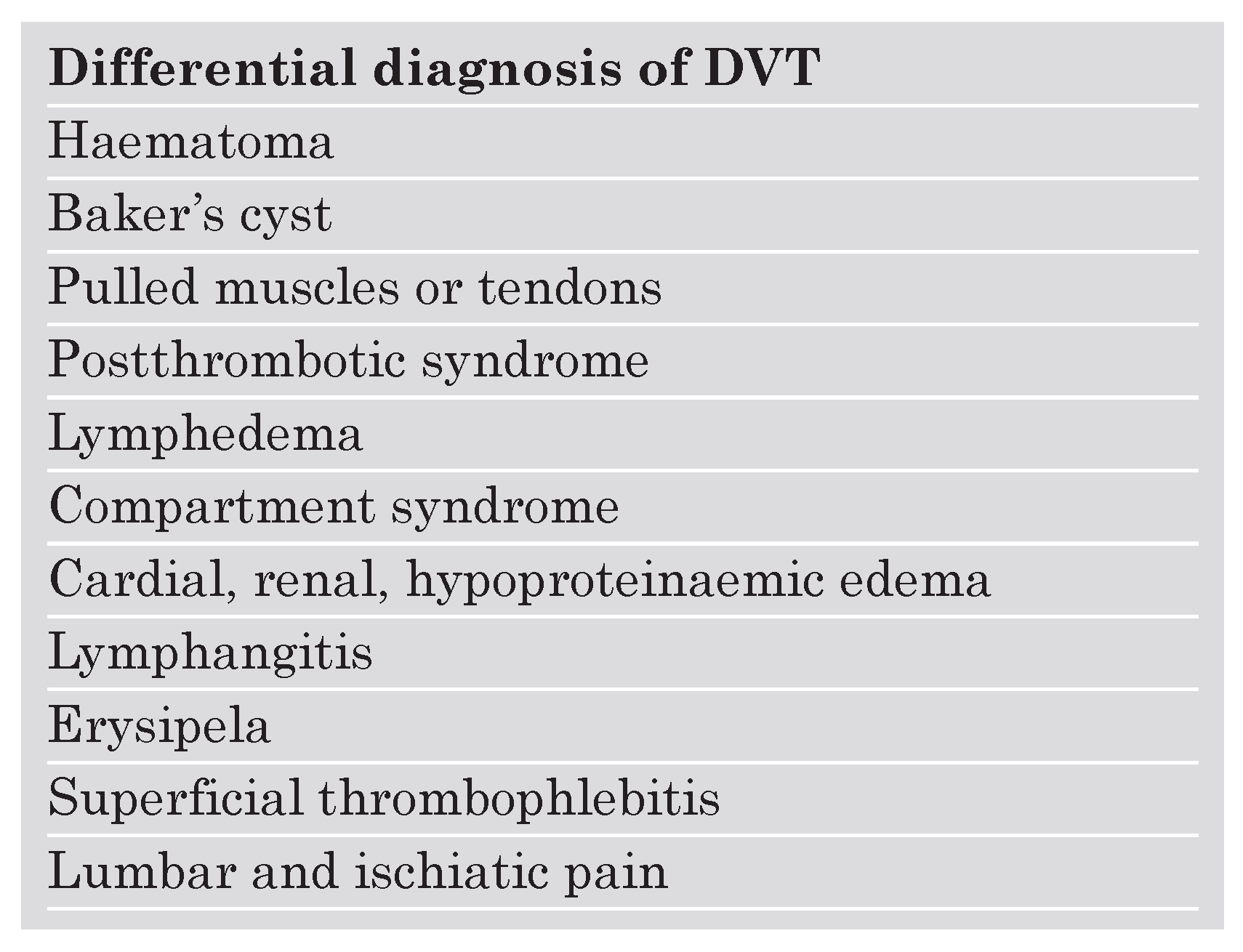
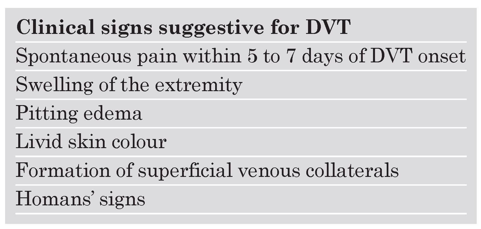
Difficulty in diagnosing DVT is based on the presence of non-specific symptoms in many patients. Symptoms are sometimes misleading due to the possibility of non-thrombotic extravascular disease mimicing signs of DVT. The differential diagnosis of pelvic and lower extremity DVT includes haematoma, ruptured Baker’s cyst, lymphedema, cancer, and postthrombotic syndrome. Common non-thrombotic differential conditions are listed in Table 1. Typical clinical signs of DVT include spontaneous leg pain, swelling of the leg, pitting edema, livid skin colour, formation of superficial venous collaterals, and tenderness along the deep venous system during overextension of the foot (Homan’s sign) (Table 2).
Based on the presence of clinical signs and adding further known individual risk factors for VTE, categories of estimated pre-test probability have been prospectively evaluated by Wells et al. using a clinical score (Table 3) [11]. Risk factors such as cancer, immobility, and postoperative states have been included in the score, whereas age, cardiac, pulmonary and renal disease, or thrombophilic disorders, such as Factor V Leiden mutation or antiphospholipid antibodies were not integrated in the score.
A further step in improving accuracy of acute symptomatic proximal DVT diagnosis on an evidence-based level was the adoption of an algorithm that combines objective testing with the estimation of clinical pre-test probability. Wells et al. suggested an algorithm based on the determination of pre-test probability and compression ultrasound (CUS) screening. A negative D-dimer test is highly sensitive for the exclusion of VTE [12] and was integrated into the algorithm as shown in Figure 1. For patients with atypical signs of DVT and a negative D-dimer test result, post-test probability is low, allowing the exclusion of DVT. When D-dimer testing is positive, CUS or colour coded duplex sonography (CCDS) should initially be performed to accurately exclude or confirm DVT (Figure 1). In patients with suspected DVT, serial CUS can be avoided if the D-dimer test is negative [13]. For high and medium pre-test probability, however, first-line sonographic assessment already allows DVT detection in the majority of patients, whereas in cases of negative sonographic findings, an additional negative D-dimer test allows exclusion of acute proximal DVT. In a patient with a high pre-test probability in combination with a positve D-dimer test and a negative CUS, magnetic resonance (MR) imaging or multislice computed tomography (CT) may be used to diagnose pelvic DVT (Figure 1).

Lower leg thrombosis

The strategy by Wells et al. focuses on the exclusion of symptomatic proximal DVT, ie the proximal venous segments of the pelvic region, groin and thigh, including the popliteal vein. Until recently, symptomatic distal DVT was considered less important because of its low risk of pulmonary embolisation. The need to diagnose and treat isolated calf vein thrombosis was controversial for many years [14]. Serial CUS testing approximately one week after presentation was considered sufficient for detecting thrombus propagation to the popliteal vein. The authors of the Seventh American College of Chest Physician (ACCP) Conference on Antithrombotic and Thrombolytic Therapy recommended antithrombotic treatment for all patients with DVT, including symptomatic distal DVT [14,15].

Asymptomatic DVT

Asymptomatic DVT is often diagnosed when DVT is found accidentally on ultrasound, CT, or MR ordered for other reasons than suspected DVT. The need for accurate diagnosis of suspected asymptomatic DVT also occurs in a clinical scenario when a patient suffers acute pulmonary embolism (PE) and concomitant DVT that was diagnosed during the same CT study that was performed to confirm PE. Justification for DVT screening in patients with acute PE is, that the treatment of lower extremity DVT includes compression therapy to diminish the risk for PTS even in asymptomatic patients. Detection of asymptomatic DVT, however, is associated with considerably lower sensitivity for both ultrasound and D-dimer testing. Orthopedic patients should be classified as a jeopardised group because postoperative asymptomatic DVT after hip or knee arthroplasty often remains undetected. CUS or CCDS have poor sensitivity to detect DVT due to wound-related leg swelling or haematoma, and assessment by contrast phlebography or non-invasively by MRI or CT phlebography is superior.

Recurrent DVT

Recurrent DVT is a frequent clinical problem as reflected by the 5-year-recurrence-rate of idiopathic DVT of approximately 25% [16]. Clinical differentiation between acute and chronic DVT is difficult. Moreover, diagnostic algorithms are not always helpful, because D-dimer testing is confounded by the presence of comorbidities. The precise thrombus extension during the primary event is important for identifying new thrombus formation. MRI can be a useful alternative for detecting recurrent DVT [17].

Upper extremity DVT

Upper extremity DVT is often seen in patients with central venous catheters (75%), it occurs spontaneously in patients with an effort-induced thrombosis (Paget von Schrötter syndrome). Patients with cancer may present with upper extremity swelling from cancer-associated venous compression of the superior vena cava or subclavian vein. CUS or CCDS might be useful as a first screening method in acute symptomatic thrombosis with sensitivities of 75 to 100% [18,19], but a low sensitivity of only approximately 30% for the central subclavian region in asymptomatic patients [20]. For both methods, the superior vena cava and the central part of the brachiocephalic vein are inaccessible. MR- or CT-phlebography are the most sensitive non-invasive diagnostic tools in this situation.

Objective testing methods

Imaging

Contrast venography

Among the imaging modalities, none has a 100% accuracy, including the “gold standard” contrast venography with 98.8% sensitivity (CI 95.6–99.8%). This procedure is invasive, technically sophisticated, requires the injection of a contrast medium and often is painful. Therefore, non-invasive diagnostic tools have replaced conventional phlebography for diagnosing symptomatic patients.

Real-time compression ultrasound and colour coded duplex sonography

Real-time compression ultrasound (CUS) and colour coded duplex sonography (CCDS) are non-invasive, cheap, easily available screening tools for acute symptomatic proximal DVT with high sensitivity and specifity for proximal symptomatic DVT, ranging from 89 to 100% for both CUS and CCDS, but only 73 to 87% for calf vein thrombosis [21]. One major disadvantage of CUS is that it does not provide acceptable accuracy for iliac or caval thrombosis. CCDS uses full compression of the target vein as principal criterion for the detection of DVT like CUS. CCDS is used at regions with restricted visibility, including the pelvic region or in the calf veins. Its main advantage over CUS is that partial thrombosis with remaining marginal flow can be detected more accurately. Both methods are operator and patient dependent. Figure 2 shows an acute thrombosis with a marginal flow in the femoral vein as visualised by CCDS.

MRI phlebography

MRI phlebography is the preferred imaging tool when the diagnosis is uncertain after a non-conclusive CUS or CCDS. The visualisation of thrombi by MRI in the pelvic region appears to be more reliable. Moreover, MRI allows detection of PE and of DVT of the lower extremities simultaneously. It is also suitable to diagnose venous compression by extravascular entities. Contraindications to MR imaging include certain metallic devices, obesity, and claustrophobia [17,22].

Contrast-enhanced multislice chest computer tomography (CT)

The main advantage of CT for diagnosing venous thromboembolism (VTE) is that it allows the detection of both PE and proximal DVT [23,24,25]. In postoperative or intensive care patients, CT may be preferred over CUS or CCDS.

Impendance plethysmography (IPG)

IPG detects increased venous outflow resistance in the deep veins of the proximal lower extremities. The main limitation of IPG is its unspecificity; it does not distinguish between venous obstruction due to DVT and obstructions from non-thrombotic lesions, such as haematoma or cancer. The method has limited sensitivity, as its application is bound to validated equipment and specific protocols. If accurately used, sensitivity reaches 90% for proximal symptomatic DVT, but only 30% for asymptomatic DVT [21]. The method is not widely used as a primary diagnostic screening tool for suspected acute DVT in Switzerland, but is considered as more suitable for the quantification of chronic venous obstruction as in PTS.

D-dimer assessment

D-dimer is a sensitive fibrin degradation protein that is released into the circulation during fibrinolysis and is used as a thrombosis marker. Other markers of activated coagulation or fibrinolysis have insufficient sensitivity to be used as screening tool for DVT. Among the various D-dimer assays, the quantitative VIDAS ELISA assay (Biomerieux, France) is the best documented and most sensitive assay. It is a fluorescence-based immunoassay that can be performed within less than one hour. The reported sensitivity ranges from 98 to 100% at a cutoff of 550 ng/ml in venographically proven DVT [12]. As an acute phase protein, it is, however, unspecific and only useful to exclude DVT by a negative result in combination with high or medium pre-test probability.

References

  1. Nordstrom, M.; Lindblad, B.; Bergqvist, D.; Kjellstrom, T. A prospective study of the incidence of deep-vein thrombosis within a defined urban population. J Internal Med 1992, 232, 155–160. [Google Scholar] [CrossRef]
  2. Anderson, F.A., Jr.; Wheeler, H.B.; Goldberg, R.J.; Hosmer, D.W.; Patwardhan, N.A.; Jovanovic, B.; et al. A population-based perspective of the hospital incidence and case-fatality rates of deep vein thrombosis and pulmonary embolism. The Worcester DVT Study. Arch Intern Med 1991, 151, 933–938. [Google Scholar] [CrossRef]
  3. Prandoni, P.; Lensing, A.W.; Cogo, A.; Cuppini, S.; Villalta, S.; Carta, M.; et al. The long-term clinical course of acute deep venous thrombosis. Ann Intern Med 1996, 125, 1–7. [Google Scholar] [CrossRef] [PubMed]
  4. Hansson, P.O.; Sörbo, J.; Eriksson, H. Recurrent venous thromboembolism after deep vein thrombosis: Incidence and risk factors. Arch Intern Med 2000, 160, 769–774. [Google Scholar] [CrossRef] [PubMed]
  5. Kearon, C. Natural history of venous thromboembolism. Circulation 2003, 107, I22–30. [Google Scholar] [CrossRef]
  6. Pengo, V.; Lensing, A.W.; Prins, M.H.; Marchiori, A.; Davidson, B.L.; Tiozzo, F.; et al.; Thromboembolic Pulmonary Hypertension Study Group Incidence of chronic thromboembolic pulmonary hypertension after pulmonary embolism. N Engl J Med 2004, 350, 2257–2264. [Google Scholar] [CrossRef]
  7. Mclachlin, J.; Richards, T.; Paterson, J.C. An evaluation of clinical signs in the diagnosis of venous thrombosis. Arch Surg 1962, 85, 738–744. [Google Scholar] [CrossRef] [PubMed]
  8. Goodacre, S.; Sutton, A.J.; Sampson, F.C. Meta-analysis: The value of clinical assessment in the diagnosis of deep venous thrombosis. Ann Intern Med 2005, 143, 129–139. [Google Scholar] [CrossRef]
  9. Levi, M.; Hart, W.; Buller, H.R. Physical examination – the significance of Homan’s sign. Ned Tijdschr Geneeskd 1999, 143, 1861–1863. [Google Scholar]
  10. Kahn, S.R. The clinical diagnosis of deep venous thrombosis. Arch Intern Med 1998, 158, 2315–2323. [Google Scholar] [CrossRef]
  11. Wells, P.S.; Hirsh, J.; Anderson, D.R.; Lensing, A.W.; Foster, G.; Kearon, C.; et al. Accuracy of clinical assessment of deep-vein thrombosis. Lancet 1995, 345, 1326–1330. [Google Scholar] [CrossRef] [PubMed]
  12. Ahearn, G.S.; Bounameaux, H. The role of the D-dimer in the diagnosis of venous thromboembolism. Semin Respir Crit Care Med 2000, 21, 521–536. [Google Scholar] [CrossRef]
  13. Kraaijenhagen, R.A.; Piovella, F.; Bernardi, E.; Verlato, F.; Beckers, E.A.; Koopman, M.M.; et al. Simplification of the diagnostic management of suspected deep vein thrombosis. Arch Intern Med 2002, 162, 907–911. [Google Scholar] [CrossRef]
  14. Büller, H.R.; Agnelli, G.; Hull, R.D.; Hyers, T.M.; Prins, M.H.; Raskob, G.E. Antithrombotic therapy for venous thromboembolic disease: The Seventh ACCP Conference on Antithrombotic and Thrombolytic Therapy. Chest 2004, 126, 401–428S. [Google Scholar] [CrossRef]
  15. Meissner, M.H.; Caps, M.T.; Bergelin, R.O.; Manzo, R.A.; Strandness, D.E., Jr. Early outcome after isolated calf vein thrombosis. Vasc Surg 1997, 26, 749–756. [Google Scholar] [CrossRef] [PubMed]
  16. Hansson, P.O.; Sörbo, J.; Eriksson, H. Recurrent venous thromboembolism after deep vein thrombosis. Incidence and risk factors. Arch Intern Med 2000, 160, 769–774. [Google Scholar] [CrossRef] [PubMed]
  17. Fraser, D.G.; Moody, A.R.; Morgan, P.S.; et al. Diagnosis of lower–limb deep venous thrombosis: A prospective blinded study of magnetic resonance direct thrombus imaging. Ann Intern Med 2002, 136, 89–98. [Google Scholar] [CrossRef]
  18. Baxter, G.M.; Kincaid, W.; Jeffrey, R.F.; Millar, G.M.; Porteous, C.; Morley, P. Comparison of colour Doppler ultrasound with venography in the diagnosis of axillary and subclavian vein thrombosis. Br J Radiol 1991, 6, 777–781. [Google Scholar] [CrossRef]
  19. Haire, W.D.; Lynch, T.G.; Lieberman, R.P.; Lund, G.B.; Edney, J.A. Utility of duplex ultrasound in the diagnosis of asymptomatic catheter-induced subclavian vein thrombosis. J Ultrasound Med 1991, 10, 493–496. [Google Scholar] [CrossRef]
  20. Prandoni, P.; Polistena, P.; Bernardi, E.; Cogo, A.; Casara, D.; Verlato, F.; et al. Upper-extremity deep vein thrombosis. Risk factors, diagnosis and complications. Arch Intern Med 1997, 157, 57–62. [Google Scholar] [CrossRef]
  21. Tapson, V.F.; Carroll, B.A.; Davidson, B.L.; Elliott, C.G.; Fedullo, P.F.; Hales, C.A.; et al. The diagnostic approach to acute venous thromboembolism. Clinical practice guideline. American Thoracic Society. Am J Respir Crit Care Med 1999, 160, 1043–1066. [Google Scholar] [PubMed]
  22. Stern, J.B.; Abehsera, M.; Grenet, D.; Friard, S.; Couderc, L.J.; Scherrer, A.; et al. Detection of pelvic vein thrombosis by magnetic resonance angiography in patients with acute pulmonary embolism and normal lower limb compression ultrasonography. Chest 2002, 122, 115–121. [Google Scholar] [CrossRef] [PubMed]
  23. Taffoni, M.J.; Ravenel, J.G.; Ackerman, S.J. Prospective comparison of indirect CT venography versus venous sonography in ICU patients. Am J Roentgenol 2005, 185, 457–462. [Google Scholar] [CrossRef]
  24. Katz, D.S.; Loud, P.A.; Bruce, D.; Gittleman, A.M.; Mueller, R.; Klippenstein, D.L.; et al. Combined CT venography and pulmonary angiography: A comprehensive review. Radiographics 2002, S3–S19; discussion S20–S24. [Google Scholar] [CrossRef]
  25. Cham, M.D.; Yankelevitz, D.F.; Shaham, D.; Shah, A.A.; Sherman, L.; Lewis, A.; et al. Deep venous thrombosis: Detection by using indirect CT venography. The Pulmonary AngiographyIndirect CT Venography Cooperative Group. Radiology 2000, 216, 744–751. [Google Scholar] [CrossRef] [PubMed]
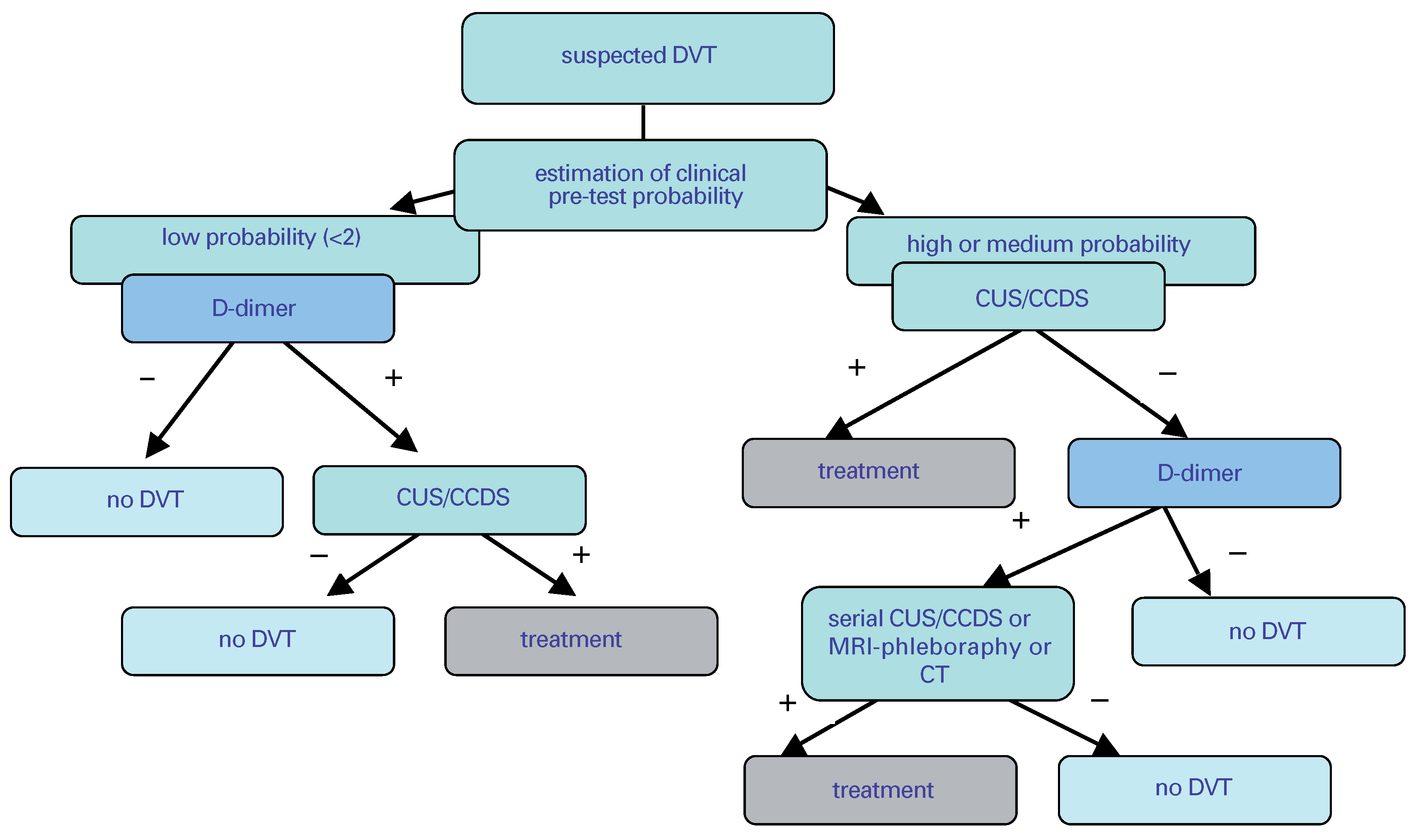
Figure 1. Diagnostic algorithm for DVT screening.
Cardiovascmed 09 00110 g001
Figure 2. Superficial vein thrombosis with marginal flow (blue) as presented by colour coded duplexsonography. The neighbouring artery is contrasted in red.
Cardiovascmed 09 00110 g002
Table 1. List of the most frequent diagnoses causing leg edema.
Cardiovascmed 09 00110 i001
Table 2. List of clinical signs that are associated with deep venous thrombosis.
Cardiovascmed 09 00110 i002
Table 3. The Wells Score for the prediction of clinical probability of DVT [11].
Cardiovascmed 09 00110 i003