Next Article in Journal
Franck’s Sign
Previous Article in Journal
Grimmige Märchen – Alternative Facts über das böse Cholesterin
 
 
Cardiovascular Medicine is published by MDPI from Volume 28 Issue 1 (2025). Previous articles were published by another publisher in Open Access under a CC-BY (or CC-BY-NC-ND) licence, and they are hosted by MDPI on mdpi.com as a courtesy and upon agreement with Editores Medicorum Helveticorum (EMH).
Review

Severe Pulmonary Embolism: Surgical Aspects

by
Oliver Reuthebuch
Clinic for Cardiac Surgery, University Hospital Basel, 4031 Basel, Switzerland
Cardiovasc. Med. 2017, 20(6), 154; https://doi.org/10.4414/cvm.2017.00486
Submission received: 21 March 2017 / Revised: 21 April 2017 / Accepted: 21 May 2017 / Published: 21 June 2017

Abstract

Severe pulmonary embolism is a life-threatening disease requiring a well-balanced therapeutic approach. It is of upmost importance to differentiate between acute pulmonary embolism and chronic pulmonary embolism in this setting. The management of acute embolism is predominantly carried out by the internal specialist, whereas the therapy of chronic pulmonary embolism is in the domain of the surgical community. Acute, untreated pulmonary embolism has a mortality of >30%, with two thirds of deaths occurring within the first 60 minutes. It is the most common cause of death without a prior clinical diagnosis because of its vague and nonspecific clinical symptoms and undefined laboratory parameters. The nature of chronic pulmonary embolism is different since it is based on recurrent pulmonary emboli without complete resolution. Five-year survival is pressure dependent, with a mere 10% survival rate in patients having a mean pulmonary arterial pressure of more than 50 mm Hg. It has an incidence of up to 3.8% in survivors of acute pulmonary embolism, as well as an incidence of >10% in patients with recurrent embolism. There are various treatment options for acute pulmonary embolism based on the haemodynamic stability or instability of the patient. If the patient is stable, anticoagulation is to be administered with a potential subsequent implantation of an inferior vena cava filter. If the patient is unstable, thrombolytic therapy has to be considered first. If this fails, mechanical embolectomy or even surgical embolectomy should be taken into account. Patients with chronic pulmonary embolism should be treated surgically with a bilateral endarterectomy of the pulmonary arteries. This is the standard and recommended treatment. The role of the emerging percutaneous pulmonary angioplasty is not yet defined and needs further evaluation. Patients suffering from acute or chronic pulmonary embolism should be transferred to a certified centre. There, specialists decide on the appropriate treatment with potential implantation of life-supporting systems (e.g., extracorporeal membrane oxygenation).

Introduction

The surgical treatment of pulmonary embolism was one of the cornerstones in the history of cardiac surgery. It was developed and advocated by Friedrich Trendelenburg in 1907, when he was director of the department for surgery at the university hospital in Leipzig, Germany [1]. Unfortunately, none of his few patients survived the “Trendelenburg operation” and it was 16 years later, in 1923, that Martin Kirschner performed the first successful surgical embolectomy in Königsberg [2]. At that time, no extracorporeal circulation (ECC) was available and thus procedural success was very unpredictable. In 1930, John Gibbon experienced, in Princeton, USA, the death of a young lady due to pulmonary embolism, which made him start developing the heart-lung machine [3]. Aher 23 years of research, in October 1953, an atrial septal defect was operated on with the first use of ECC. Another 8 years later, in 1961, Denton Cooley succeeded in performing a pulmonary embolectomy on ECC [4]. Since then, the surgical technique became more feasible and safer. However, nowadays a great armamentarium of therapeutic options is available to replace the surgical primacy: anticoagulation, thrombolytic therapy, catheter-based procedures with, for example, rotational, ultrasound or suction embolectomy. Independently of the treatment, it is of utmost importance to improve the prognosis by avoiding recurrent thromboembolism, in order to avoid the development of chronic thromboembolic pulmonary hypertension (CTEPH), as well as avoiding death.
The aim of this article is to focus on surgical modalities with descriptions of technical aspects and the weighting of surgery in specific clinical settings, as well as describing future development.

Classification of Pulmonary Embolism

Pulmonary embolism is classified into two main categories: acute and chronic. Pathogenesis, course of disease and patient stability are different in each, which therefore require alternate therapeutic approaches.
Acute pulmonary embolism is defined as obstruction of the pulmonary arteries in potentially all segments by circulating material such as thrombi, tumorous or fat tissue, foreign bodies, bacteria or even air. The mortality, if leh untreated, exceeds 30%, with two out of three patients dying within 60 minutes aher onset of symptoms. If the condition is treated, 30-day mortality reaches 4% and 1-year mortality 13%. However, clinical signs are very nonspecific, making this ailment the most common cause of death without definite diagnosis. Patients may complain of dyspnoea, tachycardia, chest pain or hypotension. Laboratory test results may encompass elevation of D-dimer (in 90% of cases), elevation of troponin and pro-brain-type natriuretic peptide (pro-BNP) levels as well as signs of hypoxaemia. Diagnosis is based on chest X-ray (“Westermark” sign, with dilatation of central pulmonary arteries), multislice computed tomography (CT) scan, magnetic resonance imaging (MRI), pulmonary angiography or echocardiography. The mainstay of the therapeutic approach is to maintain oxygenation and stabilise haemodynamic circulation [5].
Chronic pulmonary embolism is caused by single or multiple pulmonary emboli that are not resolved completely in the long run. There are two pathophysiological pathways to be considered. Firstly, thromboembolic lesions in arterial walls transform into fibrous and elastic structures. Subsequently, they endothelialise and obliterate or narrow the lumen. Secondly, an Eisenmenger-like reaction in overperfused vessels turns into vasoconstriction of small arteries, resulting in hypertrophy of the media and finally sclerosis. The incidence of chronic pulmonary embolism in survivors of acute pulmoary embolism is between 0.57 and 3.8%, whereas more than 10% of patients with recurrent acute pulmonary embolism will suffer from chronic pulmonary embolism. The overall incidence of surgical treatment in all patients with pulmonary embolism is low, at 0.1%. Five-year survival is pressure dependent: with a mean pulmonary artery pressure (mPAP) greater than 50 mm Hg it is less than 10%, with a mPAP >30 mm Hg it is 30% and with a mPAP <30 mm Hg it is 90%. The diagnostic work-up is based on echocardiography, perfusion-ventilation scintigraphy, right-heart catheterisation and bilateral pulmonary angiography. CT and MRI complete the screening process. The aim of the therapeutic approach is to bilaterally remove all thrombotic material in order to reduce pulmonary vascular resistance as well as to improve ventilatory impairment [6].

Surgical Technique

Surgery for acute or chronic pulmonary embolism differs and thus will be described for each separately. Since there is a potential role for percutaneous pulmonary balloon angioplasty, an emerging technique involving surgical teams, this procedure will be explained as a bail-out strategy in inoperable patients.

Surgical Technique for Acute Pulmonary Embolism

In the supine position the chest is opened via a median sternotomy. The aorta and both caval veins are cannulated aher full heparinisation. Extra corporeal circulation is kept normothermic, with the heart either arrested or fibrillating. The pulmonary trunk is then incised longitudinally with respect to the pulmonary valve. The emboli are grasped with forceps and carefully removed. If the emboli are located in the segmental branches, further incisions can be of help. The use of endoscopes has been described [7]. Simultaneous ventilation or even manual massage of the lungs may support mobilisation of the embolic material. Aher meticulous inspection of the arteries, the incisions are closed using 5/0 or 6/0 prolene sutures. In addition, the right atrium should be opened in order to carefully inspect the cavities and to close potential septal defects. Postoperatively, a CT scan or a Doppler echography of the venous system is recommended to exclude further thrombotic material. Full heparinisation and subsequent anticoagulation for 6 months is mandatory.

Surgical Technique for Chronic Pulmonary Embolism

The surgical technique for chronic pulmonary embolism is far more complex since it is performed under hypothermic circulatory arrest. Otherwise, visibility in the peripheral subsegmental arteries would be compromised by back-bleeding of systemic to pulmonary shunts.
In the supine position the chest is opened via a sternotomy. Aher institution of cardiopulmonary bypass the patient is cooled to about 20 °C. The aorta is clamped, the heart arrested with cardioplegia and extracorporeal circulation stopped. The pulmonary artery at the level of the bifurcation or right pulmonary artery is incised anteriorly and the correct plane for endarterectomy in the dorsal aspect of the media defined. This is a pivotal step, since too deep a plane could cause arterial perforation and a too superficial plane could result in an insufficient endarterectomy with remaining pulmonary hypertension [8]. The endarterectomy is accomplished to the level of segmental or even subsegmental arteries and the material removed (Figure 1).
During closure of the incision the patient is reperfused. In general this takes 15 minutes. In recent years, the use of endoscopic techniques to visualise the distal segments has become a supportive and beneficial adjunct. The same technique is adapted to the leh side. In general, overall circulatory arrest does not exceed 40 minutes. Finally, the heart is de-aired, the aorta unclamped and the patient rewarmed to 36°. During the rewarming additional procedures such as aortocoronary bypass, valve surgery or even combined procedures can be performed [9].

Percutaneous Pulmonary Angioplasty

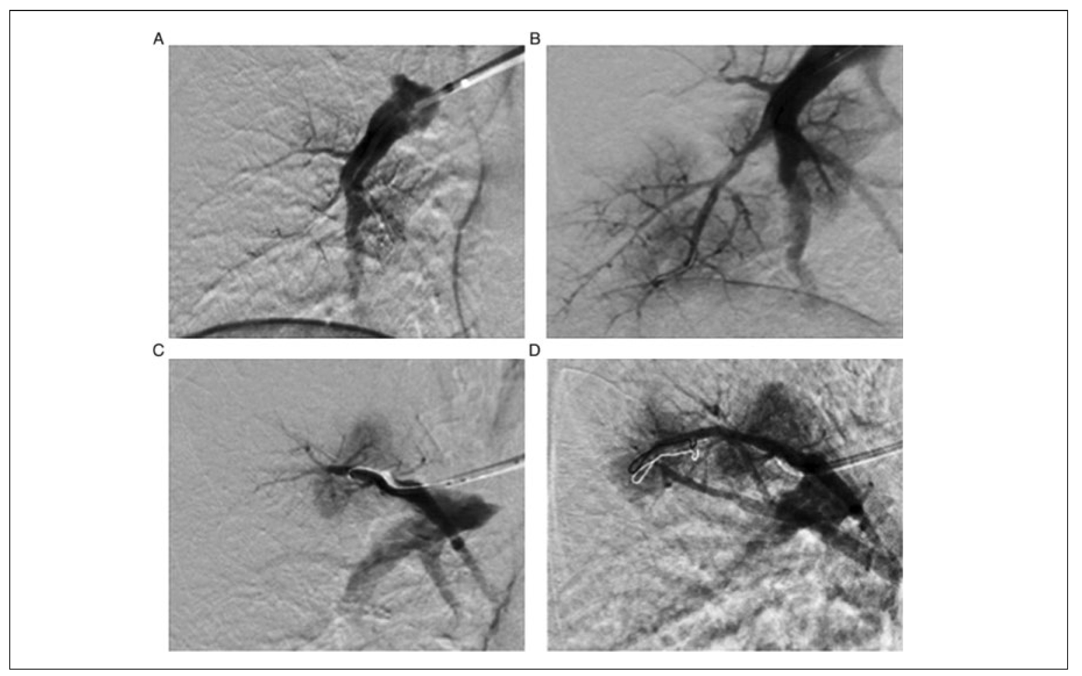
Percutaneous pulmonary angioplasty (PPA), also known as percutaneous transluminal pulmonary angioplasty (PTPA) or balloon pulmonary angioplasty (BPA), is an emerging technique and is so far indicated in patients who are not otherwise treatable. The procedure was described first in 2001 and was re-established in 2012 in Japan aher initial poor outcomes. It involves repetitive (4–8, average 4.8) catheter laboratory treatments to minimise the risk of procedure-related effects to the kidney (contrast dye) and lungs (reperfusion injury). Preprocedural pulmonary arterial angiography is performed to determine target vessels. Over a guide-wire, a balloon is positioned in the respective vessel and dilated. By using smaller balloons and limiting the inflations to one or two vascular segments, the risk for vessel rupture and reperfusion lung injury is reduced (Figure 2). Since this technique is also used by surgeons in Europe, it is described in this article [10].

Surgery in Acute Pulmonary Embolism: Indications and Outcome

Patients suffering from acute pulmonary embolism may be in a life-threatening situation. The essence of therapy is to maintain oxygenation as well as haemodynamic stabilisation. Therefore, starting possibly long-term anticoagulation is the mainstay of treatment, especially in a haemodynamically stable patient. A more intensive process, with thrombolysis or even catheter-based embolectomy or thrombus fragmentation, might be required in the event of hemodynamic instablility. This is when surgery may become a therapeutic option. According to the 2012 guidelines of the American College of Chest Physicians, surgery is a grade 2C recommendation for patients with acute pulmonary embolism and hypotension and (1) contraindications to thrombolysis, (2) failed thrombolysis or catheter-based embolectomy and (3) shock that is likely to cause death before thrombolysis can take effect [11]. However, the main prerequisite is surgical expertise and available resources. An addendum to the guidelines state that surgery has to be considered if any additional intracardiac thromboemboli, as well as a patent foramen ovale, have been diagnosed.
Stein at al. systematically reviewed 41 out of a total of 1167 reports concerning outcome of surgical therapy in patients with acute pulmonary embolism [12]. Data were acquired between 1961 and 2005. Reported mortality ranged between 6 and 64% with an average of 30%. With improvements of technique and expertise, mortality decreased from 32% (1961–1984) to 20% (1985– 2006), but remained high. Indication for surgery was haemodynamic instability in 74% of cases, previous cardiac arrest in 32% and contraindication for thrombolytic therapy in 19%. Mortality of pulmonary embolectomy aher suffering from cardiac arrest shows a loose but significant correlation. Meneveau at al. compared in-hospital course aher rescue surgical embolectomy or repeat thrombolysis in 40 nonresponders to first-time fibrinolytic therapy [13]. In-hospital course was uneventful in 79% (11/14) of surgically treated patients, but in only 31% (8/26) with repeat fibrinolysis. A tendency towards lower mortality in the surgical group could be detected (p = 0.07). Thirty-five percent of the medically treated cases had recurrent pulmonary embolism, whereas none were observed in the surgical group. All bleeding events in the repeatthrombolysis group were fatal. The authors concluded that transfer of patients with recurrent pulmonary embolism to a cardiac surgery centre should be considered.

Surgery in Chronic Thromboembolic Hypertension: Indications and Outcome

Other than in acute pulmonary embolism, surgery is so far the only definite therapeutic option for chronic thromboembolic pulmonary hypertension (CTEPH). The decision to go for a surgical procedure is based upon several aspects: (1) presence of ventilatory impairment with patients being in New York Heart Association (NYHA) functional class III to IV; (2) the preoperative pulmonary vascular resistance (PVR) should exceed 300 dyn·s–1·cm–5; (3) the thrombi need to be surgically accessible in the main, lobar or segmental arteries; (4) patients should not suffer from severe comorbidities such as obstructive or restrictive chronic lung disease or advanced secondary arteriopathy [14]. As previously mentioned, the diagnostic pathway encompasses the confirmed suspicion with echocardiogram and ventilation-perfusion scan, the confirmation of CTEPH with right heart catheterisation and pulmonary angiography (or, recently, CT pulmonary angiography or magnetic resonance angiography) and finally the risk assessment with evaluation of the haemodynamic situation and comorbidities, as well as the team experience.
Main postoperative complications in the intensive care unit comprise reperfusion oedema with an incidence of 10–15%. In these cases, adequate ventilation with tidal volumes less than 8 ml/kg bodyweight, an inspiration: expiration ratio of 3:1 as well as a PiP (peak inspiratory pressure) of <18 cm H2O is advantageous. Fluid restriction and avoidance of inotropes help stabilise the situation. In cases of failed ventilation, the implantation of an extra-corporeal membrane oxygenation has to be considered. Furthermore, rupture of arteriotomy, nosocomial pneumonia, haemoptysis and intrapulmonary bleeding (0.5–1%), as well as rare rethrombosis, may have to be dealt with [15].
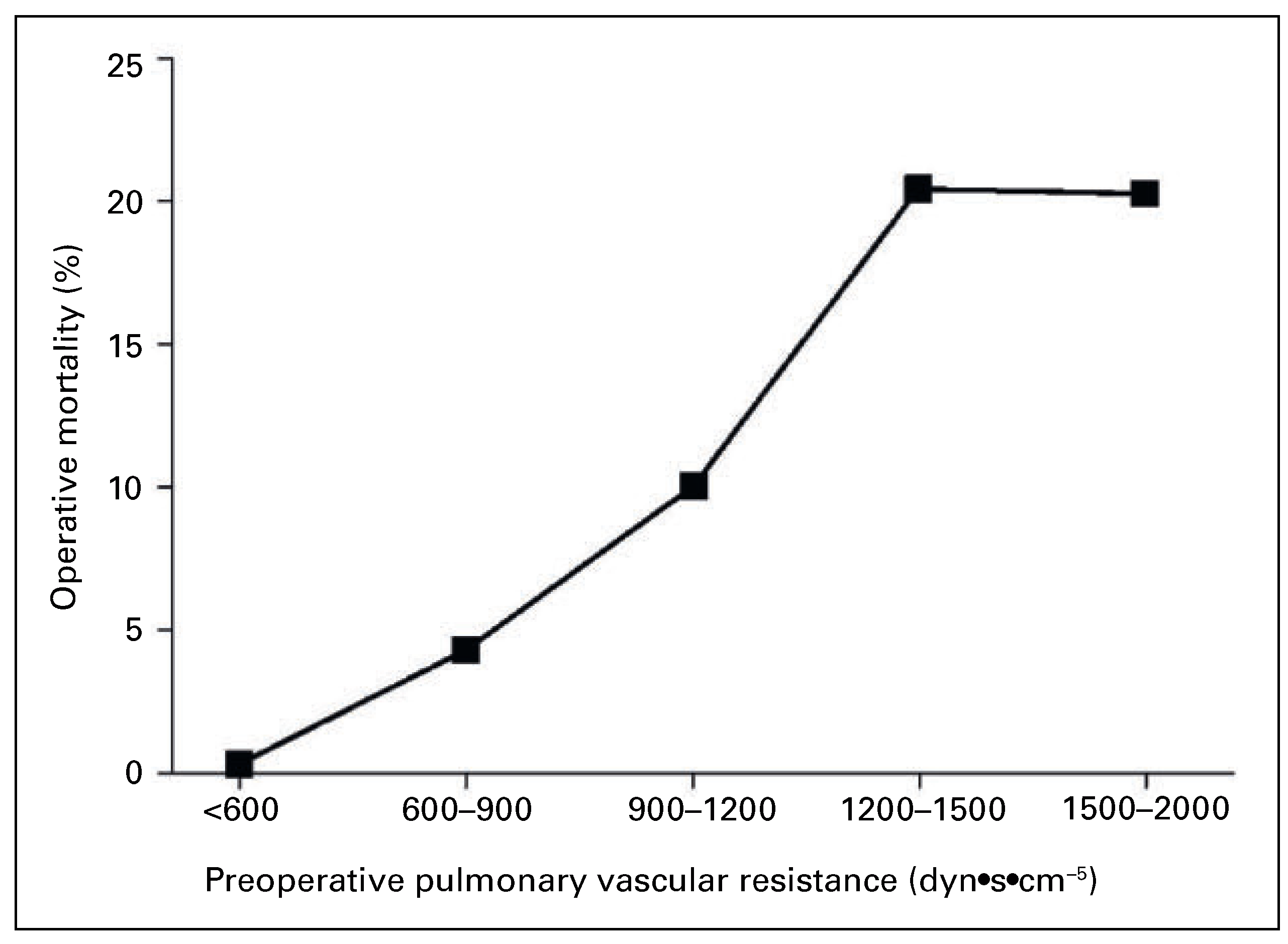
Operative mortality is highly dependent on preoperative PVR: if PVR is lower than 900 dyn·s–1·cm–5 mortality rate is below 4%, if PVR is between 900 and 1200 dyn·s–1·cm–5 mortality rate is about 10% and with PVR >1200 dyn·s–1·cm–5 20% of patients die (Figure 3). Postoperative outcome is very much dependent on persistent high pulmonary hypertension with a mean pulmonary arterial pressure of >25 mm Hg. This can be due to peripheral obstructions that were not amenable for surgical endarterectomy, or the secondary Eisenmenger-like vasculopathy [9]. Both entities may lead to right ventricular dysfunction. However, according to
Cannon et al., overall 10-year survival is approximately 75%, with even better results in patients operated on at a time with profounder surgical skills and modern techniques. These survival rates are better than aher lung transplantation [16]. Freed et al. analysed haemodynamic data preand 3 months postoperatively. He could show on the one hand a decrease of mPAP from 48 ± 12 mm Hg to 26 ±10 mm Hg and a decrease of PVR from 805 ± 365 dyn·s−1·cm−5 to 301 ± 232 dyn·s−1·cm−5, and on the other hand an increase of cardiac index from 2.0 ± 0.7 l·s−1·m−2 to 2.5 ± 0.5 l·s−1·m−2 as well as an increase of six-minute walk distance from 269 ±119 m to 367 ± 108 m [17].

Percutaneous Pulmonary Angioplasty in Chronic Thromboembolic Hypertension: Indications and Outcome

So far, BPA (balloon pulmonary angioplasty) is a bailout or supplemental strategy for patients with anatomy unsuitable for surgery, as well as for elderly, highrisk and frail patients. Beside that, nonresponders to surgical endarterectomy with remaining devastating pulmonary hypertension may benefit from this therapy. Owing to the significant side effects of this interventional procedure, other therapeutic options should have been considered first. Restricted data from clinical studies indicate that palliation is likely and cure possible [10]. Mizoguchi et al. performed BPA in 68 patients with inoperable CTEPH. With a range of two to eight procedures per patient they could reduce the risk of ballooning as well as the risk of reperfusion injury to the lung. Mean pulmonary arterial pressure decreased from 45.4 ± 9.6 to 24 ± 6.4 mm Hg (p <0.001). NYHA class improved from III to II [18]. In 2013, Andreassen et al. published an observational cohort study including 20 consecutive CTEPH patients. Each patient had a mean of 18.6 ± 6.1 BPAs performed with an average of 3.7 ± 2.1 procedures. Seven patients suffered from reperfusion injury. Mean pulmonary arterial pressure decreased from 45 ± 11 mm Hg to 33 ± 10 mm Hg (p <0.001), cardiac output increased from 4.9 ± 1.6l/min to 5.4 ± 1.9l/min (p = 0.011). NYHA classification improved from 3.0 ± 0.5 to 2.0 ± 0.5 (p <0.001). A total of 85% of patients (17/20) were alive aher 51 ± 30 months. The authors concluded that BPA may be an adjunct or alternative procedure to surgical endarterectomy in distinctive cases [19].
BPA is en emerging technique with yet comparative trials lacking and so far not fully established outside Japan. Long-term data need to be published and compared with existing and accepted treatment [10].

Conclusion

The weight of surgery as a therapeutic option in pulmonary embolism with potentially subsequent pulmonary hypertension is mainly dependent on the onset of symptoms and stability of the patient. Acute pulmonary embolism is a key indication for anticoagulation, thrombolytic therapy or interventional embolectomy. Surgery is suggested only when thrombolysis is contraindicated, has failed or the patient is in a shock situation. If additional intracardiac (floating) thromboembolisms or patent foramen ovale is detected, surgery may as well be considered.
In chronic pulmonary embolism with CTEPH, surgical pulmonary endarterectomy is the standard and recommended treatment. Though potential complications are severe, they have become rare as a result of improvement of surgical and instrumental techniques as well as experience in dedicated centers. The availability of ECMO technology should be guaranteed.
The role of percutaneous pulmonary angioplasty needs further evaluation and improvement and so far cannot replace pulmonary endarterectomy. However it may be considered as alternative therapy in otherwise untreatable patients or in cases with recurrent CTEPH. According to the heart team approach, the CTEPH team should assess operability of patients as well as alternative therapeutic options.

Disclosure Statement

No financial support and no other potential conflict of interest relevant to this article was reported.

References

  1. Trendelenburg, F. Über die operative Behandlung der Embolie der Lungenarterie. Archiv für Klinische Chirurgie 1924, 133, 312–359. [Google Scholar][Green Version]
  2. Kirschner, M. Ein durch die Trendelenburgsche Operation geheilter Fall von Embolie der Art. Pulmonalis. Archiv für klinische Chirurgie. 1924, 133, 312–359. [Google Scholar][Green Version]
  3. Gibbon, J. The Development of the Heart-Lung Apparatus. Am. J. Surg. 1978, 135, 608–619. [Google Scholar] [CrossRef] [PubMed]
  4. Cooley, D.A.; Beall, A.C. Surgical treatment of acute massive pulmonary embolism using temporary cardiopulmonary bypass. Dis. Chest. 1962, 41, 102–104. [Google Scholar] [CrossRef] [PubMed]
  5. Taylor Thompson, B. Overview of acute pulmonary embolism in adults. In: UpToDate: Waltham, MA. (Accessed on May 27, 2016).
  6. Fedullo, P.F. Overview of the treatment of chronic thromboembolic pulmonary hypertension. In: UpToDate, Waltham, MA. (Accessed on May 27, 2016).
  7. Eini, Z.M.; Houri, Z.; Cohen, I.; Sion, R.; Tamir, A.; Sasson, L.; Mandelberg, A. Massive Pulmonary Emboli in Children. Does Fiber-optic-Guided Embolectomy have a role? Review of the Literature and Report of Two Cases. Chest 2013, 143, 544–549. [Google Scholar] [CrossRef] [PubMed]
  8. Dartevelle, P.; Fadel, E.; Mussot, S.; Chapelier, A.; Hervé, P.; de Perrot, M.; et al. Chronic thromboembolic pulmonary hypertension. Eur. Respir. J. 2004, 23, 637–648. [Google Scholar] [CrossRef] [PubMed]
  9. Mayer, E.; Klepetko, W. Techniques and Outcome of Pulmonary Endarterectomy for Chronic Thromboembolic Pulmonary Hypertension. Proc. Am. Thorac. Soc. 2006, 3, 589–593. [Google Scholar] [CrossRef] [PubMed]
  10. Lang, I.M.; Madani, M. Update on Chronic Thromboembolic Pulmonary Hypertension. Circulation 2014, 130, 508–518. [Google Scholar] [CrossRef] [PubMed]
  11. Kearon, C.; Akl, E.A.; Comerota, A.J.; Prandoni, P.; Bounameaux, H.; Goldhaber, S.Z.; et al. Antithrombotic therapy for VTE disease. Antithrombotic Therpay and Prevention of Thrombosis, 9th ed: American College of Chest Physicians Evidence-Based Clinical Practice Guidelines. Chest 2012, 141 (2 Suppl), e419S–e496S. [Google Scholar] [CrossRef] [PubMed]
  12. Stein, P.D.; Alnas, M.; Beemath, A.; Patel, N. Outcome of Pulmonary Embolectomy. Am. J. Cardiol. 2007, 99, 421–423. [Google Scholar] [CrossRef] [PubMed]
  13. Meneveau, N.; Séronde, M.F.; Blonde, M.C.; Legalery, P.; et al. Management of Unsuccessful Thrombolysis in Acute Massive Pulmonary Embolism. CHEST 2006, 129, 1043–1050. [Google Scholar] [CrossRef] [PubMed]
  14. Torbicki, A. Pulmonary Thromboembolic Disease. Clinical Management of Acute and Chronic Disease. Rev. Esp. Cardiol. 2010, 63, 832–849. [Google Scholar] [CrossRef] [PubMed]
  15. Kim, N.H.; Delcroix, M.; Jenkins, D.P.; Channick, R.; Dartevelle, P.; Jansa, P.; et al. Chronic Thromboembolic Pulmonary Hypertension. JACC 2013, 62, D92–9. [Google Scholar] [CrossRef] [PubMed]
  16. Cannon, J.E.; Su, L.; Kiely, D.G.; Page, K.; Toshner, M.; Swietlik, E.; et al. Dynamic Risk Stratification of Patient Long-Term Outcome Aher Pulmonary Endarterectomy: Results From the United Kingdom National Cohort. Circulation 2016, 133, 1761–1771. [Google Scholar] [CrossRef]
  17. Freed, D.H.; Thomson, B.M.; Berman, M.; Tsui, S.S.L.; Dunning, J.; Sheares, K.K.; et al. Survival aher pulmonary thrombendarterectomy: Effect of residual pulmonary hypertension. JTCVS 2011, 141, 383–387. [Google Scholar]
  18. Mizoguchi, H.; Ogawa, A.; Munemasa, M.; Mikouchi, H.; Ito, H.; Matsubara, H. Refined Balloon Pulmonary Angioplasty for Inoperable Patients with Chronic Thromboembolic Pulmonary Hypertension. Circ. Cardiovasc. Interv. 2012, 5, 748–755. [Google Scholar] [CrossRef]
  19. Andreassen, A.K.; Ragnarsson, A.; Gude, E.; Geiran, O.; Andersen, R. Balloon pulmonary angioplasty in patients with inoperable chronic thromboembolic pulmonary hypertension. Heart 2013, 99, 1415–1420. [Google Scholar] [CrossRef] [PubMed]
Figure 1. Thromboembolic material removed from the right and left pulmonary arteries during pulmonary endarterectomy. Reprinted with permission of the American Thoracic Society. Copyright © 2017 American Thoracic Society. From: Mayer E, Klepetko W. Techniques and outcomes of pulmonary endarterectomy for chronic thromboembolic pulmonary hypertension. Proc Am Thorac Soc. 2006;3:589–93. Proceedings of the American Thoracic Society is an official journal of the American Thoracic Society.
Cardiovascmed 20 00154 g001
Figure 2. Pulmonary angiograms of right and middle lobe subsegment (A and B) and lower lobe segmental branches (C and D) before and immediately after balloon pulmonary angioplasty (From: Andreassen AK, Ragnarsson A, Gude E, Geiran O, Andersen R. Balloon pulmonary angioplasty in patients with inoperable chronic thromboembolic pulmonary hypertension. Heart. 2013;99:1415–20, reprinted with premission).
Cardiovascmed 20 00154 g002
Figure 3. Operative mortality according to preoperative PVR in patients with CTEPH undergoing pulmonary endartercectomy. Reprinted with permission of the American Thoracic Society. Copyright © 2017 American Thoracic Society. From: Mayer E, Klepetko W. Techniques and outcomes of pulmonary endarterectomy for chronic thromboembolic pulmonary hypertension. Proc Am Thorac Soc. 2006;3:589–93. Proceedings of the American Thoracic Society is an official journal of the American Thoracic Society.
Cardiovascmed 20 00154 g003