Introduction
Valve repair for mitral regurgitation represents an important advance over valve replacement with less perioperative mortality, lower perioperative complication rate, less deterioration in left ventricular function, less prosthetic material and lack of need for long-term anticoagulation [
1,
2]. However, the procedure is technically more demanding, involving some potential for suboptimal results. Unlike prosthetic valve replacement, successful valve repair requires the surgeon to understand the mechanism of the valve dysfunction, which is a primary determinant of the likelihood of successful repair, varying from 70 to 80% in patients with isolated rupture of chordae to the middle portion of the posterior leaflet to 20 to 30% in patients with extensive bileaflet prolapse. Most reports of patients having mitral valve surgery have been categorised according to the aetiology of the valve disease, that is myxomatous degeneration, ischaemia, rheumatic or congenital disease, as well as endocarditis which is not the same as its mechanism. However, a more functional classification allows exact definition of the valve dysfunction rather than description of the pathologic valve anatomy alone [
3,
4].
Preoperative diagnosis and functional classification of mitral valve regurgitation
The prerequisite for successful mitral valve repair is a precise diagnosis of the valve dysfunction before operation. This diagnosis has been facilitated by the functional classification introduced in the late 1970ies by Carpentier [
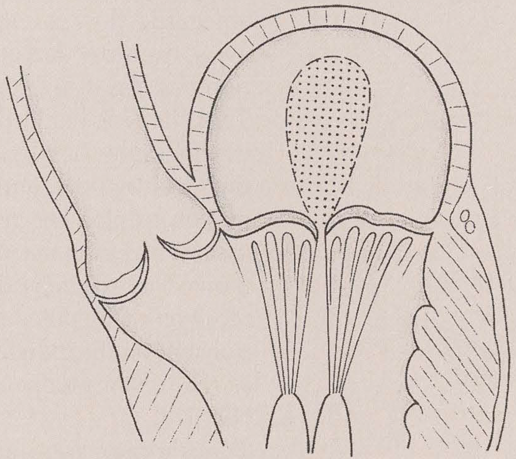
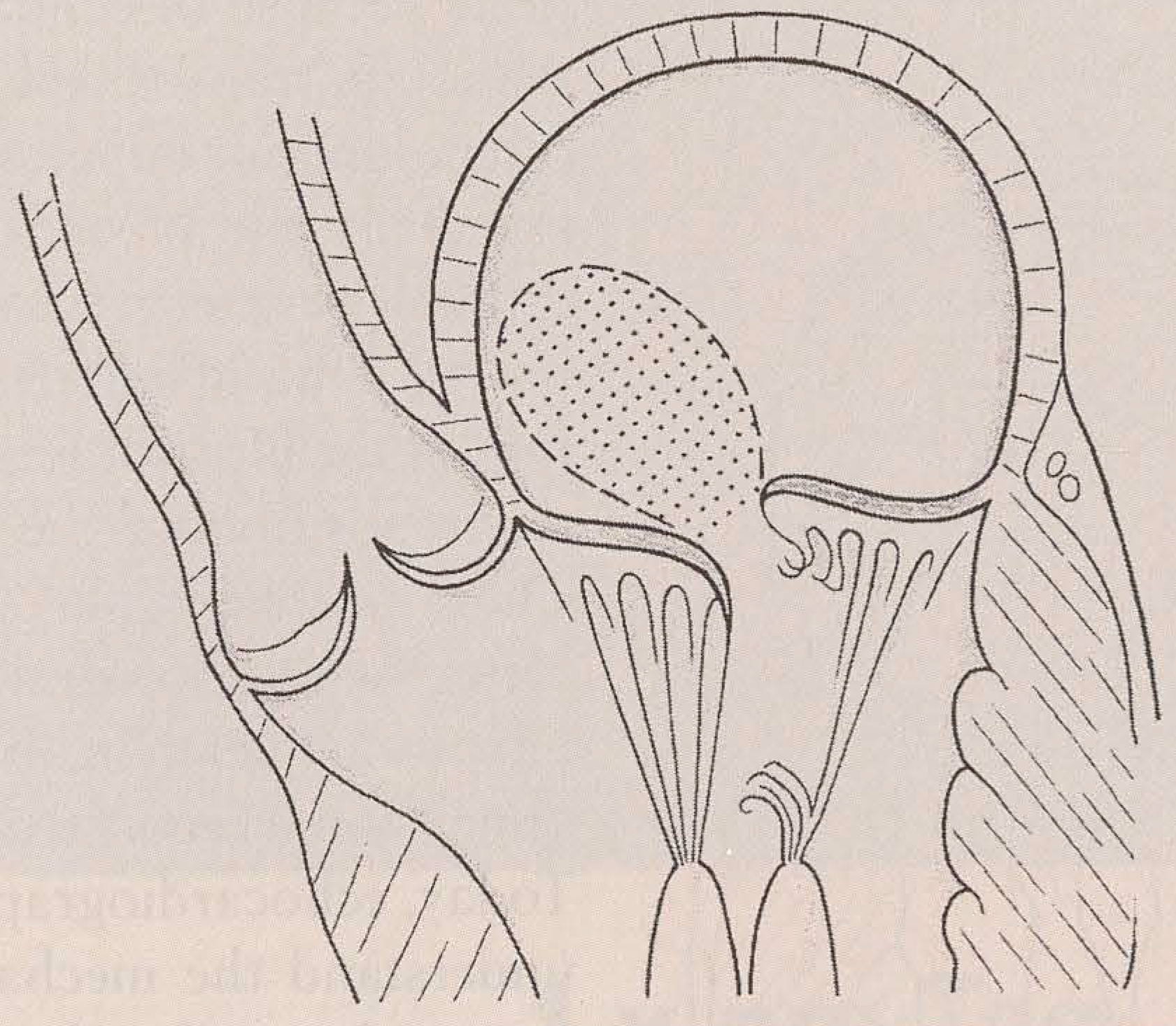
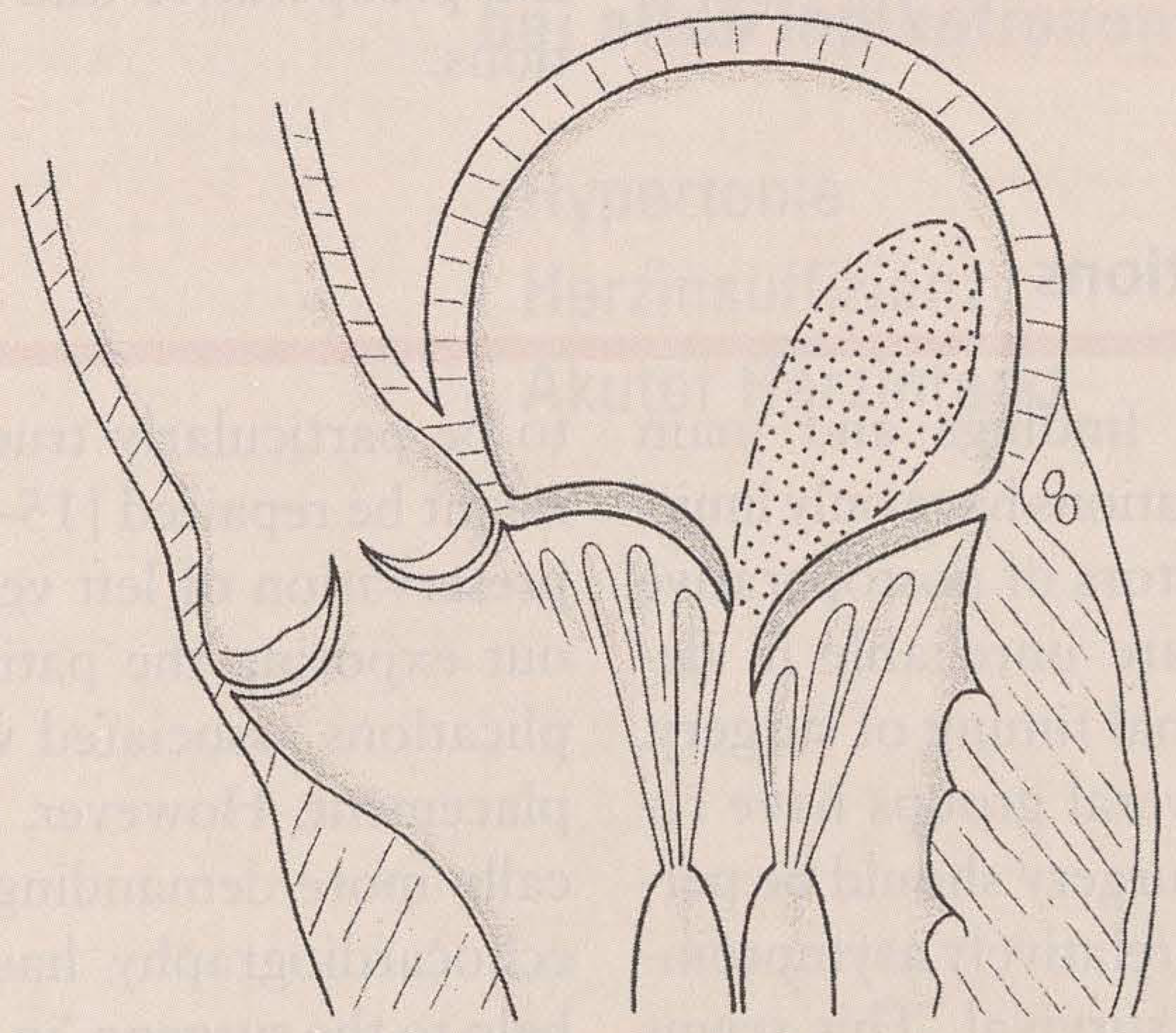
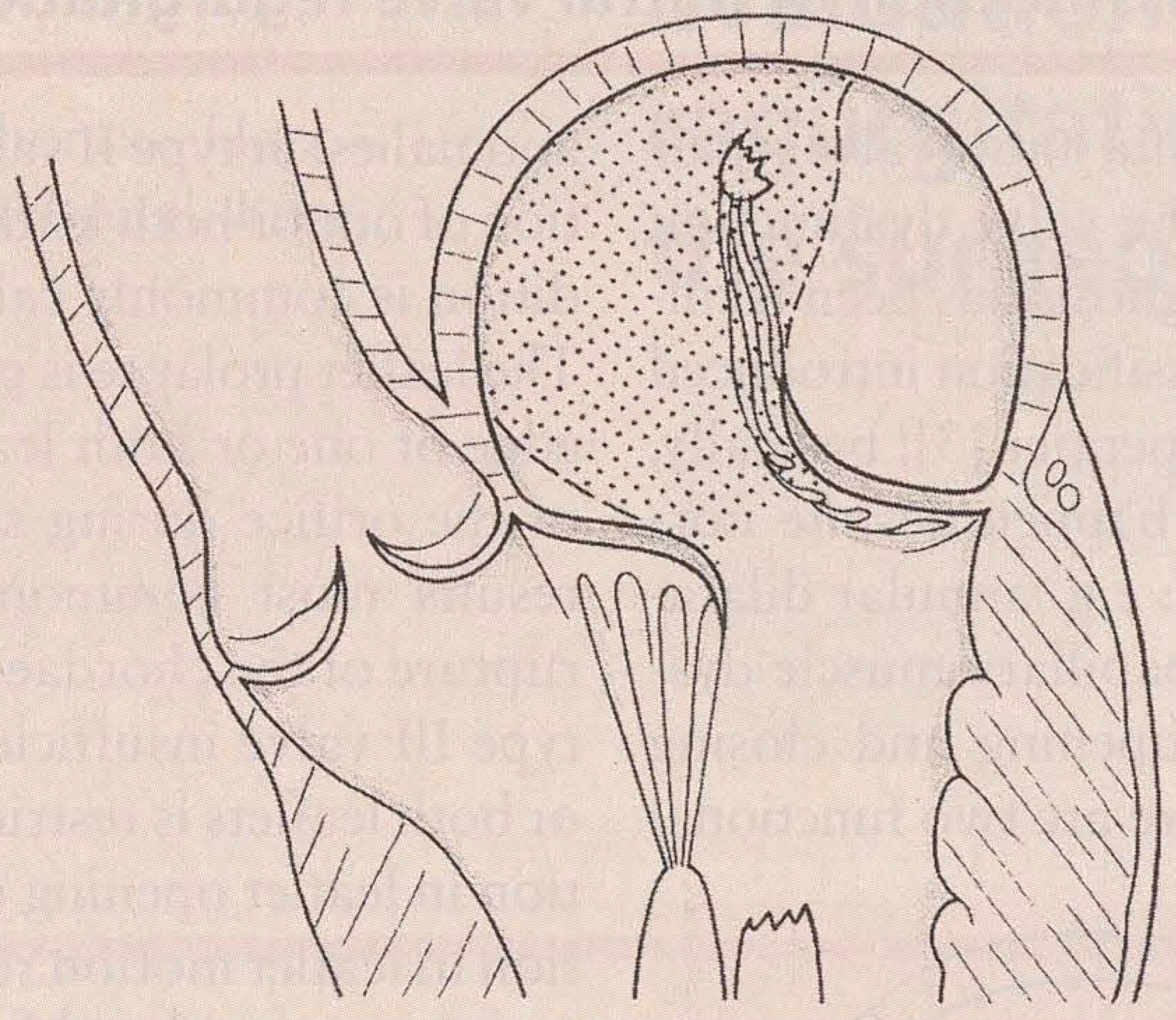
3]: basically, mitral regurgitation may happen despite normal leaflet motion (type I: e.g., annular dilatation, leaflet perforation, papillary muscle dysfunction). Based on the opening and closing motion of each leaflet, there are two functional anomalies: in type II valve insufficiency, the motion of one or both leaflets is increased, this condition is commonly called leaflet prolapse.
The leaflet prolapse is present whenever the free edge of one or both leaflets override the plane of the orifice during systole. Leaflet prolapse results most commonly from elongation or rupture of the chordae or papillary muscles. In type III valve insufficiency, the motion of one or both leaflets is restricted, usually by a limitation in leaflet opening during diastole. Restriction in leaflet motion results from commissural fusion, non perforation, thickening or shortening of the subvalvular apparatus.
Common echocardiographic assessment prior to mitral valve repair has traditionally been directed to the quantification of the regurgitation, the size and function of the left ventricle, the size of the left atrium and the presence of intraatrial thrombus material, as well as the estimation of pulmonary arterial hypertension and possible concomitant lesions (patent foramen ovale, atrial septal defect). A more specific echocardiographic approach to the mitral valve defines exact location of the prolapse (A1-A3, P1-P3), demonstrates the complete coaptation line, evaluates the chordal length and the feasibility of chordal transfer [
5,
6].
With the use of two-dimensional echocardiographic imaging from multiple planes, the leaflet motion is evaluated for the presence of excessive, normal or restricted leaflet motion according to whether the location of maximal systolic excursion of any portion of the mitral leaflet extends beyond to or below (on the ventricle side of) the mitral annulus respectively. The entire mitral apparatus is systematically scanned from the papillary muscle to the left atrium to evaluate each component for the presence of pathologic processes. Focal echogenicity indicating fibrosis or classification is noted. Hypermobile areas showing high frequency motion resulting from disruption of the integrity of the leaflet-chordal-papillary muscle continuity are also assessed. Long-axis view is used to determine which leaflet shows abnormal motion and short-axis view is used to determine which portion of the leaflet shows abnormal motion.
The valve is further studied with Doppler colour flow mapping: particular attention is given to the origin, direction and number of mitral regurgitant jets [
7-
11]. The site of the jet origin — whether along the line of leaflet coaptation or elsewhere — is recorded. For jets originating at the line of coaptation, it is noted whether this jet emanates from the anterior or posterior commissure or centrally. The primary direction of the regurgitant colour Doppler jets as they emanate from the regurgitant orifice is classified as posterior, anterior, central or multiple. As opposed to jet direction, an eccentric jet origin is diagnosed when the regurgitant jet originates from the mid-portion of the leaflet because of a perforation or cleft in the body of the leaflet. Doppler colour flow criteria for this diagnosis are a post valve jet in the left atrium, the pre-jet flow acceleration in systole displaced away from the coaptation line and a second antegrade flow stream in diastole displaced away from the antegrade flow at the coaptation line (
fig. 1-
6).
Transthoracic but also transoesophageal echo-cardiography has limited value for detecting fine details and cannot provide enough information to plan the repair procedure in every case. Therefore the ultimate choice of techniques for each individual valve repair depends more on the detailed inspection during operation and on the individual surgeon's preferences than from preoperative examinations only [
12,
13].
Transthoracic echocardiography may deliver a limited image quality and has some limitations because of echocardiographic windows. Although transoesophageal echocardiography offers superior image quality, the commissures, especially the posterio-medial are sometimes difficult to interpret even with biplane probes, but with the advent of multiplane probes, rotation of the ultrasound beam in one plane without repositioning the probe will be possible. These problems may also be resolved in the future by technologies allowing rotation of the ultrasound beam in all directions and by three-dimensional reconstruction methods [
14].
Today, echocardiography helps the surgeon to understand the mechanism of valve dysfunction and to tailor the procedure according to the preoperative and intraoperative observations.
Perioperative assessment of the need to correct ischaemic mitral valve regurgitation during myocardial revascularisation
A majority of patients suffering from congestive heart failure present some degrees of concomitant mitral valve regurgitation.
This type of mitral regurgitation is a dynamic one and depends mainly from loading conditions and from the left ventricle contractility. Mitral regurgitation may appear mild on preoperative echocardiography and during left ventriculography, but worsens usually with increased workload [
18,
19]. This type of mitral regurgitation might be best assessed by stress echocardiography in order to detect those patients prone to severe mitral regurgitation when afterload increases. Opinion is still divided on the need to correct chronic ischaemic mitral regurgitation of intermediate severity, because there is evidence that such regurgitation improves following coronary bypass grafting in those patients who demonstrate a rapid postoperative recovery of poor preoperative left ventricular function.
Intraoperative inspection and surgical techniques of reconstruction
After induction of anaesthesia, sternotomy, aortic and atrial (optionally bicaval) cannulation, institution of cardiopulmonary bypass and left atriotomy, maximal exposure of the mitral valve is accomplished by well-described techniques, including self-retaining retractor designed for mitral valve repair.
The mitral apparatus is systematically inspected, including the mitral annulus, the leaflets, the chordae tendinae and the papillary muscles. The leaflets and annulus are inspected for redundancy, vegetation, perforation, fibrosis and calcification.
Care is taken to identify which sections of the mitral leaflets are abnormal and to identify the predominance of anterior versus posterior leaflet involvement. The chordae tendinae are systematically inspected to determine the presence of chordal elongation, fibrosis, rupture, calcification or fusion. Likewise, papillary muscles are inspected to identify elongation, rupture or infarction.
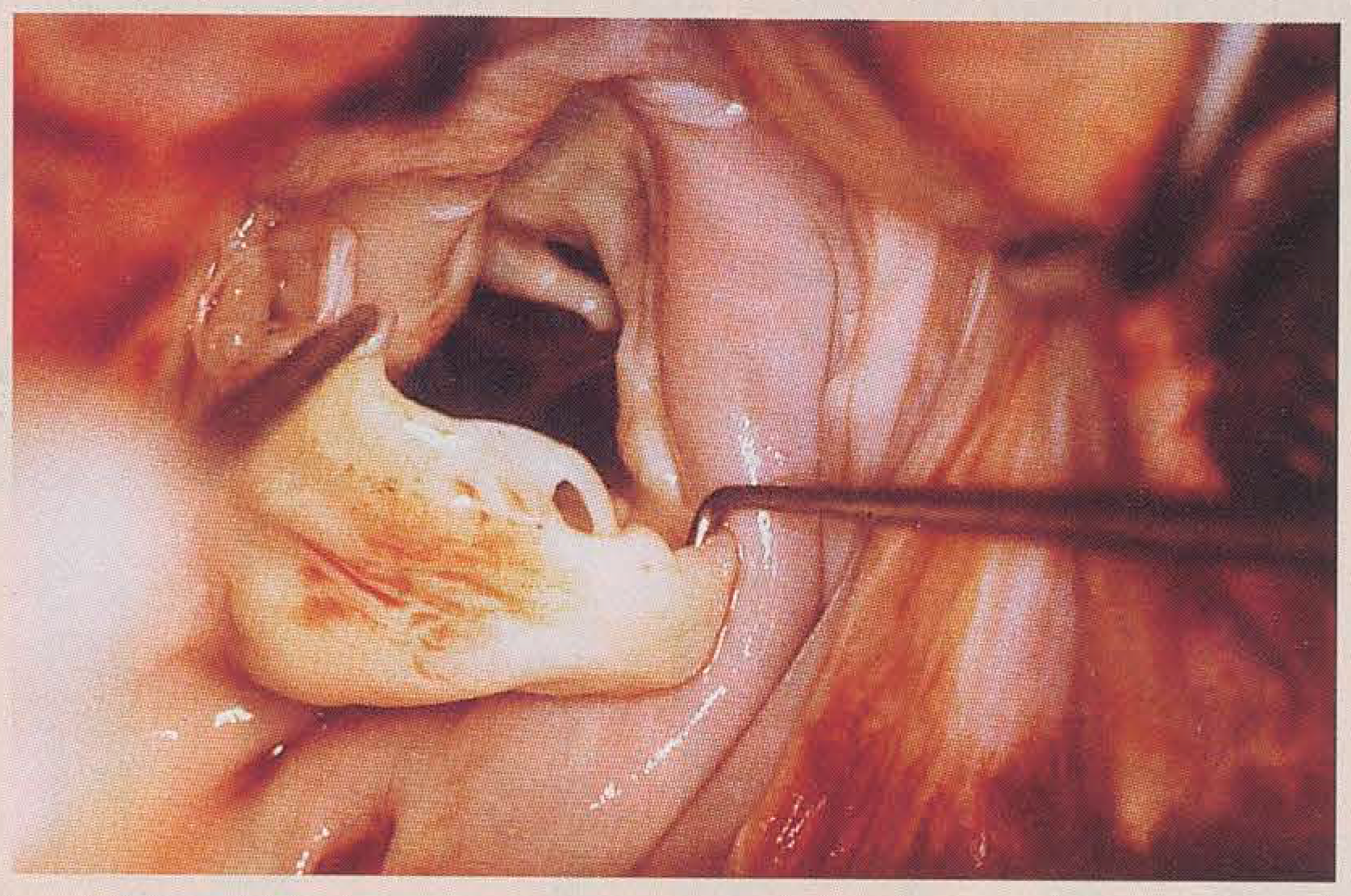
A nerve hook is used to assess leaflet range of motion by pulling each part of the leaflet edge toward the left atrium, determining whether its subvalvular restraint would allow it to go towards or past the plane of the mitral annulus. Leaflet motion is classified as restricted when the maximal extension of the leaflet edge is on the left ventricular side of the mitral annulus plane, as normal when the leaflet edge is in the annulus plane and as excessive when the leaflet edge extends into the left atrial side of the mitral annulus (
fig. 7). The disease is further categorised by the surgeon as to whether it affects the anterior or posterior leaflet or both. When there is papillary muscle infarction, the presence of elongated or disrupted papillary muscle is recorded. In patients with normal leaflet appearance and chordal restraint, the size of the mitral annulus is compared with the surface area of the anterior mitral leaflet, to identify annular dilatation in the absence of other abnormalities of the mitral apparatus. In patients with leaflet perforations — usually due to endocarditis [
20] — its presence and location are recorded.
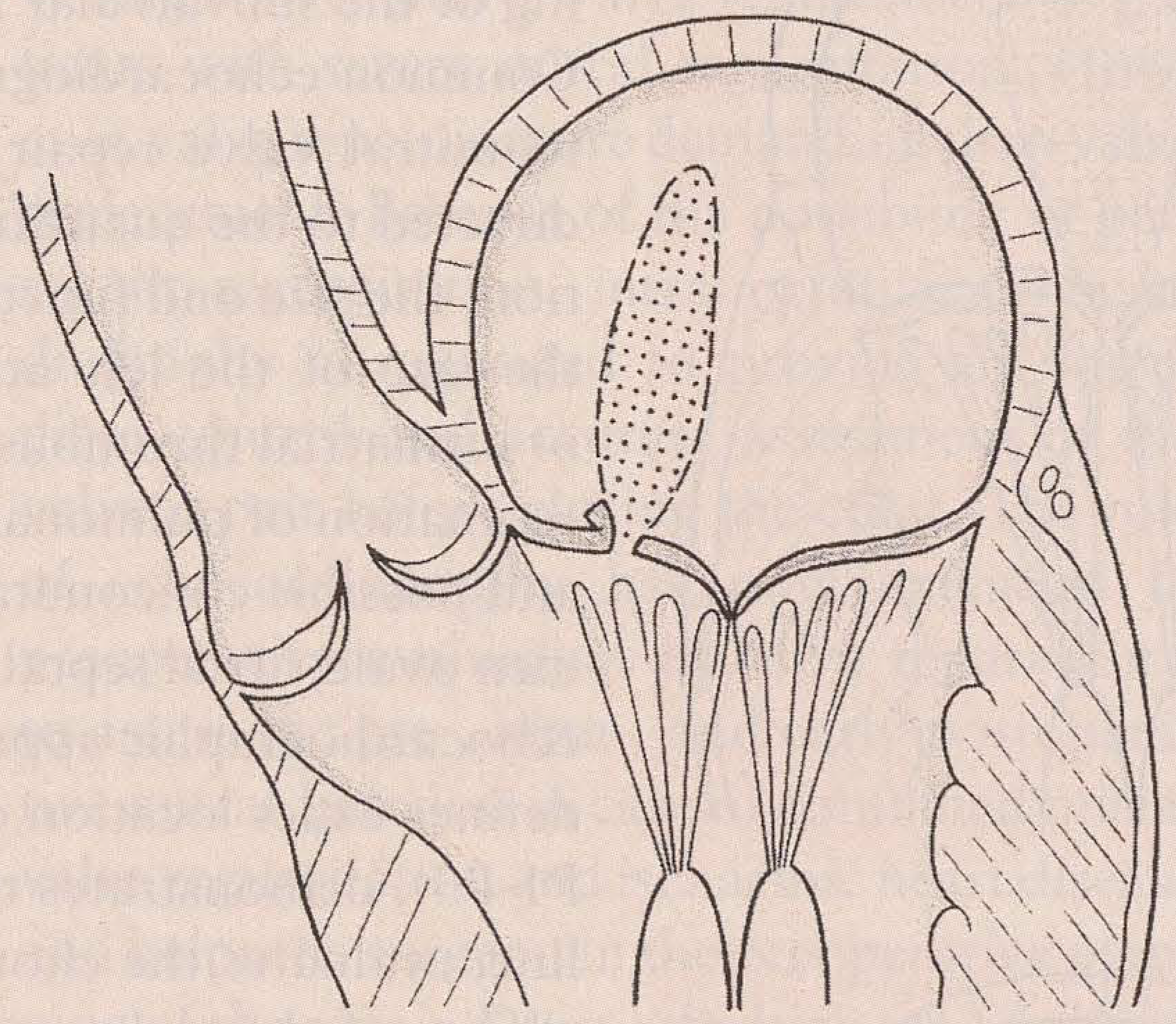
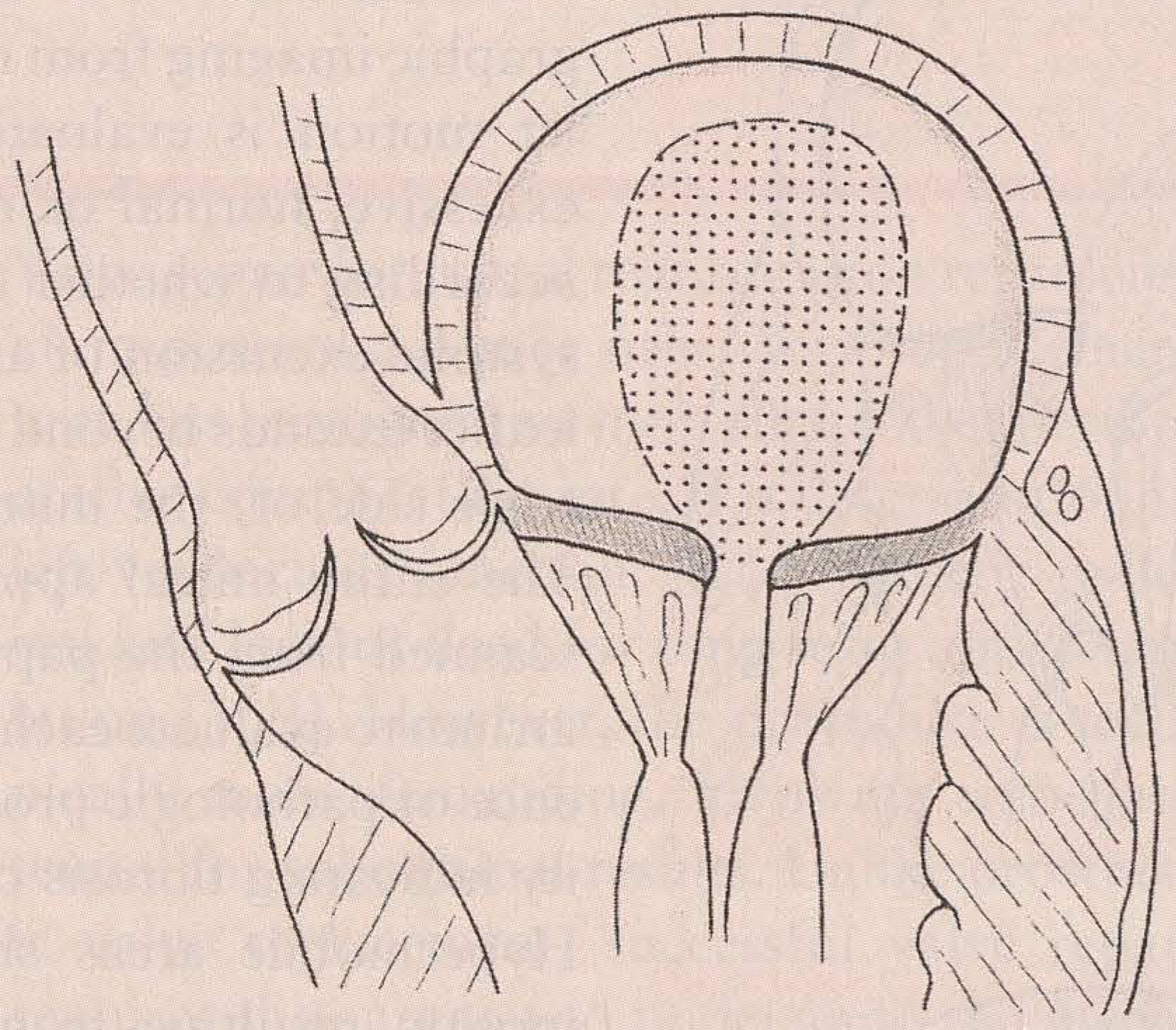
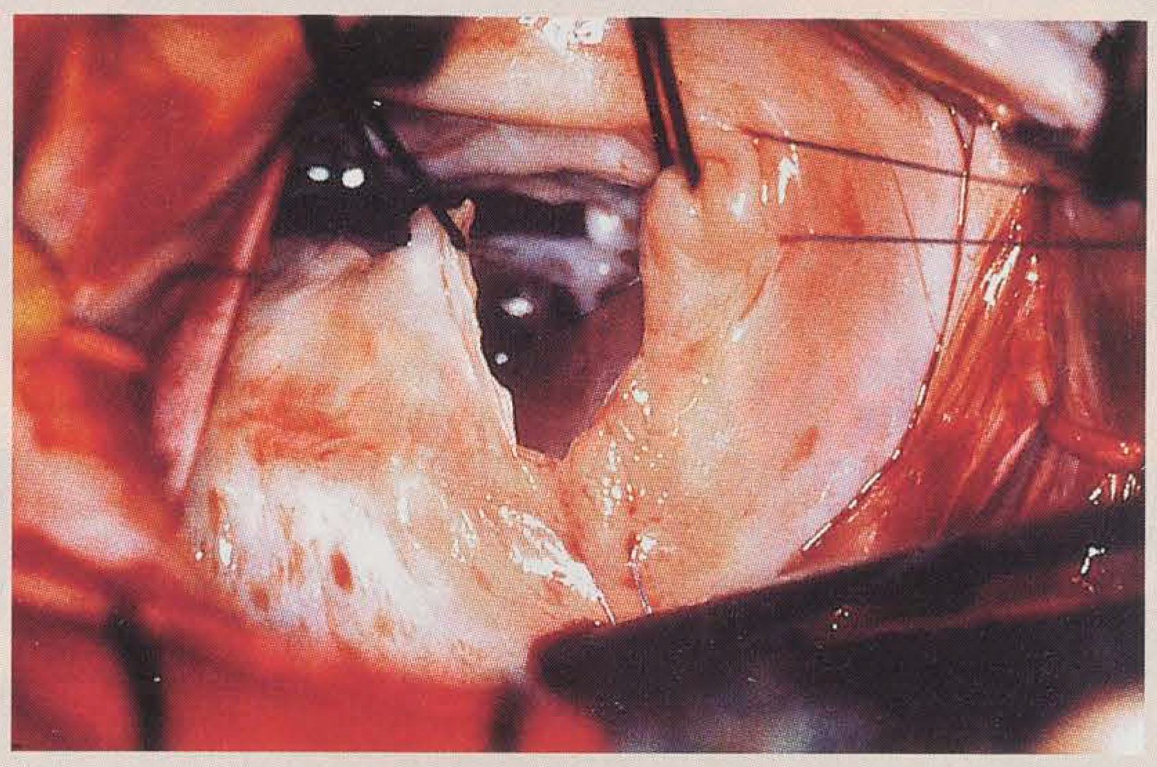
Numerous techniques are available to deal with the different pathologic abnormalities that may be responsible for mitral valve regurgitation: basically each structure of the mitral valve (leaflets, annulus, chordae tendinae and papillary muscles) may be addressed surgically (
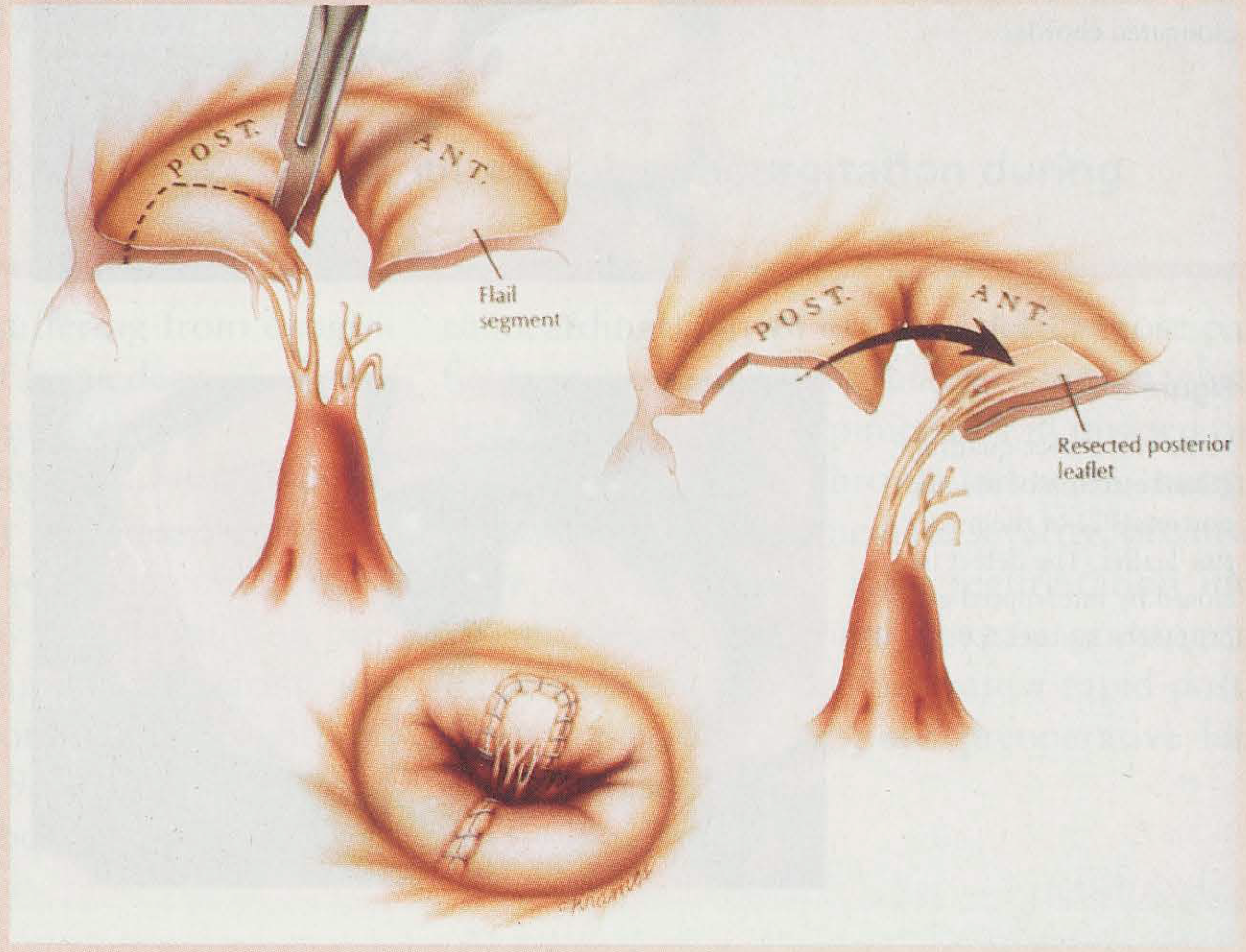
table 1). These techniques include leaflet resection (
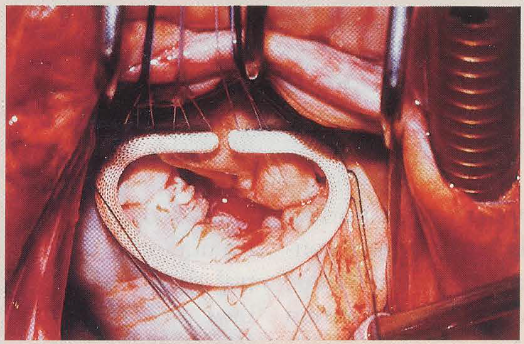
fig. 8), annular plication, annuloplasty using partial or complete ring (
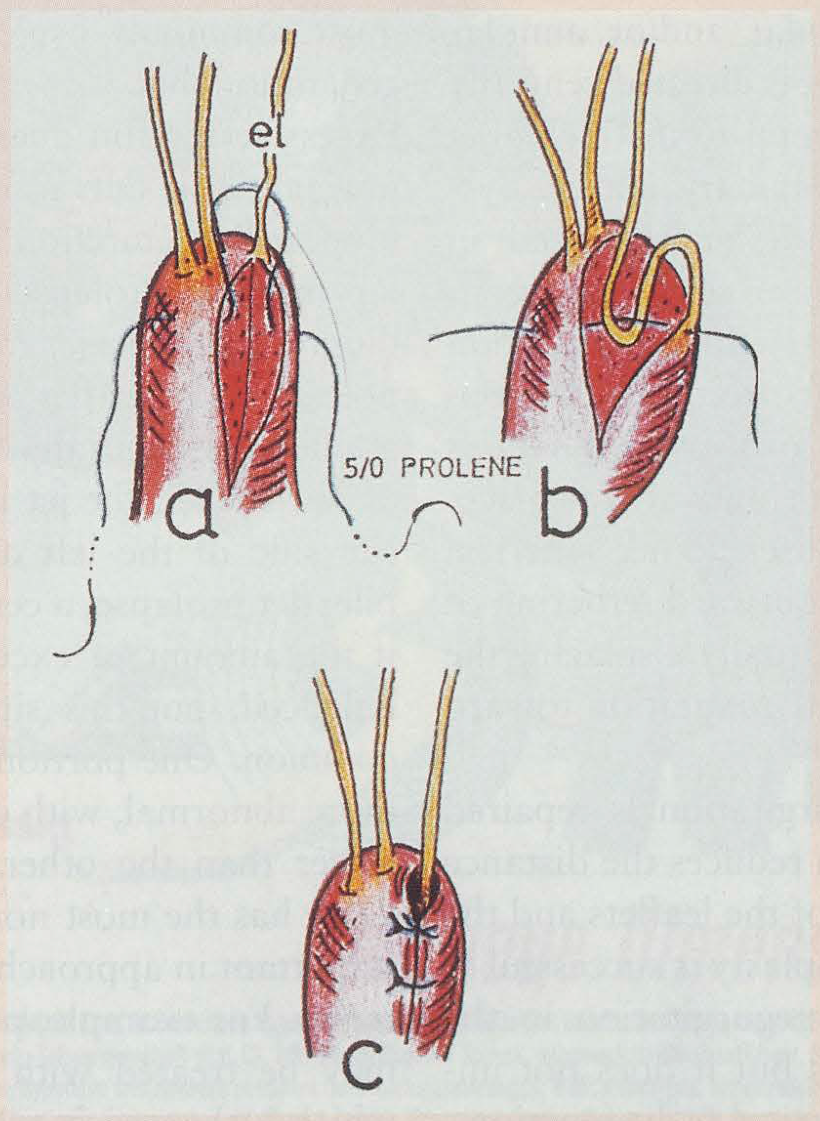
fig. 9), sliding annuloplasty, annular or leaflets patch repair, debridement of granulations and abscesses, closure of leaflet clefts and suture of perforations, chordal shortening (
fig. 10) transfer or transposition (
fig. 11), division and replacement with artificial chordae, as well as papillary muscle reimplantation [
3,
4,
12].
Table 1.
Basic technical possibilities to repair mitral valve regurgitation.
Table 1.
Basic technical possibilities to repair mitral valve regurgitation.
Isolated annular dilatation, functional mitral regurgitation due to left ventricular dilatation and dysfunction and simple posterior leaflet prolapse with ruptured or elongated chordae are the easiest pathologies to repair.
Extensive leaflet prolapse, especially if the anterior leaflet is involved, commissural prolapse, highly degenerated or calcified valves with subvalvular involvement require a more complex repair (
table 2).
Table 2.
Probability of mitral valve repair depending on the mechanisms of regurgitation and anatomic lesions.
Table 2.
Probability of mitral valve repair depending on the mechanisms of regurgitation and anatomic lesions.
With growing experience and improvement in surgical techniques, the probability to encounter a so-called "irreparable" valve has dramatically diminished; even highly complex valves with bileaflet prolapse and valve damaged by endocarditis are amenable to successful reparation. The progress realised in this field has dramatically contributed to increase the incidence of mitral valve repair, from 10 to 20% fifteen years ago to over 70% in the most recent period.
Relation of mechanism of mitral regurgitation to surgical approach in mitral regurgitation
Normal leaflet motion
Mitral regurgitation in patients with normal leaflet motion occasionally results from a congenital leaflet cleft or a leaflet perforation due to endocarditis. Repair of valve with perforation requires patch closure. Mitral regurgitation with normal leaflet motion most commonly results from left ventricular and/or annular dilatation, in which the jet is directed centrally or posteriorly [
7,
9]. Several terms including annular dilatation and papillary muscle dysfunction are unfortunately interchanged in scientific literature and are sometimes confused clinically. Ventricular annular dilatation with normal leaflets results from cardiomyopathy or any myocardial process that causes ventricular dilatation with outward displacement of the papillary muscle. This spherical ventricular shape causes outward tethering of the subvalvular support, usually displacing the most basal point of mitral coaptation toward the apex.
This type of mitral regurgitation is repaired with annuloplasty, which reduces the distance between the hinge point of the leaflets and the coaptation point. Annuloplasty is successful in reducing or eliminating regurgitation in the majority of such patients but it does not improve the underlying ventricular dysfunction.
Excessive leaflet motion
Excessive leaflet motion can result from disruption or elongation of any portion of the mitral apparatus, including the papillary muscle, the chordae tendinae or the leaflet tissue itself, all of which end in prolapse [
16]. Leaflet and chordal abnormalities in this category occur most commonly because of myxomatous degeneration but also because of endocarditis. Excessive motion due to papillary muscle abnormalities results most commonly from focal myocardial infarction at the basis of the papillary muscle. Prolapse as assessed by echocardiography reflects significant extension of any portion of the leaflet past the level of the mitral annulus. In surgically defined excessive motion of one leaflet, the jet is deflected to the opposite side of the left atrium. In patients with bileaflet prolapse, a central jet direction is seen if the amount of excessive motion is exactly balanced, but this situation is relatively uncommon. One portion of the leaflet is usually more abnormal, with one component of the jet larger than the other. Which portion of the valve has the most normal motion is critically important in approaching it correctly for valve repair. For example, posterior chordal rupture may be treated with quadrangular resection which is the easiest type of mitral valve repair. Chordal elongation may be treated by shortening and papillary muscle abnormalities may be treated using reimplantation or shortening of the involved papillary muscle. These latter procedures are technically much more demanding.
Restricted leaflet motion
Restricted leaflet motion usually results from the chronic fibrotic effects of rheumatic heart disease or less commonly from papillary muscle infarction. The regurgitant jet may be central if the systolic motion of both leaflets is equally affected. When rheumatic fibrosis predominantly affects the posterior leaflet, the relatively normal anterior leaflet overrides the restricted posterior leaflet causing a posterior jet direction. Thus, in patients with restricted leaflet motion and an asymmetric distribution of fibrosis, the jet is directed towards the most affected leaflet. This type of mitral regurgitation must be approached with techniques such as debridement, fenestration of fused chordae, commissurotomy and annuloplasty. Although commissurotomy is fairly easy, splitting of fused chordae in extensive fibrosis is a more difficult technique of repair.
Assessment of mitral valve function after repair
Echocardiography is an integral part of the optimal management of patients with mitral regurgitation before, during and after valve repair. Stewart and co-authors have reported that intraoperative echocardiography after repair of mitral valve regurgitation allows detection of failed repair in 5 to 8%, permitting further surgical treatment during the same thoracotomy [
10,
21,
22].
In the early days of mitral reconstruction, intraoperative echocardiography was performed with epicardial transducers but is now largely performed by the transoesophageal route. This latter approach has the advantages of not interfering with the surgical field and provides excellent view of the left atrium and the mitral valve and allows simultaneous assessment of the left ventricle. Intraoperative echocardiography assessing the function of the mitral valve immediately after repair helps the surgeon to detect unsatisfactory results that may be rectified during the same operation.
In particular, residual mitral regurgitation should be identified and quantified and its mechanism should be clearly elucidated.
Transoesophageal echocardiography is highly sensitive and trivial regurgitation is found in the majority of mitral valve repair [
21,
23-
25]. With the high quality of echo probes, echo-perfection is difficult to achieve and therefore the question has arisen as to how much regurgitation still constitutes an acceptable result. The group of Cleveland Clinic, using a five points scale of regurgitation showed that results were excellent but there was a trend towards late reoperation without increased mortality in those with moderate regurgitation (1+, 2+). A majority of these patients needed reoperation within two years in this series [
21]. However, some discrepancy in severity of mitral regurgitation is commonly observed between intraoperative and predischarge studies. Reasons for these discrepancies include the greater sensitivity of transoesophageal as compared with transthoracic echocardiography, postoperative changes in left ventricular function, normalisation of loading conditions, afterload reduction and cessation of inotropic support [
24].
The mechanisms of mild to moderate residual mitral regurgitation may be more important than its severity. Therefore, a residual prolapse regardless of the severity of associated regurgitation may represent an absolute indication for immediate revision whereas a moderate regurgitation through the valve closure line may be regarded as acceptable and is unlikely to progress if no area of poor coaptation can be identified. The early and late benefits of valve repair justify aggressive efforts to avoid valve replacement as often as possible. Where residual or recurrent moderate to severe mitral regurgitation occurs, revision of the repair may still be a valuable option [
22,
25].
Left ventricular outflow tract obstruction caused by systolic anterior motion of the mitral valve should also be identified. This complication may be encountered particularly when a complete annuloplasty ring has been inserted. The long posterior leaflet may extend anteriorly, allowing the coapting leaflet edge to obstruct the left ventricular outflow tract during systole. Before correcting a systolic anterior motion by additional surgical approach, positive inotropic drugs should be stopped and filling pressure optimised. Sometimes β-blockers and calcium antagonists help to manage this condition without surgical intervention. Occasionally, shortening the posterior leaflet by the sliding leaflet technique or by removing the annuloplasty ring may be the solution to cure this problem definitively [
26].
Pitfalls of intraoperative echocardiography
There are specific conditions and pitfalls associated with immediate assessment of the repaired mitral valve in patients with open chest, following weaning from extracorporeal circulation. The following conditions can alter the imaging quality: (1) dried probe in the oesophagus or air bubbles, (2) non-aspirated air from the stomach, (3) small or empty left atrium, (4) invagination of the left atrial appendage, (5) trapped air in the posterior pericardium and (6) spontaneous contrast [
12,
27]. By correcting these conditions, a transient mitral regurgitation may disappear rapidly; furthermore, immediate postoperative mitral regurgitation is closely related to the quality of the left ventricular function. Patients with long-standing heart disease and left ventricular failure prior to surgery may have residual mitral regurgitation after the heart restarts its activity because of increased afterload.
Transient mitral regurgitation is caused by functional restricted leaflet motion due to left ventricular dysfunction and low contractility, which exerts a higher than normal tension on the subvalvular apparatus, preventing the leaflet from closing normally. Subtle drug support of the LV restores usually an acceptable contractility with normal leaflet coaptation and absence of mitral regurgitation.
Early and long-term follow-up
Transthoracic echocardiography is usually adequate for follow-up of the valve repair and is usually performed before discharge in order to have a baseline observation of the function of the mitral valve and of the left ventricle. The superior quality of transoesophageal echocardiography may be required if there is any clinical doubt about mitral valve function. Morbidity and mortality from postoperative myocardial failure are high in patients with asymptomatic left ventricular impairment on predischarge echocardiography, particularly if they had a left ventricle impairment preoperatively or if they were in functional class III or IV at the time of surgery [
16,
17]. These patients should have early follow-up review by their cardiologist and if left ventricle function is poor, they should be maintained on diuretics and converting enzyme inhibitors. Follow-up echocardiography should be performed at 3 to 6 months intervals and subsequently according to the clinical condition of the patient.
Patients with less than "echo-perfect" results after mitral valve repair
Not all patients whose repair is deemed successful by the operative team have perfect result on post-cardiopulmonary bypass with no mitral regurgitation. Mild to moderate regurgitation (1+, 2+) has traditionally been considered as an acceptable result.
The observations of Fix et al. [
21] indicate that patients with less than "echo-perfect" results of mitral repair with mild to moderate mitral regurgitation during intraoperative echocardiography do not develop worse in most respects than those with an "echo-perfect" result with no residual mitral regurgitation. However, patients of this series with 1+ or 2+ mitral regurgitation on their postbypass echocardiography had a threefold increase in the annual reoperation rate for recurrent mitral valve dysfunction compared with the rate of the control group of about 2% per patient/year. Why these patients tended to require more reoperation is not completely clear. It is likely that whatever factors cause difficulty in achieving perfect repair initially are the same factors leading to greater incidence of late reoperation.
Conclusions
Actual experience with mitral valve repair seems to confirm that predictability of the techniques and stability of the results in valve reconstruction may be obtained when a comprehensive approach to the aetiology of the disease and the functional classification are respected. Thromboembolic events are much lower than after mitral valve replacement, there is no need for longterm anticoagulation and careful followup echocardiographic studies may pick up those patients that need redo-surgery because of residual or recurrent mitral regurgitation.
Echocardiography is an important adjunct in preoperative, intraoperative and postoperative assessment of the valve function and has the potential to improve clinical outcome. However, it is still possible to perform successful mitral valve repair without intraoperative echocardiography, particularly if the surgeon is very experienced and if the valve pathology is common and easy to repair.
When echocardiography is performed, the cardiologist and the surgeon should understand each other's need and use. Not only the identification of valve dysfunction is needed but imaging of the entire valve and subvalvular structures should identify the mechanisms of mitral regurgitation. The surgeon should be able to interpret these images, to correlate them to operative findings, to understand the limitation of the techniques and the influence of haemodynamic condition on echocardiographic appearance.
The necessity of intraoperative echocardiography in all cases of mitral valve repair is still controversial. There is no randomised clinical trial to assess its benefits. Whenever transoesophageal echocardiography is performed, it should be carried out under haemodynamic conditions similar to normal so as to avoid the pitfalls of overdiagnosing mitral regurgitation. The intraoperative post-repair assessment should be better performed ten to twenty minutes after weaning the patient from cardiopulmonary bypass when normal filling pressure and systemic afterload have been obtained.
Echo-perfection is not essential and it is very important for the surgeon and the cardiologist to learn what is and what is not acceptable in order to avoid unnecessary additional cardiopulmonary bypass and valve replacement: basically the mechanism more than the degree of residual mitral regurgitation is important.
Finally, the value of intraoperative echocardiography may lie primarily in (1) guiding inexperienced surgeons, (2) assessing valve competence when visual and manual assessment are equivocal, in case of complex or uncommon pathology, (3) identifying mechanism of residual regurgitation to guide a second attempt at valve repair rather than replacement, (4) assessing new techniques of repair, (5) assessing high risk patients for specific complication such as systolic anterior motion as well as (6) identifying potential cause of postoperative low cardiac output or arterial hypotension.