Next Article in Journal
SCOT—Representing Switzerland’s Future Cardiologists
Previous Article in Journal
Prevention, Epidemiology, and Prognosis of Perioperative ­Myocardial Injury
 
 
Cardiovascular Medicine is published by MDPI from Volume 28 Issue 1 (2025). Previous articles were published by another publisher in Open Access under a CC-BY (or CC-BY-NC-ND) licence, and they are hosted by MDPI on mdpi.com as a courtesy and upon agreement with Editores Medicorum Helveticorum (EMH).
Editorial

The Subcutaneous Implantable Cardioverter-Defibrillator: Pros and Cons

by
Nicolas Dayal
1,*,
Haran Burri
1,
Christian Sticherling
2 and
Angelo Auricchio
3
1
University Hospital of Geneva, Geneve, Switzerland
2
University Hospital of Basel, Basel, Switzerland
3
Cardiocentro, Lugano, Switzerland
*
Author to whom correspondence should be addressed.
Cardiovasc. Med. 2016, 19(4), 121; https://doi.org/10.4414/cvm.2016.00396
Submission received: 20 January 2016 / Revised: 20 February 2016 / Accepted: 20 March 2016 / Published: 20 April 2016

Abstract

The subcutaneous ICD (S-ICD) has recently been introduced as an alternative to transvenous devices. The system preserves venous access and reduces risk of lead issues and of systemic infection. However, pacing for antitachycardia, antibradycardia and cardiac resynchronisation therapy is not available, and the risk of inappropriate shocks is relatively high (although it is improving). This article provides an overview of this novel therapy and discusses its advantages and shortcomings.

Introduction

Since the first implantable cardioverter-defibrillator (ICD) was implanted in a human in 1980 [1], technological advances have made these devices the most effective treatment option for the prevention of sudden cardiac death in both primary [2,3] and secondary [4,5] settings. Currently, the vast majority of implanted devices are transvenous ICDs (TV-ICDs), with one or more leads implanted in the heart via the venous system and connected to a pulse generator located most frequently in the subcutaneous or submuscular tissue in the pectoral region. These systems are prone to a certain number of complications, both acute (pneumothorax, lead dislodgment, cardiac perforation, tamponade) and chronic (systemic infections, venous stenosis or occlusion and lead failure). For example, the rate of implant-related complications is estimated to be around 2–3% with TV-ICDs [6,7], rising to 6% in the case of cardiac resynchronisation therapy-defibrillator (CRT-D) devices [8]. However, it is the rate of chronic complications, most notably lead failure, that is the most troublesome, with rates of surgical revision of 2.5% at 5 years [9] and with potentially dramatic consequences such as inappropriate shocks (IAS), ineffective therapy and loss of pacing. Young, active patients are especially at risk of lead failure owing to their longer life expectancy and the increased mechanical stress placed on the leads. Moreover, lead failure constitutes, with device infection, one of the main indications for device extraction, a procedure itself linked to a relatively high morbidity (up to 2.4%) and mortality (up to 1%), as recently shown by the Electra registry [10]. In effect, lead integrity seems to be the Achilles heel of TV-ICD devices.
For these reasons, interest has grown in developing alternative novel ICD systems that reduce or eliminate vascular injury, minimise lead mechanical stress and vascular interaction, and may be more practical to implant than epicardial systems. Attention turned to subcutaneous systems, at first in the paediatric population, with initial systems consisting of standard TV-ICD leads implanted in a subcutaneous position [11], but necessitating epicardial sensing and pacing leads [12,13]. Finally, a dedicated subcutaneous ICD system (S-ICD system, Cameron Health, San Clemente, CA, USA) has been recently developed and approved for use in Europe in 2009 and the USA in 2012 [14]; such a device represents a possible alternative to TV-ICDs in patients without an indication for bradycardia pacing or cardiac resynchronisation therapy (CRT), or the need for antitachycardia pacing (ATP).

Description of the Therapy

The S-ICD Device

The S-ICD, now in its second version (Emblem S-ICD model, Figure 1) and manufactured by Boston Scientific (Marlborough, MA, USA), consists of a pulse generator and a tripolar subcutaneous lead. The current pulse generator is 20% smaller than the initial model (SQ-RX), weighing 130 g and occupying 59.5 cc of volume, and has a greater durability, with an estimated life of 7.3 years. The tripolar lumenless lead is made of polyurethane and consists of an 8 cm shocking coil banked by distal and proximal sensing electrodes.
There is a dedicated tablet-format programmer with relatively simple programming options. The device is enabled for wireless remote monitoring.

Implantation Procedure

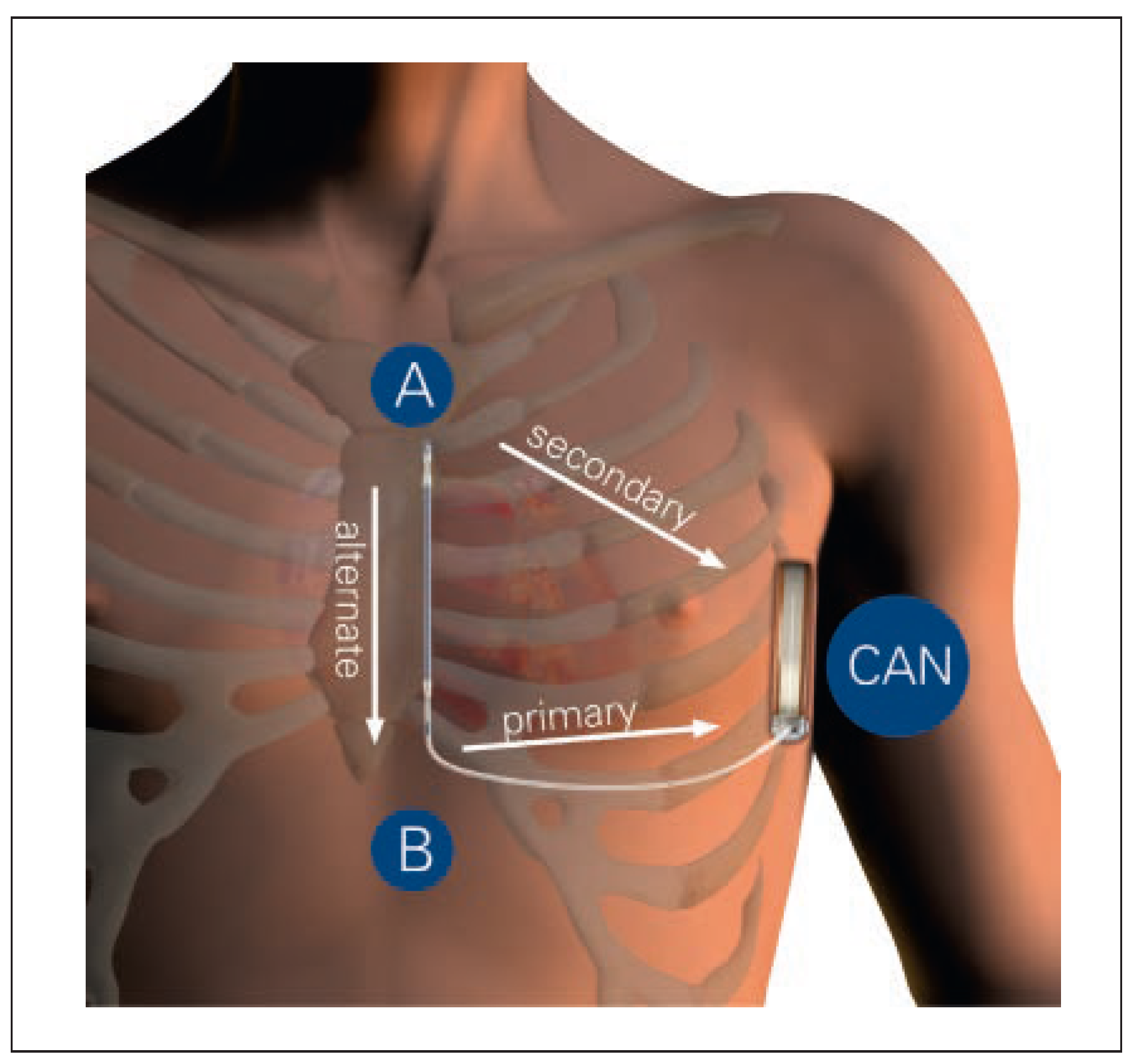
The procedure is performed under either local or general anaesthesia. Briefly, the pulse generator is placed either in the subcutaneous tissue or the intermuscular plane between the serratus and latissimus dorsi muscles, overlying the sixth rib in between the midaxillary and anterior axillary lines (Figure 2). The electrode is tunnelled to a position of maximum 1 to 2 cm to the leh of and parallel with the sternal midline via one to two parasternal incisions, one (optional) distally at the manubriosternal junction (second rib) and one proximally at the xiphoid process. Electrode stability is assured with an optional distal suture on the periostial fascia above the sternum and ribs, and with the use of an anchoring sleeve proximally at the xiphoid process. The whole implantation procedure is realised with the use of anatomical landmarks and usually without the need for fluoroscopy (<1% in the IDE study sanctioned by the US Food and Drug Administration [15]). Average procedural time in the largest worldwide registry (EF-FORTLESS registry [16]) was 69 ± 27 minutes. Figure 3A,B show a patient on the day following implant, Figure 3C,D the cosmetic result at 6-month follow-up.

Preimplantation Screening

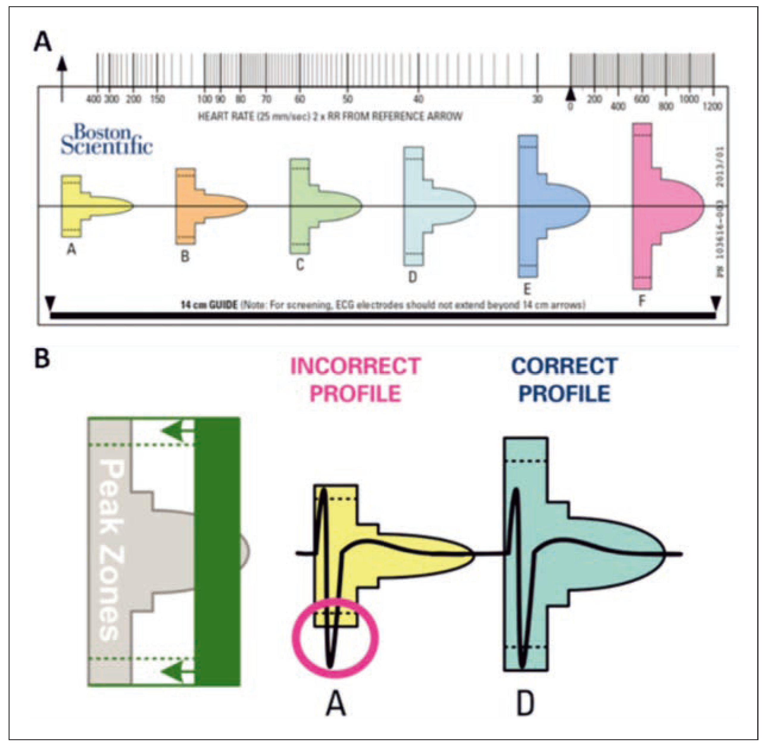
The screening procedure is an important step in assuring eligibility of the patient and potentially reducing the incidence of unwanted events such as IAS. Initial patient selection is based around the absence in the S-ICD system of durable bradycardia pacing, CRT and ATP therapy. In this respect, patients with an actual or anticipated bradycardia indication should be excluded, as well as patients fulfilling the criteria for CRT. Patients who could benefit from ATP therapy, such as those with known monomorphic ventricular tachycardia (VT) or with pathologies conferring a high risk of VT (e.g., sarcoidosis and arrhythmogenic right ventricular dysplasia) should be considered for TV-ICD. Selected candidates should undergo an electrocardio gram (ECG) screening test with a screening tool provided by the manufacturer (Figure 4A). Standard cutaneous electrodes are placed in three positions representing the two sensing electrodes and the can of the S-ICD device: 1 cm to the leh of the xiphoid process (representing electrode B in the S-ICD), 14 cm cranial to the xiphoid process on the chest wall (representing electrode A), and either the fihh or sixth intercostal space on the leh mid-axillary line (representing the can position) (Figure 2). ECG recordings in the three derivations mimicking the S-ICD sensing vectors are recorded, and the screening tool is used to verify suitability of these recordings (Figure 4B). The manufacture recommends at least one adequate vector (standing and supine) before considering S-ICD implantation, but it is desirable to have at least two adequate vectors, which will facilitate reprogramming in the event of sensing issues. Potential sources of failure include, most frequently, large or late peaking T-waves and, less frequently, low-amplitude QRS complexes that are too small to fit in the smallest window of the tool, and thus are likely to be undersensed by the device itself.
Particular attention should be paid to those cardiac disorders in which dynamic changes of the R-T wave relationship are expected, such as hypertrophic cardiomyopathy or Brugada syndrome. Indeed, vector change may cause dynamic R-wave undersensing or T-wave oversensing, to which the device cannot automatically adjust. In this particular setting, therefore, it is important to use pharmacological challenges in order to unmask potential issues and to assess precisely the most stable sensing vector, as recently demonstrated by Conte et al. [17].

Arrhythmia Detection

As mentioned, the S-ICD system uses three sensing electrodes: the distal electrode (A) located in the upper sternum, the proximal electrode (B) located at the xiphoid level and the active can (CAN) located in the lateral fihh or sixth intercostal space (see Figure 2). Three sensing vectors can be created from these electrodes: a primary vector, from electrode B to the can (resembling surface ECG lead DI), a secondary vector from electrode A to can (resembling lead DII) and, finally, an alternate vector from electrodes A to B (resembling lead aVF). The most appropriate vector to avoid noise, QRS double counting and T-wave oversensing is chosen automatically by the device, but can be manually overridden. Once the vector is chosen, detection occurs in several successive steps, depending on whether programming includes a single shock-only zone or (preferably) dual zones with both conditional and shock zones [18].
An algorithm to avoid oversensing is used, comprising threshold adaptation to the R-wave, a decay function and three double detection algorithms to avoid T-wave oversensing and double counting. A rate-based analysis is undertaken, using an average of the four last beats to detect tachyarrhythmia. In the case of rates above the programmed shock zone threshold, detection stops at this step and therapy is delivered. In the case of programming with a conditional zone, the system uses three further steps to decide on appropriateness of therapy. A static waveform analysis of the QRS complex compares the current beat with a stored template, using up to 41 points to assess correlation. A correlation of >50% classifies the rhythm as supra ventricular and prevents therapy. This cutoff is lower than for TV-ICDs (e.g., 94% in Boston Scientific’s Rhythm ID algorithm) because of the greater number of points analysed (approximately eight points in transvenous systems). In cases of noncorrelation, a dynamic waveform analysis assesses the correlation between the current tachycardia beat and the previous three tachycardia beats. In the case of polymorphism, the rhythm is classified as ventricular, whereas in the case of monomorphism, the device moves to the third and final step of morphology analysis, which analyses QRS width in relation to the stored template. In the event of prolonged QRS duration >20 ms, the rhythm is classified as ventricular.
The device uses an 18/24 beat duration criterion before charging the capacitors, with automatic extension to allow spontaneous resolution of unsustained events and with a confirmation algorithm at the end of capacitor charging before delivering therapy.
The shock zone is programmable between 170 and 250 bpm, and the conditional zone between 170 and 240 bpm. Although programming is variable in the different published studies, many used a shock zone at around 200–220 bpm [19,20,21].

Shock Delivery

Only a single shock energy of 80 J is programmable in the S-ICD, except during defibrillation threshold (DFT) testing, where shock energies of 10–80 J at 5 J intervals are programmable. DFT testing is recommended at 65 J to ensure a margin of safety [22], although the necessity of DFT testing in the S-ICD has not been studied, and some centres do not perform it systematically. Standard shock polarity is from coil to can, but the device automatically reverses polarity in the case of unsuccessful therapy, with a maximum of five shocks available. The S-ICD system has a limited post-shock pacing function, with the possibility of delivering demand pacing at 50 bpm for up to 30 seconds aher a shock in the case of asystole longer than 3.5 seconds. The system uses a 200 mA biphasic transthoracic pulse for this function. The current S-ICD model can store up to 40 treated and untreated episodes.

Advantages and Disadvantages of the S-ICD

Table 1 shows other advantages and disadvantages of S-ICDs compared with TV-ICDs. Preservation of vascular access in young patients is a major advantage. Another major advantage is the low risk of systemic infection, which is of particular interest in patients with high infectious risk such as the immunocompromised and holders of artificial valves. Another population who are likely to benefit are those with chronic kidney disease, especially those requiring dialysis, as they have a greater risk for infection and more lead extraction-related complications as a result of increased calcification around implanted leads. Concerning the issue of myocardial damage, despite higher energy shocks compared with TV-ICDs, it is estimated that only approximately 10% of this energy reaches the myocardium when delivered subcutaneously. Moreover, there seems to be less myocyte damage, as indicated in swine models showing an elevated troponin level aher TV-ICD shocks but not aher S-ICD shocks [23]. Finally, the lack of transvenous lead extraction and associated risks makes the S-ICD an attractive option for young active patients, especially those at low risk of bradycardia or monomorphic VT, such as patients with Brugada syndrome, long QT syndrome and hypertrophic cardiomyopathy.
Amongst the list of disadvantages, the absence of ATP therapy seems to be an important hurdle, as it has proven to be safe and effective for the treatment of fast VTs [24], as well as increasing quality of life and possibly lowering mortality [25]. Patients with known monomorphic VT, history of prior effective ATP therapy or at high risk for VT should not receive an S-ICD.
However, it is likely that in the future, the S-ICD will be able to communicate with leadless pacemakers to deliver ATP, if such therapy is found to be required. Leadless pacemakers may also be a solution for patients who develop a requirement for antibradycardia pacing, although this is only available as a VVIR system for the time being.
The absence of an atrial lead is also a potential disadvantage as atrial arrhythmias (which are prevalent in this patient population) will not be diagnosed. However, algorithms allowing diagnosis of atrial fibrillation (AF) based on ventricular rhythm irregularity may allow diagnosis in single-chamber devices in the future. Battery longevity, although improved compared with the previous version, is still less that reported for TV-ICDs produced by the same manufacturer. Finally, the system is currently not magnetic resonance imaging-conditional (contrary to most of the current TV-ICD systems), but will become so in the future.

Indications for the S-ICD

The 2015 European Society of Cardiology guidelines on the management of patients with ventricular arrhythmias and the prevention of sudden cardiac death [26] state that the S-ICD is a class IIa (level of evidence C) recommendation as an alternative to TV-ICDs in patients who have an ICD indication but do not require antibradycardia pacing, CRT or ATP. The guidelines also state that S-ICDs are a class IIb (level of evidence C) indication as an alternative to the TV-ICD in patients with venous access problems, aher removal of a TV-ICD for infection, or in young patients who require longterm therapy.
As mentionned above, patients at high risk of systemic infections (e.g., patients on dialysis or with valve prosthesis, etc.) are also good potential candidates.

Current Evidence

Current evidence is based on a certain number of regulatory studies [15,22] as well as several post-marketing studies, mostly multicentric [16,19,21,27,28,29]. Follow-up remains relatively short, the longest being the pooled analysis of the EFFORTLESS and IDE registries with a mean follow-up of less than 2 years [20]. Also of note, all the studies published to date tested the previous generation S-ICD (SQ-RX generator and Q-TRAK electrode). The PRAETORIAN study, the only prospective randomised trial of S-ICD versus TV-ICD, is currently recruiting patients [30]. Below are some results from published studies evaluating the S-ICD.

Eligibility for S-ICD Implantation

Several studies have evaluated the use of the ECG screening test, with rates of failure of approximately 7–8% for one adequate vector [31,32] and 15% for two vectors [33]. Each study identified specific predictors for failure, but apart from QRS duration, no two studies found the same predictors, despite the very similar populations in the studies of Groh and Olde Nordkamp. Examples of failed test predictors were: negative T-waves in surface ECG leads I, II and aVF (45% positive predictive value for failure), increased bodyweight (odds ratio [OR] of 1.5 per 10 kg overweight), hypertrophic cardiomyopathy (OR 12.6), prolonged QRS duration (especially right bundle-branch block, with an OR of 1.5 per 20 ms prolongation) and R:T ratio <3 on the surface ECG (OR 14.6). Of the three vectors tested, the primary and secondary vectors had a similar success rate of approximately 80%, whereas the success rate was only 40–50% for the alternate vector, probably because of the latter’s perpendicular nature causing low QRS amplitude.
Interestingly, according to one retrospective cohort study [34], 55% of patients with TV-ICDs are in fact potentially eligible for an S-ICD based on the absence (aher 3.4 years of follow-up) of pacing indication, episodes of ATP without subsequent shock and upgrade to CRT-D. Considering an 8% rate of failure of the ECG screening test, overall approximately 50% of all ICD patients without a pacing indication could be potentially eligible for an S-ICD.

Sensitivity and Specificity of Arrhythmia Detection

The sensitivity of the S-ICD system, or its ability to correctly diagnose ventricular tachyarrhythmias has been excellent, approaching 100% in the different studies [15,18,21], although it must be stated that prospective, long-term data are lacking.
Regarding specificity (i.e., ability to correctly distinguish supraventricular arrhythmias from VT), this was reported to be 98% in the START study, which used a library of induced ventricular and supraventricular arrhythmias with simultaneous recording of intracardiac and surface ECG tracings (simulating subcutaneous recordings), and even compared favourably to TV-ICDs [18]. In the EFFORTLESS registry [16], of the 166 delivered shocks, only 10 (6%) were due to supraventricular arrhythmias, and all of these were due to the rates falling in the zone without discriminators.

Shock Efficacy

Shock efficacy has been impressive in DFT tests, with rates of conversion ≥95% at 65 J or more. These rates are comparable to those reported with TV-ICD devices [35,36]. Mean DFT thresholds were 36.6 ± 19.8 J versus 11.1 ± 8.5 J for the TV-ICD devices [22]. During spontaneous episodes, first shock efficacy on average ranged between 88 and 92%, rising up to ≥96% aher up to five shocks [15,16]. Because of the advanced detection algorithm, time to therapy in S-ICD systems has proven to be substantially longer than in TV-ICD systems. In the pooled analysis of the EFFORTLESS and IDE studies, comprising 882 patients, the largest collection of S-ICD recipients analysed to date, mean time to therapy for spontaneous episodes was 19.2 ± 5.3 seconds. Whereas this might be considered a disadvantage, studies show that the delay allows many ventricular arrhythmias to self-terminate, thus avoiding the need for therapy from the device. In the aforementioned analysis, out of 314 ventricular events detected, 125 (40%) were episodes of unsustained VT/VF that self-terminated before therapy delivery, with no associated syncope or mortality. These findings are in accordance with recent studies in TV-ICD patients that show a lower rate of inappropriate therapy and mortality with prolonged detection times [37].
One study reported a 100% success rate of post-shock pacing in 184 cases out of 728, but in general data are scarce [15].

Inappropriate Shocks

IAS remain a major problem in all types of ICDs. Modern programming seems to have greatly reduced the rate of IAS in TV-ICDs to around 2–3% over 1 year [37,38,39]. Regarding S-ICDs, in five multicentric studies, the rate of IAS was 9.2% aher a mean follow-up of around 1 year [19,27,28,29,40]. In the EFFORTLESS registry, the IAS rate was 7%/year [16]. The rate of IAS is therefore higher with S-ICDs than with current TV-ICDs. Several important points should be mentioned. In general, whereas cumulative rates of IAS in TV-ICDs tend to increase over the years, largely owing to lead failure, rates of IAS in some S-ICDs studies such as the EFFORT-LESS registry decreased aher first IAS owing to more appropriate programming of sensing vectors.
Despite initial concerns about the S-ICDs susceptibility to external sources of oversensing (noise, myopotentials, electromagnetic interference), their rate seems relatively low (8% of all cases of IAS in the pooled study [20]) due to effective filters, whereas the vast majority of IAS are caused by oversensing of cardiac signals, notably T-wave oversensing and low-amplitude signals (60% of cases in total). In contrast to TV-ICDs, misclassification of supraventricular arrhythmias in the conditional zone only accounted for 1% of IAS. These differences can be explained by the fact that the subcutaneous signal is richer than traditional intracardiac electrograms, with the advantage of offering better morphology discrimination, but also providing larger T-waves, which are more difficult for the device to ignore without risking ventricular fibrillation underdetection.
The ongoing UNTOUCHED study will test a programming scheme designed to minimise inappropriate and unnecessary shocks in patients who have an indication for primary prevention of sudden cardiac death and low ejection fraction. The primary objective of the study is to assess the 18-month incidence of shocks in subjects implanted with Emblem S-ICD programmed with zone cutoffs at 200 and 250 bpm. The 18-month incidence rate will be compared with an objective performance criterion derived from TV-ICDs programmed to minimise shocks in the MADIT RIT study. The secondary objective is to assess perioperative complications.

Strategies to Reduce IAS

A proven strategy to reduce IAS has been the adoption of dual zone programming, with addition of a conditional zone exploiting the advanced morphology discrimination algorithm of the device [41]. This has reduced IAS rates from around 25% to approximately 10% [20,41]. Other potentially useful strategies to reduce T-wave oversensing (TWOS) and IAS include increasing the recommended number of suitable vectors on screening to more than one, as well as utilising exercise testing for screening purposes and also once the device is implanted (the reference template may even be acquired during exercise in the case of significant changes compared with during rest). In one prospective study [42], all TWOS episodes occurred during exercise or rapid AF, and exercise-testing optimisation of sensing vector and template acquisition resolved the problem in seven out of eight patients. The authors recommend achieving a maximal heart rate of at least 150 bpm, as the sensitivity of the S-ICD automatically increases at a heart rate of 148 bpm. It is important to update the template at each follow-up, as this is currently not done automatically. Finally, a recently proposed modification to the S-ICD detection algorithm reduced the rates of TWOS by nearly 40% in an experimental study [43].

Safety and Complications

Potential complications of S-ICD implantation include parasternal lead migration, pocket infection, device extrusion and haematomas. Overall complication rates vary amongst studies, but remain relatively high compared with TV-ICDs, with rates reaching 14% aher 18 months in the Dutch cohort [28]. Several points should be discussed. Firstly, as with all novel devices, a certain degree of learning curve is to be expected and has been described in some studies. In the Dutch study [28], for example, rates of complications diminished from 17 to 10% aher the first 15 implantations, and in the IDE study [15], all infections occurred in the first third of patients. Secondly, although the rates of complications in some studies have been relatively high, the actual severity of complications seems to be less important than with TV-ICDs. The rate of acute major procedure-related complications (haematoma, lead or device malpositioning/displacement) in the pooled study was 2%, comparing favourably with the rate of major in-hospital complications in TV-ICDs (1.9% for single chamber and 2.9% for dual-chamber devices [44]). Concerning infection, a dreaded complication of TV-ICD systems, no cases of systemic infection with bacteraemia have been described to date with the S-ICD, and many infections could be managed conservatively. In the pooled analysis, the rate of infection was 4.8%, but only 1.7% of patients required device removal for this indication [20]. Moreover, operators describe device extraction as a much simpler process than with TV-ICDs. The use of two incisions rather than three may lower the risk of infection, although this has not been studied. Finally, one must remember that no long-term data exist concerning lead durability.

S-ICD in Switzerland

The S-ICD was introduced in Switzerland in November 2012, and is reimbursed by healthcare insurance. In total, 40 systems have been implanted in 12 centres (as of 1 October 2015). The relatively slow uptake may be explained by the requirement for specialised training and selection of “ideal” patients, as well as the drawbacks mentioned in Table 1. However, with the recent advent of the second generation S-ICD, which has a slimmer profile, increased longevity and new algorithms with reduced risk of IAS, as well as increasing confidence with this therapy, it is likely that the numbers of implantations will increase. Boston Scientific is planning to introduce a leadless pacemaker, which will offer new possibilities in terms of antibradycardia and antitachycardia pacing in conjunction with the S-ICD, as mentioned above.

Conclusions

The S-ICD has proven to be a safe and effective device for the treatment of ventricular arrhythmias and the prevention of sudden cardiac death in patients not requiring pacing. Its unique disposition allows excellent sensitivity for discriminating supraventricular and ventricular arrhythmias, as well as good shock efficacy. Conversely, the rate of inappropriate therapy, notably a result of T-wave oversensing, remains high, and further research is needed to determine better eligibility criteria and appropriate strategies to lower this rate. The main drawbacks of the device are absence of durable pacing, resynchronisation and ATP therapy, although development of hybrid systems could potentially address some of these issues. Further research is needed to define the target population better, especially prospective, randomised studies (the PRAETO-RIAN study is underway) as well as studies in specific target groups (chronic kidney disease, for example). All in all, the S-ICD remains a promising device, and its attractiveness may rise with further technological advances.

Disclosure Statement

N.D. has no conflicts of interest to report; H.B. has received institutional fellowship support and consulting fees from Boston Scientific; C.S. has received consulting fees from Boston Scientific; A.A. has received consulting fees and speaker fees from Boston Scientific.

References

  1. Mirowski, M.; Reid, P.R.; Mower, M.M.; Watkins, L.; Gott, V.L.; Schauble, J.F.; et al. Termination of malignant ventricular arrhythmias with an implanted automatic defibrillator in human beings. N Engl J Med. 1980, 303, 322–4. [Google Scholar] [CrossRef]
  2. Moss, A.J.; Zareba, W.; Hall, W.J.; Klein, H.; Wilber, D.J.; Cannom, D.S.; et al. Prophylactic implantation of a defibrillator in patients with myocardial infarction and reduced ejection fraction. N Engl J Med. 2002, 346, 877–83. [Google Scholar] [CrossRef]
  3. Bardy, G.H.; Lee, K.L.; Mark, D.B.; Poole, J.E.; Packer, D.L.; Boineau, R.; et al. Amiodarone or an implantable cardioverter-defibrillator for congestive heart failure. N Engl J Med. 2005, 352, 225–37. [Google Scholar] [CrossRef]
  4. A comparison of antiarrhythmic-drug therapy with implantable defibrillators in patients resuscitated from near-fatal ventricular arrhythmias. The Antiarrhythmics versus Implantable Defibrillators (AVID) Investigators. N Engl J Med. 1997, 337, 1576–83. [Google Scholar]
  5. Kuck, K.H.; Cappato, R.; Siebels, J.; Ruppel, R. Randomized comparison of antiarrhythmic drug therapy with implantable defibrillators in patients resuscitated from cardiac arrest: the Cardiac Arrest Study Hamburg (CASH). Circulation. 2000, 102, 748–54. [Google Scholar] [CrossRef]
  6. Hammill, S.C.; Kremers, M.S.; Stevenson, L.W.; Heidenreich, P.A.; Lang, C.M.; Curtis, J.P.; et al. Review of the registry’s fourth year, incorporating lead data and pediatric ICD procedures, and use as a national performance measure. Heart Rhythm. 2010, 7, 1340–5. [Google Scholar] [CrossRef] [PubMed]
  7. Kirkfeldt, R.E.; Johansen, J.B.; Nohr, E.A.; Moller, M.; Arnsbo, P.; Nielsen, J.C. Risk factors for lead complications in cardiac pacing: a population-based cohort study of 28,860 Danish patients. Heart Rhythm. 2011, 8, 1622–8. [Google Scholar] [CrossRef] [PubMed]
  8. van Rees, J.B.; de Bie, M.K.; Thijssen, J.; Borleffs, C.J.; Schalij, M.J.; van Erven, L. Implantation-related complications of implantable cardioverter-defibrillators and cardiac resynchronization therapy devices: a systematic review of randomized clinical trials. J Am Coll Cardiol. 2011, 58, 995–1000. [Google Scholar] [CrossRef] [PubMed]
  9. Eckstein, J.; Koller, M.T.; Zabel, M.; Kalusche, D.; Schaer, B.A.; Osswald, S.; et al. Necessity for surgical revision of defibrillator leads implanted long-term: causes and management. Circulation. 2008, 117, 2727–33. [Google Scholar] [CrossRef]
  10. Bongiorni, M.G. Long-term results on transvenous lead extraction in Europe. European Society of Cardiology Annual Scientific -Sessions, Milano. 2015. [Google Scholar]
  11. Berul, C.I.; Triedman, J.K.; Forbess, J.; Bevilacqua, L.M.; Alexander, M.E.; Dahlby, D.; et al. Minimally invasive cardioverter defibrillator -implantation for children: an animal model and pediatric case report. Pacing Clin Electrophysiol. 2001, 24, 1789–94. [Google Scholar] [CrossRef]
  12. Luedemann, M.; Hund, K.; Stertmann, W.; Michel-Behnke, I.; Gonzales, M.; Akintuerk, H.; et al. Implantable cardioverter defibrillator in a child using a single subcutaneous array lead and an abdominal active can. Pacing Clin Electrophysiol. 2004, 27, 117–9. [Google Scholar] [CrossRef] [PubMed]
  13. Madan, N.; Gaynor, J.W.; Tanel, R.; Cohen, M.; Nicholson, S.; Vetter, V.; et al. Single-finger subcutaneous defibrillation lead and “active can”: a novel minimally invasive defibrillation configuration for implantable cardioverter-defibrillator implantation in a young child. J Thorac Cardiovasc Surg. 2003, 126, 1657–9. [Google Scholar] [CrossRef]
  14. McLeod, K.A.; McLean, A. Implantation of a fully subcutaneous ICD in children. Pacing Clin Electrophysiol. 2012, 35, e20–3. [Google Scholar] [CrossRef] [PubMed]
  15. Weiss, R.; Knight, B.P.; Gold, M.R.; Leon, A.R.; Herre, J.M.; Hood, M.; et al. -Safety and efficacy of a totally subcutaneous implantable-cardioverter defibrillator. Circulation. 2013, 128, 944–53. [Google Scholar] [CrossRef]
  16. Lambiase, P.D.; Barr, C.; Theuns, D.A.; Knops, R.; Neuzil, P.; Johansen, J.B.; et al. Worldwide experience with a totally subcutaneous implantable defibrillator: early results from the EFFORTLESS S-ICD Registry. Eur Heart J. 2014, 35, 1657–65. [Google Scholar] [CrossRef]
  17. Conte, G.; Regoli, F.R.; Moccetti, T.A.A. Subcutaneous cardioverter--defibrillator and drug-induced Brugada syndrome: the importance of repeat morphology analysis during ajmaline challenge. Eur Heart J. in press.
  18. Gold, M.R.; Theuns, D.A.; Knight, B.P.; Sturdivant, J.L.; Sanghera, R.; Ellenbogen, K.A.; et al. Head-to-head comparison of arrhythmia discrimination performance of subcutaneous and transvenous ICD -arrhythmia detection algorithms: the START study. J Cardiovasc Electrophysiol. 2012, 23, 359–66. [Google Scholar] [CrossRef]
  19. Aydin, A.; Hartel, F.; Schluter, M.; Butter, C.; Kobe, J.; Seifert, M.; et al. Shock efficacy of subcutaneous implantable cardioverter-defibrillator for prevention of sudden cardiac death: initial multicenter experience. Circ Arrhythm Electrophysiol. 2012, 5, 913–9. [Google Scholar] [CrossRef]
  20. Burke, M.C.; Gold, M.R.; Knight, B.P.; Barr, C.S.; Theuns, D.A.; Boersma, L.V.; et al. Safety and Efficacy of the Totally Subcutaneous Implantable Defibrillator: 2-Year Results From a Pooled Analysis of the IDE Study and EFFORTLESS Registry. J Am Coll Cardiol. 2015, 65, 1605–15. [Google Scholar] [CrossRef]
  21. Jarman, J.W.; Todd, D.M. United Kingdom national experience of entirely subcutaneous implantable cardioverter-defibrillator technology: important lessons to learn. Europace. 2013, 15, 1158–65. [Google Scholar] [CrossRef] [PubMed]
  22. Bardy, G.H.; Smith, W.M.; Hood, M.A.; Crozier, I.G.; Melton, I.C.; Jordaens, L.; et al. An entirely subcutaneous implantable cardioverter-defibrillator. N Engl J Med. 2010, 363, 36–44. [Google Scholar] [CrossRef]
  23. Killingsworth, C.R.; Melnick, S.B.; Litovsky, S.H.; Ideker, R.E.; Walcott, G.P. Evaluation of acute cardiac and chest wall damage after shocks with a subcutaneous implantable cardioverter defibrillator in Swine. Pacing Clin Electrophysiol. 2013, 36, 1265–72. [Google Scholar] [CrossRef]
  24. Wathen, M.S.; DeGroot, P.J.; Sweeney, M.O.; Stark, A.J.; Otterness, M.F.; -Adkisson, W.O.; et al. Prospective randomized multicenter trial of empirical antitachycardia pacing versus shocks for spontaneous rapid ventricular tachycardia in patients with implantable cardioverter-defibrillators: Pacing Fast Ventricular Tachycardia Reduces Shock Therapies (PainFREE Rx II) trial results. Circulation. 2004, 110, 2591–6. [Google Scholar]
  25. Sweeney, M.O.; Sherfesee, L.; DeGroot, P.J.; Wathen, M.S.; Wilkoff, B.L. Differences in effects of electrical therapy type for ventricular -arrhythmias on mortality in implantable cardioverter-defibrillator patients. Heart Rhythm. 2010, 7, 353–60. [Google Scholar] [CrossRef]
  26. Authors/Task Force, M.; Priori, S.G.; Blomstrom-Lundqvist, C.; Mazzanti, A.; Blom, N.; Borggrefe, M.; et al. 2015 ESC Guidelines for the -management of patients with ventricular arrhythmias and the prevention of sudden cardiac death: The Task Force for the Management of Patients with Ventricular Arrhythmias and the Prevention of Sudden Cardiac Death of the European Society of Cardiology (ESC)Endorsed by: Association for European Paediatric and Congenital Cardiology (AEPC). Eur Heart J. 2015.
  27. Kobe, J.; Reinke, F.; Meyer, C.; Shin, D.I.; Martens, E.; Kaab, S.; et al. Implantation and follow-up of totally subcutaneous versus conventional implantable cardioverter-defibrillators: a multicenter case-control study. Heart Rhythm. 2013, 10, 29–36. [Google Scholar] [CrossRef] [PubMed]
  28. Olde Nordkamp, L.R.; Dabiri Abkenari, L.; Boersma, L.V.; Maass, A.H.; de Groot, J.R.; van Oostrom, A.J.; et al. The entirely subcutaneous implantable cardioverter-defibrillator: initial clinical experience in a large Dutch cohort. J Am Coll Cardiol. 2012, 60, 1933–9. [Google Scholar] [CrossRef]
  29. Dabiri Abkenari, L.; Theuns, D.A.; Valk, S.D.; Van Belle, Y.; de Groot, N.M.; Haitsma, D.; et al. Clinical experience with a novel subcutaneous implantable defibrillator system in a single center. Clin Res Cardiol. 2011, 100, 737–44. [Google Scholar] [CrossRef]
  30. Olde Nordkamp, L.R.; Knops, R.E.; Bardy, G.H.; Blaauw, Y.; Boersma, L.V.; Bos, J.S.; et al. Rationale and design of the PRAETORIAN trial: a Prospective, RAndomizEd comparison of subcuTaneOus and tRansvenous ImplANtable cardioverter-defibrillator therapy. Am Heart J. 2012, 163, 753–60.e2. [Google Scholar] [CrossRef]
  31. Groh, C.A.; Sharma, S.; Pelchovitz, D.J.; Bhave, P.D.; Rhyner, J.; Verma, N.; et al. Use of an electrocardiographic screening tool to determine candidacy for a subcutaneous implantable cardioverter-defibrillator. Heart Rhythm. 2014, 11, 1361–6. [Google Scholar] [CrossRef] [PubMed]
  32. Olde Nordkamp, L.R.; Warnaars, J.L.; Kooiman, K.M.; de Groot, J.R.; Rosenmoller, B.R.; Wilde, A.A.; et al. Which patients are not suitable for a subcutaneous ICD: incidence and predictors of failed QRS-T-wave morphology screening. J Cardiovasc Electrophysiol. 2014, 25, 494–9. [Google Scholar] [CrossRef] [PubMed]
  33. Randles, D.A.; Hawkins, N.M.; Shaw, M.; Patwala, A.Y.; Pettit, S.J.; Wright, D.J. How many patients fulfil the surface electrocardiogram criteria for subcutaneous implantable cardioverter-defibrillator implantation? Europace. 2014, 16, 1015–21. [Google Scholar] [CrossRef]
  34. de Bie, M.K.; Thijssen, J.; van Rees, J.B.; Putter, H.; van der Velde, E.T.; Schalij, M.J.; et al. Suitability for subcutaneous defibrillator implantation: results based on data from routine clinical practice. Heart. 2013, 99, 1018–23. [Google Scholar] [CrossRef] [PubMed]
  35. Blatt, J.A.; Poole, J.E.; Johnson, G.W.; Callans, D.J.; Raitt, M.H.; Reddy, R.K.; et al. No benefit from defibrillation threshold testing in the SCD-HeFT (Sudden Cardiac Death in Heart Failure Trial). J Am Coll Cardiol. 2008, 52, 551–6. [Google Scholar] [CrossRef]
  36. Kutyifa, V.; Huth Ruwald, A.C.; Aktas, M.K.; Jons, C.; McNitt, S.; Polonsky, B.; et al. Clinical impact, safety, and efficacy of single versus dual-coil ICD leads in MADIT-CRT. J Cardiovasc Electrophysiol. 2013, 24, 1246–52. [Google Scholar] [CrossRef] [PubMed]
  37. Moss, A.J.; Schuger, C.; Beck, C.A.; Brown, M.W.; Cannom, D.S.; Daubert, J.P.; et al. Reduction in inappropriate therapy and mortality through ICD programming. N Engl J Med. 2012, 367, 2275–83. [Google Scholar] [CrossRef]
  38. Wilkoff, B.L.; Williamson, B.D.; Stern, R.S.; Moore, S.L.; Lu, F.; Lee, S.W.; et al. Strategic programming of detection and therapy parameters in implantable cardioverter-defibrillators reduces shocks in primary prevention patients: results from the PREPARE (Primary Prevention Parameters Evaluation) study. J Am Coll Cardiol. 2008, 52, 541–50. [Google Scholar] [CrossRef] [PubMed]
  39. Auricchio, A.; Schloss, E.J.; Kurita, T.; Meijer, A.; Gerritse, B.; Zweibel, S.; et al. Low inappropriate shock rates in patients with single-and dual/triple-chamber implantable cardioverter-defibrillators using a novel suite of detection algorithms: PainFree SST trial primary results. Heart Rhythm. 2015, 12, 926–36. [Google Scholar] [CrossRef]
  40. Jarman, J.W.; Lascelles, K.; Wong, T.; Markides, V.; Clague, J.R.; Till, J. Clinical experience of entirely subcutaneous implantable cardioverter-defibrillators in children and adults: cause for caution. Eur Heart J. 2012, 33, 1351–9. [Google Scholar] [CrossRef]
  41. Gold, M.R.; Weiss, R.; Theuns, D.A.; Smith, W.; Leon, A.; Knight, B.P.; et al. Use of a discrimination algorithm to reduce inappropriate shocks with a subcutaneous implantable cardioverter-defibrillator. Heart Rhythm. 2014, 11, 1352–8. [Google Scholar] [CrossRef]
  42. Kooiman, K.M.; Knops, R.E.; Olde Nordkamp, L.; Wilde, A.A.; de Groot, J.R. Inappropriate subcutaneous implantable cardioverter-defibrillator shocks due to T-wave oversensing can be prevented: implications for management. Heart Rhythm. 2014, 11, 426–34. [Google Scholar] [CrossRef]
  43. Brisben, A.J.; Burke, M.C.; Knight, B.P.; Hahn, S.J.; Herrmann, K.L.; Allavatam, V.; et al. A new algorithm to reduce inappropriate therapy in the S-ICD system. J Cardiovasc Electrophysiol. 2015, 26, 417–23. [Google Scholar] [CrossRef] [PubMed]
  44. Freeman, J.V.; Wang, Y.; Curtis, J.P.; Heidenreich, P.A.; Hlatky, M.A. Physician procedure volume and complications of cardioverter-defibrillator implantation. Circulation. 2012, 125, 57–64. [Google Scholar] [CrossRef] [PubMed]
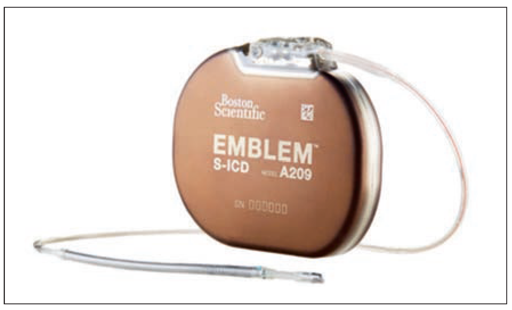
Figure 1. Emblem S-ICD model, with pulse generator and tripolar electrode. Used with permission of Boston Scientific Corporation.
Cardiovascmed 19 00121 g001
Figure 2. Recommended positioning of the S-ICD device:. the pulse generator is placed in between the mid-axillary and anterior axillary lines at the level of the sixth rib, and is connected to a tripolar electrode located 1–2 cm to the left of and parallel with the sternal midline. The electrode comprises a central shocking coil with an electrode on each end, ideally positioned distally at the manubriosternal junction (second rib) and proximally at the xiphoid process. The three possible sensing vectors are shown between the two sensing electrodes (A, B) and the active can (CAN). Used with permission of Boston Scientific Corporation.
Cardiovascmed 19 00121 g002
Figure 3. A and B: Photographs of a patient 1 day after implantation of an S-ICD device. C and D: Typical cosmetic result at 6-month follow-up. Picture taken at Cardiocentro Ticino by Dr. Auricchio.
Cardiovascmed 19 00121 g003
Figure 4. A: ECG screening tool provided by the manufacturer. B: Incorrect and correct profiles using the screening tool. An acceptable vector has the R-or S-wave within the peak zone and the QRS-T complex within the template. Used with permission of Boston Scientific Corporation.
Cardiovascmed 19 00121 g004
Table 1. Advantages and disadvantages of the S-ICD device compared with transvenous ICDs.
Cardiovascmed 19 00121 i001