Next Article in Journal
Kussmaul's Sign in Effusive Constrictive Pericarditis
Previous Article in Journal
Extreme Rocking Motion of an Infected Aortic Bioprosthesis
 
 
Cardiovascular Medicine is published by MDPI from Volume 28 Issue 1 (2025). Previous articles were published by another publisher in Open Access under a CC-BY (or CC-BY-NC-ND) licence, and they are hosted by MDPI on mdpi.com as a courtesy and upon agreement with Editores Medicorum Helveticorum (EMH).
Review

The Heart and Cirrhosis

by
Philippe Meyer
1,* and
Laurent Spahr
2
1
Service of Cardiology, Geneva University Hospitals, CH-1205 Geneva, Switzerland
2
Service of Gastroenterology, Geneva University Hospitals, CH-1205 Geneva, Switzerland
*
Author to whom correspondence should be addressed.
Cardiovasc. Med. 2015, 18(1), 20; https://doi.org/10.4414/cvm.2015.00299
Submission received: 21 October 2014 / Revised: 21 November 2014 / Accepted: 21 December 2014 / Published: 21 January 2015

Abstract

Cirrhosis has known effects on the cardiovascular system. These changes have been designated under the term cirrhotic cardiomyopathy. Half of cirrhotic patients may present this syndrome, which consists of structural, electrophysiological and functional alterations. The main features include enlargement of cardiac chambers, impaired cardiac response to stress, diastolic dysfunction and prolonged QT interval. Electrocardiogram, determination of natriuretic peptides, and echocardiography are the cornerstones of diagnosis. Pathophysiology is mostly related to systemic vasodilatation and hyperdynamic circulation. The impact of cirrhotic cardiomyopathy in the overall prognosis of patients with cirrhosis is not well documented. Management consists mainly of diuretics to control volume overload since no specific therapy has proved effective yet. Cirrhotic cardiomyopathy is generally reversible after liver transplantation.

Clinical vignette

A 66-year-old lady was admitted for progressive dyspnoea, New York Heart Association (NYHA) functional class III, and progressive lower limb oedema. She had a past history of advanced Child C cirrhosis complicated by hepatic encephalopathy, grade I oesophageal varices, and rectal varices, with a recent bleeding episode ten days before admission. She also had type II diabetes.
On physical exam, blood pressure was 105/56 mm Hg and heart rate was 66 bpm. There was an elevated jugular venous pressure at 14 cm of water. Apex beat was not displaced. Cardiac auscultation revealed a 2/6 midsystolic murmur maximal at the second leh intercostal space. Bilateral pulmonary rales were heard on pulmonary auscultation. There was bilateral lower limb pitting oedema to the thighs and clear evidence of ascites.
Laboratory tests revealed severe anaemia (haemoglobin 77 g/l), hyponatraemia (130 mmol/l), renal failure (estimated glomerular filtration rate 26 ml/min/1.73 m2), and elevated liver function tests. B-type natriuretic peptide (BNP) level was 995 ng/l. The electrocardiogram exhibited microvoltage of the QRS with a sinus rhythm at 62 bpm and a prolonged QT interval at 460 ms. Mild enlargement of all cardiac chambers was demonstrated by the transthoracic echocardiogram with a completely normal leh ventricular ejection fraction (LVEF) calculated at 64%. Doppler indices of diastolic function indicated elevated leh ventricular filling pressures (E/e’ ratio = 16).
To summarise, this patient presented a clear picture of heart failure (HF) with a normal or even supranormal LVEF, evidence of diastolic dysfunction, a surprisingly low heart rate despite severe anaemia, and a prolonged QT interval. Aher administration of furosemide and subsequent increased diuresis, dyspnoea and congestion improved dramatically. Aher 4 days, kidney function was normalised and usual spironolactone doses could be up-titrated. The patient was discharged at day 7 and remained stable in the following months.

Introduction

The heart and liver may interact in several different ways [1]. First, acute or chronic HF and especially right HF may lead to a spectrum of several liver manifestations, including cardiac cirrhosis or congestive hepatopathy. Second, chronic liver disease such as cirrhosis may affect the heart and the whole cardiovascular system, leading to a syndrome named cirrhotic cardiomyopathy [2]. This review will focus on the latter type of heart-liver interactions.
Cirrhotic cardiomyopathy was first described 15 years ago but a definition was only proposed in 2005 by an expert consensus during the annual meeting of the World Gastroenterology Organisation in Montreal [3]. Cirrhotic cardiomyopathy was defined as “a cardiac dysfunction in patients with cirrhosis characterised by impaired contractile responsiveness to stress and/or altered diastolic relaxation with electrophysiological abnormalities in the absence of other known cardiac disease” [3]. This is a rather vague definition with a lack of strict criteria. The prevalence is reported to be between 40 to 50% in patients with cirrhosis and the development of cirrhotic cardiomyopathy seems to be independent of the aetiology of liver disease [4].

Pathophysiology

Haemodynamic consequences of cirrhosis have been observed for a long time. More than 60 years ago,Kowalski and Abelman from Boston demonstrated an elevated cardiac index in patients with liver disease compared to normal controls [5]. Interestingly, this article used the term “Lannec’s cirrhosis”, which corresponds to alcoholic cirrhosis with severe atrophy. Indeed, the French physician René-Théophile-MarieHyacinthe Laennec (1785–1826), the inventor of the stethoscope, was also the first to coin the term “cirrhosis” from the Greek “Kirrhos” meaning “tawny” in English or “jaunâtre” in French according to the macroscopic appearance of the liver in this condition [6]. Many of the mechanisms involved in the development of hyperdynamic circulation and consecutively cirrhotic cardiomyopathy have now been elucidated, mainly thanks to experimental studies using animal models of cirrhosis (Figure 1).
Cirrhosis is characterised by an increase in intrahepatic vascular resistance consecutive to the development of fibrosis and the formation of regeneration nodules, which result in portal hypertension [7]. Portal hypertension, in turn, is associated with the production of vasodilators including carbon monoxide, nitric oxide, and tumour necrosis factor. In parallel, there is also a reduced degradation of these substances due to metabolic hepatic dysfunction and portosystemic shunts [4,7]. This contributes to splanchnic vasodilatation, which not only decreases global systemic vascular resistance but also creates splanchnic blood pooling. All these factors will result in hyperdynamic circulation and effective hypovolaemia. This induces baroreceptor and volume receptor activation of the renin-angiotensin-aldosterone system and of the sympathetic nervous system with an increased secretion of antidiuretic hormone contributing to the development of cirrhotic cardiomyopathy and HF [8].

Features of cirrhotic cardiomyopathy

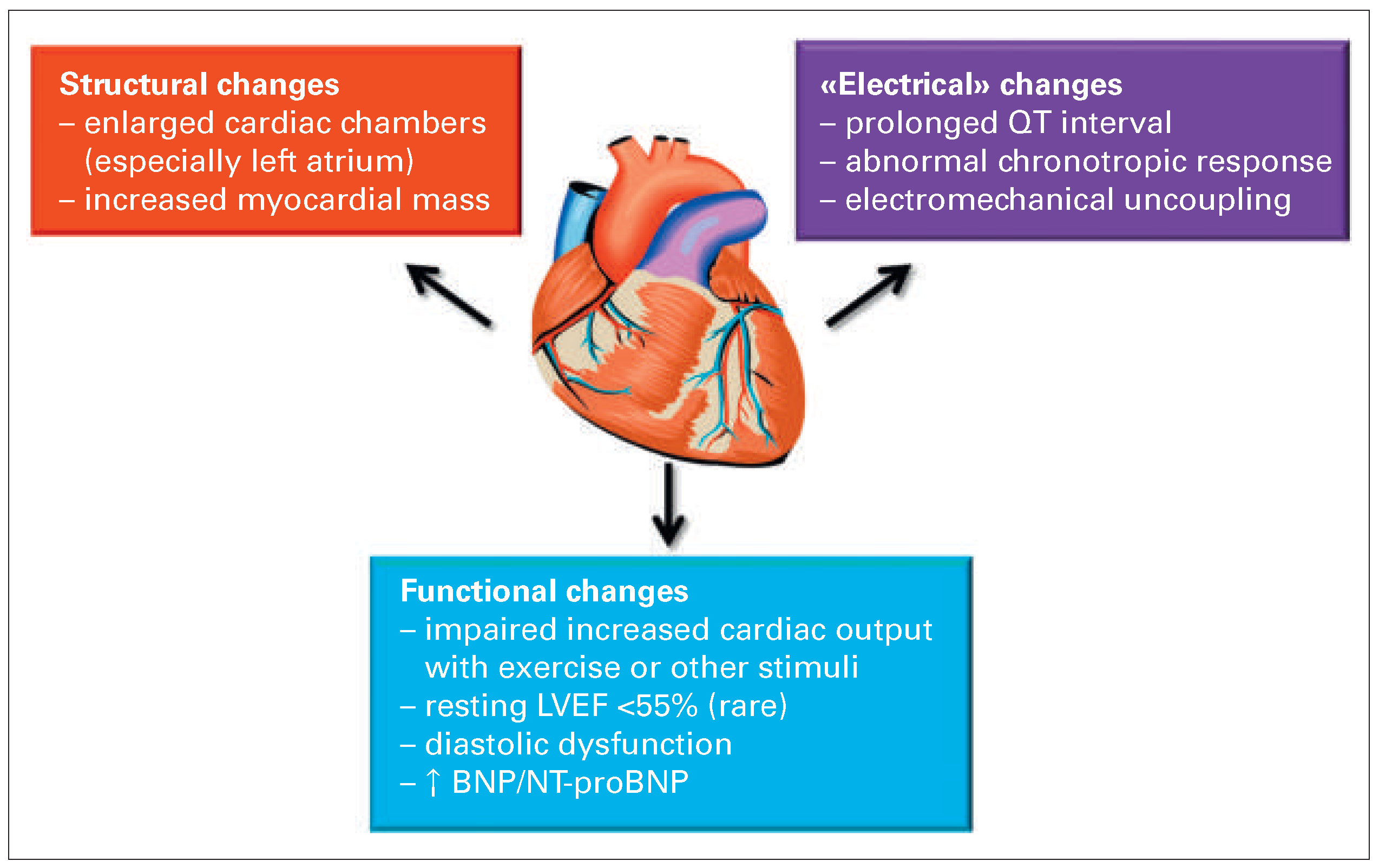
The different adaptations seen in cirrhosis can be classified in structural, electrical and functional changes (Figure 2). Structural changes include enlarged cardiac chambers and increased myocardial mass. A prolonged QT interval and an abnormal chronotropic response to stress are the main electrical changes. Finally, functional changes mainly consist of an impaired cardiac response to exercise or inotropes and diastolic dysfunction.
The most relevant features are detailed next.

Structural changes

According to an autopsy study of 135 cirrhotic patients, the most common cardiac abnormalities are leh ventricular or right ventricular dilatation and leh ventricular hypertrophy, which are encountered in more than one third of the patients [9]. Echocardiographic and magnetic resonance imaging studies show conflicting data but leh atrial dilatation and mild leh ventricular hypertrophy are the most commonly reported abnormalities [10,11]. These changes probably reflect a combination of mechanical overload due to hyperdynamic circulation and neurohormonal activation.

Impaired cardiac response to exercise

A predominant feature of cirrhotic cardiomyopathy is the impaired cardiac response to exercise. Wong et al. performed graded exercise tests on upright cycloergometers in 39 cirrhotic patients and 12 age and sex-matched healthy volunteers, in whom cardiac volumes and ejection fraction were assessed by radionuclide angiography before and during exercise [12]. This study clearly demonstrated the presence of chronotropic incompetence during exercise in cirrhotic patients with ascites, who only reached 65% of their maximal predicted heart rate. They also showed an inability to increase LVEF during exercise despite normal or even higher LVEF at rest compared to normal controls. Overall, the increase of cardiac output during exercise was severely impaired in these patients. This may explain why conditions associated with a sudden increase in cardiac output, such as an acute exacerbation of anaemia, sepsis, or creation of transjugular intrahepatic portosystemic shunt (TIPS) [13] are known to precipitate HF in patients with cirrhotic cardiomyopathy. Three main mechanisms are involved in the impaired cardiac response to exercise (Figure 3). First, there is a down-regulation of β-adrenergic receptors density in the cardiomyocytes with impaired β-adrenergic signalling owing to constant stimulation from increased levels of catecholamines [14,15]. A second mechanism is the reduced protein expression of L-type calcium channels with reduced calcium influx and subsequent impaired cardiac contractility in response to pharmacological stress [16]. Third, several mediators increased in cirrhosis such as cytokines, nitric oxide, carbon monoxide and endocannabinoids, impair intracellular signalling for contraction [1].

Diastolic dysfunction

Diastolic dysfunction is the most prominent functional alteration seen in cirrhotic cardiomyopathy. It is due to a combination of myocardial hypertrophy, fibrosis due to increased aldosterone levels and subendothelial oedema. The reported prevalence is between 45 and 56% [17].

Prolonged QT interval

Prolonged QT interval is common with a prevalence of more than 60% in advanced cirrhosis [18]. The pathophysiology is not clear: portal hypertension and portosystemic shunts are necessary but endotoxins, cytokines and increased sympathetic nervous system activity also play a role. The clinical relevance is not clear, ventricular arrhythmias seem to be rare and there is a debated effect on survival. Of course, drugs affecting the QT interval should be avoided [19].

Natriuretic peptides

Both B-type natriuretic peptide (BNP) and N-terminal proBNP (NT-proBNP) have been reported to be significantly increased in patients with advanced cirrhosis compared to controls. This increase is probably secondary to increased cardiac production of natriuretic peptides since hepatic degradation does not seem to be affected. Increased BNP and NT-proBNP were associated with the severity of both cirrhosis and cardiac dysfunction, but not to the presence of hyperdynamic circulation [20].

Clinical relevance of cirrhotic cardiomyopathy

Data on the clinical significance of cirrhotic cardiomyopathy are scarce but several publications indicate a poor prognosis for these patients. Krag et al. performed a study with 24 patients with cirrhosis and ascites in whom cardiac index was evaluated by gated single-photo emission cardiac tomography (SPECT) [21]. Cardiac index was shown to be an important predictor of renal failure and death with a cut-off value at 1.5 l/m/m2. However, a reduction of cardiac index below 1.5 l/m/m2 only occurs in terminal stages of cirrhotic cardiomyopathy evolution. In another study including 101 cirrhotic patients prior to performing a TIPS, diastolic function was evaluated by echocardiography using the E/A ratio of the mitral inflow by pulsed Doppler [22]. Diastolic dysfunction expressed as an E/A ratio ≤1 was a predictor of ascites persistence and death.

Treatment of cirrhotic cardiomyopathy

There is no specific pharmacological therapy for cirrhotic cardiomyopathy. In case of systemic or pulmonary congestion, loop diuretics should be used but with caution since patients with cirrhosis ohen have low blood pressure due to vasodilation. Nonselective β-blockers are ohen used in patients with cirrhosis for variceal bleeding prophylaxis. Propranolol and nadolol have been shown to reduce the QT interval prolongation but the potential effects on the occurrence of arrhythmia are unknown [19,23]. β-blockers also reduced cardiac output and the hepatic venous pressure gradient, which may be beneficial in the hyperdynamic state of these patients. In the presence of advanced cirrhosis with refractory ascites necessitating repeated large volume paracentesis, recent data show that nonselective β-blockers are associated with increased mortality [24]. Mineralocorticoid receptor antagonists are part of standard therapy in patients with cirrhosis and ascites. Their specific use in cirrhotic cardiomyopathy has not been studied but benefits on leh ventricular hypertrophy and dilatation have been reported [25]. Finally, angiotensinconverting enzyme inhibitors or angiotensin II receptor blockers are ohen contraindicated in patients with advanced cirrhosis because of symptomatic hypotension due to profound systemic vasodilatation. Future therapeutic strategies may consist of targeting the different pathways involved in cirrhotic cardiomyopathy including the inhibition of cytokines or other cell signalling molecules. The hyperdynamic circulation and cardiac dysfunction associated with cirrhotic cardiomyopathy seem to normalise aher liver transplantation.[26] However, there are no “cardiac” indications for liver transplantation.

Conclusions

Cirrhotic cardiomyopathy is a frequent syndrome occurring in almost 50% of patients with advanced cirrhosis. There are no strict diagnostic criteria but impaired cardiac response to stress, LV diastolic dysfunction and prolonged QT are the most prominent features. The clinical relevance of this syndrome is not well established yet, but these patients do not tolerate any conditions requiring a further increase of cardiac output. Therefore it is important for clinicians to be aware of this syndrome. There is no specific therapy but cirrhotic cardiomyopathy is almost completely reversible aher liver transplantation.

Funding/potential competing interests

No financial support and no other potential conflict of interest relevant to this article were reported.

References

  1. Moller, S.; Bernardi, M. Interactions of the heart and the liver. Eur Heart J. 2013, 34, 2804–2811. [Google Scholar] [CrossRef] [PubMed]
  2. Wiese, S.; Hove, J.D.; Bendtsen, F.; Moller, S. Cirrhotic cardiomyopathy: pathogenesis and clinical relevance. Nature reviews Gastroenterology & hepatology. 2014, 11, 177–186. [Google Scholar]
  3. Moller, S.; Henriksen, J.H. Cardiovascular complications of cirrhosis. Gut. 2008, 57, 268–278. [Google Scholar] [CrossRef] [PubMed]
  4. Timoh, T.; Protano, M.A.; Wagman, G.; Bloom, M.; Vittorio, T.J. A perspective on cirrhotic cardiomyopathy. Transplant Proc. 2011, 43, 1649–1653. [Google Scholar] [CrossRef]
  5. Kowalski, H.J.; Abelmann, W.H. The cardiac output at rest in Laennec’s cirrhosis. J Clin Invest. 1953, 32, 1025–1033. [Google Scholar] [CrossRef]
  6. Duffin, J.M. Why does cirrhosis belong to Laennec? CMAJ. 1987, 137, 393–396. [Google Scholar]
  7. Laleman, W.; Landeghem, L.; Wilmer, A.; Fevery, J.; Nevens, F. Portal hypertension: from pathophysiology to clinical practice. Liver Int. 2005, 25, 1079–1090. [Google Scholar] [CrossRef]
  8. Moller, S.; Iversen, J.S.; Henriksen, J.H.; Bendtsen, F. Reduced baroreflex sensitivity in alcoholic cirrhosis: relations to hemodynamics and humoral systems. Am J Physiol Heart Circ Physiol. 2007, 292, H2966–72. [Google Scholar] [CrossRef]
  9. Ortiz-Olvera, N.X.; Castellanos-Pallares, G.; Gomez-Jimenez, L.M.; Cabrera-Munoz, M.L.; Mendez-Navarro, J.; Moran-Villota, S.; et al. Anatomical cardiac alterations in liver cirrhosis: an autopsy study. Ann Hepato. 2011, 10, 321–326. [Google Scholar] [CrossRef]
  10. Abd-El-Aziz, T.A.; Abdou, M.; Fathy, A.; Wafaie, M. Evaluation of cardiac function in patients with liver cirrhosis. Int Med. 2010, 49, 2547–2552. [Google Scholar] [CrossRef]
  11. Moller, S.; Sondergaard, L.; Mogelvang, J.; Henriksen, O.; Henriksen, J.H. Decreased right heart blood volume determined by magnetic resonance imaging: evidence of central underfilling in cirrhosis. Hepatology. 1995, 22, 472–478. [Google Scholar]
  12. Wong, F.; Girgrah, N.; Graba, J.; Allidina, Y.; Liu, P.; Blendis, L. The cardiac response to exercise in cirrhosis. Gut. 2001, 49, 268–275. [Google Scholar] [CrossRef]
  13. Colombato, L.A.; Spahr, L.; Martinet, J.P.; Dufresne, M.P.; Lafortune, M.; Fenyves, D.; et al. Haemodynamic adaptation two months aher transjugular intrahepatic portosystemic shunt (TIPS) in cirrhotic patients. Gut. 1996, 39, 600–604. [Google Scholar] [CrossRef]
  14. Gerbes, A.L.; Remien, J.; Jungst, D.; Sauerbruch, T.; Paumgartner, G. Evidence for down-regulation of beta-2–adrenoceptors in cirrhotic patients with severe ascites. Lancet. 1986, 1, 1409–11. [Google Scholar] [CrossRef] [PubMed]
  15. Lee, S.S.; Marty, J.; Mantz, J.; Samain, E.; Braillon, A.; Lebrec, D. Desensitization of myocardial beta-adrenergic receptors in cirrhotic rats. Hepatology. 1990, 12 (Pt 1), 481–485. [Google Scholar] [CrossRef] [PubMed]
  16. Ward, C.A.; Liu, H.; Lee, S.S. Altered cellular calcium regulatory systems in a rat model of cirrhotic cardiomyopathy. Gastroenterology. 2001, 121, 1209–1218. [Google Scholar]
  17. Liu, H.; Gaskari, S.A.; Lee, S.S. Cardiac and vascular changes in cirrhosis: pathogenic mechanisms. World J Gastroenterol. 2006, 12, 837–842. [Google Scholar] [CrossRef] [PubMed]
  18. Bernardi, M.; Calandra, S.; Colantoni, A.; Trevisani, F.; Raimondo, M.L.; Sica, G.; et al. Q-T interval prolongation in cirrhosis: prevalence, relationship with severity, and etiology of the disease and possible pathogenetic factors. Hepatology. 1998, 27, 28–34. [Google Scholar] [CrossRef]
  19. Zambruni, A.; Trevisani, F.; Di Micoli, A.; Savelli, F.; Berzigotti, A.; Bracci, E.; et al. Effect of chronic beta-blockade on QT interval in patients with liver cirrhosis. J Hepatol. 2008, 48, 415–421. [Google Scholar] [CrossRef]
  20. Henriksen, J.H.; Gotze, J.P.; Fuglsang, S.; Christensen, E.; Bendtsen, F.; Moller, S. Increased circulating pro-brain natriuretic peptide (proBNP) and brain natriuretic peptide (BNP) in patients with cirrhosis: relation to cardiovascular dysfunction and severity of disease. Gut. 2003, 52, 1511–1517. [Google Scholar] [CrossRef]
  21. Krag, A.; Bendtsen, F.; Henriksen, J.H.; Moller, S. Low cardiac output predicts development of hepatorenal syndrome and survival in patients with cirrhosis and ascites. Gut. 2010, 59, 105–110. [Google Scholar] [CrossRef]
  22. Rabie, R.N.; Cazzaniga, M.; Salerno, F.; Wong, F. The use of E/A ratio as a predictor of outcome in cirrhotic patients treated with transjugular intrahepatic portosystemic shunt. Am J Gastroenterol. 2009, 104, 2458–2466. [Google Scholar] [CrossRef] [PubMed]
  23. Henriksen, J.H.; Bendtsen, F.; Hansen, E.F.; Moller, S. Acute non-selective beta-adrenergic blockade reduces prolonged frequencyadjusted Q-T interval (QTc) in patients with cirrhosis. J Hepatol. 2004, 40, 239–246. [Google Scholar] [CrossRef] [PubMed]
  24. Serste, T.; Melot, C.; Francoz, C.; Durand, F.; Rautou, P.E.; Valla, D.; et al. Deleterious effects of beta-blockers on survival in patients with cirrhosis and refractory ascites. Hepatology. 2010, 52, 1017–1022. [Google Scholar] [CrossRef] [PubMed]
  25. Pozzi, M.; Grassi, G.; Ratti, L.; Favini, G.; Dell’Oro, R.; Redaelli, E.; et al. Cardiac, neuroadrenergic, and portal hemodynamic effects of prolonged aldosterone blockade in postviral child A cirrhosis. Am J Gastroenterol. 2005, 100, 1110–1116. [Google Scholar] [CrossRef]
  26. Fouad, T.R.; Abdel-Razek, W.M.; Burak, K.W.; Bain, V.G.; Lee, S.S. Prediction of cardiac complications aher liver transplantation. Transplantation. 2009, 87, 763–770. [Google Scholar] [CrossRef]
Figure 1. Pathophysiology of cirrhotic cardiomyopathy.
Cardiovascmed 18 00020 g001
Figure 2. Main features of cirrhotic cardiomyopathy. BNP = B-type natriuretic peptide; LVEF = left ventricular ejection fraction; NT-proBNP = N terminal-proBNP.
Cardiovascmed 18 00020 g002
Figure 3. The figure reviews the most important mechanisms involved in the impaired contractile function of the cardiomyocyte in experimental cirrhotic cardiomyopathy: Desensitisation and down-regulation of β-adrenergic receptors with a decreased content of G-protein (Gai = inhibitory G-protein; Gas = stimulatory G-protein) and following impaired intracellular signalling; alterations in particular in M1 muscarinic receptors; up-regulation of cannabinoid 1-receptor stimulation; altered plasma membrane choles-terol/phospholipid ratio; increased inhibitory effects of haemo-oxygenase, carbon monoxide, nitric oxide, and tumour necrosis factor-α; reduced density of potassium channels; changed function and fluxes through L-type calcium channels; altered ratio and function of collagens and titins. Many post-receptor effects are mediated by adenylcyclase inhibition or stimulation. (Figure from: Moller S, Bernardi M. Interactions of the heart and the liver. Eur Heart J. 2013, 34, 2804–11. Copyright © 2013 Oxford University Press. Reprint with kind permission from Oxford University Press.) PKA = protein kinase A.
Cardiovascmed 18 00020 g003