Next Article in Journal
Supracardiac Total Anomalous Pulmonary Venous Connection with Right Ascending Vertical Vein
Previous Article in Journal
Recurrent Spontaneous Coronary Artery Dissection in a 46-Year-Old Woman
 
 
Cardiovascular Medicine is published by MDPI from Volume 28 Issue 1 (2025). Previous articles were published by another publisher in Open Access under a CC-BY (or CC-BY-NC-ND) licence, and they are hosted by MDPI on mdpi.com as a courtesy and upon agreement with Editores Medicorum Helveticorum (EMH).
Case Report

Percutaneous Closure of Ventricular Septal Defect Following Aortic Valve Replacement

by
Jean-Jacques Goy
1,*,
Patrick Ruchat
2,
Valérie Stoltc
3,
Léopold Schlueter
2 and
Alexandre Berger
2
1
Service de Cardiologie, Clinique Cecil, Lausanne, Switzerland
2
Service de Chirurgie Cardiovasculaire, Clinique Cecil, Lausanne, Switzerland
3
Département de Médecine, Hôpital Cantonal, Fribourg, Switzerland
*
Author to whom correspondence should be addressed.
Cardiovasc. Med. 2014, 17(9), 266; https://doi.org/10.4414/cvm.2014.00270
Submission received: 24 June 2014 / Revised: 24 July 2014 / Accepted: 24 August 2014 / Published: 24 September 2014

Abstract

Membranous ventricular septal defect (VSD) is a rare complication of surgical aortic valve replacement. We describe a complex case of iatrogenic VSD following surgical aortic valve replacement with a bioprosthetic aortic valve. VSD could be successfully closed using a retrograde approach.

Introduction

Ventricular septal defect is a rare but well known complication of surgical aortic valve replacement. When possible, percutaneous closure is the intervention of choice. Successful closure is challenging and depends on the anatomical location of the defect. We present a case of successful closure using the retrograde approach.

Case report

An 80-year-old women developed shortness of breath (NYHA III) shortly following aortic valve replacement (AVR) with a Carpentier Edwards biologic aortic prosthesis. In spite of optimal medical treatment, she remained symptomatic and echocardiographic evaluation revealed membranous ventricular septal defect (VSD) with a significant left-to-right shunt (Figure 1) and a Qp/Qs ratio of 2.1:1. She was considered at high risk for redo-surgical aortic valve replacement and was referred for percutaneous closure of this iatrogenic membranous VSD.
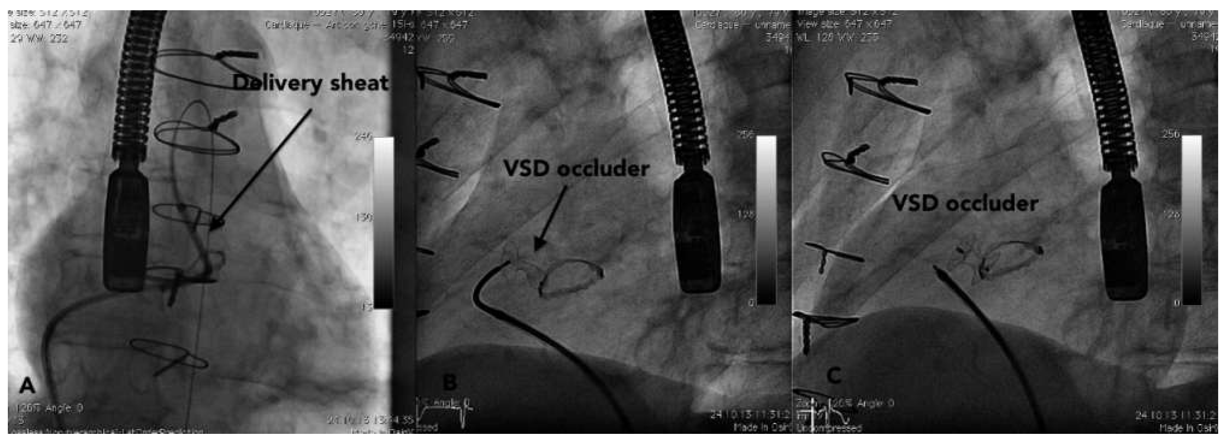
The procedure was attempted under general anaesthesia with trans-oesophageal echocardiographic (TEE) and fluoroscopic guidance. A six Fr sheath was inserted in the right femoral vein (RFV) and in the left femoral artery. Intra-procedural LV injection showed a significant left-to-right shunt in the membranous septum with a rather vertical orientation of the flow (Figure 2). A first attempt using the anterograde approach from the RFV and the right ventricle was unsuccessful, the guide wire could not go through the VSD. The retrograde approach was chosen and a six Fr left Amplatzer guiding catheter was advanced in the LV via the left femoral artery. As a 0.035 inch (Terumo) guide wire could not be stabilised in the VSD, thus a 0.014 inch angioplasty wire (Sion, Asahi,) was used to stabilise the guiding catheter and was advanced in the right ventricle through the VSD, then further into the left pulmonary artery (Figure 2). A 0.035 inch wire (Terumo) followed the 0.014 inch guide wire and could be advanced in the left pulmonary artery (Figure 2). A six Fr lasso (En Snare, Merit Medical) was inserted into the left pulmonary artery through the right femoral vein. The 0.035 wire was snared in the pulmonary artery and exteriorised from the RFV completing the arterial-venous loop (Figure 2). The seven Fr Amplatzer Torq sheath was advanced from the right femoral vein over the Terumo wire across the VSD and the aortic prosthetic valve. As the occluder could not be deployed in the left ventricle due to the position of the VSD, it had to be opened in the ascending aorta (Figure 3) and then pulled back across the prosthesis to be applied on the left side of the VSD. Then, the occluder could be delivered from the delivering sheath and took its final position through the VSD (Figure 3).
The patient developed a 2:1 Mobitz two A-V block in the hours following the procedure. A permanent DDD pacemaker was inserted. She was discharged 2 days later with no significant residual shunt confirmed by TTE (Figure 4). At the 3 three month follow-up, the patient remained asymptomatic with no residual shunt or valve dysfunction as confirmed by CT scan and TTE (Figure 4).

Discussion

Ventricular septal defect is a rare complication of AVR. A left-to-right shunt can result in significant LV overload and symptoms of heart failure. Shunts <1.5 are often well tolerated, although larger shunts need to be corrected. Closure is also needed to prevent endocarditis, pulmonary hypertension, arrhythmias, and LV dysfunction. Surgery, for many years, has been considered as the gold-standard treatment but with a significant morbidity and mortality especially in patients with prior sternotomies [1]. Percutaneous VSD closure has been successfully used in pediatric patients in the 1980s [2]. Only a few cases of VSD secondary to AVR have been reported in the literature, but were usually treated successfully using either an anterograde transseptal or retrograde approach [3,4]. Of course in patients with mechanical AVR, only the trans-septal approach could be used. In patients with a biological prosthesis, the retrograde approach seems to be the route of choice, since it is simpler to manipulate the catheters. The most challenging part of the intervention in our case was the deployment of the device. Due to the anatomical location of the VSD, close to the aortic prosthesis and being mainly vertical, the positioning of the occluder in the LV before deployment in the VSD was impossible. Because of the position of the guiding sheath through the VSD, the distal disk had to be opened in the aortic prosthesis and then pulled back through the septum, with the risk of damage to the aortic prosthesis. The proximal disc on the right side of the ventricular septum was released with a partially undeployed distal disc. The occluder took its final position only when it detached from the guiding sheath. This confirms that the final position of the occluder is always unpredictable until its final release. A movable core of the device would be beneficial to better predict final position of the device. Although the procedure can be performed with fluoroscopy only, the use of general anaesthesia and trans-oesophagal echocardigoraphy seems beneficial since it helps to localise the device and improves positioning.
Post AVR heart block occurs in up to 4%–6% [5] of the cases and requires pacemaker implantation. In our patient, the A-V block was not present after surgery but only following VSD closure. The mechanism is probably mechanical due to compression of the conduction pathways by the device. Percutaneous closure is a safe and effective treatment of iatrogenic VSD following AVR. Anterograde or retrograde approach should be chosen on case by case evaluation.

Funding/potential competing interests:

No financial support and no other potential conflict of interest relevant to this article were reported.

References

  1. Holzer, R.; Latson, L.; Hijazi, Z.M. Device closure of iatrogenic membranous ventricular septal defects after prosthetic aortic valve replacement using the Amplatzer membranous ventricular septal defect occluder. Catheter Cardiovasc Interv. 2004, 62, 276–280. [Google Scholar] [CrossRef] [PubMed]
  2. Koneti, N.R.; Sreeram, N.; Penumatsa, R.R.; Arramraj, S.K.; Karunakar, V.; Trieschmann, U. Transcatheter retrograde closure of perimembranous ventricular septal defects in children with the Amplatzer duct occluder II device. J Am Coll Cardiol. 2012, 60, 2421–2422. [Google Scholar] [CrossRef] [PubMed][Green Version]
  3. Noble, S.; Ibrahim, R. Transcatheter membranous ventricular septal defect closure through a mechanical aortic prosthesis using the Amplatzer membranous ventricular septal defect occluder. Catheter Cardiovasc Interv. 2009, 73, 167–172. [Google Scholar] [CrossRef] [PubMed]
  4. Klein, A.J.; Garcia, J.A.; Carroll, J.D. Percutaneous closure of an iatrogenic ventricular septal defect following mechanical aortic valve replacement using the transseptal technique. Catheter Cardiovasc Interv. 2007, 70, 1018–1024. [Google Scholar] [CrossRef] [PubMed]
  5. Butera, G.; Carminati, M.; Chessa, M.; et al. Transcatheter closure of perimembranous VSD: Early and long-term results. J Am Coll Cardiol. 2007, 50, 1189–1195. [Google Scholar] [CrossRef] [PubMed]
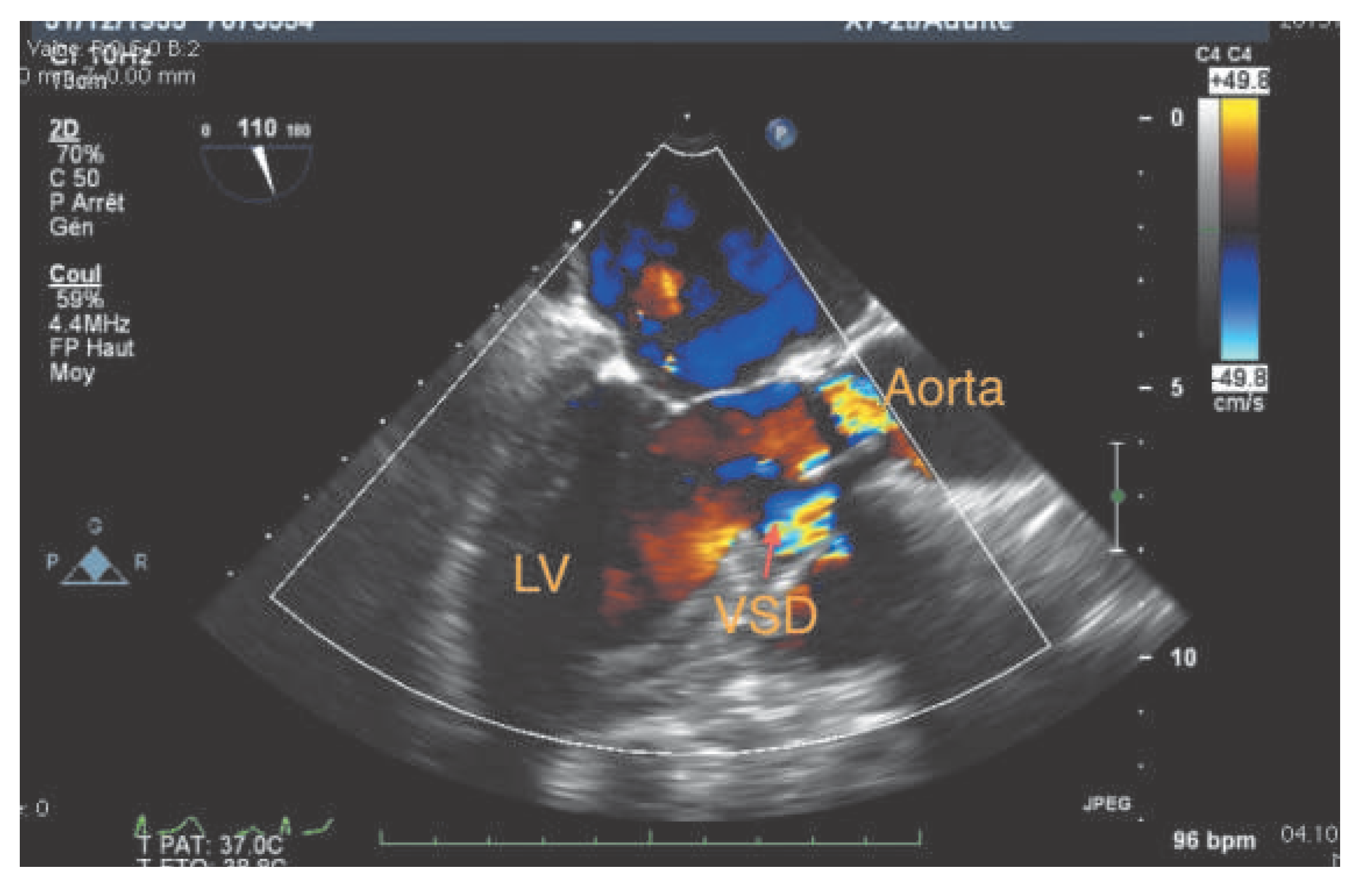
Figure 1. VSD visible below the aortic prosthesis.
Cardiovascmed 17 00266 g001
Figure 2. a: Angiographic aspect of the VSD (RV = right ventricle, LV = left ventricle). b: AL 2 guiding catheter in the left ventricle and 0.014” guidewire in the pulmonary artery through the VSD. c: Lasso in the pulmonary artery to snare the .035” Terumo wire.
Cardiovascmed 17 00266 g002
Figure 3. a: Delivery catheter across the VSD and the aortic prosthesis. The Terumo (arterio-venous loop) is still in place. b: Delivery catheter across the VSD and the aortic prosthesis. The Terumo (arterio-venous loop) is still in place. c: Final position of the VSD occluder.
Cardiovascmed 17 00266 g003
Figure 4. a: Final TEE view showing a complete occlusion of the VSD. b: CT scan three months after implantation showing the occluder still in the correct position without residual shunt.
Cardiovascmed 17 00266 g004