Next Article in Journal
Pregnancy-Associated Cardiomyopathies
Previous Article in Journal
Percutaneous Closure of Ventricular Septal Defect Following Aortic Valve Replacement
 
 
Cardiovascular Medicine is published by MDPI from Volume 28 Issue 1 (2025). Previous articles were published by another publisher in Open Access under a CC-BY (or CC-BY-NC-ND) licence, and they are hosted by MDPI on mdpi.com as a courtesy and upon agreement with Editores Medicorum Helveticorum (EMH).
Interesting Images

Supracardiac Total Anomalous Pulmonary Venous Connection with Right Ascending Vertical Vein

by
Himanshu Mahla
1,*,
Kavya Mallikarjun
2,
Usha Mandikal Kodanda Rama Sastry
2,
Jayarangnath Mahimarangaiah
2 and
Cholenahally Nanjappa Manjunath
1
1
Department of Cardiology, Sri Jayadeva Institute of Cardiovascular Sciences and Research, Bangalore, Karnataka, India
2
Department of Pediatric Cardiology, Sri Jayadeva Institute of Cardiovascular Sciences and Research, Bangalore, Karnataka, India
*
Author to whom correspondence should be addressed.
Cardiovasc. Med. 2014, 17(9), 273; https://doi.org/10.4414/cvm.2014.00265
Submission received: 24 June 2014 / Revised: 24 July 2014 / Accepted: 24 August 2014 / Published: 24 September 2014

Case Report

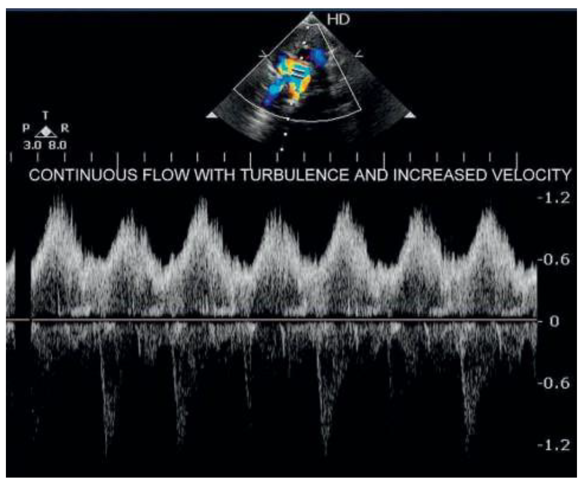
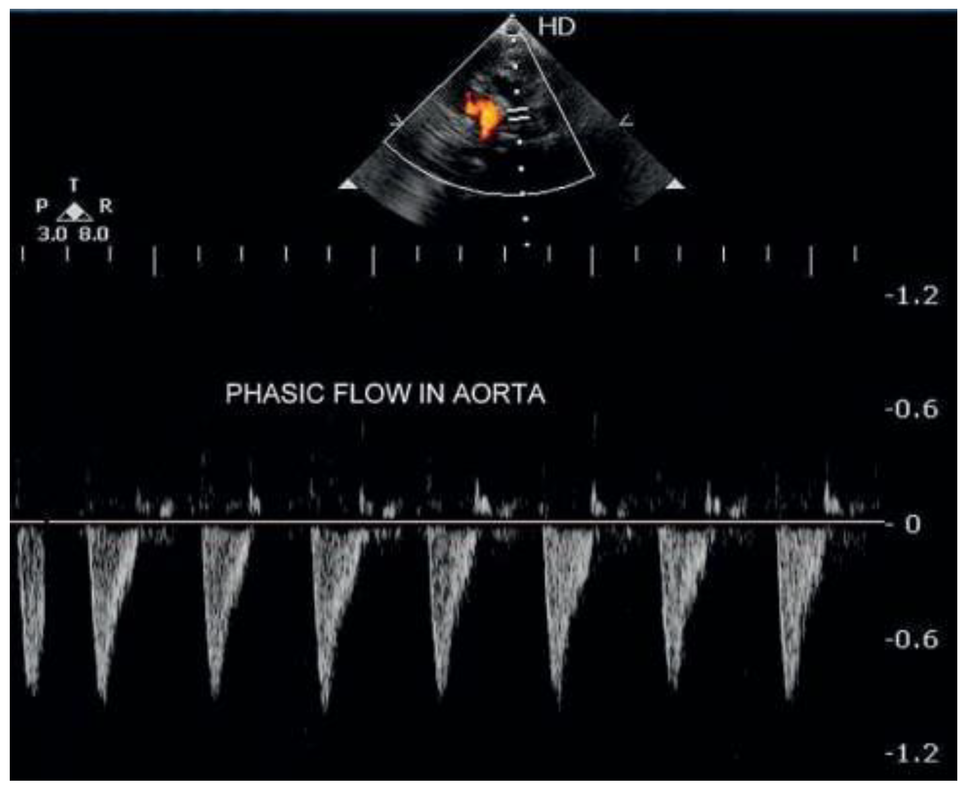
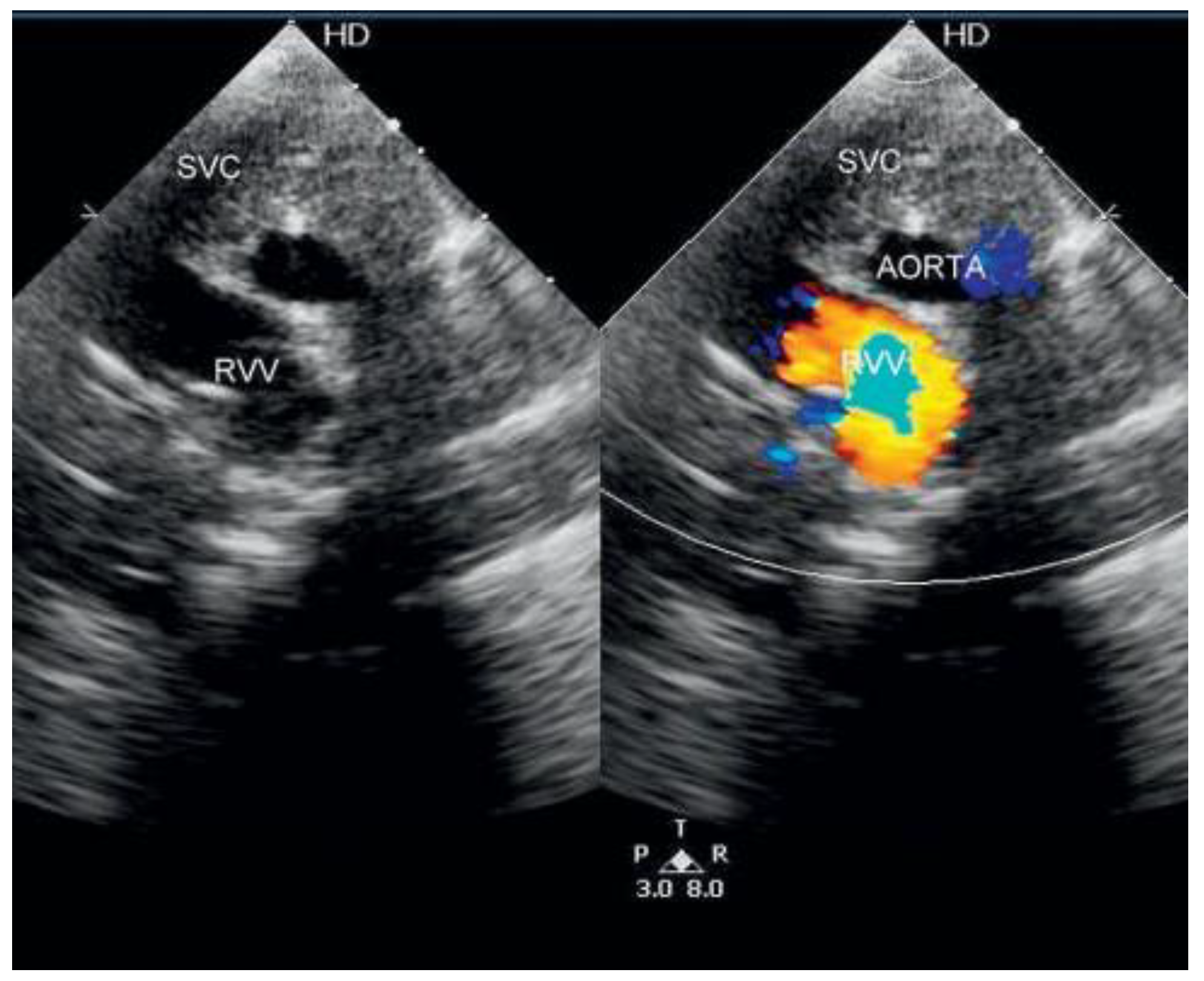
A 20-day-old neonate was referred to our tertiary care institute for evaluation of increased precordial activity and feeding difficulty. The birth weight was 2.6 kilograms. On examination the child had tachycardia, increased precordial activity, loud pulmonary component of second heart sound and ejection systolic murmur in the pulmonary area. The transthoracic echocardiogram revealed common chamber (confluence) posterior to left atrium and ostium secundum atrial septal defect with right-to-left shunt and right ventricular dysfunction (Figure 1; video 1*). All four pulmonary veins were connected to a common chamber and draining via right sided ascending vertical vein to right superior vena cava (SVC), one centimetre above the SVC-right atrium (SVC-RA) junction (Figure 2, Figure 3, Figure 4 and Figure 5; video 2, 3 and 4*). There was obstruction across the pathway with severe pulmonary arterial hypertension and no other associated anomalies. The pathway of drainage was confirmed by cardiac computed tomography (figure 5). It was further confirmed upon surgery. The child underwent surgical correction and was doing well postoperatively. Total anomalous pulmonary venous connection (TAPVC) is classified as supracardiac, cardiac, infracardiac, or mixed forms [1]. Supracardiac TAPVC [2] occurs due to developmental arrest or atresia of the common pulmonary vein. The common pulmonary vein connects the embryonic lung buds with the left atrium. Collateral channels for pulmonary venous drainage develop via the fetal cardinal venous system. Usually, only one of these channels persists to become the vertical vein that connects the pulmonary venous confluence to the SVC or innominate vein. If left cardinal venous system persists, left vertical vein (LVV) will be present which is usually the case. It usually connects to innominate vein. Rarely the right cardinal venous system persists which results in right vertical vein (RVV). RVV usually has an oblique course and drains to SVC or SVC-RA junction [3]. Anomalous pulmonary venous connections have wide variability and separate vertical veins can also be seen in heterotaxy syndromes [4]. TAPVC without any associated anomalies but with a right ascending vertical vein is a rare association.

Funding

This research received no external funding.

Acknowledgments

We are thankful to Dr Neha Godara for the kind support. Video files: You can find the videos on http://www.cardiovascmed.ch/for-readers/ multimedia. Video 1: Transthoracic subcostal four chamber view showing dilated right atrium, right ventricle, ostium secundum atrial septal defect with confluence posterior to left atrium. Video 2: Transthoracic high parasternal view showing superior vena cava (SVC), aorta and pulmonary artery (from right to left ) and continuous turbulence in lower portion of SVC. Video 3: Transthoracic modified suprasternal long axis view showing the drainage of right vertical vein (RVV) to right superior vena cava (SVC). Video 4: Transthoracic modified suprasternal long axis view showing the drainage of right vertical vein (RVV) to right superior vena cava (SVC) and a point source of turbulence, the probable site of obstruction.

Potential Competing Interests

No other potential conflict of interest relevant to this article were reported.

References

  1. Craig, J.M.; Darling, R.C.; Pothney, W.B. Total anomalous pulmonary venous drainage on right side of heart; report of 17 autopsied cases not associated with other major cardiovascular anomalies. Lab Invest. 1957, 6, 44–64. [Google Scholar] [PubMed]
  2. Brown, D.W.; Geva, T. Anomalies of the pulmonary veins. In Moss and Adams’ Heart Disease in Infants, Children, and Adolescents, 8th ed.; Allen, H., Driscoll, D., Shaddy, R., Feltes, T., Eds.; Lippincott Williams & Wilkins: Philadelphia, PA, USA, 2013; pp. 823–825. [Google Scholar]
  3. Lehner, A.; Kozlik-Feldmann, R.; Herrmann, F.; et al. An unusual form of supracardiac total anomalous pulmonary venous return via a right-sided vertical vein in a heterotaxy syndrome case. Pediatr Cardiol. 2012, 33, 1200–1202. [Google Scholar] [CrossRef] [PubMed]
  4. Grosse-Wortmann, L.; Friedberg, M.K. Bilateral vertical veins from a common confluence in supracardiactotal anomalous pulmonary venous connection. Circulation 2008, 118, e103–e104. [Google Scholar] [CrossRef] [PubMed][Green Version]
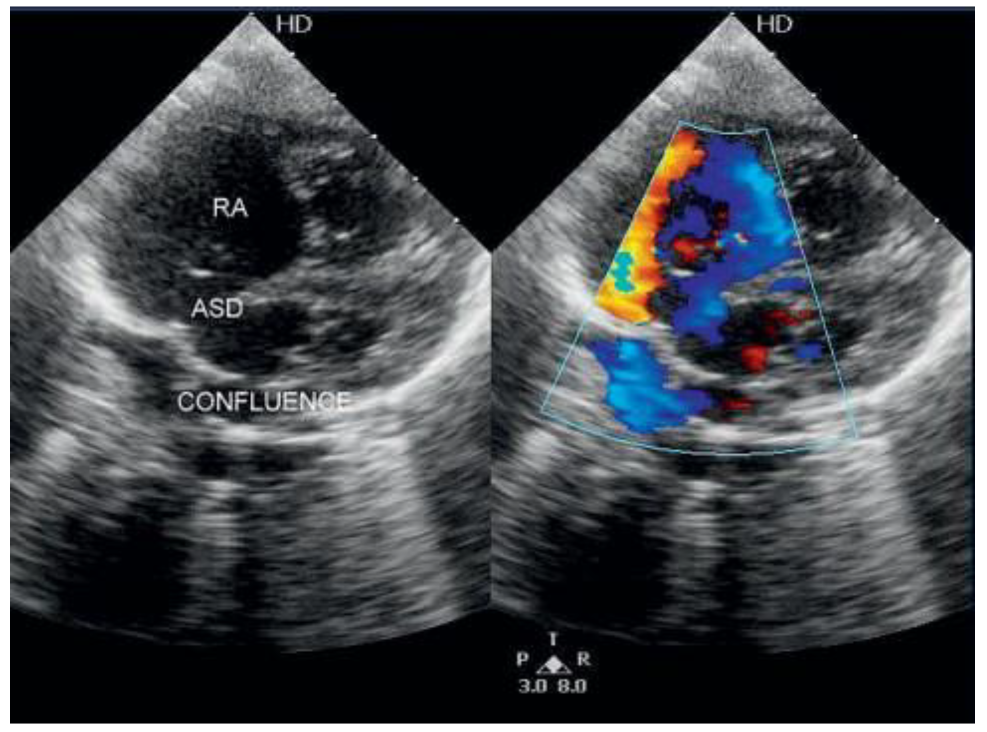
Figure 1. Transthoracic subcostal four chamber view showing dilated right atrium, right ventricle, ostium secundum atrial septal defect with confluence posterior to left atrium. RA = right atrium; ASD = atrial septal defect.
Cardiovascmed 17 00273 g001
Figure 2. Transthoracic suprasternal long axis view with pulse wave Doppler at the junction of right ascending vertical vein (RVV) with superior vena cava (SVC), showing continuous flow with increased velocity.
Cardiovascmed 17 00273 g002
Figure 3. Transthoracic suprasternal long axis view with pulse wave Doppler kept to left of right vertical vein (RVV) showing phasic and pulsatile flow of aorta.
Cardiovascmed 17 00273 g003
Figure 4. Transthoracic modified suprasternal long axis view showing the drainage of right vertical vein (RVV) to right superior vena cava (SVC).
Cardiovascmed 17 00273 g004
Figure 5. Cardiac CT with 3D reconstruction showing right vertical vein (RVV) joining right superior vena cava (SVC) with an almost 90 degree turn.
Cardiovascmed 17 00273 g005