Next Article in Journal
The Infamous Coronary Stent Saga
Previous Article in Journal
The Early Development of Cardiology in Zurich—A Personal Account Part II
 
 
Cardiovascular Medicine is published by MDPI from Volume 28 Issue 1 (2025). Previous articles were published by another publisher in Open Access under a CC-BY (or CC-BY-NC-ND) licence, and they are hosted by MDPI on mdpi.com as a courtesy and upon agreement with Editores Medicorum Helveticorum (EMH).
Review

Left Atrial Appendage Device Closure as Non-Pharmacological Prevention of Thromboembolism in Atrial Fibrillation

Cardiology, Cardiovascular Department, Bern University Hospital, CH-3010 Bern, Switzerland
Cardiovasc. Med. 2014, 17(9), 252; https://doi.org/10.4414/cvm.2014.00277
Submission received: 24 June 2014 / Revised: 24 July 2014 / Accepted: 24 August 2014 / Published: 24 September 2014

Abstract

The fact that non-valvular (non-rheumatic) atrial fibrillation creates detectable thrombi in 17% of patients without oral anticoagulation and that 16% of the 17% (over 90% relatively) reside in the left atrial appendage (LAA), suggest that patients without an LAA or with an occluded LAA do not need oral anticoagulation. This has already led to surgical LAA exclusion for decades and to catheter-based LAA occlusion for the past 13 years. Currently 5 different technical approaches are approved in European countries, but only 2 are widely used, the Amplatzer and the Watchman devices. With the latter, randomised data showed superiority in terms of embolism protection, bleeding, and survival compared to vitamin K antagonists (VKA) after 4 years in a randomised trial. The clinical results in thousands of patients with Watchman and Amplatzer occluders suggest, moreover, that they are at least competitive with non-VKA oral anticoagulants. Non-pharmacological prevention of thromboembolism with device implantation or, in case of another indication for heart surgery during that intervention, may be discussed with every patient with atrial fibrillation as an alternative to oral anticoagulation.

Introduction

It is known from autopsy, cardiac surgery, and trans-oesophageal echocardiography (TOE) that 17% of patients with non-valvular (basically non-rheumatic) atrial fibrillation (AF) have clots in the left atrium (LA). Over 90% of these clots (16% of 17%) reside in the left atrial appendage (LAA) [1]. Hence, the presence of 1% of blood clots in patients with no LAA or an occluded LAA provides a risk much too small to warrant chronic oral anticoagulation either with vitamin K antagonists (VKA) or non-VKA oral anti-coagulants (NOAC). The bleeding risk more than outweighs the potential benefit.
AF is the most common clinically significant rhythm disorder with an increasing prevalence parallel to the increasing average age of our population. The decision of whether to treat a given patient with VKA, NOAC, or LAA occlusion has to be taken almost daily by cardiologists, internists, and other physicians.

Techniques of left atrial appendage occlusion

Excision or ligation of the LAA has been part of some surgical procedures for decades [2,3]. However, these interventions were mostly combined with mitral valve replacement using metallic implants so that the patients needed to stay under oral anticoagulation regardless. No thorough appreciation of the value of LAA exclusion was therefore performed or even possible.
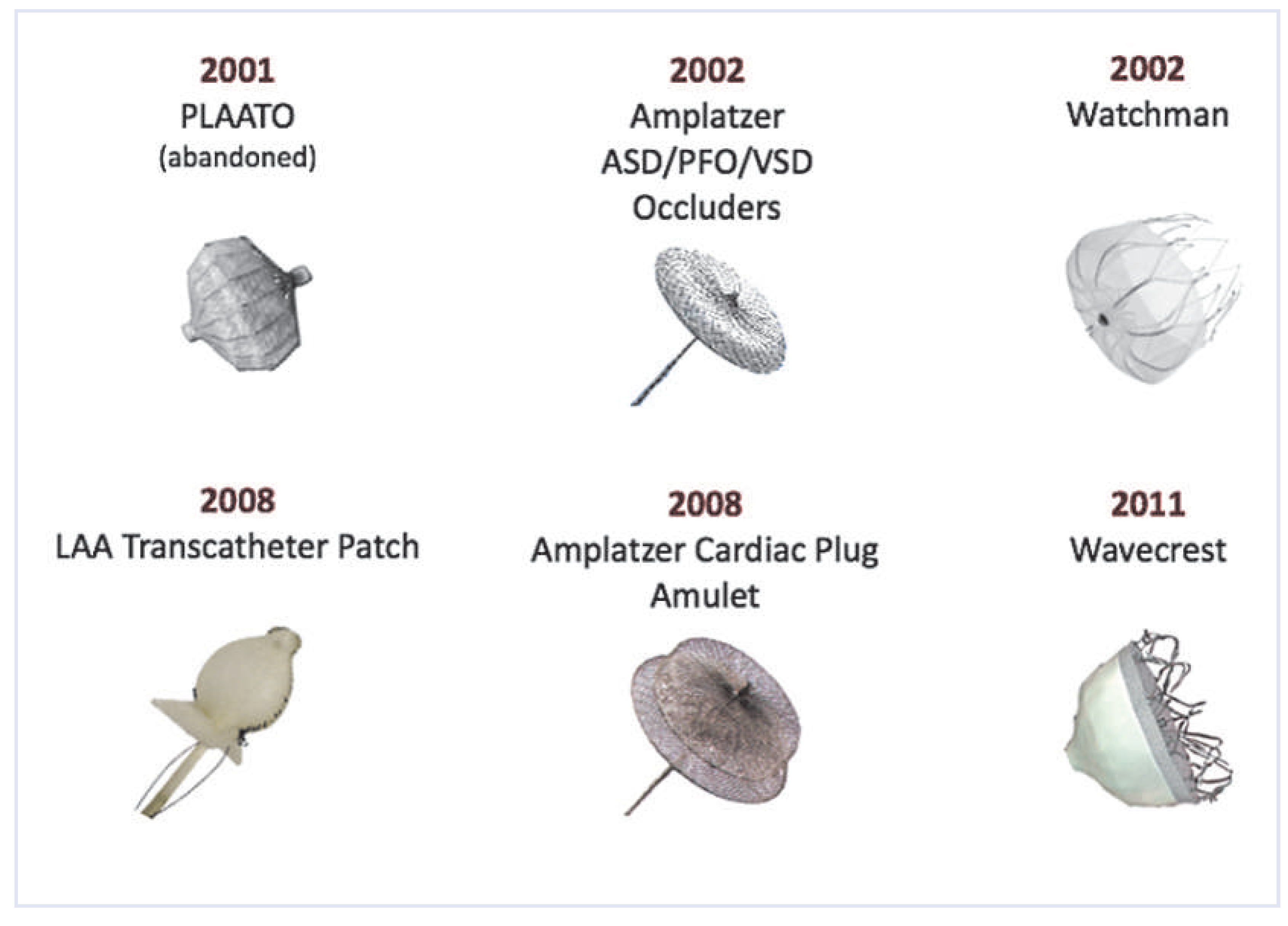
The electrophysiologist Michael Lesh developed a plug device that could be implanted percutaneously, intrigued by the fact that the LAA was easily accessible with catheters used for AF ablation in the LA. He attended the first implantation of his Percutaneous Left Atrial Appendage Transcatheter Occluder (PLAATO, abandoned, Medtronic, Mansfield, MA, USA) on August 30, 2001, by Horst Sievert in Frankfurt, Germany [4]. The results of patients treated with this device were superior to what could be expected with VKA [5,6]. The procedure itself, however, was rather intricate, partly due to bulky catheters.
On June 15, 2002, we introduced a more simple technique applicable in a conscious patient under local anaesthesia and without echocardiographic guidance. This technique used various Amplatzer devices (St. Jude, Plymouth, MS, USA) that were market leaders in the closure of atrial septal defects (ASD) and patent foramen ovale (PFO) [7]. Figure 1 shows the 1-year follow-up echocardiogram of the world’s first patient.
Later the same year, the Watchman device (Boston Scientific, Marlborough, MA, USA) was introduced into clinical medicine (first case on August 12, 2002, performed by Peter Sick) [8]. This device and the Amplatzer devices, in particular the Amplatzer Cardiac Plug (ACP), a modification of the original Amplatzer devices introduced in 2008, currently dominate the market [9,10]. The LAA Transcatheter Patch (Custom Medical Devices, Athens, Greece) and the WaveCrest device (Johnson and Johnson, Diamond Bar, CA, USA), the two other devices that are clinically used in Europe, are depicted in Figure 2. On rare occasions a hybrid procedure using a left atrial approach combined with a pericardial approach is used [11,12,13]. This technique is more in competition with mini-invasive surgery [14] than with interventional cardiology.

Percutaneous technique for left atrial appendage occlusion

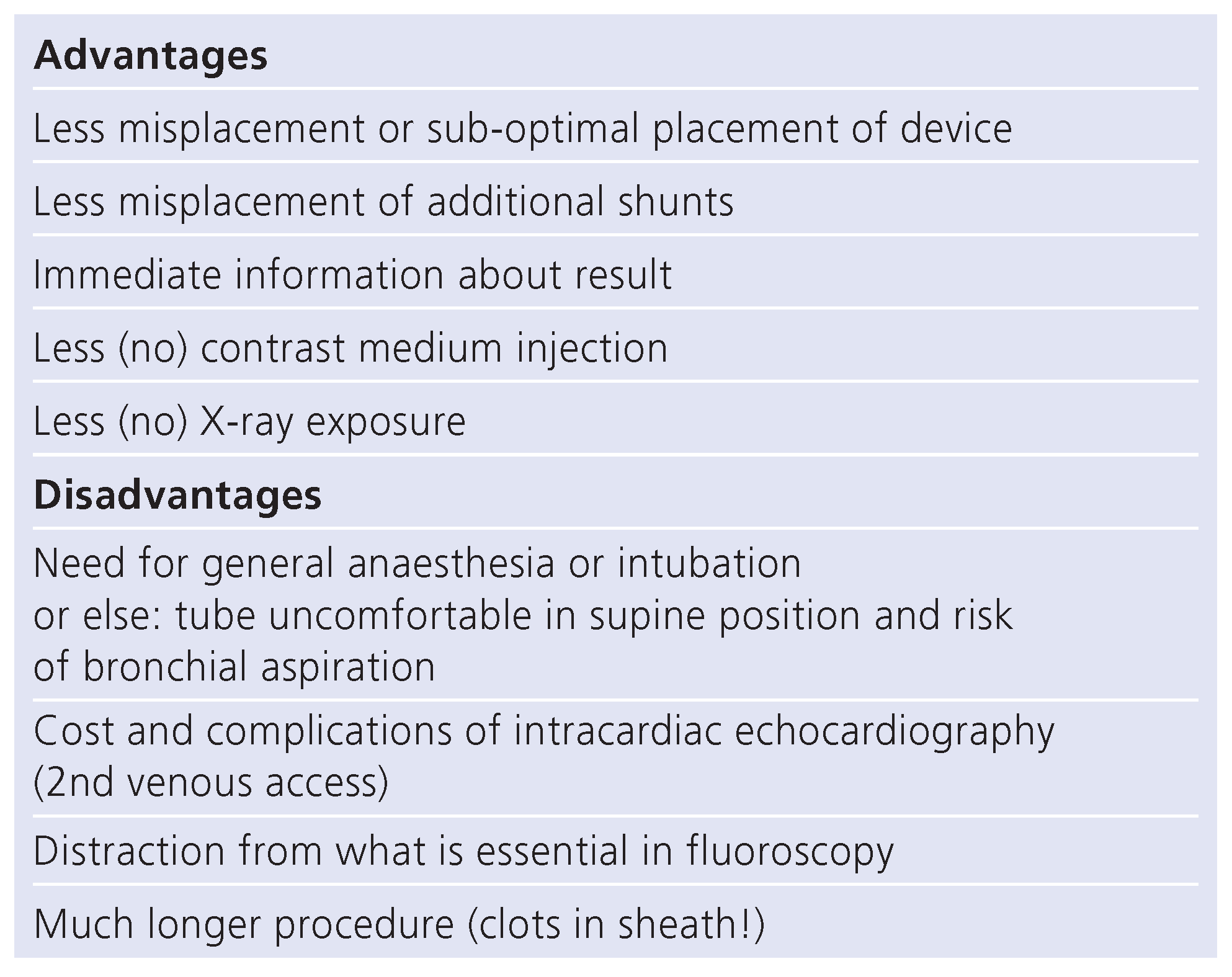
While most centres prefer general anaesthesia with endotracheal intubation and TOE guidance, simplifications have been suggested [15]. TOE guidance is possible with deep sedation but bronchial aspiration has to be watched out for (Table 1). Intracardiac echocardiography (ICE) has also been recommended [16]. Our group has had good experience with the most frugal of all approaches [10].
Most patients undergo a TOE before the procedure to rule out mobile clots in the LAA. It is possible to perform the procedure without prior TOE but this requires a contrast medium injection into the LA at a distance of the LAA to assess for thrombus [17]. While most operators prefer a trans-septal puncture through the septum primum low and posterior in the fossa ovalis, a PFO and, even better, an ASD when present can be used for LA access in most cases. The LAA occlusion can be combined with a variety of other interventions, most commonly coronary angiography, yielding an associated procedure in 41%, 2 associated procedures in 32%, 3 in 6%, and 4 or more in 1% at our centre (Figure 3). We finish the LAA occlusion with closing a pre-existing atrial septal passage using the same delivery sheath for a second Amplatzer (ASD or PFO) occluder.
A specifically shaped sheath (13 French = 4.3 mm inner and 5.5 mm outer diameter) is gently introduced into the left atrial appendage and the device is implanted through it. This is performed under heparin (e.g., 5000 units before beginning the procedure) and antibiotic protection (e.g., 1 g of cephalosporin before the procedure and at least once after the procedure). Stable position of the device and absence of pericardial effusion is usually confirmed by transthoracic echocardiography a few hours later (after mobilisation) and the patient is often discharged the same day. The latest moment of device embolisation appears to be the mobilisation of the patient leading to markedly altered position of the heart. Either oral anticoagulation for a few weeks followed by acetylsalicylic acid (ASA) or, at our centre, clopidogrel for 1 month and ASA for 5 months with or without loading doses are used after discharge. We recommend a TOE at 3 to 6 months to assure proper closure of the LAA and absence of mobile thrombi or of thrombi protruding into the LA.

Complications of left atrial appendage occlusion

Device embolisation and cardiac perforation with pericardial bleeding are the most feared complications and occur in about 1 to 3% each. They appear to be deviceindependent and to occur less frequently with more experienced operators. However, the technique is intricate and the learning curve is rather flat. Late device embolisations that have been reported were probably misinterpreted early device embolisations that were clinically silent (as practically all device embolisations are) and were detected only late. Late pericardial effusions, however, do occur, probably more due to pericardial reactions than to late perforations. Nonetheless, late erosion for instance of a pulmonary artery by the retainer hook of an LAA occluder has been reported [18]. Late mobile thrombi on the device detected either by routine TOE follow-up or after a clinical event occur in 3 to 5%. They may need a period of VKA or NOAC if feasible for the patients. Thrombectomy, perhaps with surgical explantation of the device, is an alternative but has rarely been reported.

Clinical results of left atrial appendage occlusion

The results of 2 randomised trials with the Watchman device (PROTECT AF, and PREVAIL) [15] and registry publications of thousands of patients treated with Watchman or Amplatzer devices [19,20,21] attest reasonable safety and surprising efficacy. In particular, the Watchman device proved to reduce mortality over 4 years when compared to Warfarin treatment [20]. A large registry using the Amplatzer device with over 1000 patients showed a mortality curve congruent with that of the PROTECT AF trial [21]. This suggests that both currently leading techniques may be not only superior to VKA but are probably also competitive with NOAC. A respective randomised trial with the Amplatzer device is ongoing.
A cost analysis projected that about 50 000 US dollars have to be invested for 1 quality-adjusted year of life with LAA closure compared to VKA while this costs 90 000 US dollars with dabigatran, the hitherto most widely used NOAC [22].

Conclusion

Clinical results with LAA occlusion confirm the theory that eliminating the LAA as a possible source of systemic emboli in AF provides at least as good a protection against systemic emboli as oral anticoagulation with VKA and probably also with NOAC and yields a net benefit because the constant bleeding risk (growing with age) with any type of oral anticoagulation is virtually eliminated. Hence, this option may be discussed with every patient with atrial fibrillation irrespective of whether or not anti-thrombotic treatment has already been started. This has yet to be reflected in guidelines where LAA closure is at best mentioned as an alternative for oral anticoagulation in patients with contraindications to it or unwilling to take it. Moreover, as both methods fail to provide complete prevention against systemic embolism, a combination of them can be considered.

Funding/potential competing interests:

Speaker honoraria and research grants to the institution of St. Jude Medical. No other potential conflict of interest relevant to this article was reported.

References

  1. Blackshear, J.; Odell, J. Appendage obliteration to reduce stroke in cardiac surgical patients with atrial fibrillation. Ann Thorac Surg. 1996, 61, 755–759. [Google Scholar] [CrossRef] [PubMed]
  2. Madden, J. Resection of the left auricular appendix; a prophylaxis for recurrent arterial emboli. J Am Med Assoc. 1949, 140, 769–772. [Google Scholar] [CrossRef] [PubMed]
  3. Bonow, R.; Carabello, B.; Chatterjee, K.; de Leon, A.J.; Faxon, D.; Freed, M.; et al. ACC/AHA 2006 guidelines for the management of patients with valvular heart disease: A report of the American College of Cardiology/American Heart Association Task Force on Practice Guidelines (writing Committee to Revise the 1998 guidelines for the management of patients with valvular heart disease) developed in collaboration with the Society of Cardiovascular Anesthesiologists endorsed by the Society for Cardiovascular Angiography and Interventions and the Society of Thoracic Surgeons. J Am Coll Cardiol. 2006, 48, e1–e148. [Google Scholar] [PubMed]
  4. Sievert, H.; Lesh, M.; Trepels, T.; Omran, H.; Bartorelli, A.; Della Bella, P.; et al. Percutaneous left atrial appendage transcatheter occlusion to prevent stroke in high-risk patients with atrial fibrillation: Early clinical experience. Circulation 2002, 105, 1887–1889. [Google Scholar] [CrossRef] [PubMed]
  5. Ostermayer, S.; Reisman, M.; Kramer, P.; Matthews, R.; Gray, W.; Block, P.; et al. Percutaneous left atrial appendage transcatheter occlusion (PLAATO system) to prevent stroke in high-risk patients with non-rheumatic atrial fibrillation: Results from the international multi-center feasibility trials. J Am Coll Cardiol. 2005, 46, 9–14. [Google Scholar] [CrossRef] [PubMed]
  6. Bayard, Y.; Omran, H.; Neuzil, P.; Thuesen, L.; Pichler, M.; Rowland, E.; et al. PLAATO (Percutaneous Left Atrial Appendage Transcatheter Occlusion) for prevention of cardioembolic stroke in non-anticoagulation eligible atrial fibrillation patients: Results from the European PLAATO study. EuroIntervention 2010, 6, 220–226. [Google Scholar] [CrossRef] [PubMed]
  7. Meier, B.; Palacios, I.; Windecker, S.; Rotter, M.; Cao, Q.; Keane, D.; et al. Transcatheter left atrial appendage occlusion with Amplatzer devices to obviate anticoagulation in patients with atrial fibrillation. Catheter Cardiovasc Interv. 2003, 60, 417–422. [Google Scholar] [CrossRef] [PubMed]
  8. Sick, P.; Schuler, G.; Hauptmann, K.; Grube, E.; Yakubov, S.; Turi, Z.; et al. Initial worldwide experience with the Watchman left atrial appendage system for stroke prevention in atrial fibrillation. J Am Coll Cardiol. 2007, 49, 1490–1495. [Google Scholar] [CrossRef] [PubMed]
  9. Freixa, X.; Chan, J.L.; Tzikas, A.; Garceau, P.; Basmadjian, A.; Ibrahim, R. The Amplatzer Cardiac Plug 2 for left atrial appendage occlusion: Novel features and first-in-man experience. EuroIntervention 2013, 8, 1094–1098. [Google Scholar] [CrossRef] [PubMed]
  10. Nietlispach, F.; Gloekler, S.; Krause, R.; Shakir, S.; Schmid, M.; Khattab, A.A.; et al. Amplatzer left atrial appendage occlusion: Single center 10-year experience. Catheter Cardiovasc Interv. 2013, 82, 283–289. [Google Scholar] [CrossRef] [PubMed]
  11. Lee, R.; Bartus, K.; Yakubov, S. Catheter-based left atrial appendage (LAA) ligation for the prevention of embolic events arising from the LAA: Initial experience in a canine model. Circ Cardiovasc Interv. 2010, 3, 224–229. [Google Scholar] [CrossRef] [PubMed]
  12. Bartus, K.; Bednarek, J.; Myc, J.; Kapelak, B.; Sadowski, J.; Lelakowski, J.; et al. Feasibility of closed-chest ligation of the left atrial appendage in humans. Heart Rhythm. 2011, 8, 188–193. [Google Scholar] [CrossRef] [PubMed]
  13. Shetty, R.; Leitner, J.; Zhang, M. Percutaneous catheter-based left atrial appendage ligation and management of periprocedural left atrial appendage perforation with the LARIAT suture delivery system. J Invasive Cardiol. 2012, 24, E289–93. [Google Scholar] [PubMed]
  14. Friedman, P.A.; Asirvatham, S.J.; Dalegrave, C.; Kinoshita, M.; Danielsen, A.J.; Johnson, S.B.; et al. Percutaneous epicardial left atrial appendage closure: Preliminary results of an electrogram guided approach. J Cardiovasc Electrophysiol. 2009, 20, 908–915. [Google Scholar] [CrossRef] [PubMed]
  15. Meier, B.; Blaauw, Y.; Khattab, A.; Lewalter, T.; Sievert, H.; Tondo, C.; et al. Catheter Based Left Atrial Appendage Occlusion, Scientific document supported by the European Association of Percutaneous Coronary Interventions (EAPCI) and the European Heart Rhythm Association (EHRA). EuroInterv, 2014; in print. [Google Scholar]
  16. Santoro, G.; Meucci, F.; Stolcova, M.; Rezzaghi, M.; Mori, F.; Balmieri, C.; et al. Percutaneous left atrial appendage occlusion with patients with nonvalvular atial fibrillation: Implantation and op to four-years follow-up of the Amplatzer cardiac Plug. EuroInterv, 2014; in print. [Google Scholar]
  17. Nietlispach, F.; Krause, R.; Khattab, A.; Gloekler, S.; Schmid, M.; Wenaweser, P.; et al. Ad hoc percutaneous left atrial appendage closure. J Invasive Cardiol. 2013, 25, 683–686. [Google Scholar] [PubMed]
  18. Hannazawa, K.; Brunelli, M.; Saenger, J.; Grosse, A.; Santi, R.; Lauer, B.; et al. Clouse proximity between pulmonary artery and left atrial appendage leading to perforation of the artery tamponade and death after appendage closure you using cardiac plug device. Interv J Cardiol. 2014; in print. [Google Scholar]
  19. Park, J.; Bethencourt, A.; Sievert, H.; Santoro, G.; Meier, B.; Walsh, K.; et al. Leithauser B. Left atrial appendage closure with Amplatzer Cardiac Plug in atrial fibrillation: Initial European experience. Catheter Cardiovasc Interv. 2011, 77, 700–706. [Google Scholar] [PubMed]
  20. Reddy, V. Watchman LAA closure device reduces the risk of ischemic stroke in patients with AF entirely without anticoagulation. The ASAPlavix (ASAP) Registry. In Proceedings of the Presentation of Transcatheter Therapeutics (TCT), Miami, Florida, USA, November 2012. [Google Scholar]
  21. Tzikas, A.; Shakir, S.; Sievert, H.; Omran, H.; Berti, S.; Santoro, G.; et al. Left atrial appendage occlusion for stroke prevention in atrial fibrillation: Multicenter experience with the Amplatzer Cardiac Plug. EuroInterv, 2014; in print. [Google Scholar]
  22. Singh, S.M.; Micieli, A.; Wijeysundera, H.C. Economic evaluation of percutaneous left atrial appendage occlusion, dabigatran, and warfarin for stroke prevention in patients with nonvalvular atrial fibrillation. Circulation 2013, 127, 2414–2423. [Google Scholar] [CrossRef] [PubMed]
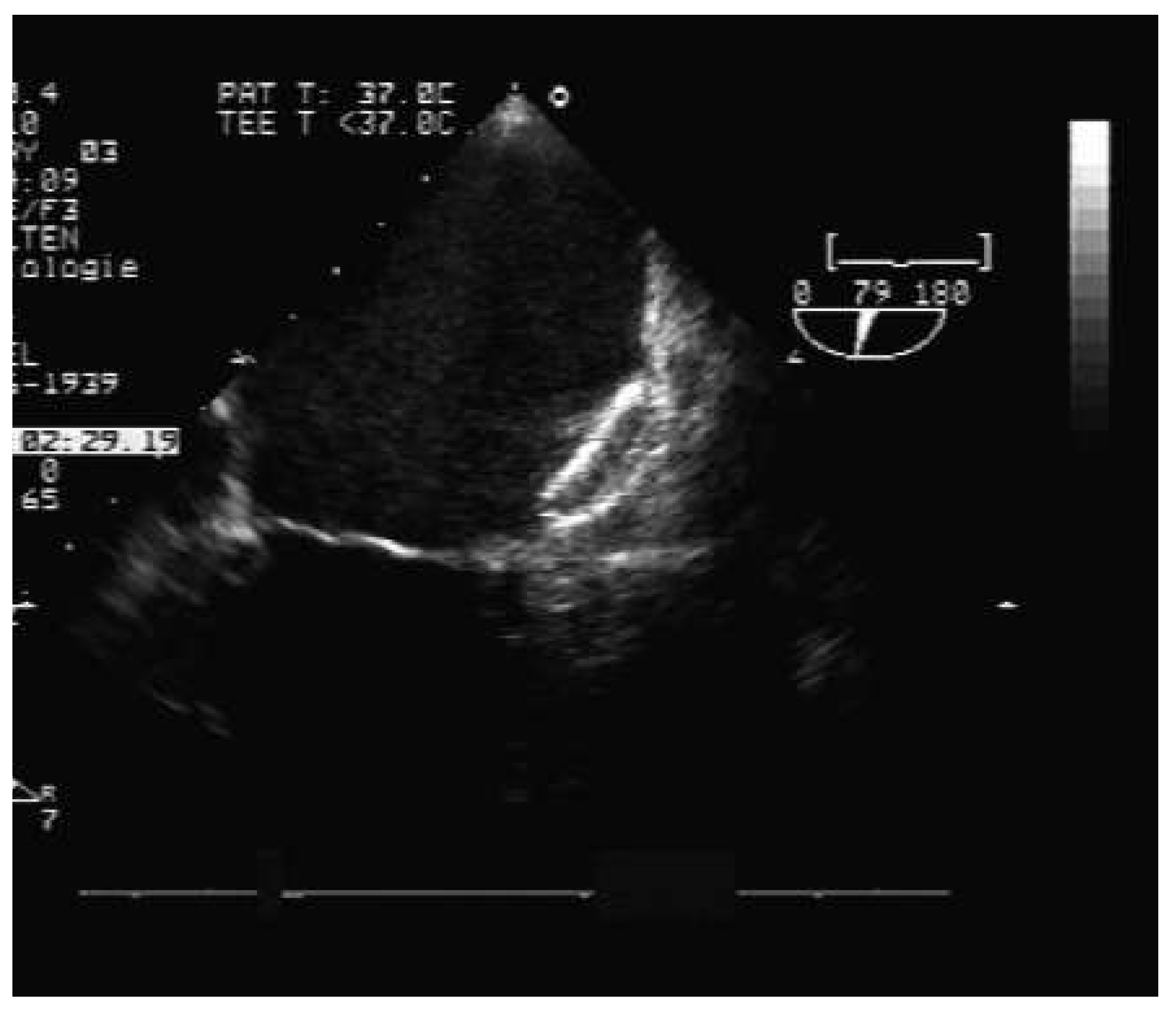
Figure 1. Trans-oesophageal echocardiography of the world’s first patient with a left atrial appendage occlusion under non-sedated local anaesthesia and without echocardiographic guidance. The patient was a 63-yearold butcher with atrial fibrillation. He could not take oral anticoagulants for professional reasons. The procedure was performed on June 15, 2002, using an Amplatzer 30 mm Atrial Septal Occluder and the patient has had no further need for anticoagulation in a clinically uneventful course for over 12 years.
Cardiovascmed 17 00252 g001
Figure 2. Left atrial appendage occluders used in Europe with their year of introduction. The Amplatzer devices are the most commonly used ones followed by the Watchman devices.
Cardiovascmed 17 00252 g002
Figure 3. Procedures associated to left atrial appendage occlusion at our institution in 413 cases. AF = atrial fibrillation; ASD = atrial septal defect; PCI = percutaneous coronary intervention; PFO = patent foramen ovale; RF = radio frequency; TAVI = transcatheter aortic valve implantation.
Cardiovascmed 17 00252 g003
Table 1. Advantages and disadvantages of echocardiographic guidance (trans-oesophageal or intracardiac) for left atrial appendage closure.
Cardiovascmed 17 00252 i001