Next Article in Journal
A Tachycardia with QRS Duration Narrower Than That During Sinus Rhythm
Previous Article in Journal
Early Versus Late Initial Echocardiographic Assessment in Infective Endocarditis: Similar Findings and No Difference in Clinical Outcome
 
 
Cardiovascular Medicine is published by MDPI from Volume 28 Issue 1 (2025). Previous articles were published by another publisher in Open Access under a CC-BY (or CC-BY-NC-ND) licence, and they are hosted by MDPI on mdpi.com as a courtesy and upon agreement with Editores Medicorum Helveticorum (EMH).
Interesting Images

Valentine in the Heart

by
Panneerselvam Arunkumar
* and
Ramasamy Palanimuthu
Department of Cardiology, PSG Institute of Medical Sciences and Research, Peelamedu, Coimbatore 641004, India
*
Author to whom correspondence should be addressed.
Cardiovasc. Med. 2012, 15(10), 295; https://doi.org/10.4414/cvm.2012.00108
Submission received: 24 July 2012 / Revised: 24 August 2012 / Accepted: 24 September 2012 / Published: 24 October 2012

Case Report

A 45-year-old male with recent anterior wall infarction underwent coronary angiogram. His left coronary system showed insignificant stenosis with thrombus. Incidentally, the right coronary artery revealed a branch arising from the posterolateral left ventricular branch of right coronary artery (RCA) and anastomosing with the sinus nodal artery arising from the proximal RCA. This anastomosis gave a Valentines shaped heart to the coronary artery (Figure 1). This branch arising from distal RCA in the region of atrioventricular nodal artery has also been called “right posterior” sinus nodal artery. This dual blood supply SA node makes it relatively resistant to ischaemia.

Funding/Potential Competing Interests

No financial support and no other potential conflict of interest relevant to this article were reported.

References

  1. Gavaliatsis, I.P. “Right posterior” sinus node artery. Int J Cardiol. 2000, 75, 301–303. [Google Scholar] [CrossRef] [PubMed]
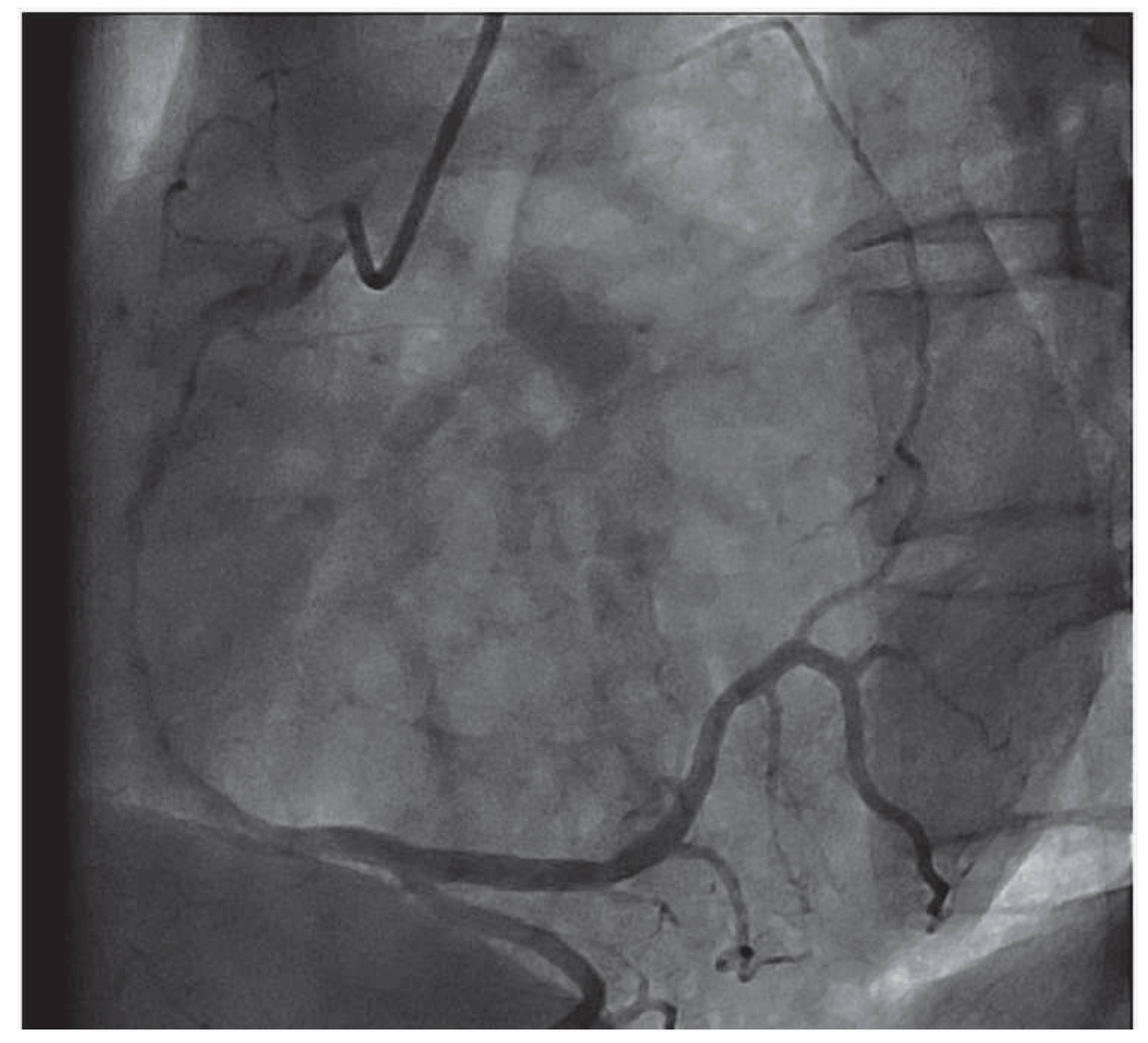
Figure 1. Right coronary angiogram demonstrating the communication between posterolateral LV branch of right coronary artery and SA nodal artery. This collateral circulation gives a heart shaped appearance to the right coronary circulation.
Cardiovascmed 15 00295 g001