Next Article in Journal
Cardiac Thrombus as a Primary Manifestation of Polycythemia Vera
Previous Article in Journal
Moderne Technologien in der Ablation des Vorhofflimmerns
 
 
Cardiovascular Medicine is published by MDPI from Volume 28 Issue 1 (2025). Previous articles were published by another publisher in Open Access under a CC-BY (or CC-BY-NC-ND) licence, and they are hosted by MDPI on mdpi.com as a courtesy and upon agreement with Editores Medicorum Helveticorum (EMH).
Interesting Images

Curing Heart Failure with Cathether Ablation?

by
Marcus Mutschelknauss
1,
Peter Rickenbacher
2 and
Michael Kühne
1,*
1
Division of Cardiology/Electrophysiology, University of Basel Hospital, 4031 Basel, Switzerland
2
Division of Cardiology, Department of Internal Medicine, University Hospital Bruderholz, Bruderholz, Switzerland
*
Author to whom correspondence should be addressed.
Cardiovasc. Med. 2010, 13(9), 288; https://doi.org/10.4414/cvm.2010.01528
Submission received: 15 June 2010 / Revised: 15 July 2010 / Accepted: 15 August 2010 / Published: 15 September 2010

1. Case Presentation

A 63-year-old Japanese tourist presented to the emergency department with increasing shortness of breath and palpitations over the previous two weeks. On physical examination the patient showed signs of acute heart failure. Left ventricular ejection fraction was 35% and B-type natriuretic peptide was elevated (BNP 900 pg/mL, normal < 50 pg/mL). The 12-lead ECG (Figure 1) showed regular narrow-complex tachycardia with a heart rate of 176 bpm and an RP-interval of 170 ms. Inverted P-waves in the inferior leads and positive P-waves in lead V1 were visible. Immediate recurrence occurred after termination of the tachycardia with adenosine. After additional treatment with flecainide the patient was converted to stable sinus rhythm. The 12-lead ECG in sinus rhythm after termination showed no signs of preexcitation. The patient elected to undergo radiofrequency ablation (RFA) of the arrhythmia before returning to Japan.

2. Questions

  • What is the differential diagnosis of the 12-lead ECG (Figure 1)?
  • How do the intracardiac electrograms (Figure 2) allow further differentiation of the arrhythmia?
  • What is the mechanism of heart failure?

3. Commentary

In patients with or without preexcitation in sinus rhythm, given the RP-interval and the inverted P-waves in the inferior leads, the differential diagnosis of the 12-lead ECG shown in Figure 1 chiefly features three arrhythmias: atypical atrioventricular nodal reentrant tachycardia (AVNRT), atrial tachycardia originating from the inferior parts of the atria, and orthodromic reciprocating tachycardia (ORT) using a concealed accessory pathway.
During the electrophysiological study the patient was in sinus rhythm with normal AH- and HV-intervals. No sustained tachycardia could be induced despite administration of isoproterenol. There was one non-sustained episode of tachycardia, as shown in Figure 2A. The figure shows the tachycardia with the earliest atrial activation (see asterisk) in the distal coronary sinus (CS 1-2). This eccentric activation sequence rules out atypical AVNRT. However, left atrial tachycardia and ORT still feature in the differential diagnosis. The Figure 2B shows that there is also an eccentric activation sequence with ventricular pacing, the earliest atrial activation occurring in the distal coronary sinus. This evidences the presence of an accessory pathway, but does not prove its involvement in the documented tachycardia. If the tachycardia is sustained, an entrainment manceuvre should be performed from the ventricle to rule out atrial tachycardia by demonstrating a VAV-response. However, on the basis of the available evidence, transseptal puncture and ablation of the left lateral accessory pathway was performed. Ablation was followed by ventriculo-atrial dissociation during ventricular pacing, proving successful elimination of accessory pathway conduction.
In our patient, ORT, although usually considered benign, was complicated by transient left ventricular dysfunction during a prolonged episode of tachyarrhythmia with a heart rate of approximately 175 bpm for several weeks, suggesting tachycardia-mediated cardiomyopathy as the cause of the patient’s signs of heart failure. The pathophysiological mechanism of tachycardia-mediated cardiomyopathy is poorly understood. The hypotheses include depletion of myocardial energy stores, abnormalities of myocardial blood flow, neurohumoral changes or altered myocardial calcium handling [1,2]. Left ventricular ejection fraction in our patient was completely normal four weeks after the procedure, and the patient has remained free of arrhythmia after a follow-up of three months.

Conflicts of Interest

The authors have no conflict of interest to disclose.

References

  1. Khasnis, A.; Jongnarangsin, K.; Abela, G.; Veerareddy, S.; Reddy, V.; Thakur, R. Tachycardia-induced cardiomyopathy: A review of literature. Pacing Clin Electrophysiol. 2005, 28, 710–721. [Google Scholar] [CrossRef] [PubMed]
  2. Gopinathannair, R.; Sullivan, R.; Olshansky, B. Tachycardia-mediated cardiomyopathy: Recognition and management. Curr Heart Fail Rep. 2009, 6, 257–264. [Google Scholar] [CrossRef] [PubMed]
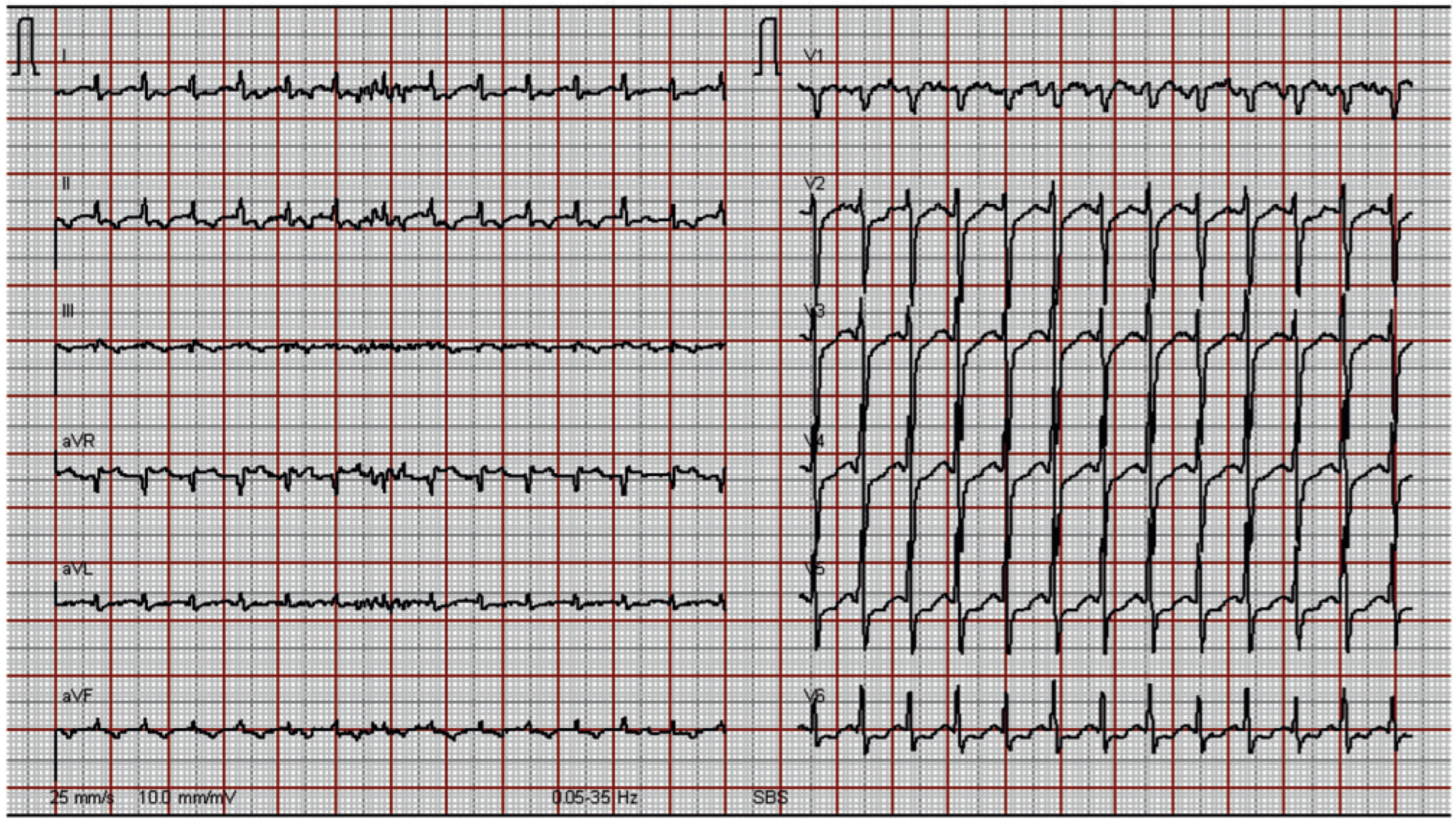
Figure 1. 12-lead admission ECG showing regular narrow-complex tachycardia with a heart rate of 176 bpm.
Cardiovascmed 13 00288 g001
Figure 2. (A) Two beats of a non-sustained episode of narrow-complex tachycardia. Shown are four surface leads and intracardiac electrograms recording the high right atrium (HRA), the His bundle (HIS), the coronary sinus (CS) 1,2 (distal) to CS 9,10 (proximal), and the right ventricular apex (RVA). The asterisk denotes the earliest atrial activation in the distal coronary sinus. (B) Corresponding surface leads and intracardiac electrograms during ventricular pacing at a cycle length of 600 ms.
Cardiovascmed 13 00288 g002