Next Article in Journal
Krankenversicherungsgesetz, die Versicherungen und die ärztliche Ethik—Jenseits Evidenzbasierter Medizin?
Previous Article in Journal
Interventional Cardiology in Switzerland 2006
 
 
Cardiovascular Medicine is published by MDPI from Volume 28 Issue 1 (2025). Previous articles were published by another publisher in Open Access under a CC-BY (or CC-BY-NC-ND) licence, and they are hosted by MDPI on mdpi.com as a courtesy and upon agreement with Editores Medicorum Helveticorum (EMH).
Review

Coronary CT Is Overused, Angiography Remains the Standard †

Cardiovascular Department, University Hospital, Berne, Switzerland
This article summarises a lecture at the annual meeting of the Swiss Society of Cardiology in Geneva, June 2007.
Cardiovasc. Med. 2008, 11(6), 196; https://doi.org/10.4414/cvm.2008.01337
Submission received: 27 March 2008 / Revised: 27 April 2008 / Accepted: 27 May 2008 / Published: 27 June 2008
Computerised tomography (CT) was first used to screen for coronary artery disease (CAD) by using a calcium score produced by electron beam computerised tomography (EBCT). After an initial boom, the interest waned until CT angiography became available with 16and more recently 64-slice technology. Stunning pictures of the major epicardial coronary arteries can be produced and luminal narrowings can be reconstructed. However, the enthusiasm of those running such equipment is not justified for a number of banes of the technique. First, the coronary arteries can be seen at best down to the second-degree vessels. Smaller vessels do simply not appear on the pictures. Second, there are plenty of artefacts. Even if they are recognised as such, each of them renders a segment of the coronary tree non-assessable. Third, calcium (virtually omnipresent in people beyond 50 years of age) forfeits analysis of the vessel adjacent to it. Forth, the heart rate needs to be regular and in the physiological range for any gating process. Fifth, the patients have to be able to hold their breaths for 15–30 s while constrained in an uncomfortable position and surrounding. And last but not least the radiation dose applied is prohibitive for a screening method. Irradiation is higher than for a conventional coronary angiogram including multivessel angioplasty. There is hope, that radiation can be reduced by dual source CT apparatus or prospective ECG gating. Yet, even if the x-ray hazard can be significantly curbed, the method remains invalidated by oversensitivity. Most people in the typical screening ages will have irregularities in CT coronary angiography. Hence, they will all be sent on to conventional coronary angiography so that the intermediate step of CT angiography should be skipped. In young people, where normal coronary arteries prevail, CT angiography screening for CAD will produce a malignant tumor in about 1 out of 200 cases. This being conveyed to people asking for it will likely stop them asking for it.

Historical development

Selective coronary angiography started on October 30th, in 1958, when Mason Sones inadvertently injected dye directly into the right coronary artery of a patient rather than into the ascending aorta as customary at the time (Figure 1) [1]. The quality of the picture was certainly not up to today’s standards. Nonetheless, it is superior to the current resolution of computerised tomography (CT) angiography.
Interest for CT technology in context with coronary artery disease first rose more than 10 years ago when electron beam computerised tomography (EBCT) was advocated for screening for coronary calcifications pinpointing clinically relevant coronary artery disease [2]. The technique boomed for a while in commercially oriented medical environments. It practically always found “disease” in the population interested in the test, ie, middle-aged (mostly affluent) men worried about their risk for myocardial infarction. It has long been known to cardiologists and eventually also to the remainder of the doctors that calcium in the wall of the coronary arteries be part of aging as are wrinkles in the face. Performing EBCT was a win-win situation that did, however, carry the risk of exploding healthcare costs. The patients were typically first alarmed but in most cases this was unveiled as false alarm with subsequent tests, be they exercise stress electrocardiography, myocardial scintigraphy, or coronary angiography. The patients (nonpatients that is) easily reconciled with the initial exam although it had lifted the flag for all the wrong reasons. They felt like having been given a second life by the subsequent tests acquitting them from the verdict of coronary artery disease. The few in whom real disease was found were even more pleased with the CT test as they regarded it as a life saviour having uncovered the problem in time for being taken care of before irreversible damage occurred.
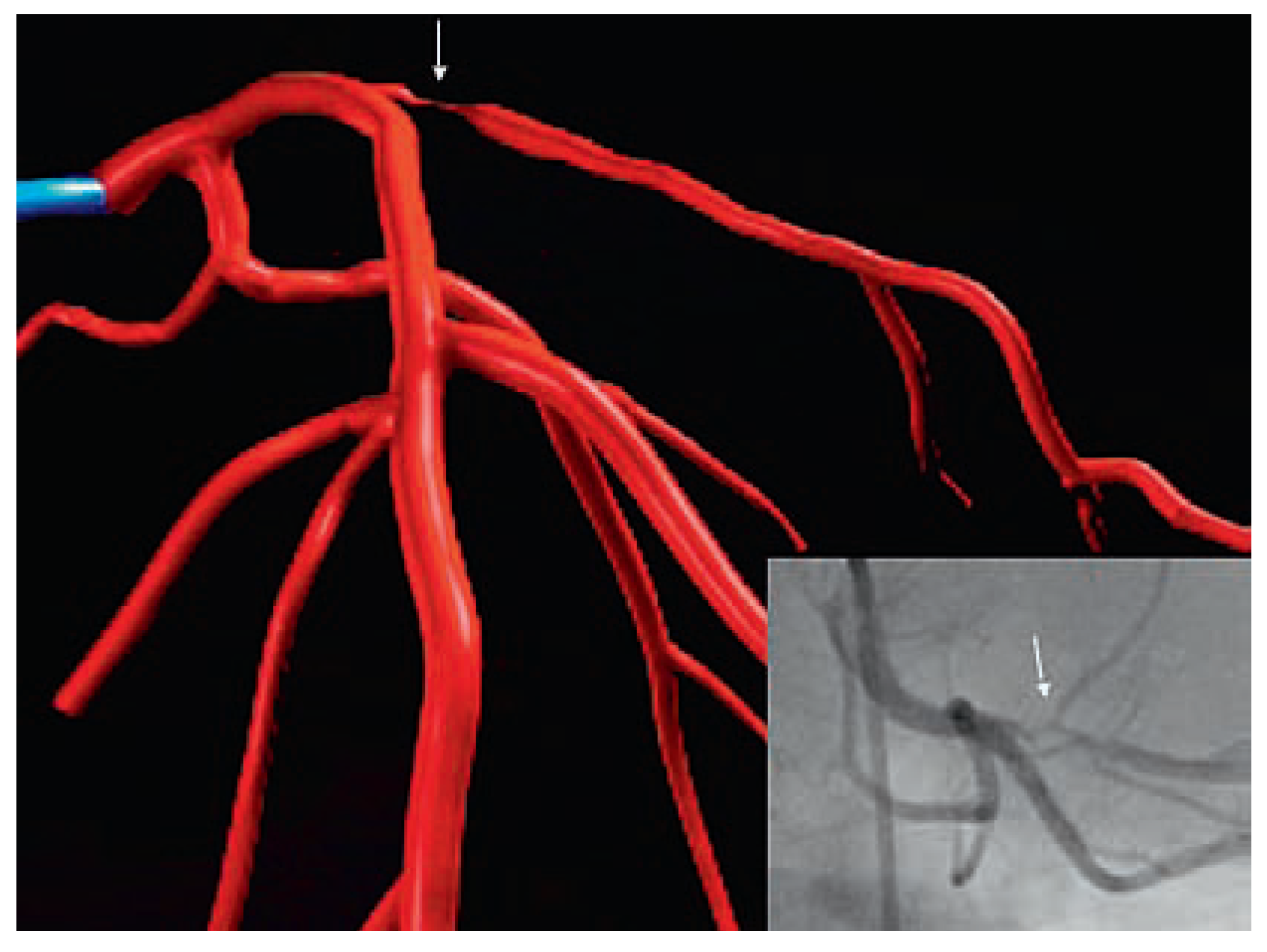
The industry once launched was unstoppable. First 4-slice CT angiography ensued, then 16-slice, then 64-slice, now 256-slice or double source, and this is not the end. The relief depictions of the coronary tree conjuring up 3-dimensionality were true eye catchers. Easily people were distracted from the fact that only the main vessels were seen, artefacts abounded, the lumen (what really keeps the patients alive) was less conspicuously seen, and finally, angiography could rival CT in terms of plasticity and colour (Figure 2) but did not have to because it had better things to offer.

Technicalities

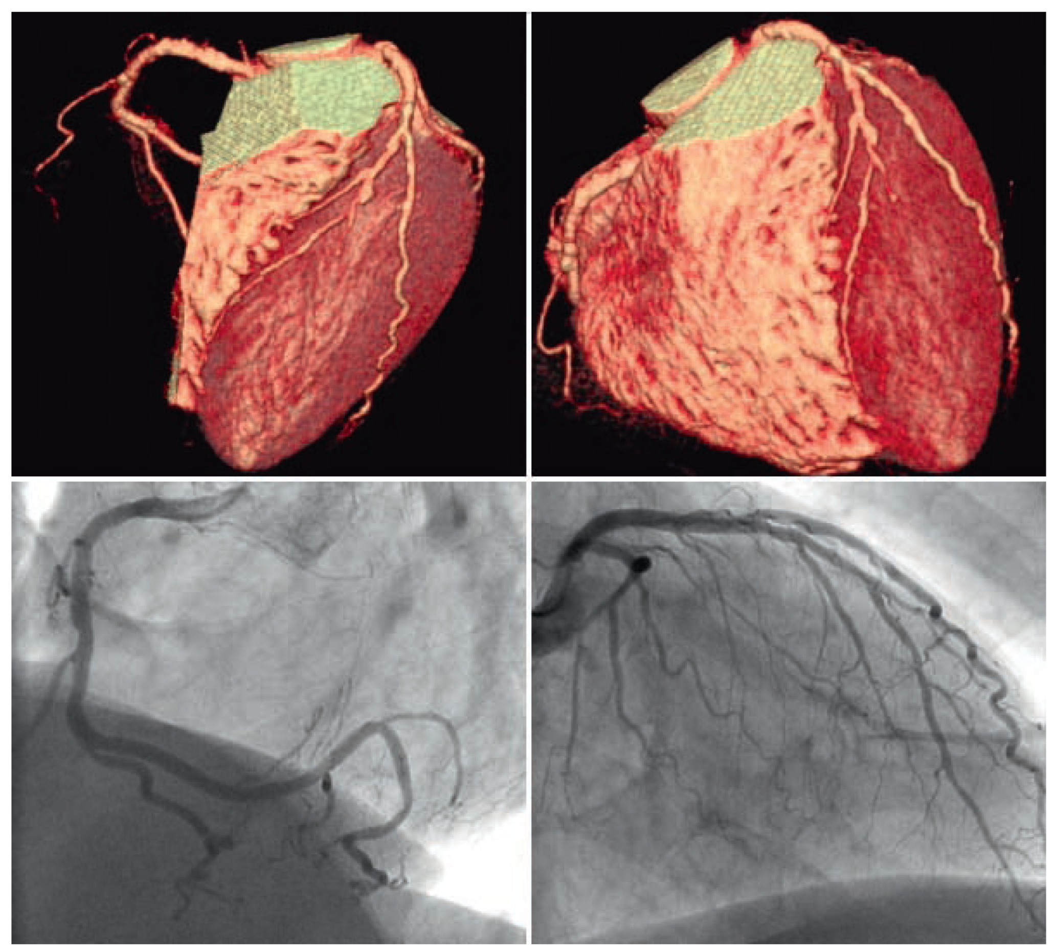
The main advantage of angiography consists in the fact that it projects the entire heart onto a plane, thereby lossless reducing 3 dimensions to 2 dimensions. Every picture point is placed in an analogue fashion. Its position is therefore unaltered and genuine. Computerised pictures, be they from CT or magnetic resonance (MR) are also 2-dimensional. Yet, they are recalculated from a number of slices (0.5 mm) cut through the object. As the picture should be real-time or at least close to, powerful computers are required. On one hand, they can add a 3-dimensional flair to the picture; on the other hand, they are bound to miscalculate and creating pixel displacements or even gross artefacts. Figure 3 depicts, what happens, when a CT shows what is not really there which is rather the rule than the exception.

Diagnostic performance

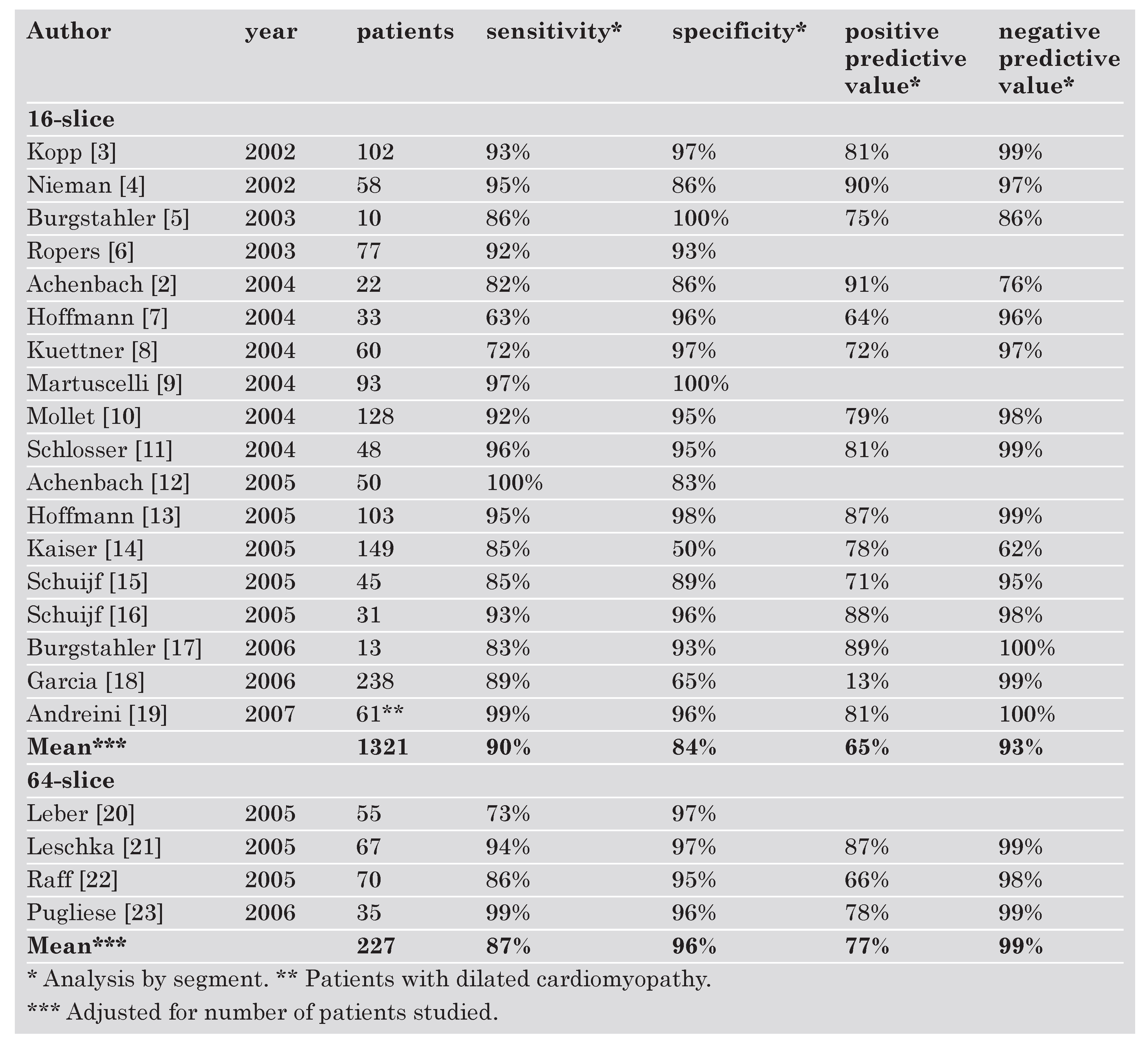
Table 1 [2,3,4,5,6,7,8,9,10,11,12,13,14,15,16,17,18,19,20,21,22,23] depicts results of a selection of studies comparing coronary CT to the gold standard, ie, coronary angiography. Even abstracting from the fact, that angiography is possible in all patients while CT can only be done in patients capable of holding their breaths for about 20 s while being in a fairly slow sinus rhythm, specificity and positive predictive value of CT are severely handicapped by artefacts such as the ones in the case of Figure 1. A comparative study between the Siemens Sensation 64-slice and the Toshiba Aquilion 64-slice equipment in 150 and 42 patients, respectively, showed comparable but poor positive predictive values of 46% and 40%, respectively [24].
Cardiovascmed 11 00196 i001

Associated techniques

Functional techniques can be associated to coronary angiography with table mounted bicycle exercise testing, flow velocity measurements, pressure assessments, and detailed collateral studies. CT has been combined with stress single photon emission computed tomography (SPECT) or positron emission tomography (PET) [25] depicting myocardial perfusion or viability. These two informations are not obtainable by conventional angiography. However, those adjunctive methods can be combined with angiography serially.

Hazards and complications

The complications of diagnostic coronary angiography have been estimated at a mortality of 0.05% and a rate of significant cardio-cerebral complications of 0.4% around 1990 in over 20 000 patients [26,27]. The respective figures around 2000 in an equally large patient population were 0.03% and 0.05% [28]. Both procedures require a venous access, CT for contrast administration, angiography (in most laboratories) for safety reasons and for injection of auxiliary drugs. Angiography in addition requires an arterial access that, admittedly, can cause local problems all of which may not necessarily show up in complication data banks. CT, on the other hand, is subject to problems like claustrophobia and poor collaboration in terms of breath holding and fidgeting. Both procedures can be done within a few hours in out-patients. Angiography requires some post interventional surveillance.
The contrast medium load in terms of quantity and toxicity is comparable. For CT, 80 mL to 130 mL are standard. It is easier to reduce contrast medium with conventional angiography than with CT-angiography. With biplane equipment, 3 coronary injections (1 right and 2 left) of 10 mL each and a left ventricular injection of 15 mL suffice, amounting to a total of 45 mL.
The positive and negative sides of the competing techniques are depicted in Table 2 [18,29,30].

Radiation exposure

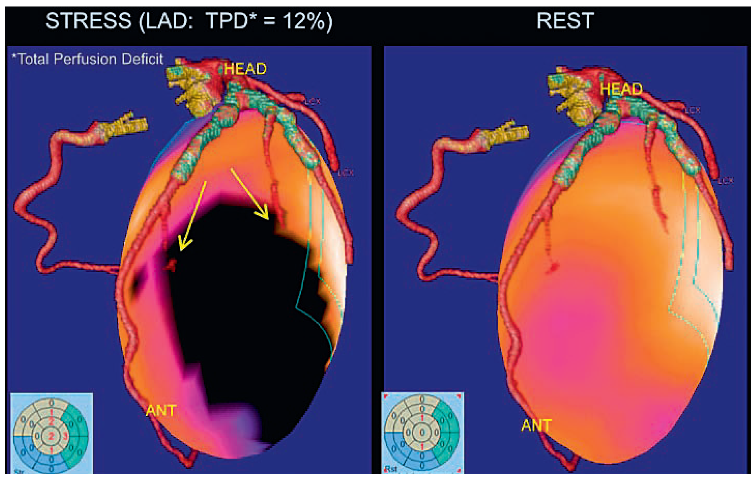
Simple calcium scoring imparts 1–2 mSv on the patient [31]. However, calcium scoring has been abandoned because calcium is invariably present in people older than 50 years and it is difficult to create an algorithm that tells significant (relevant for lumen) from insignificant (eg, adventitial) calcifications. The recent report showing a good correlation of coronary calcification detected by CT with all-cause mortality appears rather trivial, irrespective of the adjustment for age [32]. Calcification (not unlike grey hair) is a surrogate of biological (not necessary chronological) aging and therefore impending death. For a multislice coronary angiography, at least 10 mSv have to be invested, with the current 64-slice machines the exposure approaches 20 mSv. This is bound to increase when moving to 256-slice equipment although the use of a dual source may curb the radiation exposure somewhat. Conventional coronary angiography (monoplane or biplane) ranges in the realm of 5 mSv [33]. This allows adding therapy (percutaneous coronary intervention) and still staying below the radiation dose of CT angiography. For comparison, men are exposed to an annual dose of about 2 mSv from natural sources and radiation workers are monitored not to exceed a dose of 15 mSv per year [31]. Combining CT angiography with a functional test such as SPECT or PET doubles the dose to 30–40 mSv. Applying this combined technique (Figure 4) raises ethical questions considering the fact that radiation-free alternatives are available for the functional part (MR and stress or exercise echocardiography) and the anatomical information can be collected with 1/10th of the dose.
It has been calculated, that screening about 600 patients with multislice CT coronary angiography will produce one new malignancy. This number increases if the screening field is enlarged (venous or particularly mammary artery bypass grafts) or functional tests are associated. Breast cancer in women is the most common tumor produced. It can be calculated that one in 150 20-year-old women subjected to a 64-slice CT for screening for coronary artery disease (not a likely scenario) will pay for it with a cancer. This ratio decreases with age but is still about 1 in 300 if the screening is done at the age of 50 years [34].

Conclusions

The dictum says that the fact that something can be done does not necessarily mean that it should be done. Multislice CT angiography provides colourful and relief pictures of the coronary tree. A true stenosis is detected most (but not all) of the times but artefacts are abundant. Just screening for calcium will raise rather than soothe unwarranted worries. Doing a full CT angiogram will only acquit a patient older than 50 years from the suspicion of having coronary artery disease if the interpreter is experienced and bold enough to take the many artefacts on his account. Less experienced operators (particularly radiologists not privy to the patient’s history and global situation) may be unwilling to take the responsibility of parting with a clean bill of health in light of the artefacts and vessels that they cannot really see. The indication for coronary angiography is the consequence even in patients who basically dropped in from the street to have a check-up and would not have wound up with coronary angiography if they had done a conventional exercise test after a thorough history and physical examination in a cardiologist’s office. Those who have true disease are likely to be properly detected and consequently sent for angiography. If only they had not already spent their yearly allowance of x-ray exposure before getting to the one-stop-shop they should have stopped by in the first place. MR coronary angiography is lagging behind in resolution but we have to wait for it, to find a proper tool for mass screening for coronary artery disease.
What remains for CT is the diagnosis of large structures such as valves, congenital anomalies, and perhaps the distinction between extremes in coronary artery disease. In the workup of dilated cardiomyopathy, for instance, coronary artery disease is either advanced (postmyocardial infarction heart failure) or non existent (idiopathic heart failure). Maybe there, a possible playground opens up for CT coronary angiography despite its poor accuracy and prohibitive radiation dose [19]. And then again, maybe not.

References

  1. Sones, F.M.J.; Shirey, E.K.; Proudfit, W.L.; Westcott, R.N. Cine-coronary arteriography (abstract). Circulation. 1959, 20, 773. [Google Scholar]
  2. Achenbach, S.; Moshage, W.; Bachmann, K. Coronary angiography by electron beam tomography. Herz. 1996, 21, 106–117. [Google Scholar] [PubMed]
  3. Kopp, A.F.; Schroeder, S.; Kuettner, A.; Baumbach, A.; Georg, C.; Kuzo, R.; et al. Non-invasive coronary angiography with high resolution multidetector-row computed tomography. Results in 102 patients. Eur Heart J. 2002, 23, 1714–1725. [Google Scholar] [CrossRef] [PubMed]
  4. Nieman, K.; Cademartiri, F.; Lemos, P.A.; Raaijmakers, R.; Pattynama, P.M.; de Feyter, P.J. Reliable noninvasive coronary angiography with fast submillimeter multislice spiral computed tomography. Circulation. 2002, 106, 2051–2054. [Google Scholar] [CrossRef] [PubMed]
  5. Burgstahler, C.; Kuettner, A.; Kopp, A.F.; Herdeg, C.; Martensen, J.; Claussen, C.D.; et al. Non-invasive evaluation of coronary artery bypass grafts using multi-slice computed tomography: Initial clinical experience. Int J Cardiol. 2003, 90, 275–280. [Google Scholar] [CrossRef] [PubMed]
  6. Ropers, D.; Baum, U.; Pohle, K.; Anders, K.; Ulzheimer, S.; Ohnesorge, B.; et al. Detection of coronary artery stenoses with thin-slice multi-detector row spiral computed tomography and multiplanar reconstruction. Circulation. 2003, 107, 664–666. [Google Scholar] [CrossRef] [PubMed]
  7. Hoffmann, M.H.; Shi, H.; Schmid, F.T.; Gelman, H.; Brambs, H.J.; Aschoff, A.J. Noninvasive coronary imaging with MDCT in comparison to invasive conventional coronary angiography: A fast-developing technology. Am J Roentgenol. 2004, 182, 601–608. [Google Scholar] [CrossRef] [PubMed]
  8. Kuettner, A.; Trabold, T.; Schroeder, S.; Feyer, A.; Beck, T.; Brueckner, A.; et al. Noninvasive detection of coronary lesions using 16–detector multislice spiral computed tomography technology: Initial clinical results. J Am Coll Cardiol. 2004, 44, 1230–1237. [Google Scholar] [PubMed][Green Version]
  9. Martuscelli, E.; Romagnoli, A.; D’Eliseo, A.; Tomassini, M.; Razzini, C.; Sperandio, M.; et al. Evaluation of venous and arterial conduit patency by 16-slice spiral computed tomography. Circulation. 2004, 110, 3234–3238. [Google Scholar] [CrossRef] [PubMed]
  10. Mollet, N.R.; Cademartiri, F.; Nieman, K.; Saia, F.; Lemos, P.A.; McFadden, E.P.; et al. Multislice spiral computed tomography coronary angiography in patients with stable angina pectoris. J Am Coll Cardiol. 2004, 43, 2265–2270. [Google Scholar] [CrossRef] [PubMed]
  11. Schlosser, T.; Konorza, T.; Hunold, P.; Kuhl, H.; Schmermund, A.; Barkhausen, J. Noninvasive visualization of coronary artery bypass grafts using 16–detector row computed tomography. J Am Coll Cardiol. 2004, 44, 1224–1229. [Google Scholar] [CrossRef] [PubMed]
  12. Achenbach, S.; Ropers, D.; Pohle, F.K.; Raaz, D.; von Erffa, J.; Yilmaz, A.; et al. Detection of coronary artery stenoses using multi-detector CT with 16 ? 0.75 collimation and 375 ms rotation. Eur Heart J. 2005, 26, 1978–1986. [Google Scholar] [CrossRef] [PubMed]
  13. Hoffmann, M.H.; Shi, H.; Schmitz, B.L.; Schmid, F.T.; Lieberknecht, M.; Schulze, R.; et al. Noninvasive coronary angiography with multislice computed tomography. JAMA. 2005, 293, 2471–2478. [Google Scholar] [CrossRef] [PubMed]
  14. Kaiser, C.; Bremerich, J.; Haller, S.; Brunner-La Rocca, H.P.; Bongartz, G.; Pfisterer, M.; et al. Limited diagnostic yield of noninvasive coronary angiography by 16–slice multi-detector spiral computed tomography in routine patients referred for evaluation of coronary artery disease. Eur Heart J. 2005, 26, 1987–1992. [Google Scholar] [CrossRef] [PubMed]
  15. Schuijf, J.D.; Bax, J.J.; Salm, L.P.; Jukema, J.W.; Lamb, H.J.; van der Wall, E.E.; et al. Noninvasive coronary imaging and assessment of left ventricular function using 16-slice computed tomography. Am J Cardiol. 2005, 95, 571–574. [Google Scholar] [CrossRef] [PubMed]
  16. Schuijf, J.D.; Bax, J.J.; Jukema, J.W.; Lamb, H.J.; Vliegen, H.W.; van der Wall, E.E.; et al. Noninvasive evaluation of the coronary arteries with multislice computed tomography in hypertensive patients. Hypertension. 2005, 45, 227–232. [Google Scholar] [CrossRef] [PubMed][Green Version]
  17. Burgstahler, C.; Beck, T.; Kuettner, A.; Drosch, T.; Kopp, A.F.; Heuschmid, M.; et al. Non-invasive evaluation of coronary artery bypass grafts using 16-row multi-slice computed tomography with 188 ms temporal resolution. Int J Cardiol. 2006, 106, 244–249. [Google Scholar] [CrossRef] [PubMed]
  18. Garcia, M.J.; Lessick, J.; Hoffmann, M.H. Accuracy of 16-row multidetector computed tomography for the assessment of coronary artery stenosis. JAMA. 2006, 296, 403–411. [Google Scholar] [CrossRef] [PubMed]
  19. Andreini, D.; Pontone, G.; Pepi, M.; Ballerini, G.; Bartorelli, A.L.; Magini, A.; et al. Diagnostic accuracy of multidetector computed tomography coronary angiography in patients with dilated cardiomyopathy. J Am Coll Cardiol. 2007, 49, 2044–2050. [Google Scholar] [CrossRef] [PubMed]
  20. Leber, A.W.; Knez, A.; von Ziegler, F.; Becker, A.; Nikolaou, K.; Paul, S.; et al. Quantification of obstructive and nonobstructive coronary lesions by 64-slice computed tomography: A comparative study with quantitative coronary angiography and intravascular ultrasound. J Am Coll Cardiol. 2005, 46, 147–154. [Google Scholar] [CrossRef] [PubMed]
  21. Leschka, S.; Alkadhi, H.; Plass, A.; Desbiolles, L.; Grunenfelder, J.; Marincek, B.; et al. Accuracy of MSCT coronary angiogra- phy with 64-slice technology: First experience. Eur Heart J. 2005, 26, 1482–1487. [Google Scholar] [CrossRef]
  22. Raff, G.L.; Gallagher, M.J.; O’Neill, W.W.; Goldstein, J.A. Diagnostic accuracy of noninvasive coronary angiography using 64-slice spiral computed tomography. J Am Coll Cardiol. 2005, 46, 552–557. [Google Scholar] [CrossRef]
  23. Pugliese, F.; Mollet, N.R.; Runza, G.; van Mieghem, C.; Meijboom, W.B.; Malagutti, P.; et al. Diagnostic accuracy of non-invasive 64-slice CT coronary angiography in patients with stable an- gina pectoris. Eur Radiol. 2006, 16, 575–582. [Google Scholar] [CrossRef]
  24. Cademartiri, F.; Schuijf, J.D.; Pugliese, F.; Mollet, N.R.; Jukema, J.W.; Maffei, E.; et al. Usefulness of 64-slice multislice compu- ted tomography coronary angiography to assess in-stent restenosis. J Am Coll Cardiol. 2007, 49, 2204–2210. [Google Scholar] [CrossRef]
  25. Lüscher, T.F.; Kaufmann, P.; Corti, R.; Trigo-Trindade, P. «Car- diac Imaging»: Die Qual der Wahl? Kardiovaskuläre Medizin. 2007, 10, 121–125. [Google Scholar]
  26. Johnson, L.W.; Lozner, E.C.; Johnson, S.; Krone, R.; Pichard, A.D.; Vetrovec, G.W.; et al. Coronary arteriography 1984–1987: A re- port of the Registry of the Society for Cardiac Angiography and Interventions. I. Results and complications. Cathet Cardiovasc Diagn. 1989, 17, 5–10. [Google Scholar] [CrossRef]
  27. Wyman, R.M.; Safian, R.D.; Portway, V.; Skillman, J.J.; McKay, R.G.; Baim, D.S. Current complications of diagnostic and therapeu- tic cardiac catheterization. J Am Coll Cardiol. 1988, 12, 1400–1406. [Google Scholar] [CrossRef]
  28. West, R.; Ellis, G.; Brooks, N. Complications of diagnostic car- diac catheterisation: Results from a confidential inquiry into cardiac catheter complications. Heart. 2006, 92, 810–814. [Google Scholar] [CrossRef]
  29. Mollet, N.R.; Cademartiri, F.; van Mieghem, C.A.; Runza, G.; McFadden, E.P.; Baks, T.; et al. High-resolution spiral computed tomography coronary angiography in patients referred for diagnostic conventional coronary angiography. Circulation. 2005, 112, 2318–2323. [Google Scholar] [CrossRef] [PubMed]
  30. Schwitter, J. Abklärung der Koronaren Herzkrankheit mittels Herz-MR oder Mehrzeilen-CT. Kardiovaskuläre Medizin. 2007, 10, 126–138. [Google Scholar]
  31. Hunold, P.; Vogt, F.M.; Schmermund, A.; Debatin, J.F.; Kerkhoff, G.; Budde, T.; et al. Radiation exposure during cardiac CT: Ef- fective doses at multi-detector row CT and electron-beam CT. Radiology. 2003, 226, 145–52. [Google Scholar] [CrossRef]
  32. Budoff, M.J.; Shaw, L.J.; Liu, S.T.; Weinstein, S.R.; Mosler, T.P.; Tseng, P.H.; et al. Long-term prognosis associated with coro- nary calcification: Observations from a registry of 25,253 pa- tients. J Am Coll Cardiol. 2007, 49, 1860–1870. [Google Scholar] [CrossRef]
  33. Coles, D.R.; Smail, M.A.; Negus, I.S.; Wilde, P.; Oberhoff, M.; Karsch, K.R.; et al. Comparison of radiation doses from multislice computed tomography coronary angiography and conventio- nal diagnostic angiography. J Am Coll Cardiol. 2006, 47, 1840–1845. [Google Scholar] [CrossRef] [PubMed]
  34. Einstein, A.; Henzlova, M.; Rajagopalan, S. Estimating risk of cancer associated with radiation exposure from 64-slice com- puted tomography coronary angiography. JAMA. 2007, 298, 317–323. [Google Scholar] [CrossRef] [PubMed]
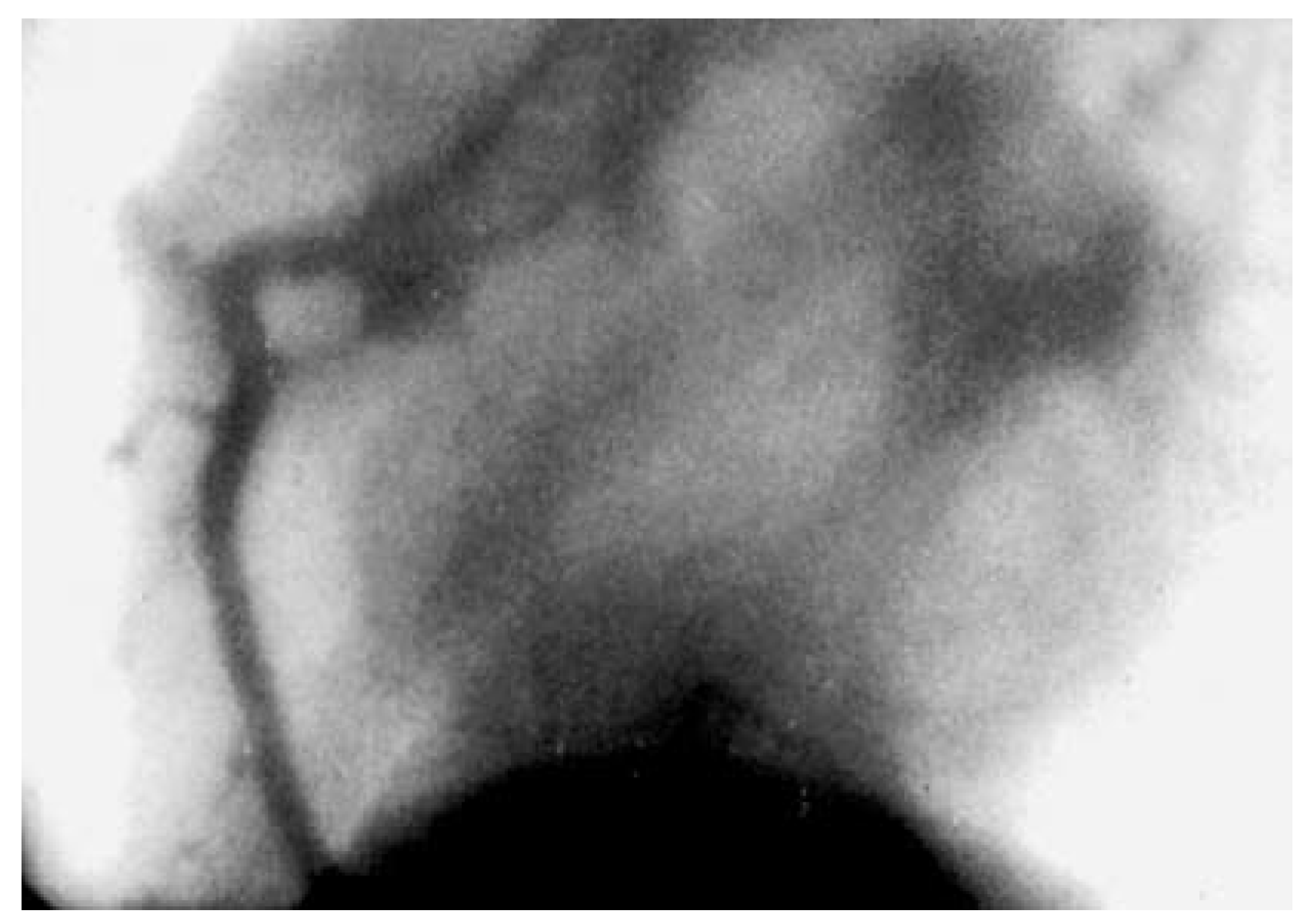
Figure 1. First selective coronary angiogram of 1958.
Cardiovascmed 11 00196 g001
Figure 2. Tight lesion (arrow) in the left coronary artery shown by traditional angiography (insert) and 3-dimensional reconstruction using conventional modern X-ray equipment.
Cardiovascmed 11 00196 g002
Figure 3. Top: CT done for screening for coronary artery disease in a 54-year-old asymptomatic Hungarian business man who was subsequently urgently flown to Switzerland for immediate angiography and therapy. Bottom: Angiography revealed only minor wall irregularities and uncovered the CT as blatantly false positive.
Cardiovascmed 11 00196 g003
Figure 4. Integrated 64-slice CT-angiography and SPECT-imaging. The left picture indicated the myocardial perfusion deficit of the diseased (stop of flow) diagonal and marginal branches (arrows) which provides additional information to the purely anatomical picture at the right which shows that the proximal stents are patent. (With courtesy: Syntermed Software and C.A. Santana, MD, Emory University, Atlanta, GA, USA.).
Cardiovascmed 11 00196 g004
Table 1. Quality of CT coronary angiography.
Cardiovascmed 11 00196 i001
Table 2. Comparison of CT and conventional coronary angiography.
Cardiovascmed 11 00196 i002