Article

Correlation of Air Pollution and Prevalence of Acute Pulmonary Embolism in Northern Thailand

by
Chaiwat Bumroongkit
1,
Chalerm Liwsrisakun
1,*,
Athavudh Deesomchok
1,
Chaicharn Pothirat
1,
Theerakorn Theerakittikul
1,
Atikun Limsukon
1,
Konlawij Trongtrakul
1,
Pattraporn Tajarernmuang
1,
Nutchanok Niyatiwatchanchai
1,
Juntima Euathrongchit
2,
Juthamas Inchai
1 and
Warawut Chaiwong
1
1
Division of Pulmonary, Critical Care, and Allergy, Department of Internal Medicine, Faculty of Medicine, Chiang Mai University, Chiang Mai 50200, Thailand
2
Department of Radiology, Faculty of Medicine, Chiang Mai University, Chiang Mai 50200, Thailand
*
Author to whom correspondence should be addressed.
Int. J. Environ. Res. Public Health 2022, 19(19), 12808; https://doi.org/10.3390/ijerph191912808
Submission received: 30 August 2022 / Revised: 4 October 2022 / Accepted: 5 October 2022 / Published: 6 October 2022
(This article belongs to the Special Issue Environmental Risk Assessment in Public Health)

Abstract

:
Background: The relationship between the level of air pollution and acute pulmonary embolism (APE) has had inconsistent results. Objective: This study aimed to analyze the relationship between the high level of air pollution exposure and APE. Methods: A ten-year retrospective cohort, single-center study was performed on patients diagnosed with APE from October 2010 to December 2020. The association between air pollution and monthly APE case diagnosis was analyzed. Results: A total number of 696 patients was included. The effect of every 10 µg/m3 increment of particulate matters with an aerodynamic diameter < 10 µm (PM10) on total monthly APE cases (unprovoked PE and provoked PE) was increased significantly at lag 4, 5 and 6 months with adjusted RR (95% CI) of 1.06 (1.01, 1.12), p = 0.011, 1.07 (1.01, 1.13), p = 0.021 and 1.06 (1.01, 1.12), p = 0.030, respectively. Adjusted RR for APE was significantly increased for PM10 in the second tertile ((adjusted RR (95% CI) 1.76 (1.12, 2.77)), p = 0.014. Conclusions: We conclude that PM10 is associated with an increased prevalence of APE cases. The policy for tighter control of air pollution in our country is needed to reduce the impact of air pollutants on people’s health.

1. Introduction

The harmful effects of various ambient air pollution on human health, particularly in developing countries, have been persistently reported in many studies [1,2,3,4]. Both long-term and short-term exposures to ambient particulate matters (PMs), including PM with an aerodynamic diameter < 10 µm (PM10) and PM with an aerodynamic diameter < 2.5 µm (PM2.5), and gaseous pollution, including nitrogen dioxide (NO2), volatile organic compounds (VOCs), including benzene, carbon monoxide (CO), sulfur dioxide (SO2) and ozone (O3), increase the risk of respiratory admission, cardiovascular admission, cardiovascular mortality and all-cause mortality [5,6,7]. There was evidence of associations between air pollutants and cardiovascular problems, such as myocardial infarction, stroke, heart failure, and atrial fibrillation [8,9,10,11]. A meta-analysis of studies conducted in China to assess the mortality effects of short-term exposure to air pollution showed that each 10 µg/m3 increase in PM2.5 was associated with a 0.38% (95% CI; 0.31, 0.45) increase in total mortality, a 0.51% increase in respiratory mortality (95% CI; 0.30, 0.73) and a 0.44% (95% CI; 0.33, 0.54) increase in cardiovascular mortality [12]. In the respiratory aspect, air pollutants are the cause and aggravating factor of many respiratory diseases, such as chronic obstructive pulmonary disease (COPD), asthma and lung cancer [13]. Increased ambient O3, NO2, PM2.5 and SO2 levels were consistently associated with increased hospital admission for asthma and pneumonia in various studies conducted in Hong Kong and Taipei [14,15,16,17].
Venous thromboembolism (VTE), a condition with blood clots in the deep venous system, is composed of pulmonary embolism (PE), deep venous thrombosis (DVT) or both. DVT is defined as a disease with blood clots in the deep vein, most commonly in the lower extremities. Acute pulmonary embolism (APE) refers to a situation where the pulmonary artery contains blood clots that most likely slip from DVT. APE, as a complication of DVT, was the most severe form of VTE [18]. Although exposure to PM has been strongly linked to cardiovascular and respiratory disorders, the effect on VTE, including PE and DVT, is still uncertain [19].
In Chiang Mai Province, Northern Thailand, air pollutant levels usually exceed the safety threshold during January and April, which are winter and pre-summer times in Thailand. The source of air pollutant emissions in our area comes from forest fires and the burning of agricultural remains after harvest. Our previous study demonstrated the effect of increasing PM10 concentrations from seasonal smog on asthma and COPD exacerbations [20]. Interestingly, there is growing evidence of an association between VTE and air pollution in many areas of the world. Nevertheless, the relationship between the level of air pollution and VTE had inconsistent results [21,22,23,24,25,26]. Therefore, our study aimed to analyze the relationship between the level of air pollution exposure and acute pulmonary embolism (APE) in Chiang Mai, Northern Thailand.

2. Materials and Methods

2.1. Study Design and Population

A retrospective data collection was conducted. All Asian patients, aged ≥15 years, who were admitted to Chiang Mai University Hospital between October 2010 and December 2020 with ICD-10 coding I 26.0 (pulmonary embolism with acute cor pulmonale) and I 26.9 (pulmonary embolism without acute cor pulmonale) were recruited into the study. All patients live in the northern part of Thailand. All medical records were reviewed to confirm the diagnosis of APE.
Diagnosis of APE was made when the presence of one of the following radiologic criteria was confirmed by a radiologist.
  • Demonstration of thrombus in the pulmonary artery and its branches by computed tomography pulmonary angiography (CTPA).
  • Demonstration of thrombus in the pulmonary artery and its branches by CT chest with contrast.
Patients who had predisposing factors for PE including active cancers were defined as provoked PE. Those with no predisposing factors were defined as unprovoked PE.
A total of 1560 patient records was collected from the ICD-10 coding diagnosis between October 2010 and December 2020; 864 were excluded due to age under 15 years, non-Asian ethnicities, repeated cases and no CT confirmation of diagnosis. A total number of 696 patients was included in the study.
Patients’ data including demographics, risk factors, date of diagnosis, clinical data and underlying conditions were recorded. Only cases with the first diagnosis of APE were included for analysis. This study was conducted at Faculty of Medicine, Chiang Mai University, Chiang Mai, Thailand.

2.2. Air Pollution Data

The levels of air pollutants were measured at sampling stations of the Pollution Control Department, Ministry of National Resources and Environment which are located in municipal areas of the Muang District, Chiang Mai Province, Thailand. The analysis methods were the non-dispersive infrared detection for CO concentrations, the pararosaniline technique for SO2 concentrations, the chemiluminescence technique for NO2 and O3 concentrations and the gravimetric technique for PM10 and PM2.5 concentrations [27]. The data were reported as monthly average concentrations of pollutants. The PM2.5 levels were officially reported after July 2016 in Thailand. Therefore, the correlation between PM2.5 and APE was not analyzed. We use the average 24 h air quality guidelines level of WHO (45 µg/m3 of PM10) for reference to the monthly average PM10 in this study [28].
The study was approved by the Research Ethics Committee of the Faculty of Medicine, Chiang Mai University (Institutional Review Board (IRB) approval number: MED-2564-08294).

2.3. Statistical Analysis
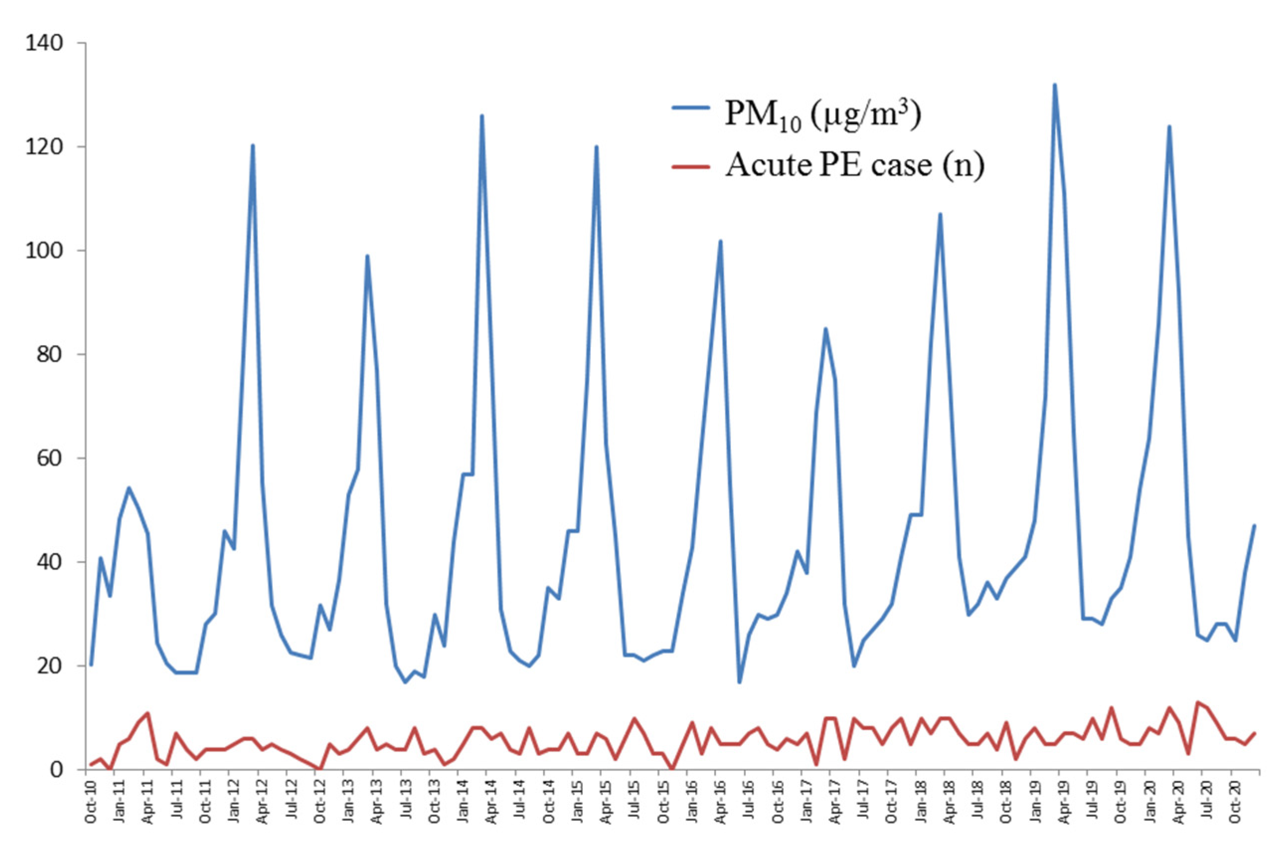
Results for numerical values were expressed as means ± standard deviation (SD) or median, IQR (Interquartile range) and those for categorical data were expressed as absolute frequencies and percentages. The association between a monthly number of acute PE cases and PM10 concentrations was analyzed by the application of general linear models (GLMs) with Poisson distribution. Poisson models with log links are often called log-linear models and are used for frequency data. To determine the association between the effects of PM10 on acute PE admissions, Poisson regression was used for analysis after adjustment for SO2, NO2, CO and O3. To assess the lag structure between the concentration of PM10 level and the number of acute PE cases, we initially examined separate models for each lag from zero to seven months after the APE diagnosis. The lag time zero (lag 0) is the month of an increase of 10 µg/m3 of PM10. Finally, risk regression analysis is applied to the data to estimate risk ratios (RRs) with 95% confidence intervals (CIs) of the independent variables in the constructed model. For case-crossover design, to allow comparison across pollutants, the monthly PM10, CO, NO2, O3 and SO2 concentrations were divided into tertiles and frequencies of cases were compared using Fisher exact test. The risk of PE due to high levels of pollutants was evaluated by comparing tertiles of each pollutant level by using the first tertile as a control point and calculating the RR and the 95% CI. All tests for statistical significance were two tailed and p-values below 0.05 were considered statistically significant. Statistical analysis was performed using a software package (StataCorp, College Station, TX, USA).

3. Results

A total number of 696 patients was included in the study. The patients’ characteristics and risk factors are demonstrated in Table 1. Most of them (58.9%) were female. The mean age was 57.7 ± 15.7 years old (min = 15, max = 98). Clinical suspicion of APE before CT confirmation was made in 468 of 696 patients (67.2%), while 228 patients (32.8%) were incidental PE. Provoked PE was found in 560 (80.5%) and unprovoked PE was found in 136 (19.5%). Unprovoked and provoked PE data are shown in Table 2.
The levels of PM10, SO2, NO2, CO and O3 were 45.4 ± 27.5 (17.2–132) µg/m3, 0.9 ± 0.6 (0.0–3.0) ppb, 9.6 ± 4.5 (1.0–23.0) ppb, 0.5 ± 0.2 (0.1–1.0) ppm and 24.3 ± 9.9 (6.0–47.0) ppb, respectively (Table 3). During the study period, there were 46 months of low PM10 (monthly average < 45 µg/m3) and 77 months of high PM10 (monthly average ≥ 45 µg/m3). The PM10 concentrations in low PM10 months and high PM10 months were 28.8 ± 7.4 and 72.5 ± 25.5 µg/m3, respectively (Table 4). The concentrations of other pollutants were also significantly higher in high-PM10 months.
The monthly average of total APE cases, presented as median (IQR), was significantly higher in high-PM10 months (6 (5, 8)) than in low-PM10 months (5 (3, 7)), p = 0.013. The monthly average of unprovoked PE and provoked PE cases was insignificantly higher in high-PM10 months (Table 5). The effect of every 10 µg/m3 increase in PM10 on monthly total APE cases, unprovoked PE and provoked PE is demonstrated in Table 6. The total monthly APE cases increased significantly at lag 4, 5 and 6 months with adjusted RR (95% CI) of 1.06 (1.01, 1.12), p = 0.011; 1.07 (1.01, 1.13), p = 0.021 and 1.06 (1.01, 1.12), p = 0.030, respectively. Provoked PE cases increased significantly at lag 3, 4, 5 and 6 months with adjusted RR (95% CI) of 1.04 (1.01, 1.07), p = 0.004; 1.06 (1.03, 1.09), p < 0.001; 1.04 (1.01, 1.07), p = 0.004 and 1.04 (1.01, 1.07), p = 0.004, respectively. The risk of unprovoked PE was also increased but not statistically significant. Variation in monthly average PM10 and monthly acute PE cases throughout the study period are shown in Figure 1.
The tertiles of the air pollutant levels and the number of confirmed APE cases during the study period are shown in Table 7. There was a higher prevalence of APE cases in the second and third tertiles of PM10, SO2, CO and O3 than in the first tertile but not in the NO2 pollutant data. The relative risk for APE associated with exposure to elevated air pollutants is demonstrated in Table 8. When using the first tertile of air pollutants as a reference, the adjusted RR for APE was significantly increased for PM10 in the second (adjusted RR (95% CI) 1.76 (1.12, 2.77), p = 0.014) but not for NO2, CO and O3. For SO2, we could not divide data into tertiles; therefore, we used the cut-off median value as the lower range (0.0–0.9 ppb) and the higher range (≥1.0 ppb) for comparison. The higher-range SO2 had a non-significant increase in APE risk (adjusted RR (95% CI) 1.28 (0.85, 1.94), p = 0.236).

4. Discussion

Our study found that exposure to high concentrations of PM10 was significantly correlated with the number of confirmed APE cases. We used the reference level of PM10 as WHO air quality guideline recommendations [28]. In high-PM10 months (≥45 µg/m3), a monthly average of total pulmonary embolism cases was higher than low-PM10 (<45 µg/m3) months, p = 0.013. The results of our study are in agreement with those in previous studies, which showed an association between high concentrations of air pollutants and VTE incidence [29,30]. Baccarelli et al. proposed that the mechanism of PM-induced DVT triggered the pro-thrombotic state [25,31]. Although the numbers of unprovoked PE in our study did not meet statistical significance in high-PM10 months, the tendency of higher cases in those months might suggest the role of PM as a provoking factor in the unprovoked PE group. The trend was also observed in the number of provoked PE in high-PM10 months.
In Northern Thailand, illegal forest invasion for the cultivation of maize has been practiced for more than ten years [32]. Mass burning of agricultural waste preparing for the next cropping, joining together with forest firing in the dry season during January and April, which were winter and summer seasons in Thailand, has occurred since 2006. After that, air pollution became a major problem in Chiang Mai annually [32] for more than 15 years. At present, the problem is worse than ever, especially between March and April every year. Miguel-Díez et al. found that the hospitalization rate for PE was high in association with the autumn and winter seasons, lower temperatures and high levels of NO2 and O3 [33]. In contrast, Nimako et al. reported that the higher temperature was correlated with the high incidence of idiopathic PE [34]. However, the climate and temperature in Thailand are not as varied as in Western countries. We did not consider that climate change alone was the most important associated factor. A systematic review and meta-analysis concluded that the VTE rate was significantly high during winter time when it was usually coincident with increased PM in the same geographical areas [35].
To find the associations between the effect of an increase of every 10 µg/m3 of PM10 and total monthly acute PE cases, unprovoked PE and provoked PE, Poisson regression was used for analysis after adjustment for SO2, NO2, CO and O3. To assess the lag structure between the concentration of PM10 level and monthly PE cases, we initially examined separate models for each lag from lag time zero (lag 0), which is the month of PM10 measurement, to the lag later months, until no statistical significance was demonstrated. From our study, we found an association between every 10 µg/m3 increase in PM10 and increased risk of total APE cases after 4 months, 5 months and 6 months. The provoked PE risks also increased after 3, 4, 5 and 6 months. For unprovoked PE, the adjusted RR was also increased but without statistical significance. These findings are in agreement with the results in the study of Baccarelli et al., who found that exposure to a higher annual average PM10 level in the previous year was associated with shortened prothrombin time in DVT cases and every increment in 10 μg/m3 of PM10 increased DVT risk for 70% [25]. We supposed that the pro-thrombotic enhancement effect of PM to cause PE might take time after exposure.
Our study correlated the tertile of the air pollutant level and several confirmed APE cases and demonstrated a higher prevalence of APE cases in the second and third tertile of PM10, CO and O3 than in the first tertile. However, we did not find that NO2 and SO2 were significantly related to APE. These results were different from the study of Miguel-Diez et al. [26], which showed a relationship between short-term exposure to NO2 and the development of unprovoked PE. When using the first tertile of air pollutants as a reference, the adjusted RR of APE in the second and the third tertile was increased for PM10 but not for SO2, NO2, CO and O3. However, Gwon et al. [36] demonstrated that long-term exposures not only to PM10 but also to SO2 and O3 were correlated with the occurrence of VTE. However, not all studies replicated the same positive findings, making the association between exposure to air pollution and VTE development somewhat uncertain. Colais et al. found a strong relationship between short-term NO2 exposure and the development of PE, but not with PM10 and O3 [37]. A large prospective study conducted in the U.S. provided no association between short-term or long-term traffic-related air pollution exposure and the risk of VTE [38]. A study in Beijing by Li et al. showed that exposure to PM2.5, PM10, SO2 and CO increased PE admission [39]. A recent meta-analysis by Miao et al. demonstrated that exposure to CO, SO2, PM10 and PM2.5 had no significant association with PE. However, NO2 and O3 modestly increased the risk of PE [40]. Although the exact pathogenesis of air pollutants to VTE development was not well known, the possible hypotheses were pro-inflammatory effects and oxidative stress on alveolar epithelial cells and macrophages [41], endothelial malfunction [42], activation of the coagulation pathways [43,44,45,46,47], platelet aggregation [48] and in situ thrombus formation [49]. In addition, multifactorial and unknown factors, such as genetic heterogeneity risk for VTE [50], might be involved in the occurrence of pollution-associated PE and made the results of this relationship inconsistent.
Our study had some strengths. Firstly, we enrolled only Asian patients. Therefore, the heterogeneity in the study population might be minimal. In addition, our study was less affected by other confounding factors, including national policy or the effects of traffic-associated air pollution. Although the policy on reducing air pollution has been implemented by the Thai government since 1992, the air pollution remains a serious problem in the northern region of Thailand between January and April of every year. Moreover, the air pollution caused by traffic was not a major problem in the northern part of Thailand, including Chiang Mai, where most of these regions are rural areas [32]. Therefore, the temporal changes in air pollution and traffic regulations may not be affected by the results of this study. Secondly, our study is a large retrospective cohort. Thirdly, our air pollutant data were recorded completely for ten years for analysis of both short-term and long-term exposure effects of air pollutants. However, there are some limitations to this study. Firstly, the number of pollutants analyzed was limited, so we could not obtain complete data on PM2.5, which might be a better representative of a thrombosis-associated pollutant than PM10. Secondly, we did not evaluate the prevalence of DVT. Therefore, the rate of overall VTE induced by pollution was not known. Thirdly, the meteorological parameters, including temperature, humidity and wind speed, were not included as confounding factors for the analysis. Fourthly, occupational status was not included for the analysis. Therefore, this should be counted as a confounding factor in a future study.

5. Conclusions

Our study shows a significant association between a high level of PM10 and an increased prevalence of APE cases. This study provides evidence in support of an association between exposure to particulate air pollution and the risk of VTE. The policy for higher control of air pollution in our country is needed to reduce its impact on our citizens’ health.

Author Contributions

Conceptualization, C.B.; Methodology, C.B., C.L. and W.C.; Software, W.C.; Validation, C.B., C.L., A.D., W.C. and C.P.; Formal Analysis, C.B., W.C.; Investigation, C.B., T.T., A.L., K.T., P.T., J.I., N.N., A.D, W.C., C.L., J.E. and C.P.; Resources, C.B., W.C., C.L. and C.P.; Data Curation, C.B., A.D., W.C. and C.P.; Writing—Original Draft Preparation, C.B.; Writing—Review and Editing, C.B., C.L. and W.C.; Visualization, A.D., W.C., C.L. and C.P.; Supervision, C.L. and C.P.; Project Administration, C.B., C.L., W.C. and C.P. All authors have read and agreed to the published version of the manuscript.

Funding

This research received no external funding.

Institutional Review Board Statement

The study was approved by the Research Ethics Committee of the Faculty of Medicine, Chiang Mai University (Institutional Review Board (IRB) approval number: MED-2564-08294).

Informed Consent Statement

Not applicable.

Data Availability Statement

The data that support the findings of this study are available on request from the corresponding author.

Acknowledgments

The authors wish to acknowledge the contribution of the staff of the Division of Pulmonary, Critical Care and Allergy, Department of Internal Medicine, Faculty of Medicine, Chiang Mai University to this trial. Special thanks are due to the Pollution Control Department for the air quality data. The authors would like to thank Ruth Leatherman, Research Administration Section, Faculty of Medicine, Chiang Mai University, for native English proofreading.

Conflicts of Interest

The authors declare no conflict of interest.

References

  1. Lim, S.S.; Vos, T.; Flaxman, A.D.; Danaei, G.; Shibuya, K.; Adair-Rohani, H.; Amann, M.; Anderson, H.R.; Andrews, K.G.; Aryee, M.; et al. A comparative risk assessment of burden of disease and injury attributable to 67 risk factors and risk factor clusters in 21 regions, 1990–2010: A systematic analysis for the Global Burden of Disease Study 2010. Lancet 2012, 380, 2224–2260. [Google Scholar] [CrossRef] [Green Version]
  2. Mannucci, P.M.; Harari, S.; Martinelli, I.; Franchini, M. Effects on health of air pollution: A narrative review. Intern. Emerg. Med. 2015, 10, 657–662. [Google Scholar] [CrossRef]
  3. Landrigan, P.J. Air pollution and health. Lancet Public Health 2017, 2, e4–e5. [Google Scholar] [CrossRef] [Green Version]
  4. Mannucci, P.M.; Franchini, M. Health effects of ambient air pollution in developing countries. Int. J. Environ. Res. Public Health 2017, 14, 1048. [Google Scholar] [CrossRef]
  5. Dominici, F.; Peng, R.D.; Bell, M.L.; Pham, L.; McDermott, A.; Zeger, S.L.; Samet, J.M. Fine particulate air pollution and hospital admission for cardiovascular and respiratory diseases. J. Am. Med. Assoc. 2006, 295, 1127–1134. [Google Scholar] [CrossRef] [Green Version]
  6. Raaschou-Nielsen, O.; Andersen, Z.J.; Jensen, S.S.; Ketzel, M.; Sørensen, M.; Hansen, J.; Loft, S.; Tjønneland, A.; Overvad, K. Traffic air pollution and mortality from cardiovascular disease and all causes: A Danish cohort study. Environ. Health 2012, 11, 60. [Google Scholar] [CrossRef] [Green Version]
  7. Samoli, E.; Atkinson, R.W.; Analitis, A.; Fuller, G.W.; Green, D.C.; Mudway, I.; Anderson, H.R.; Kelly, F.J. Associations of Short-term Exposure to Traffic-related Air Pollution with Cardiovascular and Respiratory Hospital Admissions in London, UK. Occup. Environ. Med. 2016, 73, 300–307. [Google Scholar] [CrossRef] [Green Version]
  8. Rajagopalan, S.; Al-Kindi, S.G.; Brook, R.D. Air Pollution and Cardiovascular Disease: JACC State-of-the-Art Review. J. Am. Coll. Cardiol. 2018, 72, 2054–2070. [Google Scholar] [CrossRef]
  9. Scheers, H.; Jacobs, L.; Casas, L.; Nemery, B.; Nawrot, T.S. Long-term exposure to particulate matter air pollution is a risk factor for stroke. Stroke 2015, 46, 3058–3066. [Google Scholar] [CrossRef] [Green Version]
  10. Von Klot, S.; Peters, A.; Aalto, P.; Bellander, T.; Berglind, N.; Ippoliti, D.D.; Elosua, R.; Hörmann, A.; Kulmala, M.; Lanki, T.; et al. Ambient air pollution is associated with increased risk of hospital cardiac readmissions of myocardial infarction survivors in five European cities. Circulation 2005, 112, 3073–3079. [Google Scholar] [CrossRef]
  11. Solimini, A.G.; Renzi, M. Association between air pollution and emergency room visits for atrial fibrillation. Int. J. Environ. Res. Public Health 2017, 14, 661. [Google Scholar] [CrossRef]
  12. Shang, Y.; Sun, Z.; Cao, J.; Wang, X.; Zhong, L.; Bi, X.; Li, H.; Liu, W.; Zhu, T.; Huang, W. Systematic review of Chinese studies of short-term exposure to air pollution and daily mortality. Environ. Int. 2013, 54, 100–111. [Google Scholar] [CrossRef]
  13. Jiang, X.Q.; Mei, X.D.; Feng, D. Air pollution and chronic airway diseases: What should people know and do? J. Thorac. Dis. 2016, 8, E31–E40. [Google Scholar]
  14. Qiu, H.; Yu, I.T.; Tian, L.; Wang, X.; Tse, L.A.; Tam, W.; Wong, T.W. Effects of coarse particulate matter on emergency hospital admissions for respiratory diseases: A time-series analysis in Hong Kong. Environ. Health Perspect. 2012, 120, 572–576. [Google Scholar] [CrossRef] [Green Version]
  15. Ko, F.W.; Tam, W.; Wong, T.W.; Lai, C.K.; Wong, G.W.; Leung, T.F.; Ng, S.S.; Hui, D.S. Effects of air pollution on asthma hospitalization rates in different age groups in Hong Kong. Clin. Exp. Allergy 2007, 37, 1312–1319. [Google Scholar] [CrossRef]
  16. Cheng, M.H.; Chen, C.C.; Chiu, H.F.; Yang, C.Y. Fine particulate air pollution and hospital admissions for asthma: A case-crossover study in Taipei. J. Toxicol. Environ. Health 2014, 77, 1075–1083. [Google Scholar] [CrossRef] [PubMed]
  17. Qiu, H.; Tian, L.W.; Pun, V.C.; Ho, K.F.; Wong, T.W.; Yu, I.T. Coarse particulate matter associated with increased risk of emergency hospital admissions for pneumonia in Hong Kong. Thorax 2014, 69, 1027–1033. [Google Scholar] [CrossRef] [Green Version]
  18. Cordeanu, E.M.; Lambach, H.; Heitz, M.; Di Cesare, J.; Mirea, C.; Faller, A.M.; Cavaro, A.C.; Frantz, A.S.; Gaertner, S.; Schini-Kerth, V.; et al. Pulmonary Embolism and Coexisting Deep Vein Thrombosis: A Detrimental Association? J. Clin. Med. 2019, 8, 899. [Google Scholar] [CrossRef] [Green Version]
  19. Franchini, M.; Mengoli, C.; Cruciani, M.; Bonfanti, M.; Mannucci, P.M. Association between particulate air pollution and venous thromboembolism: A systematic literature review. Eur. J. Intern. Med. 2016, 27, 10–13. [Google Scholar] [CrossRef]
  20. Pothirat, C.; Tosukhowong, A.; Chaiwong, W.; Liwsrisakun, C.; Inchai, J. Effects of seasonal smog on asthma and COPD exacerbations requiring emergency visits in Chiang Mai, Thailand. Asian Pac. J. Allergy Immunol. 2016, 34, 284–289. [Google Scholar]
  21. Franchini, M.; Guida, A.; Tufano, A.; Coppola, A. Air pollution, vascular disease and thrombosis: Linking clinical data and pathogenic mechanisms. J. Thromb. Haemost. 2012, 10, 2438–2451. [Google Scholar] [CrossRef]
  22. Crous-Bou, M.; Harrington, L.B.; Kabrhel, C. Environmental and genetic risk factors associated with venous thromboembolism. Semin. Thromb. Hemost. 2016, 42, 808–820. [Google Scholar] [CrossRef]
  23. Dales, R.E.; Cakmak, S.; Vidal, C.B. Air pollution and hospitalization for venous thromboembolic disease in Chile. J. Thromb. Haemost. 2010, 8, 669–674. [Google Scholar] [CrossRef] [PubMed]
  24. Spiezia, L.; Campello, E.; Bon, M.; Maggiolo, S.; Pelizzaro, E.; Simioni, P. Short-term exposure to high levels of air pollution as a risk factor for acute isolated pulmonary embolism. Thromb. Res. 2014, 134, 259–263. [Google Scholar] [CrossRef] [PubMed]
  25. Baccarelli, A.; Martinelli, I.; Zanobetti, A.; Grillo, P.; Hou, L.F.; Bertazzi, P.A.; Mannucci, P.M.; Schwartz, J. Exposure to particulate air pollution and risk of deep vein thrombosis. Arch. Intern. Med. 2008, 168, 920–927. [Google Scholar] [CrossRef] [PubMed]
  26. de Miguel-Diez, J.; Blasco-Esquivias, I.; Rodriguez-Matute, C.; Bedate-Diaz, P.; Lopez-Reyes, R.; Fernandez-Capitan, C.; Garcia-Fuika, S.; Lobo-Beristain, J.L.; Garcia-Lozaga, A.; Quezada, C.A.; et al. Correlation between short-term air pollution exposure and unprovoked lung embolism. Prospective observational (Contamina-TEP Group). Thromb. Res. 2020, 192, 134–140. [Google Scholar] [CrossRef]
  27. Air Quality and Noise Management Bureau, 2004. Pollution Control Department, Ministry of National Resources and Environment Homepage. Available online: http://www.pcd.go.th/info (accessed on 15 May 2022).
  28. WHO Global Air Quality Guidelines: Particulate Matter (PM2.5 and PM10), Ozone, Nitrogen Dioxide, Sulfur Dioxide and Carbon Monoxide. Executive Summary. Available online: https://apps.who.int/iris/handle/10665/345329 (accessed on 10 April 2022).
  29. Martinelli, N.; Girelli, D.; Cigolini, D.; Sandri, M.; Ricci, G.; Rocca, G.; Olivieri, O. Access rate to the emergency department for venous thromboembolism in relationship with coarse and fine particulate matter air pollution. PLoS ONE 2012, 7, e34831. [Google Scholar] [CrossRef] [PubMed] [Green Version]
  30. Chiu, H.H.; Whittaker, P. Venous thromboembolism in an industrial north American city: Temporal distribution and association with particulate matter air pollution. PLoS ONE 2013, 8, e68829. [Google Scholar] [CrossRef] [Green Version]
  31. Baccarelli, A.; Martinelli, I.; Pegoraro, V.; Melly, S.; Grillo, P.; Zanobetti, A.; Hou, L.; Bertazzi, P.A.; Mannucci, P.M.; Schwartz, J. Living near major traffic roads and risk of deep vein thrombosis. Circulation 2009, 119, 3118–31124. [Google Scholar] [CrossRef] [Green Version]
  32. Pengchai, P.; Chantara, S.; Sopajaree, K.; Wangkarn, S.; Tengcharoenkul, U.; Rayanakorn, M. Seasonal variation, risk assessment and source estimation of PM 10 and PM10-bound PAHs in the ambient air of Chiang Mai and Lamphun, Thailand. Environ. Monit. Assess. 2009, 154, 197–218. [Google Scholar] [CrossRef] [PubMed]
  33. de Miguel-Díez, J.; Jiménez-García, R.; López de Andrés, A.; Hernández-Barrera, V.; Carrasco-Garrido, P.; Monreal, M.; Jiménez, D.; Jara-Palomares, L.; Álvaro-Meca, A. Analysis of environmental risk factors for pulmonary embolism: A case-crossover study (2001–2013). Eur. J. Intern. Med. 2016, 31, 55–61. [Google Scholar] [CrossRef] [PubMed]
  34. Nimako, K.; Poloniecki, J.; Draper, A.; Rahman, T. Seasonal variability and meteorological factors: Retrospective study of the incidence of pulmonary embolism from a large United Kingdom teaching hospital. Respir. Care 2012, 57, 1267–1272. [Google Scholar] [CrossRef] [Green Version]
  35. Dentali, F.; Ageno, W.; Rancan, E.; Donati, A.V.; Galli, L.; Squizzato, A.; Venco, A.; Mannucci, P.M.; Manfredini, R. Seasonal and monthly variability in the incidence of venous thromboembolism. A systematic review and a meta-analysis of the literature. Thromb. Haemost. 2011, 106, 439–447. [Google Scholar]
  36. Gwon, J.G.; Lee, S.A.; Park, K.Y.; Oh, S.U.; Kim, J.S.; Seo, H.M. Long-Term Exposure to Air Pollution and Incidence of Venous Thromboembolism in the General Population: A Population-Based Retrospective Cohort Study. J. Clin. Med. 2022, 11, 3517. [Google Scholar] [CrossRef]
  37. Colais, P.; Serinelli, M.; Faustini, A.; Stafoggia, M.; Randi, G.; Tessari, R.; Chiusolo, M.; Pacelli, B.; Mallone, S.; Vigotti, M.A.; et al. Air pollution and urgent hospital admissions in nine Italian cities. Epidemiol. Prev. 2009, 33, 77–94. [Google Scholar] [PubMed]
  38. Kan, H.; Folsom, A.R.; Cushman, M.; Rose, K.M.; Rosamond, W.D.; Liao, D.; Lurmann, F.; London, S.J. Traffic exposure and incident venous thromboembolism in the Atherosclerosis Risk in Communities (ARIC) Study. J. Thromb. Haemost. 2011, 9, 672–678. [Google Scholar] [CrossRef] [Green Version]
  39. Li, Z.; Zhang, Y.; Yuan, Y.; Yan, J.; Mei, Y.; Liu, X.; Xu, Q.; Shi, J. Association between exposure to air pollutants and the risk of hospitalization for pulmonary embolism in Beijing, China: A case-crossover design using a distributed lag nonlinear model. Environ. Res. 2022, 204, 112321. [Google Scholar] [CrossRef]
  40. Miao, H.; Li, X.; Wang, X.; Nie, S. Air pollution increases the risk of pulmonary embolism: A meta-analysis. Rev. Environ. Health 2021, 37, 259–266. [Google Scholar] [CrossRef]
  41. Li, X.Y.; Gilmour, P.S.; Donaldson, K.; MacNee, W. Free radical activity and proinflammatory effects of particulate air pollution (PM10) in vivo and in vitro. Thorax 1996, 51, 1216–1222. [Google Scholar] [CrossRef] [Green Version]
  42. Mills, N.L.; Donaldson, K.; Hadoke, P.W.; Boon, N.A.; MacNee, W.; Cassee, F.R.; Sandström, T.; Blomberg, A.; Newby, D.E. Adverse cardiovascular effects of air pollution. Nat. Clin. Pract. Cardiovasc. Med. 2009, 6, 36–44. [Google Scholar] [CrossRef] [PubMed]
  43. Baccarelli, A.; Zanobetti, A.; Martinelli, I.; Grillo, P.; Hou, L.; Giacomini, S.; Bonzini, M.; Lanzani, G.; Mannucci, P.M.; Bertazzi, P.A.; et al. Effects of exposure to air pollution on blood coagulation. J. Thromb. Haemost. 2007, 5, 252–260. [Google Scholar] [CrossRef]
  44. Budinger, G.R.; McKell, J.L.; Urich, D.; Foiles, N.; Weiss, I.; Chiarella, S.E.; Gonzalez, A.; Soberanes, S.; Ghio, A.J.; Nigdelioglu, R.; et al. Particulate matter-induced lung inflammation increases systemic levels of PAI-1 and activates coagulation through distinct mechanisms. PLoS ONE 2011, 6, e18525. [Google Scholar] [CrossRef] [Green Version]
  45. Emmerechts, J.; Jacobs, L.; Van Kerckhoven, S.; Loyen, S.; Mathieu, C.; Fierens, F.; Nemery, B.; Nawrot, T.S.; Hoylaerts, M.F. Air pollution-associated procoagulant changes: The role of circulating microvesicles. J. Thromb. Haemost. 2012, 10, 96–106. [Google Scholar] [CrossRef] [Green Version]
  46. Franchini, M.; Mannucci, P.M. Thrombogenicity and cardiovascular effects of ambient air pollution. Blood 2011, 118, 2405–2412. [Google Scholar] [CrossRef] [PubMed] [Green Version]
  47. Milojevic, A.; Wilkinson, P.; Armstrong, B.; Bhaskaran, K.; Smeeth, L.; Hajat, S. Short-term effects of air pollution on a range of cardiovascular events in England and Wales: Case-crossover analysis of the MINAP database, hospital admissions and mortality. Heart 2014, 100, 1093–1098. [Google Scholar] [CrossRef]
  48. Rudez, G.; Janssen, N.A.; Kilinc, E.; Leebeek, F.W.; Gerlofs-Nijland, M.E.; Spronk, H.M.; ten Cate, H.; Cassee, F.R.; de Maat, M.P. Effects of ambient air pollution on hemostasis and inflammation. Environ. Health Perspect. 2009, 117, 995–1001. [Google Scholar] [CrossRef] [PubMed] [Green Version]
  49. Nemmar, A.; Hoylaerts, M.F.; Hoet, P.H.; Dinsdale, D.; Smith, T.; Xu, H.; Vermylen, J.; Nemery, B. Ultrafine particles affect experimental thrombosis in an in vivo hamster model. Am. J. Respir. Crit. Care Med. 2002, 166, 998–1004. [Google Scholar] [CrossRef] [PubMed]
  50. Angchaisuksiri, P.; Atichartakarn, V.; Aryurachai, K.; Archararit, N.; Rachakom, B.; Atamasirikul, K.; Tiraganjana, A. Risk factors of venous thromboembolism in Thai patients. Int. J. Hematol. 2007, 86, 397–402. [Google Scholar] [CrossRef] [PubMed]
Figure 1. Variation in monthly average PM10 and monthly acute PE cases throughout the study period (October 2010–December 2020).
Ijerph 19 12808 g001
Table 1. Demographic data, clinical and underlying conditions.
Demographic Data (n = 696)Mean ± SD or n (%)
Age (years) (Range)57.7 ± 15.7 (15–98)
Male sex286 (41.1)
Female sex410 (58.9)
Clinical type
 Suspected PE468 (67.2)
 Incidental PE228 (32.8)
 Provoked PE560 (80.5)
 Unprovoked PE136 (19.5)
Underlying conditions
 Hypertension305 (43.8)
 DM114 (16.4)
 Renal diseases69 (9.9)
 Thalassemia and hematologic diseases45 (6.5)
 COPD38 (5.5)
 Cirrhosis31 (4.5)
 CAD with prior myocardial infarction30 (4.3)
 Other chronic lung problem28 (4.0)
 Active smoking24 (3.4)
 Chronic alcohol drinking24 (3.4)
 Connective tissue disease22 (3.2)
 OSA15 (2.2)
 Obesity (BMI ≥ 30 kg/m2)12 (1.7)
 Nephrotic syndrome11 (1.6)
 Post-splenectomy10 (1.4)
 HIV9 (1.3)
 Vasculitis3 (0.4)
 Pregnancy2 (0.3)
 Known prothrombotic state68 (9.8)
Known prothrombotic state (N = 68)
 Protein C deficiency31 (45.6)
 Protein S deficiency12 (17.6)
 AT III deficiency6 (8.8)
 lupus anticoagulant15 (22.1)
 Anticardiolipin2 (2.9)
Note: Data are mean ± standard deviation (SD) or n (%). Abbreviations: AT, antithrombin-III; DM, diabetes mellitus; COPD, chronic obstructive pulmonary disease; CAD, coronary arterial disease; OSA, obstructive sleep apnea; HIV, human immunodeficiency viral infection; PE, pulmonary embolism.
Table 2. Provoked and unprovoked PE.
Characteristicsn (%)
Unprovoked136 (19.5)
Provoked PE560 (80.5)
 Active malignancy388 (55.7)
 Immobility—total body immobilization211 (30.4)
 Surgery or trauma requiring endotracheal or epidural anesthesia within the last 4 weeks141 (20.3)
 Indwelling venous catheter23 (3.3)
 Recent significant trauma12 (1.7)
 Oral contraceptives/Estrogen therapy27 (3.9)
  Protein C deficiency31 (4.4)
  Protein S deficiency12 (1.7)
  AT III deficiency6 (0.9)
  lupus anticoagulant15 (2.1)
  Antiphospholipid 2 (0.3)
 Right side endocarditis2 (0.3)
 Long travel history > 6 h10 (1.4)
Note: Data are n (%). Abbreviations: AT, antithrombin-III; PE, pulmonary embolism.
Table 3. Monthly pollutant data (October 2010 to December 2020).
PollutantsMean ± SDMin–Max
PM10 (µg/m3)45.4 ± 27.517.0–132
SO2 (ppb)0.9 ± 0.60.0–3.0
NO2 (ppb)9.6 ± 4.51.0–23.0
CO (ppm)0.5 ± 0.20.1–1.0
O3 (ppb)24.3 ± 9.96.0–47.0
Note: Data are mean ± standard deviation (SD). Abbreviations: PM10, particulate matters with diameter of less than 10 microns; CO, carbon monoxide; SO2, sulfur dioxide; NO2, nitrogen dioxide; O3, ozone.
Table 4. Pollutant data between high-PM10 period (≥45 µg/m3) and low-PM10 periods (<45 µg/m3) recommended by WHO air quality guidelines 2021.
PollutantsLow PM10 (n = 46)High PM10 (n = 77)p-Value
PM10 (µg/m3)28.8 ± 7.472.5 ± 25.5<0.001
SO2 (ppb)0.8 ± 0.6 1.0 ± 0.80.031
NO2 (ppb)7.2 ± 2.313.5 ± 4.7<0.001
CO (ppm)0.4 ± 0.10.6 ± 0.2<0.001
O3 (ppb)18.6 ± 6.033.7 ± 7.4<0.001
Note: Data are mean ± standard deviation (SD). Abbreviations: PM10, particulate matters with diameter of less than 10 microns; CO, carbon monoxide; SO2, sulfur dioxide; NO2, nitrogen dioxide; O3, ozone.
Table 5. Acute pulmonary emboli case during high-PM10 months (≥45 µg/m3) and low-PM10 months (<45 µg/m3).
VariablesLow PM10High PM10p-Value
Monthly average pulmonary emboli case5.0 (3.0, 7.0)6.0 (5.0, 8.0)0.013
Monthly average unprovoked pulmonary emboli case1.0 (0.0, 2.0)1.0 (1.0, 2.0)0.111
Monthly average provoked pulmonary emboli case4.0 (2.0, 6.0)4.5 (3.0, 6.0)0.678
Notes: Results are expressed as median (IQR).
Table 6. Associations between every 10 µg/m3 increase in PM10 and monthly APE cases (total), unprovoked PE and provoked PE (October 2010–December 2020).
OutcomesAdjusted RR # (95% CI)p-Value
Total acute PE cases
 Lag 0 month1.00 (0.92, 1.10)0.865
 Lag 1 month1.00 (0.95, 1.06)0.919
 Lag 2 month1.02 (0.98, 1.06)0.402
 Lag 3 month1.02 (0.98, 1.06)0.275
 Lag 4 month1.06 (1.01, 1.12)0.011
 Lag 5 month1.07 (1.01, 1.13)0.021
 Lag 6 month1.06 (1.01, 1.12)0.030
 Lag 7 month1.01 (0.98, 1.04)0.550
Unprovoked PE
 Lag 0 month1.00 (0.94, 1.07)0.887
 Lag 1 month1.03 (0.97, 1.09)0.361
 Lag 2 month1.03 (0.97, 1.09)0.280
 Lag 3 month1.05 (0.99, 1.10)0.109
 Lag 4 month1.05 (0.99, 1.11)0.111
 Lag 5 month1.03 (0.97, 1.09)0.310
 Lag 6 month1.03 (0.97, 1.09)0.328
 Lag 7 month1.02 (0.96, 1.09)0.445
Provoked PE
 Lag 0 month1.02 (0.98, 1.05)0.326
 Lag 1 month1.02 (0.99, 1.04)0.237
 Lag 2 month1.03 (0.99, 1.06)0.091
 Lag 3 month1.04 (1.01, 1.07)0.004
 Lag 4 month1.06 (1.03, 1.09)<0.001
 Lag 5 month1.04 (1.01, 1.07)0.004
 Lag 6 month1.04 (1.01, 1.07)0.004
 Lag 7 month1.00 (0.97, 1.03)0.773
Note: RR, relative risk; #, adjusted for other pollutants (CO, SO2, NO2 and O3). Abbreviations: PM10, particulate matters with diameter of less than 10 microns; RR, relative risk; PE, pulmonary embolism.
Table 7. Tertiles of exposure to air pollutants of study population during months of enrollment.
PollutantsTertilesNo. of Casesp-Value
PM10 (μg/m3)≤26.01160.045
26.1–54.9349
≥55.0181
SO2 (ppb)0.0–0.92090.366
≥1.0487
NO2 (ppb)≤7.93410.349
8.0–11.9179
≥12.0176
CO (ppm)≤0.32860.053
0.33–0.59130
≥0.6087
O3 (ppb)≤15.01610.471
15.1–38.9341
≥39.0194
Abbreviations: PM10, particulate matters with diameter of less than 10 microns; CO, carbon monoxide; SO2, sulfur dioxide; NO2, nitrogen dioxide; O3, ozone.
Table 8. Relative risk for APE associated with exposure to elevated air pollutants.
PollutantsTertilesUnadjusted RR
(95% CI)
p-ValueAdjusted RR #
(95% CI)
p-Value
PM10 (μg/m3)≤26.0Ref. Ref.
26.1–54.91.12 (0.93, 1.35)0.2251.76 (1.12, 2.77)0.014
≥55.01.13 (0.91, 1.39)0.2711.62 (0.90, 3.05)0.105
SO2 (ppb) *0.0–0.9Ref. Ref.
≥1.00.85 (0.73, 1.00)0.0571.28 (0.85, 1.94)0.236
NO2 (ppb)≤7.9Ref. Ref.
8.0–11.90.72 (0.60, 0.87)<0.0010.75 (0.51, 1.09)0.140
≥12.00.75 (0.63, 0.90)0.0020.83 (0.48, 1.45)0.518
CO (ppm)≤0.32Ref. Ref.
0.33–0.590.82 (0.63, 1.08)0.1650.73 (0.53, 1.02)0.068
≥0.601.01 (0.75, 1.36)0.9390.76 (0.48, 1.20)0.243
O3 (ppb)≤15Ref. Ref.
15.1–38.90.96 (0.79, 1.15)0.6420.76 (0.52, 1.11)0.163
≥39.01.02 (0.83, 1.26)0.8350.92 (0.56, 1.49)0.728
Note: * SO2 data could not be divided into tertiles; we used cut-off median value as lower range and higher range. Abbreviations: PM10, particulate matters with diameter of less than 10 microns; CO, carbon monoxide; SO2, sulfur dioxide; NO2, nitrogen dioxide; O3, ozone; # adjusted for all pollutants (PM10, CO, SO2, NO2 and O3).
Publisher’s Note: MDPI stays neutral with regard to jurisdictional claims in published maps and institutional affiliations.

