Article

Limited Role of Rhamnolipids on Cadmium Resistance for an Endogenous-Secretion Bacterium

1
Shandong Key Laboratory of Water Pollution Control and Resource Reuse, School of Environmental Science and Engineering, Shandong University, Qingdao 266237, China
2
Sino-French Research Institute for Ecology and Environment (ISFREE), School of Environmental Science and Engineering, Shandong University, Qingdao 266237, China
*
Author to whom correspondence should be addressed.
Int. J. Environ. Res. Public Health 2022, 19(19), 12555; https://doi.org/10.3390/ijerph191912555
Submission received: 15 September 2022 / Revised: 27 September 2022 / Accepted: 28 September 2022 / Published: 1 October 2022

Abstract

:
Rhamnolipids, a type of biosurfactant, represent a potential strategy for both enhancing organismic resistance and in situ remediation of heavy metals contaminations. In-depth study of the mechanism of rhamnolipids synthesis in response to heavy metals stress, is indispensable for a wide use of biosurfactant-secreting microbes in bioremediation. In this study, we employed the wild-type and the rhlAB deficient strain (ΔrhlAB) of Pseudomonas aeruginosa, a prototypal rhamnolipids-producing soil microorganism, to investigate its responses to cadmium resistance based on its physicochemical, and physiological properties. Compared with the wild-type strain, the ΔrhlAB were more sensitive to Cd-stress at low Cd concentration (<50 mg/L), whereas there was little difference in sensitivity at higher Cd concentrations, as shown by spot titers and cell viability assays. Secreted rhamnolipids reduced intracellular Cd2+ accumulation to alleviate Cd2+ stress, whereas endogenous rhamnolipids played a limited role in alleviating Cd2+ stress. Synthesized rhamnolipids exhibited a higher critical micelle concentration (CMC) (674.1 mg/L) and lower emulsification index (4.7%) under high Cd-stress, while these parameters showed no obvious changes. High Cd-stress resulted in high hydrophilic wild-type bacterial surface and lower bioremediation ability. This study could advance a deeper understanding of the mechanism of cadmium resistance and provide a theoretical foundation for the application of biosurfactant and biosurfactant-secreted bacterium in contaminant bioremediation.

Graphical Abstract

1. Introduction

Increasingly serious cadmium (Cd) pollution has been caused by industrialization and technological development with large amounts of waste released [1,2,3]. Heavy metals can cause non-biodegradable bioaccumulation and have remarkable toxicity to organisms, leading to severe damage to biology and ecosystems in low concentrations [4,5,6]. For example, cadmium (Cd) can accumulate in the food chain and be a cause of great public concern, such as the Cd-related disease (Itai-Itai disease) in Japan [7,8].
Several remediation technologies have been developed for heavy metal contaminants, such as precipitation, membrane filtration, electrochemistry, adsorption, and phytoremediation [9,10,11,12]. However, most of these have limited practical applications due to high costs, energy requirements, and toxic secondary pollutions [13,14]. As a cost-effective and green strategy, bioremediation through biological scavenging of heavy metals by microorganisms, can be considered as an attractive alternative to those physiochemical processes [15,16,17]. In general, bioremediation can be performed both in situ and ex situ strategies. Compared to ex situ strategies, in situ bioremediation is more economical and effective but seriously limited by the microbial inhibition of heavy metals. Therefore, it is very meaningful to enhance the microbial resistance to heavy metals during in situ bioremediation.
Biosurfactants can combine with metal ions and have been successfully utilized in the elimination of heavy metals in the environment [9,18]. As a kind of anionic glycolipid biosurfactant, rhamnolipids provide various coordination sites with a strong affinity for heavy metal-rhamnolipid vesicles, and thus modify the bioavailability and mobilization of heavy metals, leading to high removal efficiency [19,20,21]. It is worth noting that rhamnolipids could enhance microbial resistance to Cd2+. For example, minimal inhibition concentration of Cd2+ from 92 μg/L to 246 μg/L for Escherichia coli [22]. Moreover, rhamnolipids could preferentially bond with Cd2+ compared with other heavy metals (Cd = Cr > Pb = Cu > Ni) [23]. Rhamnolipids are generally performed in situ (secreted by microbe in contaminated sites) and ex situ (industrial production) strategies [10,18]. Compared to in situ strategies, ex situ rhamnolipids seem to have a higher economic cost due to their higher natural biodegradability. It is worth noting that in situ rhamnolipids have been treated as potential applications for amplifying microbial pollutants resistance.
As a ubiquitous microbe that secretes rhamnolipids, Pseudomonas aeruginosa is commonly used for in situ bioremediation, and it has also been reported to secrete rhamnolipids as secondary metabolites [24,25,26]. Moreover, P. aeruginosa presents a high resistance to many heavy metals, especially to Cd [27,28,29,30]. Therefore, P. aeruginosa would be an optimal microorganism for rhamnolipids synthesis and the in situ remediation of Cd pollution. However, both the physicochemical properties and the structures of rhamnolipids are closely related to the cultivation environment of P. aeruginosa, which directly affect its remediation capacity [23,31]. Previous studies has focused on rhamnolipid secretion and associated gene expression at different growth stages under heavy metal stress and the effect of exogenous rhamnolipid administration to alleviate heavy metal toxicity in plants and microorganisms [10,22,32,33]. However, the endogenous rhamnolipids’ response to Cd2+ stress has not been fully explored, especially in regard to bacterial physicochemical and biochemical properties.
This study aimed to elucidate the interactions among microbe-endogenous rhamnolipids-Cd, including the role of endogenous rhamnolipids on bacterial resistance and the impact of cadmium on rhamnolipids’ properties. To further investigate the response of rhamnolipids synthesis on Cd2+ stress, we constructed the ΔrhlAB (rhlAB deficiency) strain (no rhamnolipids secretion) via the single-step double-recombination approach [34]. We then examined the Cd2+ distribution, cell metabolism, and biological activity of the wild-type and the ΔrhlAB strains and the physicochemical properties of secreted rhamnolipids response to Cd2+ stress. This research provides important theoretical support for the wide applications of biosurfactant-secreted microbes in bioremediation.

2. Materials and Methods

2.1. Bacterial Strains and Growth Conditions

The strains and plasmids used in this study are listed in Supplementary Information Table S1. All bacteria were grown from single-colony isolates in the Luria–Bertani (LB) broth at 37 °C and 180 rpm. When required, antibiotics were added to the media at the following final concentrations: 60 μg/mL gentamicin (Gm) for P. aeruginosa and 20 μg/mL Gm for E. coli WM 3064. Then, 0.3 mM 2,6-diaminopimelic acid (DAP) was obtained for the growth of E. coli WM 3064.

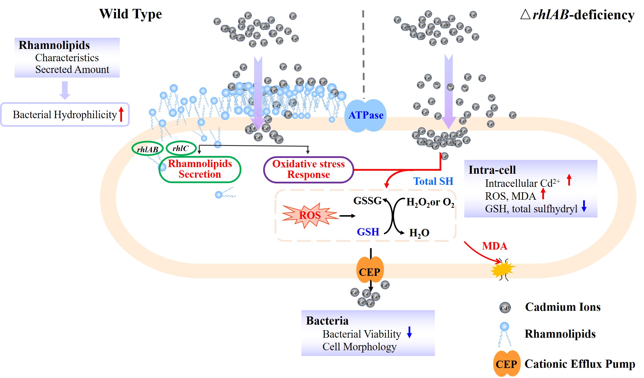
2.2. Plasmid Construction and Gene Deletion Mutant

The biosynthesis of rhamnolipids mainly relates to rhamnosyltransferase-1 (RhlAB) encoded by rhlAB operon. RhlA is involved in the synthesis of fatty acids, and rhlB catalyzed the synthesis of mono-rhamnolipids [35,36]. In this study, we deleted rhlAB genes to cut off the synthetic pathways of rhamnolipids and obtained the ΔrhlAB strain that could not secrete rhamnolipids. Fragments of 400~500 bp gene for the upstream or downstream of the rhlAB were amplified by gene splicing overlap extension (SOE-PCR) (primers listed in Table S2) based on P. aeruginosa genomic as template DNA. The fragments of the desired mutant allele were inserted into the suicide plasmid pEX18GM (Figure S1). This resulting deletion vector was manipulated with E. coli DH5α, then transformed into the donor strain E. coli WM3064 (auxotroph) and mobilized into P. aeruginosa by conjugation [34]. The ΔrhlAB cells were selected through two selections (Gm resistance selection and counter-selected by 15% sucrose). The ΔrhlAB strain was confirmed by PCR, sequencing, and blue agar tests with no rhamnolipids secretion (details are described in Supplementary Information).

2.3. Detection Analysis of Rhamnolipids

In order to analyze rhamnolipids productions, the wild-type bacteria grown with different Cd2+ concentrations were collected in the stationary phase and centrifuged for 20 min at 10,000× g to remove bacterial cells. The supernatant liquids were filtered through 0.22 μm filter membranes twice to wipe out residual cells. The cell-free supernatant was acidified to pH ≈ 2 and incubated at 4 °C overnight to precipitate rhamnolipids. The precipitated rhamnolipids were removed via centrifugation (10,000× g, 20 min, 4 °C) and re-dissolved in sterilized deionized water and then extracted with chloroform–methanol (2:1, v/v). Semi-purified rhamnolipids were obtained via evaporating the organic phases in a rotary evaporator (RE-52AA, Shanghai Yarong Biochemistry Instrument Factory) at 50 °C. The harvested rhamnolipids were lyophilized into powder and stored at desiccator for the following experiments.
To quantifying rhamnolipids, the amounts of rhamnose moiety were first determined using a anthrone-sulfuric acid colorimetric assay with L-rhamnose as the standard substance [37]. The rhamnolipids concentrations were obtained via the multiplication of the values of rhamnose moiety amounts by 3.4, which was the coefficient of the relation between purified rhamnolipids and rhamnose [22,38].

2.4. Characteristics Analysis of Rhamnolipids

The obtained rhamnolipids were analyzed with surface tension measurement, emulsifying activity, critical micelle concentration (CMC), and FTIR-ATR [18]. Surface tension assays were performed at room temperature using a tension meter (JK99C, Shanghai, China). Emulsification activities were measured by emulsification index for five days. CMCs were determined with a breaking point of the surface tension versus a series of rhamnolipids concentrations. The details were described in the Supplementary Information.

2.5. Distribution of Cd on Bacterial Media

To ascertain Cd2+ processing ability, wild-type and ΔrhlAB of P. aeruginosa were grown in the LB media with a series of Cd2+ concentrations (5 and 200 mg/L). For each group, a supernatant was taken in the stationary phase and centrifuged at 5000 rpm for 10 min. The supernatant was collected to measure the amounts of supernatant Cd2+ ions (CSup). The precipitate was washed three times with EDTA to remove the Cd2+ bonded to the bacterial surface, and the washed cells were collected by centrifugation to determine intracellular Cd2+ (CB). Meanwhile, the control group was also prepared without bacteria cells to determine the total Cd2+ (CTotal). All samples were acids digested with HClO4:HNO3 (1:9), and then the amounts of Cd2+ were analyzed with an atomic fluorescence spectrophotometer (AFS-933, Beijing Jitian Instrument Co., Ltd., Beijing, China). The amounts of Cd2+ absorbed on the bacterial surfaces (CSur) were calculated as follows:
CSur = CTotal − CSup − CB
The percentage of each part (CSup, CB, and CSur) was calculated by dividing the Cd2+ amount of each part by the CTotal and then multiplied by 100.

2.6. Bacterial Surface Properties Analysis

Bacterial hydrophobicity was determined with bacterial adherence to hydrocarbon tests (BATHs) [39]. Briefly, P. aeruginosa were cultured in the LB medium with different Cd2+ concentrations, and cells were collected in the stationary phase via centrifugation at 5000 rpm for 15 min and washed with NaCl solution (0.85%, pH = 7.2~7.4) three times. Then, bacterial pellets were resuspended with 0.85% NaCl solution and adjusted OD600 ≈ 0.5 as the ODinitial. N-dodecane at equal volume was mixed with the suspension and vortexed for 90 s. The mixture was then placed at room temperature for 30 min, and the absorbance of the inorganic phase was measured at 600 nm as the ODfinal. The bacterial hydrophobicity was calculated with the following equation:
Hydrophobicity (%) = 100% × (1 − ODfinal/ODinitial)
Cellular morphology. Wild -type and ΔrhlAB of P. aeruginosa were cultured in the LB medium with different Cd2+ concentrations (0, 5, 200 mg/L Cd). The cells were harvested in the stationary phase and then fixed, dehydrated, and coated with gold film, followed by observation with a field emission scanning electron microscope (Quanta 250 FEG, FEI, Hillsboro, OR, USA).

2.7. Bacterial Viability Assays

Spot Titer Assays. Wild-type and ΔrhlAB strains of P. aeruginosa were harvested from LB media without Cd2+ and then resuspended with 0.85% NaCl solution to adjust OD600 to 1.0. Serial 5-fold dilutions (10−2 to 10−7 for 0 to 50 mg/L Cd, and 100 to 10−5 for 100 and 200 mg/L Cd, respectively) of 2 μL bacterial suspensions were used on the LB-agar plates with a series of Cd2+ concentrations and then the plates were incubated at 37 °C overnight [40].
Colony-Forming Unit (CFU) assays. Bacterial cells were harvested in the stationary phase and resuspended with 0.85% NaCl solution contained different Cd2+ concentrations (5, 20, and 200 mg/L). Samples were taken at 0 h and 18 h for CFU assays, named CFU0 and CFU18, respectively. Bacterial viability was calculated as the percentage of living cells using Equation (3) as follows:
Bacterial viability = CFU18/CFU0 × 100%

2.8. Bacterial Biochemical Characteristic

Oxidative stress assays. Two type strains of P. aeruginosa were harvested in the stationary phase, which were exposed to Cd2+ at different dosages (0, 5, and 200 mg/L). The cells were collected and resuspended with 0.85% NaCl solution to similar OD600. Reactive oxygen species (ROS), MDA, GSH, and ATPase were determined in order to explore different responses of cells to Cd stress. In brief, ROS were tested with ROS Assay Kits (Beyotime Institute of Biotechnology, Shanghai, China) using a microplate spectrophotometer (Spark, Tecan Austria GmbH, Salzburg, Austria) with an excitation wavelength of 488 nm and emission wavelength of 525 nm. The oxidative damage to cells (MDA) was estimated using the thiobarbituric acid reactive substances (TBARS) method [41]. ATPase activity, GSH, and total sulfhydryl (total-SH) assays were measured with ATPase activity test kits, reduced glutathione (GSH) assay kits, total sulfhydryl (-SH) measurement kits (Nanjing Jiancheng Bioengineering Institute, Nanjing, China), respectively. Bacterial total proteins were measured with the BCA measurement kits (Nanjing Jiancheng Bioengineering Institute, Nanjing, China).

2.9. Statistical Analysis

The data of Cd distribution, cells viability, bacterial surface hydrophobicity, and bacterial biochemical properties among different treatment were assessed by two-way AVOVA analysis (factor 1 = Cd concentration, factor 2 = bacterial type). The data concerning rhamnolipids yields and surface tension assays were evaluated by one-way ANOVA. The statistically significant differences of the data described as follows: (none) indicated no difference (p > 0.05), (*) denoted p < 0.05; (**) denoted p < 0.01, (***) denoted p < 0.005.

3. Results and Discussion

3.1. Effect of Cd2+ on Cells Viability

To explore the response of rhamnolipids to Cd2+ stress, the two-step allelic exchange approach was adopted to knockout the rhlAB gene of P. aeruginosa, and the ΔrhlAB strain was detected by PCR analysis, rhamnolipids secretion, and blue agar tests (Figure 1A and Figure S2).
Biological activities under Cd2+ stress were assessed by bacterial viability and spot titer to determine bacterial sensitivity to Cd2+. The ΔrhlAB strain showed lower bacterial viability (68.5%, 56.0%, and 49.4% for 5, 20, and 200 mg/L Cd, respectively) than wild-type (91.6%, 62.8%, and 63.6% for 5, 20, and 200 mg/L Cd, respectively) (Figure 1B). In addition, spot titer assays provided a visually observable indication of the sensitivity of cells to Cd2+ (Figure 1C). The ΔrhlAB strain presented as being more sensitive to low doses of Cd2+ (<50 mg/L) than the wild-type, which is consistent with bacterial viability tests. However, at high Cd2+ concentrations (>50 mg/L), slight differences in sensitivity were observed for two types of strains. Thus, rhamnolipids were able to enhance the bacterial resistance to Cd2+ at low doses, with limited increases at high Cd2+ levels.

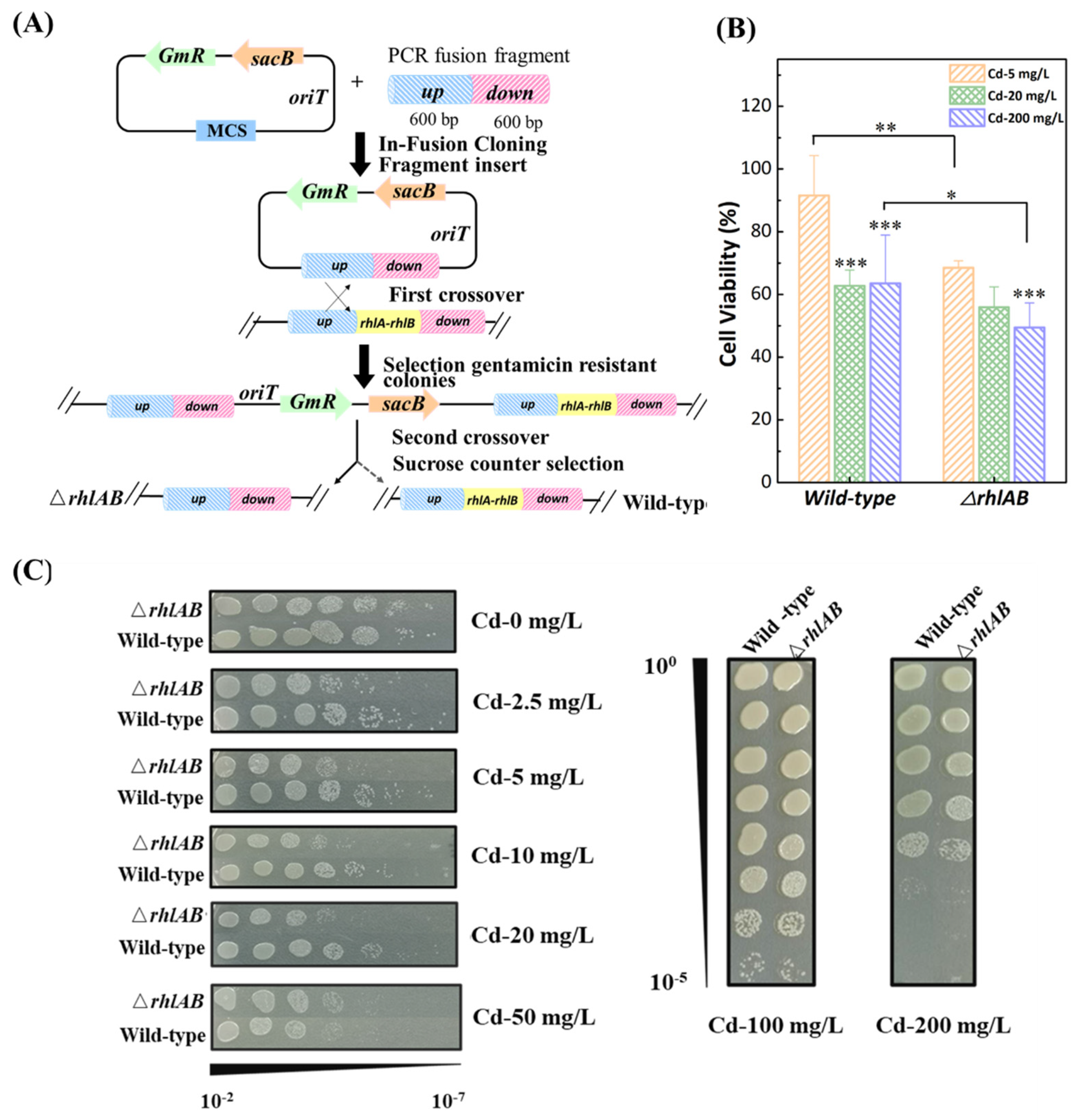
3.2. Distribution of Cd2+ in Bacteria

To investigate the effect of rhamnolipid synthesis on bacterial susceptibility to Cd2+, the distributions of Cd2+ on the surface, inside the cell, and in the supernatant of the two strains were determined (Figure 2A and Figure S3). Cadmium was mainly distributed on the bacterial surface of the two strains. The percentage of cadmium increased significantly from 48.9% to 62.1% (5 mg/L and 200 mg/L Cd2+, respectively) on the wild-type bacterial surfaces but remained stable (52.1% and 51.4%) in the ΔrhlAB cells. As a result, more intracellular Cd2+ was detected in ΔrhlAB cells than in wild-type strains. We evaluated intracellular Cd2+ per dry weight to eliminate the effect of bacterial biomass on Cd accumulation. As shown in Figure 2B, the amounts of Cd2+ were 59.1 mg/g and 406.1 mg/g in the wild-type strain while those were 75.3 mg/g and 1025.8 mg/g in the ΔrhlAB strain. These results suggested that mutant cells take up more Cd2+ into the cells, so rhamnolipids might play a significant role in blocking the entry of Cd2+ into the cells. This is mainly because rhamnolipids form a complex with Cd2+ rapidly within 15 min and remain stable for at least 27 h [42]. The morphologies of two strains with and without Cd2+ treatment were shown in SEM images (Figure 2C). They revealed that the ΔrhlAB strain looked fuller than wild-type cells at high Cd2+ concentration, which might be related to the large amount of Cd2+ entering the cells. Rhamnolipids combined with extracellular Cd2+ effectively reduced intracellular Cd2+ accumulation.

3.3. Effect of Cd2+ on Bacterial Characteristics

Cadmium exposure altered cell-surface properties. As shown in Figure 2B, the hydrophobicity of the wild-type was higher than that of the ΔrhlAB strain and decreased with increasing Cd2+ concentration, whereas that of the ΔrhlAB strain remained stable. This is mainly due to the role of rhamnolipids. Rhamnolipids are secreted around the cells and thus alter the hydrophobicity of the bacterial surface [43].
Since Cd2+ inhibited bacterial growth and rhamnolipids alleviated the negative effects of Cd2+ on cells, it seemed reasonable to assume that Cd2+ stress disrupted the metabolism in P. aeruginosa, including the biosynthesis and secretion strategy of endogenous rhamnolipids. To prove this, we examined the response of P. aeruginosa to Cd2+ stress.

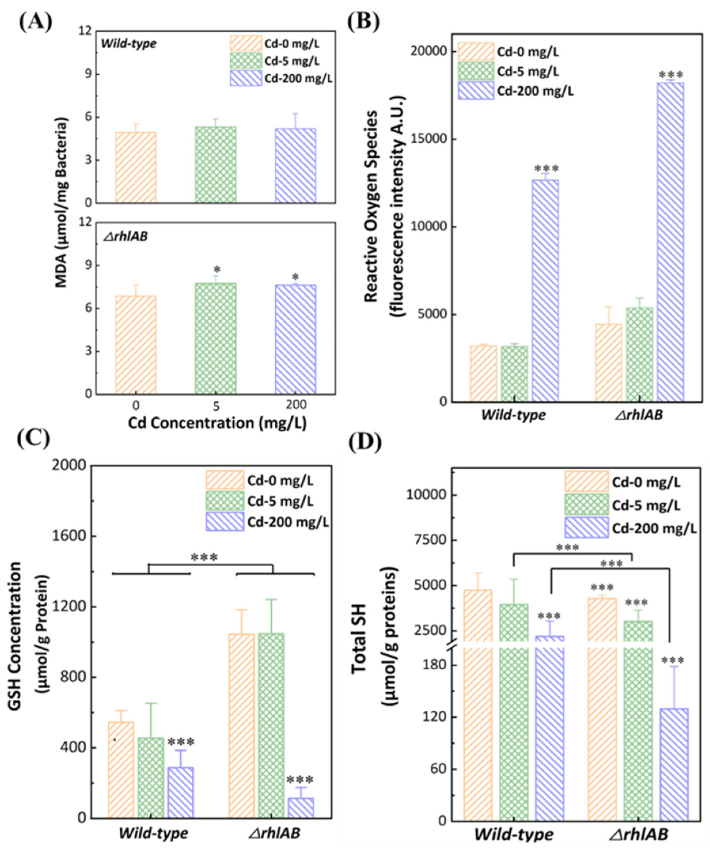
3.4. Response of Antioxidant Systems to Cd2+ Stress

MDA is a byproduct of bacterial membrane oxidation that reflects the degree of peroxidation of membrane lipids under oxidative stress [44,45]. As shown in Figure 3A, there was no obvious change in the wild-type with or without Cd2+. However, MDA levels were significantly increased in the ΔrhlAB strain in the presence of Cd2+ (p < 0.05), suggesting that more severe membrane damage occurred in the ΔrhlAB strain. These results indicated that the rhlAB gene might contribute to the protection of bacterial membrane integrity. In addition, ROS levels increased obviously in both strains at 200 mg/L Cd2+, whereas the ΔrhlAB strain exhibited a much higher ROS level than the wild-type strain (Figure 3B). This indicated that the ΔrhlAB strain was exposed to higher levels of Cd2+ due to more intracellular Cd2+. As shown in Figure S4, there were no obvious changes in Ca2+/Mg2+ ATPase and K+/Na+ ATPase activities in the wild-type strain with the presence of Cd2+, but the activities in the ΔrhlAB strain with Cd2+ decreased significantly, leading to a decrease in the ATP level in cells [46]. Generally, the suppression of energy metabolism could function as a control mechanism to reduce the generation of ROS [47].
Glutathione (GSH) is a tripeptide molecule that still widely existed in bacteria, which is important in the antioxidant defense system and effectively prevents oxidative stress from ROS [48]. Compared to wild-type strains, a higher GSH level was detected in ΔrhlAB strains in the absence or presence of 5 mg/L Cd2+ (Figure 3C). For cells exposed to 200 mg/L Cd, GSH levels for wild-type and ΔrhlAB strains decreased to 288.4 μmol/g protein, and 113.7 μmol/g protein, respectively. It indicated an inadequate resistance ability of ΔrhlAB strains for more serious oxidative stress at higher Cd2+ concentrations. These results confirmed that rhamnolipids could obviously enhance bacterial resistance to Cd2+ at low dosages and the enhancement was limited at high Cd2+ stress.
In P. aeruginosa cells, cysteine-rich metallothionein proteins (MT) with cysteine residues, calculated by total sulfhydryl groups (total-SH), play an important role in the bioremediation of heavy metals contamination [49]. In this study, total-SH contents were measured to confirm that the wild-type strain exhibited a higher ability to alleviate heavy metal stress. For the wild-type strain, the amounts of total-SH without Cd2+ (4746.3 μmol/g protein) or with 5 mg/L Cd2+ (3946.1 μmol/g protein) were similar and decreased obviously with 200 mg/L Cd2+ (2183.7 μmol/g protein) (Figure 3D). Additionally, the total-SH content at 200 mg/L Cd2+ decreased sharply to 129.6 μmol/g protein in the ΔrhlAB strain. This was mainly attributed to more Cd2+ entering the cells of the ΔrhlAB strain and combining with sulfhydryl groups, which was consistent with the distribution of Cd2+ in the cells.
In this work, we observed the protective mechanisms of P. aeruginosa from the Cd2+ through the bacterial membrane into the cytoplasm. When Cd2+ stress was not high enough to kill cells, the Cd2+ efflux systems could protect the cells from Cd2+, resulting in an unconspicuous change in the bacterial antioxidant systems (Figure 3) [28,50,51]. When the Cd2+ load was high, sulfhydryl complexed with intracellular Cd2+ in MT, which could relieve the cells from the high Cd2+ load. Meanwhile, due to the high Cd2+ load, more ROS were generated, which could not be completely removed by GSH, promoting membrane depolarization, leading to more severe cell damage. It was worth noting that rhamnolipids could bind with Cd2+ to prevent Cd2+ from entering the cells, and thus reduced the Cd2+ load on the cells.

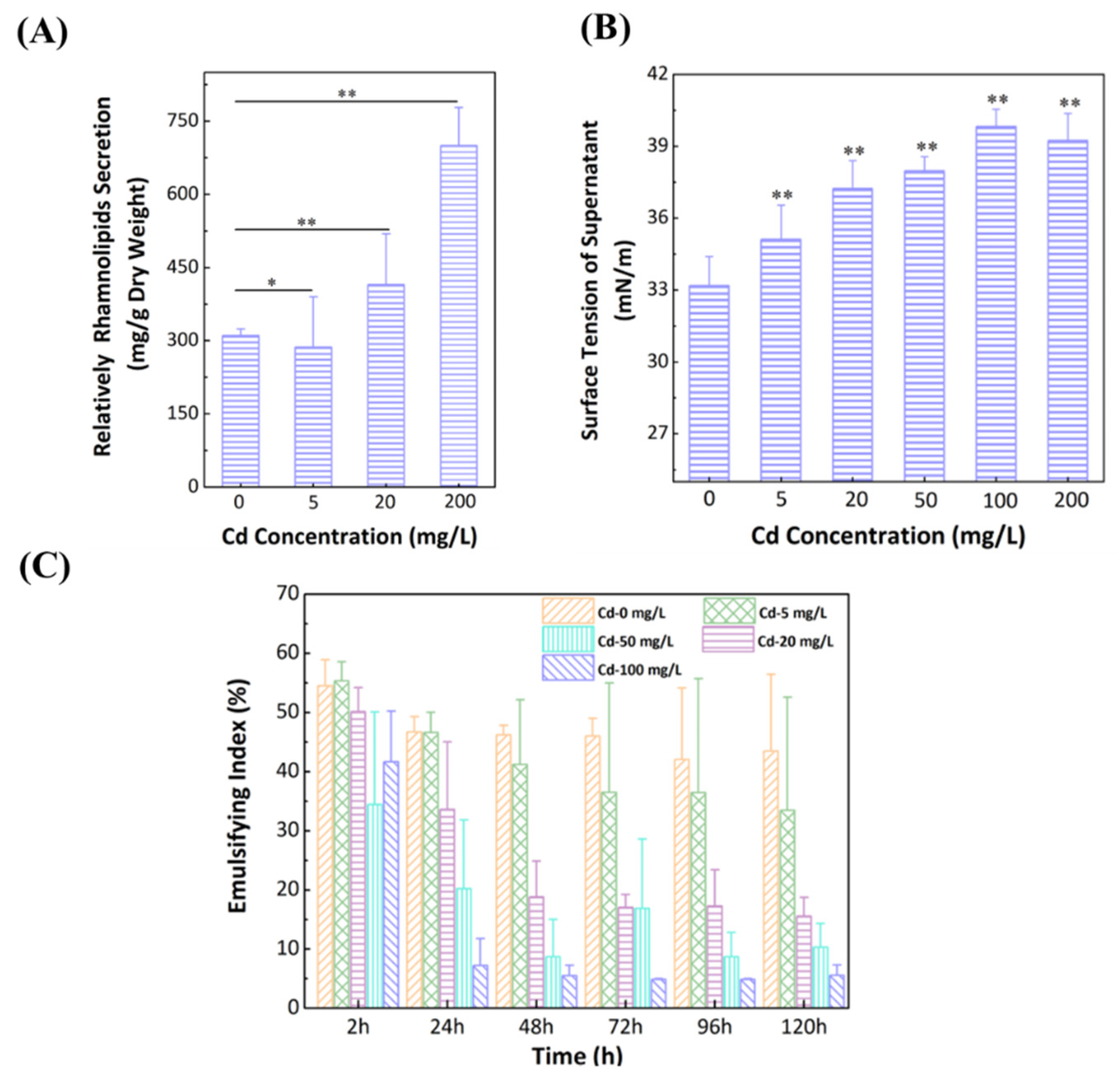
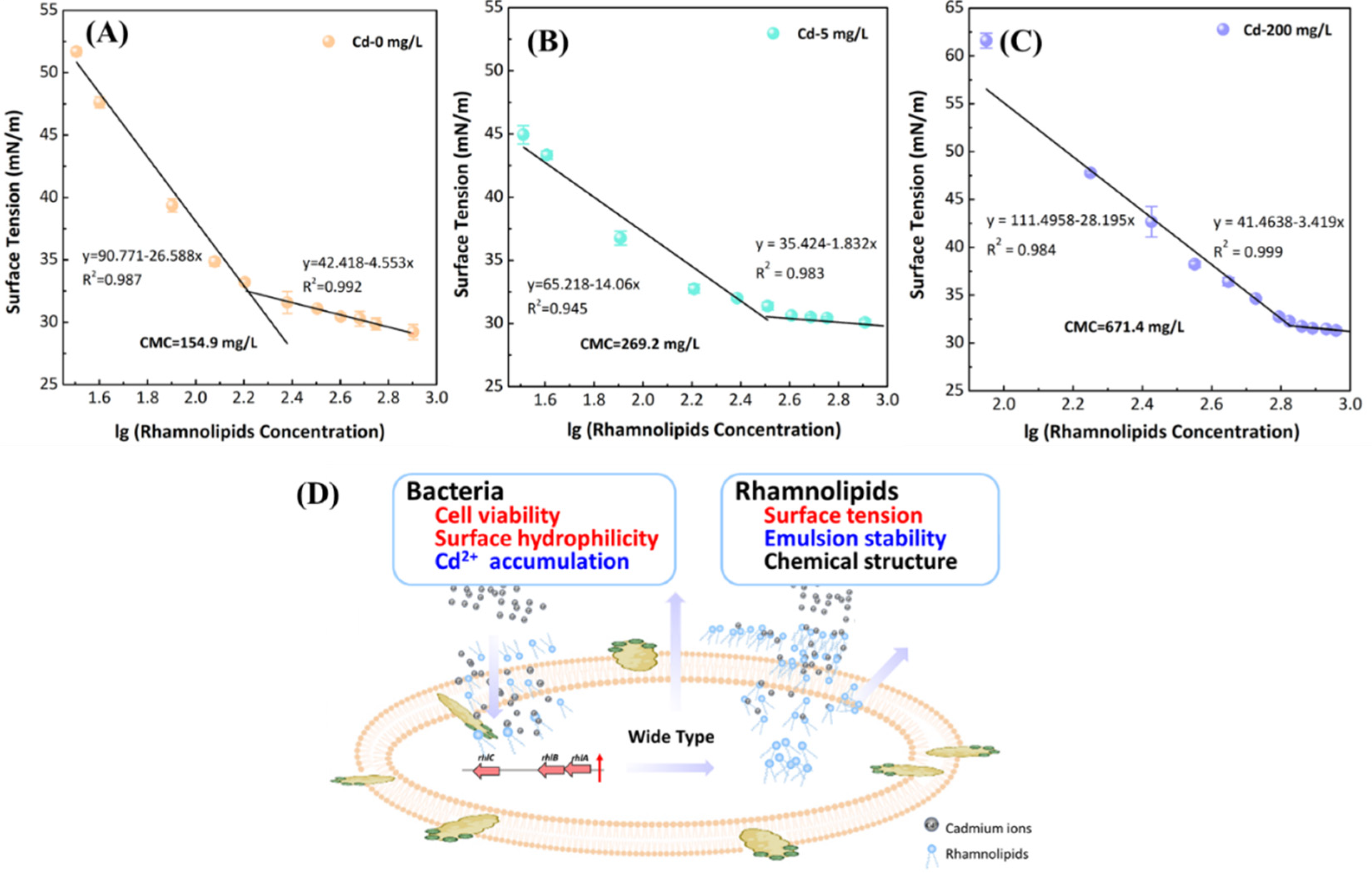
3.5. Effect of Cd2+ on Secretion of Rhamnolipids
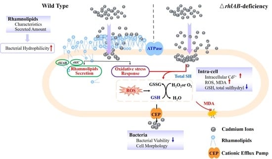
The secretion of rhamnolipids from the wild-type strain were assessed with a range of Cd2+ concentrations. As shown in Figure 4A, Cd stress obviously promoted the production of rhamnolipids per dry biomass, which increased by 143% at 200 mg/L compared with that without Cd2+. It has been reported that heavy metal cations can form a complex in a binding pocket, consisting of a carboxylate moiety in the fatty lipid and the hydroxy moiety in the rhamnose [52]. In this study, heavy metal stress affected the secretion of rhamnolipids as the extracellular polymeric substances and thus promoted complex formation with Cd2+, which had a beneficial effect of alleviating heavy metal stress [16].
In this experiment, the physicochemical properties of rhamnolipids were studied based on emulsification activity and surface tension. The secretion of rhamnolipids decreased the surface tension (33.2 mN/m) of media without Cd2+, compared to the fresh medium (47.5 ± 0.5 mN/m) (Figure 4B). In addition, the increas in Cd2+ concentration enhanced surface tensions (35.1, 37.2, and 39.2 mN/m for 5, 20, and 200 mg/L Cd2+, respectively). It indicated that Cd2+ could impact the stability of the secreted rhamnolipids. The emulsification activities of the biosurfactants were measured with a range of water-immiscible substrates, and the emulsion ability was found to be with hydrocarbons. Secreted rhamnolipids without Cd2+ were capable of stabilizing emulsions with an emulsification index (EI) in the range of 46.7% at 24 h to 43.5% at 120 h, whereas the EI value dropped gradually dropped to 4.7% at 100 mg/L (Figure 4C). The low emulsification activity implied that the toxicity of Cd2+ to cells, leading to a lower ability to remove hydrophobic organic compound–heavy metal complex contaminations [53].
The chemical structures of the rhamnolipids with Cd were analyzed by FTIR (Figure S5). The peaks were assigned to hydroxyl, rhamnose, and lipid backbones, the main characteristics of glycolipids. Fewer spectral bands in the presence of Cd2+ (Table S3) indicated that lipids functional groups appeared to be less or that the fatty acid chains became shorter under Cd2+ stress. The absorption intensities of the spectral bands in the presence of Cd2+ were weaker than that of the Cd-null sample, as the decreased intensity for the strong and broad bonds of the free-stretching hydroxyl (O-H) due to it complexation with Cd2+.
To further investigate the effect of Cd2+ loading on the rhamnolipid secretion, we also examined the changes in CMC of rhamnolipids in the presence of Cd2+. The CMC values of rhamnolipids were 154.9 mg/L, 269.2 mg/L, and 671.4 mg/L for the cells cultured under Cd2+ concentration of 0, 5, and 200 mg/L, respectively (Figure 4A–C). The CMC value of rhamnolipids relates to superficial characteristics, such as surface tension and emulsification activity. However, the value depends on the structure of rhamnolipids, e.g., chain length, number of rhamnoses, etc. [33,54].
Nitschke et al. reported that the major components of rhamnolipids are RhaC10C10 and Rha2C10C10 in P. aeruginosa [55]. However, the composition of rhamnolipids mixtures differs by the presence of unsaturated bonds, fatty acid chains, and the size of hydrophilic groups, which affect the surface character of the mixture. The length of the fatty acid chains and the number of rhamnose units played an important role in the CMC value, affecting hydrophobicity and emulsifying activity [31,56]. Therefore, we concluded that Cd2+ actively interacted with rhamnolipids and altered the structural and functional properties of rhamnolipids, which were affected by cultural conditions [57,58]. Our results suggested that Cd2+ stress led to shorter fatty acid chains of rhamnolipids, a higher ratio of di-rhamnolipids to mono-rhamnolipids, and a lower proportion of hydrophobic groups, resulting in higher hydrophilicity and lower emulsifying activity and higher concentration is needed to form the micelle aggregation [25].
Rhamnolipids biosynthesis pathway begins with the de novo synthesis of rhamnose and fatty acids, which are ubiquitous in the bacterium followed by three consecutive enzymatic reactions specific in rhamnolipids-secreting bacterium. Our results suggested that Cd stress effects rhlA, rhlB, and rhlC expression (Figure S6) and rhamnolipids might also be a endogenous factor in response to heavy metals stress. A strong correlation between rhamnolipids production and heavy metal stress has also been documented for other P. aeruginosa strains. The ratio of di-rhamnolipids to mono-rhamnolipids increased obviously under Cd2+ stress [32]. This may explain the increase of bacterial surface hydrophilicity in the presence of Cd2+ in this study.
Briefly, the interactions among bacteria, endogenous rhamnolipids, and cadmium were demonstrated in Figure 5D as follows: (1) the endogenous rhamnolipids with more rhamnose tails could form a complex with additional cadmium, reduce intracellular Cd2+ accumulation, decrease oxidative stress response, and improve bacterial cell viability; (2) the altered physicochemical properties of endogenous rhamnolipids, such as increased surface tension and decreased emulsification activity, was attributed to the changed structures of rhamnolipids under Cd2+ stress; and (3) the altered properties of rhamnolipids resulted in a more hydrophilic bacterial surface. These changes negatively affected the utilization of endogenous rhamnolipids.

4. Conclusions

Biosurfactants secreted by microorganisms can probably improve the removal of contaminants and reduce environmental hazards. Together with our results, this study confirmed the potential of employing rhamnolipids and rhamnolipids-secreted bacteria in Cd2+ remediation. Cadmium stress could affect rhamnolipid production and increase the complexation of Cd2+ on bacterial surfaces, resulting in the protection of cells from Cd2+ stress. For the wild-type cells, Cd stress altered the structure of rhamnolipid structures, leading to higher CMC and hydrophilicity, and this discrimination prejudiced emulsification ability. Once Cd entered the cytoplasm, the oxidative stress system had no obvious effects on the membrane integrity of the wild-type due to other stress mitigation mechanisms available, such as GSH, total-SH, ATPase activity, cation efflux pump, etc. For the mutant cells, the lack of rhamnolipid secretion led to greater intracellular Cd accumulation, which motivated fearful oxidative stress that could not be alleviated by detoxification mechanisms and impaired cell viability as a result. As with higher Cd loading, intracellular Cd accumulation increased; however, the cell viability of the wild-type was slightly higher than that of the mutant. The GSH content and total-SH content for the mutants were much lower than in the wild-type cells. These findings suggested that rhamnolipids had a limited effect on improving Cd2+ resistance. This study enriched the theoretical knowledge on the use of P. aeruginosa to enhance the bioremediation of heavy metals bioremediation and alleviate stress in contaminated environments.

Supplementary Materials

The following are available online at https://www.mdpi.com/article/10.3390/ijerph191912555/s1, Figure S1. The schematic of pEX18GM-△rhlAB plasmid construction. Figure S2. DNA gel electrophoresis (A), rhamnolipids secretions (B), and CTAB-MB plate (C) to verified the △rhlAB strain could not secreted rhamnolipids. Figure S3. Distribution of cadmium for the wild-type and the △rhlAB strains under different Cd2+ concentrations (5, 200 mg/L). Figure S4. ATPase activity (Na+/K+, and Ca2+/Mg2+) assays for the wild-type and the △rhlAB strains which expressed in U per mg proteins. Figure S5. ATR-FTIR assays for semi-purified rhamnolipids from the wild-type under a series of Cd2+ concentrations (0, 5, 50, 100, 200 mg/L). Figure S6. Expression of genes of the wild type under the stress of Cd. Table S1. Plasmids and bacteria used in this study. Table S2. The primers and sequences for rhlA-rhlB deletion and qPCR. Table S3. Assignments of FTIR spectra of rhamnolipids. References [32,59,60,61,62] are cited in the supplementary materials

Author Contributions

S.X.: Conceptualization, Methodology, Investigation, Formal analysis, Writing—Original draft, and Writing—Review and Editing. Z.Y.: Conceptualization, Formal analysis, Supervision, and Writing—Review and Editing. C.S.: Formal analysis and Supervision. H.T.: Formal analysis, Writing—Review and Editing, and Supervision. S.W.: Project administration, Supervision, and Funding acquisition. All authors have read and agreed to the published version of the manuscript.

Funding

This work was supported by the National Natural Science Foundation of China (U20A20146, 21808127) and Qilu Youth Talent Programme of Shandong University.

Institutional Review Board Statement

Not applicable.

Informed Consent Statement

Not applicable.

Data Availability Statement

All data used in this study are included in this published article and its supplementary information file.

Acknowledgments

The authors thank Xiaoxue Wang and Yunxue Guo from South China Sea Institute of Oceanology, Chinese Academy of Sciences for providing pEX18GM, E. coli WM3064 and P. putida. We would like to thank Sen Wang, Haiyan Yu, Xiaomin Zhao, and Yuyu Guo from State Key laboratory of Microbial Technology of Shandong University for their assistance in the application of the scanning electron microscope.

Conflicts of Interest

The authors declare no conflict of interest.

References

  1. Módenes, A.N.; de Abreu Pietrobelli, J.M.T.; dos Santos, G.H.F.; Borba, C.E.; da Silva Sá Ravagnani, M.A.; Espinoza-Quiñones, F.R. Multi-component mathematical model based on mass transfer coefficients for prediction of the Zn and Cd ions biosorption data by E. densa in a continuous system. J. Environ. Chem. Eng. 2018, 6, 5141–5149. [Google Scholar] [CrossRef]
  2. Hamid, Y.; Tang, L.; Sohail, M.I.; Cao, X.; Hussain, B.; Aziz, M.Z.; Usman, M.; He, Z.-l.; Yang, X. An explanation of soil amendments to reduce cadmium phytoavailability and transfer to food chain. Sci. Total Environ. 2019, 660, 80–96. [Google Scholar] [CrossRef] [PubMed]
  3. da Silva Oliveira, A.; Bocio, A.; Trevilato, T.M.B.; Takayanagui, A.M.M.; Domingo, J.L.; Segura-Muñoz, S.I. Heavy metals in untreated/treated urban effluent and sludge from a biological wastewater treatment plant. Environ. Sci. Pollut. Res. Int. 2007, 14, 483. [Google Scholar] [CrossRef] [PubMed]
  4. Tu, C.; Wei, J.; Guan, F.; Liu, Y.; Sun, Y.H.; Luo, Y.M. Biochar and bacteria inoculated biochar enhanced Cd and Cu immobilization and enzymatic activity in a polluted soil. Environ. Int. 2020, 137, 105576. [Google Scholar] [CrossRef] [PubMed]
  5. Lim, S.-R.; Schoenung, J.M. Human health and ecological toxicity potentials due to heavy metal content in waste electronic devices with flat panel displays. J. Hazard. Mater. 2010, 177, 251–259. [Google Scholar] [CrossRef] [PubMed]
  6. Zhang, Q.; Liu, H.; Liu, F.; Ju, X.; Dinis, F.; Yu, E.; Yu, Z. Source identification and superposition effect of heavy metals (HMs) in agricultural soils at a high geological background area of Karst: A case study in a typical watershed. Int. J. Environ. Res. Public Health 2022, 19, 11374. [Google Scholar] [CrossRef]
  7. Liu, X.; Hu, Q.; Yang, J.; Huang, S.; Wei, T.; Chen, W.; He, Y.; Wang, D.; Liu, Z.; Wang, K.; et al. Selective cadmium regulation mediated by a cooperative binding mechanism in CadR. Proc. Natl. Acad. Sci. USA 2019, 116, 20398–20403. [Google Scholar] [CrossRef] [Green Version]
  8. Zhu, Y.; Ma, J.; Chen, F.; Yu, R.; Hu, G.; Zhang, S. Remediation of Soil polluted with Cd in a postmining area using thiourea-modified biochar. Int. J. Environ. Res. Public Health 2020, 17, 7654. [Google Scholar] [CrossRef]
  9. Mishra, S.; Lin, Z.; Pang, S.; Zhang, Y.; Bhatt, P.; Chen, S. Biosurfactant is a powerful tool for the bioremediation of heavy metals from contaminated soils. J. Hazard. Mater. 2021, 418, 126253. [Google Scholar] [CrossRef]
  10. Agnello, A.C.; Bagard, M.; van Hullebusch, E.D.; Esposito, G.; Huguenot, D. Comparative bioremediation of heavy metals and petroleum hydrocarbons co-contaminated soil by natural attenuation, phytoremediation, bioaugmentation and bioaugmentation-assisted phytoremediation. Sci. Total Environ. 2016, 563–564, 693–703. [Google Scholar] [CrossRef]
  11. Yang, K.; Zhu, L.; Zhao, Y.; Wei, Z.; Chen, X.; Yao, C.; Meng, Q.; Zhao, R. A novel method for removing heavy metals from composting system: The combination of functional bacteria and adsorbent materials. Bioresour. Technol. 2019, 293, 122095. [Google Scholar] [CrossRef] [PubMed]
  12. Asad, S.A.; Farooq, M.; Afzal, A.; West, H. Integrated phytobial heavy metal remediation strategies for a sustainable clean environment—A review. Chemosphere 2019, 217, 925–941. [Google Scholar] [CrossRef] [PubMed]
  13. Arora, P.K. Bacilli-mediated degradation of xenobiotic compounds and heavy metals. Front. Bioeng. Biotechnol. 2020, 8, 570307. [Google Scholar] [CrossRef] [PubMed]
  14. Mishra, S.; Lin, Z.; Pang, S.; Zhang, W.; Bhatt, P.; Chen, S. Recent advanced technologies for the characterization of xenobiotic-degrading microorganisms and microbial communities. Front. Bioeng. Biotechnol. 2021, 9, 632059. [Google Scholar] [CrossRef]
  15. Ghaith, E.-S.I.; Rizvi, S.; Namasivayam, C.; Rahman, P. Removal of Cd++ from contaminated water using bio-surfactant modified ground grass as a bio-sorbent. In Proceedings of the 2019 Advances in Science and Engineering Technology International Conferences (ASET), Dubai, United Arab Emirates, 26 March–10 April 2019; IEEE: Piscataway, NJ, USA, 2019; pp. 1–7. [Google Scholar]
  16. Qin, H.; Hu, T.; Zhai, Y.; Lu, N.; Aliyeva, J. The improved methods of heavy metals removal by biosorbents: A review. Environ. Pollut. 2020, 258, 113777. [Google Scholar] [CrossRef]
  17. Wen, J.; McLaughlin, M.J.; Stacey, S.P.; Kirby, J.K. Aseptic hydroponics to assess rhamnolipid-Cd and rhamnolipid-Zn bioavailability for sunflower (Helianthus annuus): A phytoextraction mechanism study. Environ. Sci. Pollut. Res. 2016, 23, 21327–21335. [Google Scholar] [CrossRef]
  18. da Rocha Junior, R.B.; Meira, H.M.; Almeida, D.G.; Rufino, R.D.; Luna, J.M.; Santos, V.A.; Sarubbo, L.A. Application of a low-cost biosurfactant in heavy metal remediation processes. Biodegradation 2019, 30, 215–233. [Google Scholar] [CrossRef]
  19. Yang, Z.; Zhang, Z.; Chai, L.; Wang, Y.; Liu, Y.; Xiao, R. Bioleaching remediation of heavy metal-contaminated soils using Burkholderia sp. Z-90. J. Hazard. Mater. 2016, 301, 145–152. [Google Scholar] [CrossRef]
  20. Liu, G.; Zhong, H.; Yang, X.; Liu, Y.; Shao, B.; Liu, Z. Advances in applications of rhamnolipids biosurfactant in environmental remediation: A review. Biotechnol. Bioeng. 2018, 115, 796–814. [Google Scholar] [CrossRef]
  21. Sachdev, D.P.; Cameotra, S.S. Biosurfactants in agriculture. Appl. Microbiol. Biotechnol. 2013, 97, 1005–1016. [Google Scholar] [CrossRef] [Green Version]
  22. Bondarenko, O.; Rahman, P.K.S.M.; Rahman, T.J.; Kahru, A.; Ivask, A. Effects of rhamnolipids from Pseudomonas aeruginosa DS10-129 on iuminescent bacteria: Toxicity and modulation of cadmium bioavailability. Microb. Ecol. 2010, 59, 588–600. [Google Scholar] [CrossRef] [Green Version]
  23. Juwarkar, A.A.; Dubey, K.V.; Nair, A.; Singh, S.K. Bioremediation of multi-metal contaminated soil using biosurfactant—A novel approach. Indian J. Microbiol. 2008, 48, 142–146. [Google Scholar] [CrossRef] [PubMed] [Green Version]
  24. Zhao, F.; Yuan, M.; Lei, L.; Li, C.; Xu, X. Enhanced production of mono-rhamnolipid in Pseudomonas aeruginosa and application potential in agriculture and petroleum industry. Bioresour. Technol. 2021, 323, 124605. [Google Scholar] [CrossRef] [PubMed]
  25. Zhao, F.; Han, S.; Zhang, Y. Comparative studies on the structural composition, surface/interface activity and application potential of rhamnolipids produced by Pseudomonas aeruginosa using hydrophobic or hydrophilic substrates. Bioresour. Technol. 2020, 295, 122269. [Google Scholar] [CrossRef] [PubMed]
  26. Eslami, P.; Hajfarajollah, H.; Bazsefidpar, S. Recent advancements in the production of rhamnolipid biosurfactants by Pseudomonas aeruginosa. RSC Adv. 2020, 10, 34014–34032. [Google Scholar] [CrossRef]
  27. Chakraborty, J.; Das, S. Characterization and cadmium-resistant gene expression of biofilm-forming marine bacterium Pseudomonas aeruginosa JP-11. Environ. Sci. Pollut. Res. 2014, 21, 14188–14201. [Google Scholar] [CrossRef]
  28. Brocklehurst, K.; Megit, S.; Morby, A.P. Characterisation of CadR from Pseudomonas aeruginosa: A Cd (II)-responsive MerR homologue. Biochem. Biophys. Res. Commun. 2003, 308, 234–239. [Google Scholar] [CrossRef]
  29. Abbas, S.; Rafatullah, M.; Hossain, K.; Ismail, N.; Tajarudin, H.; Khalil, H.A. A review on mechanism and future perspectives of cadmium-resistant bacteria. Int. J. Environ. Sci. Technol. 2018, 15, 243–262. [Google Scholar] [CrossRef]
  30. Singh, R.; Bishnoi, N.R.; Kirrolia, A. Evaluation of Pseudomonas aeruginosa an innovative bioremediation tool in multi metals ions from simulated system using multi response methodology. Bioresour. Technol. 2013, 138, 222–234. [Google Scholar] [CrossRef]
  31. Jain, R.M.; Mody, K.; Mishra, A.; Jha, B. Isolation and structural characterization of biosurfactant produced by an alkaliphilic bacterium Cronobacter sakazakii isolated from oil contaminated wastewater. Carbohydr. Polym. 2012, 87, 2320–2326. [Google Scholar] [CrossRef]
  32. Neilson, J.W.; Zhang, L.; Veres-Schalnat, T.A.; Chandler, K.B.; Neilson, C.H.; Crispin, J.D.; Pemberton, J.E.; Maier, R.M. Cadmium effects on transcriptional expression of rhlB/rhlC genes and congener distribution of monorhamnolipid and dirhamnolipid in Pseudomonas aeruginosa IGB83. Appl. Microbiol. Biotechnol. 2010, 88, 953–963. [Google Scholar] [CrossRef] [PubMed]
  33. Chen, J.; Wu, Q.; Hua, Y.; Chen, J.; Zhang, H.; Wang, H. Potential applications of biosurfactant rhamnolipids in agriculture and biomedicine. Appl. Microbiol. Biotechnol. 2017, 101, 8309–8319. [Google Scholar] [CrossRef] [PubMed]
  34. Hmelo, L.R.; Borlee, B.R.; Almblad, H.; Love, M.E.; Randall, T.E.; Tseng, B.S.; Lin, C.; Irie, Y.; Storek, K.M.; Yang, J.J. Precision-engineering the Pseudomonas aeruginosa genome with two-step allelic exchange. Nat. Protoc. 2015, 10, 1820–1841. [Google Scholar] [CrossRef] [PubMed] [Green Version]
  35. Zhao, F.; Shi, R.; Ma, F.; Han, S.; Zhang, Y. Oxygen effects on rhamnolipids production by Pseudomonas aeruginosa. Microb. Cell Factories 2018, 17, 39. [Google Scholar] [CrossRef] [PubMed] [Green Version]
  36. Setoodeh, P.; Jahanmiri, A.; Eslamloueyan, R.; Niazi, A.; Ayatollahi, S.S.; Aram, F.; Mahmoodi, M.; Hortamani, A. Statistical screening of medium components for recombinant production of Pseudomonas aeruginosa ATCC 9027 rhamnolipids by nonpathogenic cell factory Pseudomonas putida KT2440. Mol. Biotechnol. 2014, 56, 175–191. [Google Scholar] [CrossRef]
  37. Somani, B.L.; Khanade, J.; Sinha, R. A Modified Anthrone Sulfuric-Acid Method for the Determination of Fructose in the Presence of Certain Proteins. Anal. Biochem. 1987, 167, 327–330. [Google Scholar] [CrossRef]
  38. Benincasa, M.; Abalos, A.; Oliveira, I.; Manresa, A. Chemical structure, surface properties and biological activities of the biosurfactant produced by Pseudomonas aeruginosa LBI from soapstock. Antonie Van Leeuwenhoek 2004, 85, 1–8. [Google Scholar] [CrossRef]
  39. Subbiahdoss, G.; Reimhult, E. Biofilm formation at oil-water interfaces is not a simple function of bacterial hydrophobicity. Colloids Surf. B Biointerfaces 2020, 194, 111163. [Google Scholar] [CrossRef]
  40. Matthews, L.A.; Simmons, L.A. The Bacillus subtilis PriA Winged Helix Domain Is Critical for Surviving DNA Damage. J. Bacteriol. 2022, 204, e00539-21. [Google Scholar] [CrossRef]
  41. Dolezalova, E.; Lukes, P. Membrane damage and active but nonculturable state in liquid cultures of Escherichia coli treated with an atmospheric pressure plasma jet. Bioelectrochemistry 2015, 103, 7–14. [Google Scholar] [CrossRef]
  42. Tan, H.; Champion, J.T.; Artiola, J.F.; Brusseau, M.L.; Miller, R.M. Complexation of Cadmium by a Rhamnolipid Biosurfactant. Env. Sci. Technol. 1994, 28, 2402–2406. [Google Scholar] [CrossRef]
  43. Mohanty, S.; Mukherji, S. Surfactant aided biodegradation of NAPLs by Burkholderia multivorans: Comparison between Triton X-100 and rhamnolipid JBR-515. Colloids Surf. B Biointerfaces 2013, 102, 644–652. [Google Scholar] [CrossRef] [PubMed]
  44. He, T.-F.; Zhang, Z.-H.; Zeng, X.-A.; Wang, L.-H.; Brennan, C.S. Determination of membrane disruption and genomic DNA binding of cinnamaldehyde to Escherichia coli by use of microbiological and spectroscopic techniques. J. Photochem. Photobiol. B Biol. 2018, 178, 623–630. [Google Scholar] [CrossRef] [PubMed]
  45. Yun, O.; Liu, Z.-W.; Zeng, X.-A.; Han, Z. Salmonella typhimurium resistance on pulsed electric fields associated with membrane fluidity and gene regulation. Innov. Food Sci. Emerg. Technol. 2016, 36, 252–259. [Google Scholar] [CrossRef]
  46. Hamida, R.S.; Ali, M.A.; Goda, D.A.; Khalil, M.I.; Al-Zaban, M.I. Novel biogenic silver nanoparticle-induced reactive oxygen species inhibit the biofilm formation and virulence activities of methicillin-resistant Staphylococcus aureus (MRSA) Strain. Front. Bioeng. Biotechnol. 2020, 8, 433. [Google Scholar] [CrossRef]
  47. Simão, F.; Matté, A.; Matté, C.; Soares, F.M.S.; Wyse, A.T.S.; Netto, C.A.; Salbego, C.G. Resveratrol prevents oxidative stress and inhibition of Na+K+-ATPase activity induced by transient global cerebral ischemia in rats. J. Nutr. Biochem. 2011, 22, 921–928. [Google Scholar] [CrossRef]
  48. Wang, X.; Fan, L.; Cheng, L.; Sun, Y.; Wang, X.; Zhong, X.; Shi, Q.; Gong, F.; Yang, Y.; Ma, Y.; et al. Biodegradable nickel disulfide nanozymes with GSH-depleting function for high-efficiency photothermal-catalytic antibacterial therapy. iScience 2020, 23, 101281. [Google Scholar] [CrossRef] [PubMed]
  49. Benhalima, L.; Amri, S.; Bensouilah, M.; Ouzrout, R. Heavy metal resistance and metallothionein induction in bacteria isolated from Seybouse river, Algeria. Appl. Ecol. Env. Res. 2020, 18, 1721–1737. [Google Scholar] [CrossRef]
  50. Lee, S.W.; Glickmann, E.; Cooksey, D.A. Chromosomal locus for cadmium resistance in Pseudomonas putida consisting of a cadmium-transporting ATPase and a MerR family response regulator. Appl. Env. Microb. 2001, 67, 1437–1444. [Google Scholar] [CrossRef] [Green Version]
  51. Hassan, M.E.T.; van der Lelie, D.; Springael, D.; Romling, U.; Ahmed, N.; Mergeay, M. Identification of a gene cluster, czr, involved in cadmium and zinc resistance in Pseudomonas aeruginosa. Gene 1999, 238, 417–425. [Google Scholar] [CrossRef]
  52. Rondeau, P.; Sers, S.; Jhurry, D.; Cadet, F. Sugar interaction with metals in aqueous solution: Indirect determination from infrared and direct determination from nuclear magnetic resonance spectroscopy. Appl. Spectrosc. 2003, 57, 466–472. [Google Scholar] [CrossRef]
  53. Tang, S.; Yin, H.; Zhou, S.; Chen, S.; Peng, H.; Liu, Z.; Dang, Z. Simultaneous Cr(VI) removal and 2,2′,4,4′-tetrabromodiphenyl ether (BDE-47) biodegradation by Pseudomonas aeruginosa in liquid medium. Chemosphere 2016, 150, 24–32. [Google Scholar] [CrossRef]
  54. Zeng, Z.T.; Liu, Y.; Zhong, H.; Xiao, R.; Zeng, G.M.; Liu, Z.F.; Cheng, M.; Lai, C.; Zhang, C.; Liu, G.S.; et al. Mechanisms for rhamnolipids-mediated biodegradation of hydrophobic organic compounds. Sci. Total Env. 2018, 634, 1–11. [Google Scholar] [CrossRef]
  55. Nitschke, M.; Costa, S.G.V.A.O.; Contiero, J. Rhamnolipid surfactants: An update on the general aspects of these remarkable biomolecules. Biotechnol. Prog. 2005, 21, 1593–1600. [Google Scholar] [CrossRef]
  56. Helvacı, Ş.Ş.; Peker, S.; Özdemir, G. Effect of electrolytes on the surface behavior of rhamnolipids R1 and R2. Colloids Surf. B Biointerfaces 2004, 35, 225–233. [Google Scholar] [CrossRef]
  57. Kłosowska-Chomiczewska, I.E.; Mędrzycka, K.; Hallmann, E.; Karpenko, E.; Pokynbroda, T.; Macierzanka, A.; Jungnickel, C. Rhamnolipid CMC prediction. J. Colloid Interface Sci. 2017, 488, 10–19. [Google Scholar] [CrossRef]
  58. Costa, S.G.V.A.O.; Nitschke, M.; Lépine, F.; Déziel, E.; Contiero, J. Structure, properties and applications of rhamnolipids produced by Pseudomonas aeruginosa L2-1 from cassava wastewater. Process Biochem. 2010, 45, 1511–1516. [Google Scholar] [CrossRef]
  59. Patel, K.; Patel, M. Improving bioremediation process of petroleum wastewater using biosurfactants producing Stenotrophomonas sp. S1VKR-26 and assessment of phytotoxicity. Bioresour. Technol. 2020, 315, 123861. [Google Scholar] [CrossRef]
  60. Datta, P.; Tiwari, P.; Pandey, L.M. Isolation and characterization of biosurfactant producing and oil degrading Bacillus subtilis MG495086 from formation water of Assam oil reservoir and its suitability for enhanced oil recovery. Bioresour. Technol. 2018, 270, 439–448. [Google Scholar] [CrossRef]
  61. Zhu, L.; Qi, H.-y.; Kong, Y.; Yu, Y.-w.; Xu, X.-y. Component analysis of extracellular polymeric substances (EPS) during aerobic sludge granulation using FTIR and 3D-EEM technologies. Bioresour. Technol. 2012, 124, 455–459. [Google Scholar] [CrossRef]
  62. Bazire, A.; Dheilly, A.; Diab, F.; Morin, D.; Jebbar, M.; Haras, D.; Dufour, A. Osmotic stress and phosphate limitation alter production of cell-to-cell signal molecules and rhamnolipid biosurfactant by Pseudomonas aeruginosa. Fems. Microbiol. Lett. 2005, 253, 125–131. [Google Scholar] [CrossRef] [PubMed] [Green Version]
Figure 1. Construction of the ΔrhlAB strain using the two-step allelic exchange by pEX18GM (A), cell viability tests (B), and spot titer assays (C) of P. aeruginosa wild-type and ΔrhlAB strains under a series of Cd concentrations in the LB media. Differences were considered significant at p < 0.05, and * presented p < 0.05, ** presented p < 0.01, *** presented p < 0.005.
Ijerph 19 12555 g001
Figure 2. Intracellular cadmium amount per dry biomass (A), bacterial hydrophobicity (B), and SEM images (C) for wild-type and ΔrhlAB strains exposed to a series of Cd concentrations. Differences were considered significant at p < 0.05, and * presented p < 0.05, *** presented p < 0.005.
Ijerph 19 12555 g002
Figure 3. Bacterial membranes damaged (MDA, A), reactive oxygen species (ROS, B), reductive glutathione (GSH, C), and total sulfhydryl group (Total-SH, D) amounts for wild-type and ΔrhlAB strains. Differences were considered significant at p < 0.05, *** presented p < 0.005.
Ijerph 19 12555 g003
Figure 4. Rhamnolipids secretion (A), surface tensions (B), and emulsification ability tests (C) for the wild-type strain with different Cd concentrations. Differences were considered significant at p < 0.05, and * presented p < 0.05, ** presented p < 0.01.
Ijerph 19 12555 g004
Figure 5. Critical micelle concentrations (A, B, and C for 0, 5, and 200 mg/L Cd2+, respectively) of rhamnolipids extracted from wild-type cells with different Cd concentrations; schematic diagram for the effect of Cd2+ on wild-type bacterial cells and rhamnolipids; words in red represent enhancement, blue words represent reduction (D).
Ijerph 19 12555 g005
Publisher’s Note: MDPI stays neutral with regard to jurisdictional claims in published maps and institutional affiliations.

