Article

Molecular Verification of Bloom-forming Aphanizomenon flos-aquae and Their Secondary Metabolites in the Nakdong River

1
Nakdong River Environment Research Center, National Institute of Environmental Research, Goryeong 40103, Korea
2
School of Applied Biosciences, Kyungpook National University, Daegu 41566, Korea
*
Author to whom correspondence should be addressed.
Int. J. Environ. Res. Public Health 2018, 15(8), 1739; https://doi.org/10.3390/ijerph15081739
Submission received: 25 June 2018 / Revised: 30 July 2018 / Accepted: 10 August 2018 / Published: 13 August 2018

Abstract

:
Aphanizomenon spp. have formed harmful cyanobacterial blooms in the Nakdong River during spring, autumn, and now in winter, and the expansion of blooming period and area, associated with the global warming is predicted. The genus Aphanizomenon has been described to produce harmful secondary metabolites such as off-flavors and cyanotoxins. Therefore, the production of harmful secondary metabolites from the Aphanizomenon blooms in the Nakdong River needs to be monitored to minimize the risk to both water quality and public health. Here, we sampled the cyanobacterial blooms in the Nakdong River and isolated ten Aphanizomenon strains, morphologically classified as Aphanizomenon flos-aquae Ralfs ex Bornet et Flahault 1888. Phylogenetic analysis using 16S rRNA and internal transcribed spacer (ITS) region nucleotide sequences confirmed this classification. We further verified the harmful secondary metabolites-producing potential of A. flos-aquae isolates and water samples containing cyanobacterial blooms using PCR with specific primer sets for genes involved in biosynthesis of off-flavor metabolites (geosmin) and toxins (microcystins, saxitoxins and cylindrospermopsins). It was confirmed that these metabolite biosynthesis genes were not identified in all isolates and water samples containing only Aphanizomenon spp. Thus, it is likely that there is a low potential for the production of off-flavor metabolites and cyanotoxins in Aphanizomenon blooms in the Nakdong River.

1. Introduction

Harmful cyanobacterial blooms (cyanoHAB) are one of the largest global issues regarding the water quality of fresh waters. They cause various water utilization problems, including reduction of the value of drinking water sources by causing odors or producing toxic substances harmful to livestock and people and by interfering with recreational activities through formation of cyanobacterial scums on the water surface. It has been predicted that the amount and duration of cyanoHAB will increase along with recent climate changes [1,2]. Moreover, global warming is associated with the apparent spread of some nostocalean cyanobacteria from tropical to temperate latitudes [3,4].
The Nakdong River is the second largest river in Korea, originating from Hwangji of the Taebaek Mountains and running 525 km to the South Sea, covering 23,817 km2 of basin area. In addition to serving as the agricultural and industrial water source, this river serves as the drinking water source for the 13 million people who live nearby [5]. However, large cities as well as various agricultural industrial complexes in the basin have polluted the Nakdong River, and there have been reports of cyanoHAB in the downstream section of the Nakdong River since the 1990s [6,7]. After the construction of the eight weirs along the river in 2012, there has been cyanoHAB formation every summer [8,9]. In addition, cyanoHAB, which has been known to only occur during seasons with high water temperature, was recently observed to persist even during the winter, and the nostocalean cyanobacteria, Aphanizomenon spp., were the most dominant [10]. Aphanizomenon spp. are characterized by high ecological plasticity and their spread to colder areas and the development of blooms under the scenario of global warming is predicted. Thus, the prevention and management of Aphanizomenon blooms will be a major challenge to scientists and water managers in future [11].
The genus Aphanizomenon, a filamentous cyanobacterium that belongs to the order Nostocales, has been known as the causative algae for cyanoHAB and most types of cyanotoxins including paralytic shellfish poisoning (PSP) toxins such as saxitoxins (SXTs), microcystins (MCs), cylindrospermopsins (CYNs) and anatoxins since the late 19th century and its occurrence rate has increased in recent decades [11,12,13]. It has also been reported that these species produce various secondary metabolites including off-flavor metabolites such as geosmin [14]. The type species of Aphanizomenon is Aphanizomenon flos-aquae (A. flos-aquae) Ralfs ex Bornet et Flahault 1888. In the past, most species were identified as A. flos-aquae and determined to be the causative species for the production of cyanotoxins [15,16,17]. However, detailed morphological classification and phylogenetic studies have reclassified Aphanizomenon spp. into various morphologically similar species such as A. gracile and A. issatschenkoi, rather than A. flos-aquae later [18,19,20]. Furthermore, recent report on potentially harmful A. flos-aquae in Korean river [21] indicates the geographic variability of intra-specific genotype. In addition, laboratory culturing may also change the original cyanobacterial morphological features making a correct identification even harder. Thus, it is necessary to use the polyphasic approaches by combining morphological and molecular methods based on the DNA sequence information of the genes to identify the causative cyanobacteria in blooms and their production potential for harmful secondary metabolites.
The occurrence of the Aphanizomenon spp. cyanoHAB, which has the potential to produce harmful substances such as cyanobacterial toxins or off-flavor metabolites, in drinking water sources such as the Nakdong River [8,9,10], does not only call for an urgent response but also call for the critical verification of the harmful substances-producing potential of cyanoHAB-causing cyanobacteria. However, the identity of toxin producers can only be confirmed by isolating and identifying strains and by analytically confirming the presence of both the harmful metabolites and the genetic identity of the monoculture [11].
The present study aimed to precisely identify the causative cyanobacteria for cyanoHAB in the Nakdong River and evaluate their potential to produce harmful secondary metabolites to minimize the risk to both water quality and public health using molecular biology methods. Furthermore, it helps to select specific primer sets and PCR methods that can be used as monitoring tools for early detection of A. flos-aquae and cyanobacterial secondary metabolites.

2. Materials and Methods

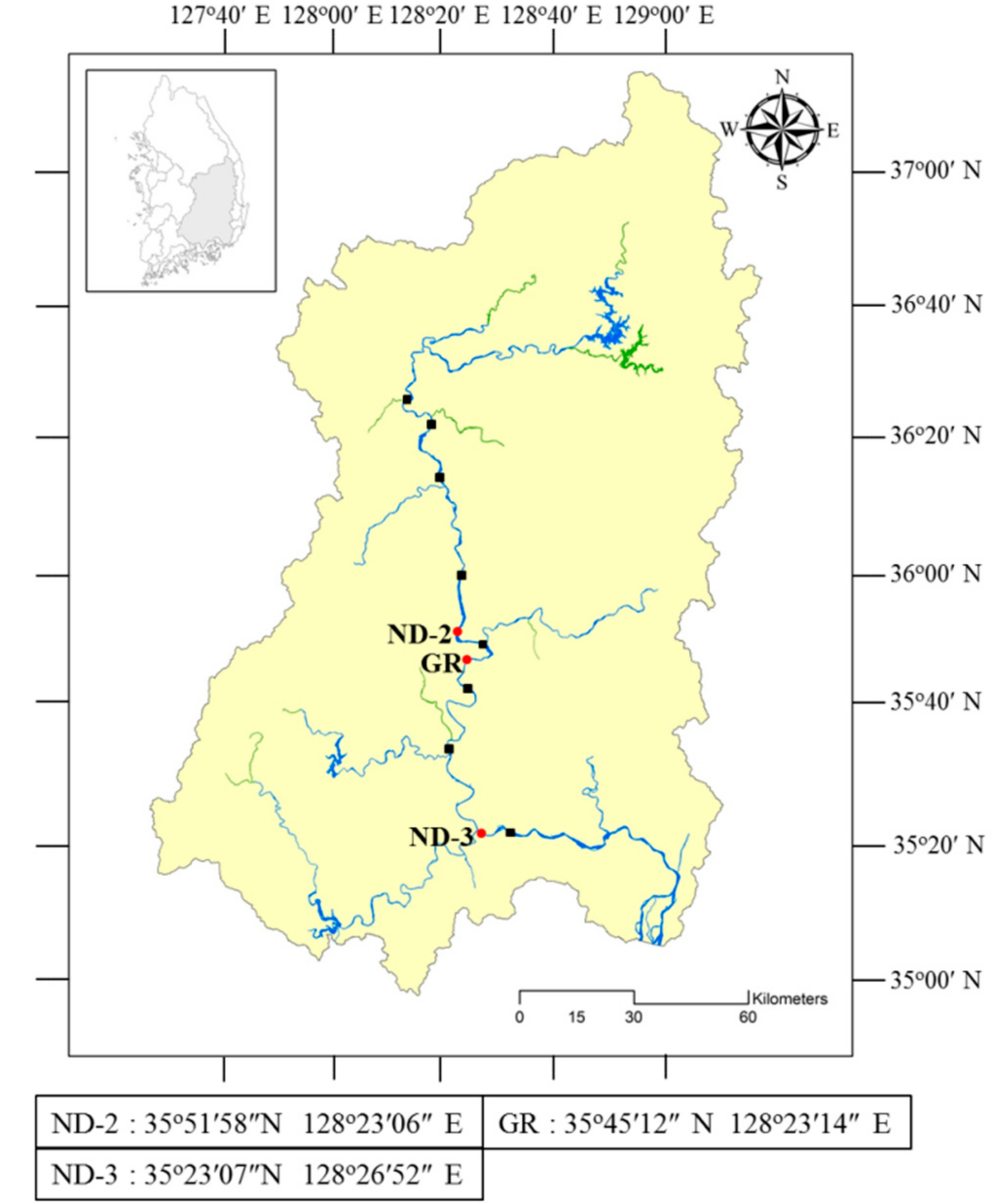
2.1. Sampling and Phytoplankton Analysis

For molecular verification of cyanobacterial secondary metabolites production potential, surface water samples were collected monthly from two stations (ND-2, ND-3) in the Nakdong River, where the Harmful Cyanobacterial Bloom Alert System of the Nakdong River has been operating [22] from April to December, 2016 (Figure 1). The water samples were filtered through a 0.45 μm MicronSep nitrocellulose membrane disk (GVS Life Sciences, Findlay, OH, USA) and then stored in a cryogenic freezer (−80 °C) until molecular analysis. The water samples for phytoplankton analysis were preserved by adding Lugol’s solution (final concentration 0.3%). Phytoplankton was classified at the genus or species level under a microscope [23,24], counted using a Sedgwick-Rafter counting chamber and expressed as phytoplankton cell density as cells per mL of water.

2.2. Isolation and Culture of Aphanizomenon Strains

To isolate Aphanizomenon strains, cyanoHAB samples were collected at ND-2 and ND-3 on November 26 and 28, 2016 and at GR station on June 8, 2017 (Figure 1). The collected samples were examined under an optical microscope (Zeiss Axio Imager M1, Göttingen, Germany) using a cavity microscopic slide glass on which a droplet of CB liquid medium [25] for cyanobacteria was placed, and a colony of Aphanizomenon spp. was identified. The identified colonies were diluted with CB liquid medium until a single trichome was separated. The single trichome identified by the microscope was transferred to a culture bottle containing a CB liquid medium and cultured by shaking in an incubator at 20 °C with 40 μEm−2s−1 and L/D =14/10 h conditions. For each isolated strain, images were taken under the microscope, and cell size was measured, followed by subculture in a CB liquid medium every 4 weeks. To isolate genomic DNA from the Aphanizomenon isolates, the strains cultured in the CB liquid medium were filtered through a 0.45-μm MicronSep nitrocellulose membrane disk (GVS Life Sciences, Findley, OH, USA) and then stored in a cryogenic freezer (−80 °C) until use.

2.3. Isolation of Genomic DNA

Genomic DNA was isolated from cyanobacterial cells using the QIAGEN DNeasy Plant Mini Kit (QIAGEN, Hilden, Germany). A lysis buffer was added to filtered samples stored in the cryogenic freezer (−80 °C), followed by disruption of cells with a tissue grinder, and genomic DNA was extracted by centrifugation of the disrupted cells within the mini spin column. The concentration and purity of the DNA were measured using an Infinite M200 PRO Microplate Reader (Tecan Austria GmbH, Grödig, Austria). The isolated genomic DNA was stored at −20 °C.

2.4. Phylogenetic Analysis

For phylogenetic analysis of Aphanizomenon isolates, the nucleotide sequences of 16S rRNA and internal transcribed spacer region (ITS) between 16S rRNA and 23S rRNA were used. For sequence analyses of 16S rRNA and ITS region, CY16S-F1/CY16S-R2 and CY16S-F3/CY16S-R1 primers and APHITS-F/ATPITS-R primers were designed based on the sequence information of 16S rRNA and ITS in A. flos-aquae obtained from the GenBank database registered in the US NCBI (National Center for Biotechnology Information, NIH, Bethesda, MD, USA) (Table 1).
For amplification of the 16S rRNA and ITS region, PCR was performed at 95°C for 3 min, followed by 30 cycles of 95 °C for 30 s, 55 °C for 50 s and 72 °C for 1 min, and then treatment at 72 °C for 10 min, thus producing 840 and 763 bp PCR products for 16S rRNA and 312 and 541 bp PCR products for ITS. For nucleotide sequence analysis, the amplified PCR products were subjected to TA cloning using M13F and M13R primers, and then sent to Macrogen (Seoul, Korea) for sequencing. Multiple alignment of sequencing results with ClustalW in the BioEdit program resulted in a 1463 bp sequence for 16S rRNA and 266 and 495 bp sequences for ITS.
For phylogenetic analysis of 16S rRNA and 16S-23S rRNA ITS sequences, the sequence information of the 16S rRNA for Aphanizomenon spp., Dolichospermum spp., and Cylindrospermopsis raciborskii (C. raciborskii) and the ITS information of Aphanizomenon spp. were downloaded from the GenBank database of NCBI. Nucleotide sequences with a length of at least 1400 bp and 260 bp were selected for 16S rRNA and ITS, respectively, followed by ClustalW multiple alignment in the BioEdit program [32], which was used to construct a phylogenetic tree by the Neighbor-joining (NJ), Maximum likelihood (ML), and Maximum parsimony (MP) analyses using MEGA6 [33], with 1000 repetitions of bootstrap. For NJ, Kimura 2-parameter method [34] was used as a nucleotide substitution model. For ML, Kimura 2-parameter method with nearest-neighbor-interchange (NNI) heuristic search method was used. Microcystis novacekii (Accession No. AB035551) and Microcystis aeruginosa (Accession No. AB638217) was used as an outgroup to build 16S rRNA and 16S-23S rRNA ITS phylogenetic tree, respectively.

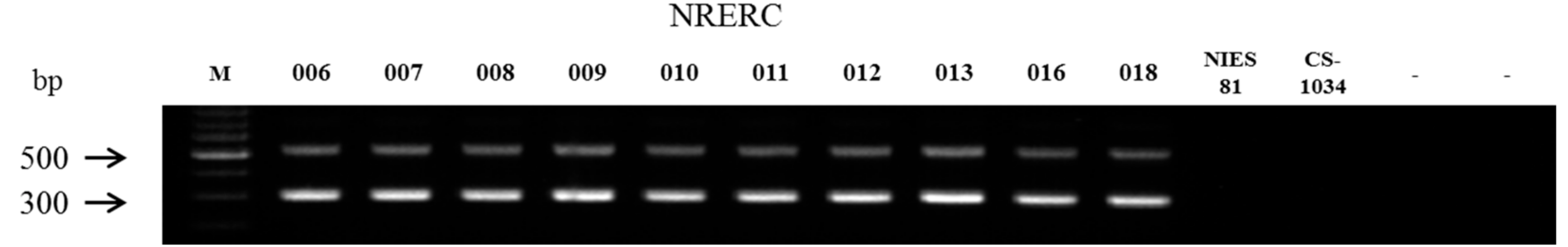
2.5. Molecular Assays for Off-Flavor and Toxin Production

To evaluate the off-flavor-producing potential of field samples and Aphanizomenon isolates from the Nakdong River, the biosynthetic gene of geosmin (geoA) was analyzed. Some of the biosynthetic genes of MCs (mcyA-Cd, mcyB), SXTs (sxtA, sxtI) and CYNs (cyrA, cyrJ), were also analyzed. PCR was performed on the DNA extracts using the primer pairs presented in Table 1. The PCR reaction mixture was composed of 1.0 μL of DNA template, 12.5 μL of EmeraldAmp PCR Master Mix (Takara, Kusatsu, Japan), and 1 μL of primers (10 pmol) in a final volume of 25 μL. The PCR conditions for gene amplification were according to the protocols described by the authors (Table 1).
The positive control used for verification of geoA gene was from Dolichospermum planctonicum (D. planctonicum, basionym Anabaena planctonica) NRERC-101 (Nakdong River Environment Research Center, NRERC, Goryeong, Korea), isolated from the Nakdong River, which had been confirmed to produce geosmin by instrumental analysis (gas chromatography/mass spectrometry, GC/MS) [35]. The positive control for mcyA-Cd and mcyB in the MC synthetase gene cluster was M. flos-aquae NRERC-205 (NRERC), which was isolated from the Nakdong River and had been confirmed to produce MCs through instrumental analysis (liquid chromatography/mass spectrophotometry, LC/MS). The genomic DNA of SXT-producing A. gracile NH-5 [18] was used as a positive control for sxtA and sxtI in the SXT gene cluster. The genomic DNA of CYN-producing C. raciborskii (CS-1101, Australian National Algae Culture Collection, ANACC, CSIRO, Clayton South, Australia) was used as a positive control for cyrA and cyrJ among the CYN gene cluster. The genomic DNA of Escherichia coli 5299 (Korean Collection for Type Culture, KCTC, Daejeon, Korea) and deionized water (DW) were used as negative controls and subjected to PCR amplification under the same condition used for amplification of each target gene. The size of the PCR products was checked by gel electrophoresis using 3% and 1.5% agarose gels and SiZer-50 (iNtRON, 24072) and SiZer-100 (iNtRON, 24073) DNA markers, respectively.

3. Results

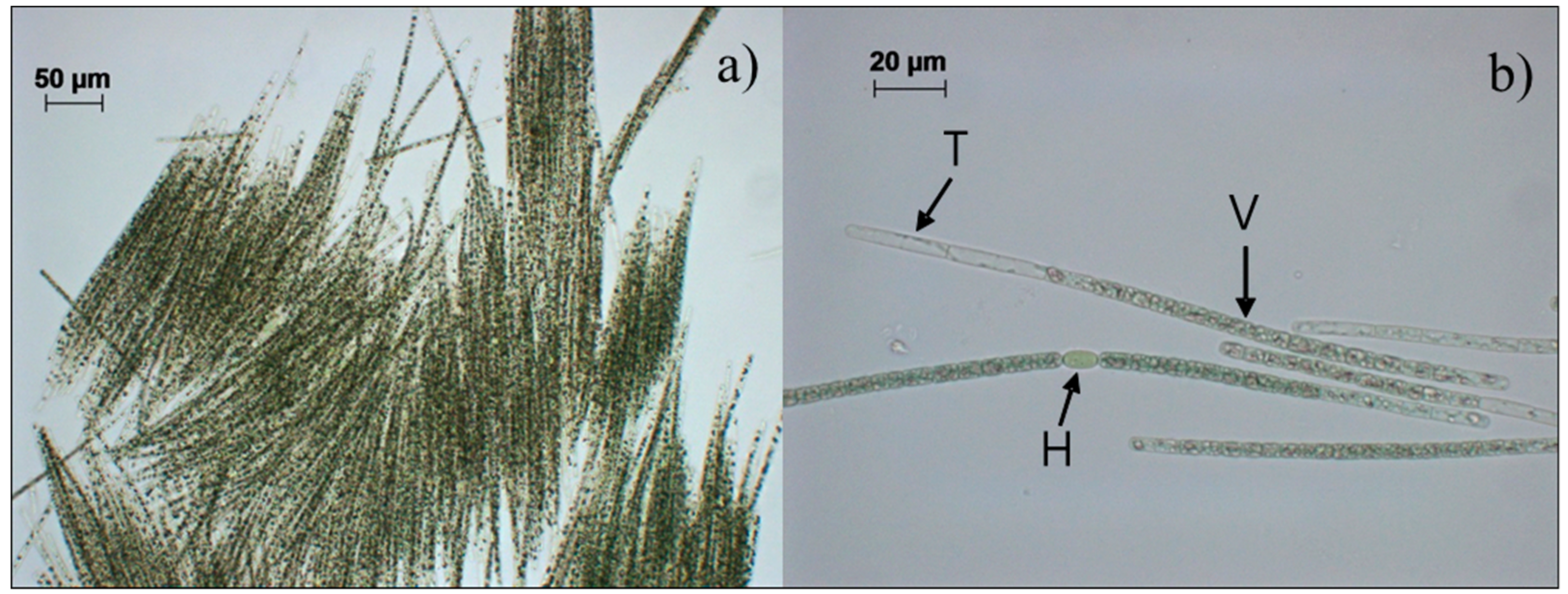
3.1. Identification of Aphanizomenon Isolates by Morphological Characteristics

Ten strains were isolated from CyanoHAB samples from the Nakdong River. Based on their morphological characteristics such as band-like fascicles with multiple trichomes, and almost hyaline apical cells, usually with characteristic remnants of cytoplasm in the form of an irregular lengthwise string, heterocytes solitary in a trichome, cylindrical, and akinetes, long cylindrical, distant from heterocytes [20], all strains were identified as A. flos-aquae Ralfs ex Bornet et Flahault 1888 (Figure 2, Table 2). All isolates were designated as NRERC-006, -007, -008, -009, -010, -011, -012, -013, -016 and -018 by the NRERC culture collection (NRERC, Goryeong, Korea). The size of vegetative cells of A. flos-aquae isolates in the present study was 4–10.6 × 3.5–5.8 μm.

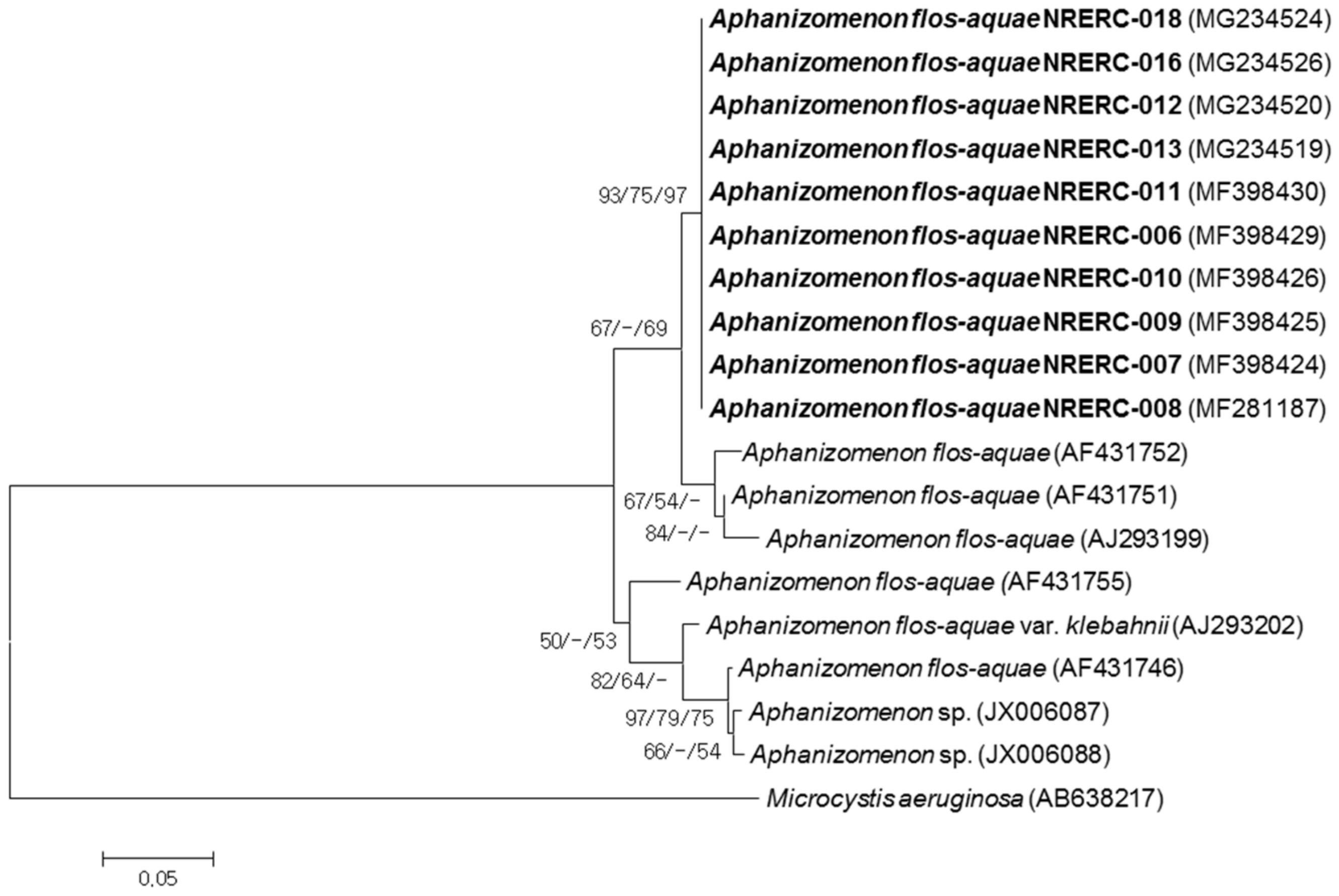
3.2. Phylogenetic Analyses of 16S rRNA and ITS Sequences

For phylogenetic analysis using 16S rRNA sequences from the ten strains of A. flos-aquae, 1463 bp of the 16S rRNA sequences were obtained using the CY16S-F1/CY16S-R2 and CY16S-F3/CY16S-R1 primers designed in the present study, and the results were registered in the GenBank database of NCBI (accession numbers are listed in Table 2).
BLAST analysis was performed with nucleotide sequences, including over 1400 bp from Aphanizomenon spp. and from similar species registered in the GenBank database of NCBI as well as the nucleotide sequences of the ten A. flos-aquae strains isolated in the present study. In the analysis, the ten isolates were classified as A. flos-aquae. They also showed over 99% sequence homology with 16S rRNA sequences from other A. flos-aquae strains (NCBI GenBank Accession No. KY327795, KY327796) isolated from the Nakdong River. Among the A. flos-aquae strains isolated in other countries, the 16S rRNA sequences of the present study showed 99% sequence homology with strains isolated from France (NCBI GenBank Accession No. AJ293123), Finland (AJ630441), and the UK (HE975013), and 98% sequence homology with those from Germany (KM019949), Lithuania (KM363406), and Thailand (AB862162).
Overall branching pattern was similar in all the phylogenetic analyses (NJ, ML, MP). The strains isolated from the Nakdong River, including the A. flos-aquae isolates in the present study were supported by bootstrap values up to 80% and formed a single cluster. This was different from the clusters of A. flos-aquae or A. gracile isolated in other countries with 98~100% bootstrap support. In addition, Dolichospermum strains, Anabaena strains and Sphaerospermopsis aphanizomenoides (synonym, Aphanizomenon aphanizomenoides) were classified into a different group with >99% bootstrap support (Supplementary Material Figure S1).
In the present study, a primer set (APHITS-F/ATPITS-R primers) was designed to amplify the 16S-23S rRNA ITS region in the A. flos-aquae strains and to analyze its nucleotide sequence, which identified two characteristic types of bands with a size of 266 bp (type-1) and 495 bp (type-2) (Figure 3, Supplementary Material Table S1). The results were deposited in the GenBank database of NCBI (Table 2). It was previously reported that A. flos-aquae isolated in the Baltic Sea also had two ITS types of different sizes [36]. The ITS nucleotide sequences of the ten A. flos-aquae isolates in the present study showed 100% homology regardless of the collection site and date. However, when A. gracile (formerly A. flos-aquae f. gracile) NIES81 and Chrysosporum ovalisporum (Ch. ovalisporum, synonym Aphanizomenon ovalisporum) CS-1034 were subjected to PCR amplification under the same conditions, no PCR product was generated (Figure 3).
Of the two 16S-23S rRNA ITS types, the nucleotide sequence of ITS type-2, the longer ITS, was registered in the GenBank database of NCBI for the first time. Because the sequences of ITS type-2 of other A. flos-aquae isolates were not available in the database, we attempted to search for the ITS nucleotide sequence of A. flos-aquae in the GenBank database of NCBI, using the 266-bp DNA sequence of ITS type-1 as a query. To date, no ITS DNA sequence of A. flos-aquae isolated in Korean water systems is present in the GenBank database of NCBI except for that of the ten isolates in the present study. Hence, the ITS nucleotide sequences of A. flos-aquae strains isolated in Europe and the US were used to construct a phylogenetic tree using the NJ, ML and MP analyses. As a result, the A. flos-aquae strains isolated in the present study formed a cluster, and some strains isolated in Finland and France were separated as several clusters with >50% support (Figure 4). BLAST analysis found 89, 97, 94 and 93% sequence homology with ITS regions from A. flos-aquae strains isolated in Finland (AF431746, AF431751, AF431752, and AF431755, respectively), whereas there was 96% and 89% sequence homology with strains isolated in France (AJ293199 and AJ293202, respectively). In addition, there was 89% sequence homology with the strains from the US (JX006087 and JX006088).

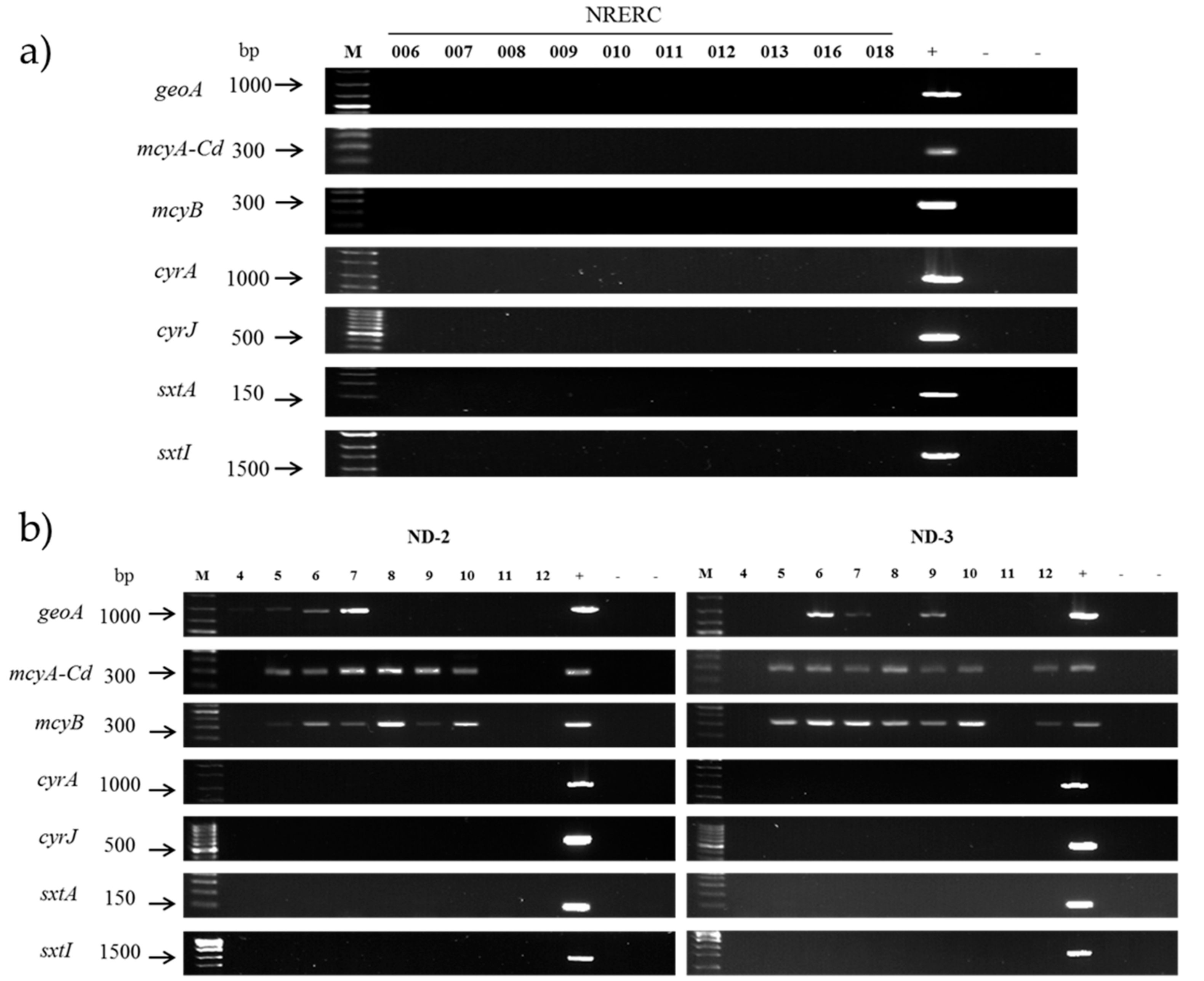
3.3. Evaluation of Secondary Metabolite Production Potential

To examine the potential of the A. flos-aquae strains to produce geosmin, PCR amplification was conducted with geo78F/geo982R primers for the geoA gene, but the 905 bp band was not found during electrophoresis. In contrast, PCR amplification using genomic DNA of D. planctonicum NRERC-101 resulted in the 905 bp band (Figure 5a). When the DNA sequences of the PCR product were BLAST searched in GenBank, they showed 99% homology with that of Anabaena ucrainica CHAB2155 (Accession No. HQ404997), confirming the presence of the geosmin synthase gene [35]. As a negative control, the genomic DNA of E. coli yielded no band in the corresponding size on electrophoresis. Therefore, it was confirmed that the ten A. flos-aquae strains isolated in the present study had no geosmin synthase gene.
The genomic DNA of the A. flos-aquae isolates was subjected to PCR using the mcyA-Cd and mcyB primer sets, which failed to amplify the 303 and 320 bp expected band. In contrast, PCR with positive control (M. flos-aquae NRERC-205) showed 303 and 320 bp PCR products on electrophoresis (Figure 5a). The PCR products of mcyB gene from M. flos-aquae NRERC-205 were sequenced and then searched against GenBank database using BLAST, which found 98% homology with the mcyB of M. aeruginosa UV027 (Accession No. AY034602) and M. aeruginosa PCC 7813 (Accession No. AY034601), indicating that the PCR product could be mcyB. Negative controls using E. coli and distilled water yielded no band on electrophoresis as in the A. flos-aquae isolates. From these results, it was confirmed that the ten A. flos-aquae isolates had no mcyA-Cd and mcyB.
The genomic DNA of the A. flos-aquae isolates was subjected to PCR using the sxtA and sxtI primer set, which failed to amplify the 125- and 1669 bp expected bands as judged based on electrophoresis. In contrast, PCR amplification using genomic DNA of STX producing A. gracile NH-5 [17] under the same conditions produced the 125- and 1669 bp bands (Figure 5a), indicating that the ten A. flos-aquae isolates had no sxtA and sxtI.
To examine the presence of the CYN biosynthesis gene in A. flos-aquae, cyrA and cyrJ primer sets were used for PCR. The A. flos-aquae isolates failed to yield 1105–1179 bp and 578 bp bands that corresponded to the expected sizes of the PCR products. However, CYN-producing C. raciborskii CS-1101 produced 1105–1179 bp and 578 bp PCR products. The PCR products from C. raciborskii CS-1101 were sequenced and searched in GenBank database using BLAST. CyrA showed 99% homology with cyrA of C. raciborskii cyDB-1 (Accession No. KJ139707) and 96% homology with aoa gene cluster of A. ovalisporum (Accession No. AF395828). CyrJ showed 99% homology with cyrJ of C. raciborskii cyDB-1 (Accession No. KJ139693) and 94% homology with cyrJ of A. ovalisporum ILC-164 (Accession No. KJ139694). Therefore, it was confirmed that the ten A. flos-aquae isolates had no cyrA and cyrJ.
Monthly water samples from April to December in 2016 in the Nakdong River showed higher cyanobacterial cell density in the ND-3 station on the downstream than ND-2 station on the upstream (Table 3). Aphanizomenon spp. was present in all months in the range of 43–15,080 cells mL−1 in the ND-3 station. The cell density of Microcystis spp. was 18–71,625 cells mL−1 in the ND-2 and ND-3 stations but was not detected in April and November in both stations. Dolichospermum spp. showed the lowest cell density among the three genera (23–602 cells mL−1). PCR amplification for the geosmin synthase gene using genomic DNA of water samples yielded a 905 bp band in June and July in the ND-2 and ND-3 stations and September in the ND-3 station, corresponding to the months in which Dolichospermum spp. occurred in the river (Figure 5b). The PCR products were sequenced, searched in GenBank database using BLAST and identified as the geosmin synthase gene. All samples, positive for geosmin synthase gene, contained Dolichospermum spp. and Microcystis spp., but Aphanizomenon spp. presence did not correspond to the geosmin synthase gene. All samples, that did not yield PCR product did not contain Dolichospermum spp. regardless of Microcystis spp. and Aphanizomenon spp. cell density. Thus, the geosmin synthase gene of the samples might originate from co-occurring Dolichospermum spp. and not from Aphanizomenon spp.
PCR amplification with primer sets targeting partial regions of the MC synthetase gene cluster, the SXT gene cluster, and the CYN gene cluster using genomic DNA from water samples showed different results according to the target gene (Figure 5b). Samples collected from May to October showed the 303 and 320 bp band of the mcyA-Cd and mcyB PCR product in the ND-2 and ND-3 stations similar to the sample collected in December in the ND-3 station. All samples showing the mcyA-Cd and mcyB PCR product contained Microcystis spp., whereas samples without mcyA-Cd and mcyB PCR product did not contain Microcystis spp. The samples collected in ND-2 in May, July, August and October did not contain Aphanizomenon spp., whereas the samples collected in April in both stations and in November in the ND-3 station did not yield the mcyA-Cd and mcyB PCR product. Combined with the results of Aphanizomenon and Microcystis isolates, the mcyA-Cd and mcyB gene of the field samples might originate from co-occurring Microcystis spp. and not from Aphanizomenon spp. None of the water samples showed the expected band of PCR products of the sxtA, sxtI, cyrA and cyrJ primer sets on electrophoresis (Figure 5b) regardless of the cell density of major cyanobacterial genera.

4. Discussion

A. flos-aquae is known to be the Aphanizomenon species blooming in the mainstream of the Nakdong River during spring and autumn [8,9]. Thus, the ten Aphanizomenon isolates, which were identified as A. flos-aquae by morphological and phylogenetic analysis, were confirmed as the Aphanizomenon species causing cyanoHAB in the Nakdong River. However, there were only 1–3 bp differences in the 16S rRNA nucleotide sequence and 100% sequence homology in the ITS among the ten strains isolated from three separate sites in spring and winter blooms, indicating a highly similar genotype of A. flos-aquae regardless of location and season of blooming in the Nakdong River.
The 16S rRNA gene is considered a molecular clock in the bacterial evolutionary process and is the most widely used genetic marker [11]. However BLAST analysis showed little variation in the 16S rRNA sequences of A. flos-aquae, regardless of geographical location. Increasing attention has been paid to the ITS region between 16S and 23S rRNA regions because it has a higher discerning power for the classification of allied species of cyanobacteria due to its high sequence variation [37]. In particular, the nucleotide sequence of rRNA ITS in cyanobacteria is an appropriate region to investigate intra-species variations [38]. The 89–97% sequence homology among the A. flos-aquae strains from the Nakdong River, Europe, and the US indicates higher intra-species variation of the nucleotide sequence in ITS regions than in 16S rRNA which showed 98–99% sequence homology among the A. flos-aquae strains. Thus, it is expected that the ITS region can be used to trace the geographical variations of A. flos-aquae nucleotide sequences. Additionally, the ITS primer set (APHITS-F/ATPITS-R) designed in the present study amplified the ITS nucleotide sequence in A. flos-aquae, but not those in A. gracile NIES81 and Ch. ovalisporum CS-1034 indicating this ITS primer set has potential to use molecular marker for A. flos-aquae molecular identification, but needs confirmation through the amplification with various Aphanizomenon species and other Nostocalean species.
The number of earthy/musty odor and taste episodes in treated water and customer complaints have increased worldwide, including Korea, during the last two decades [39,40]. Geosmin has been designated as a water quality surveillance indicator for drinking water in Korea. The occurrence of geosmin in the mainstream section of the Nakdong River, which serves as a drinking water source, is a significant issue, and determining the causative cyanobacteria can be the first step to control off-flavor substances. The level of geosmin in the Nakdong River is particularly high in spring [22]. During this season, the dominant cyanobacteria are mostly Aphanizomenon spp. and Dolichospermum spp., of which Aphanizomenon spp. has a higher dominance and has recently began blooming from autumn to early winter, in addition to spring [9,10]. Hence, if Aphanizomenon spp. possess the geosmin synthase gene, it will likely increase the level of off-flavor in the water. However, between Aphanizomenon spp. and Dolichospermum spp. isolated from the Nakdong River, the present study found the geosmin synthase gene only in Dolichospermum spp. and not in Aphanizomenon spp. Moreover, the geosmin synthase gene was amplified in the field samples with Dolichospermum spp. and not in the field samples with only Aphanizomenon spp. (April in the ND-2 and ND-3, November in the ND-3) Therefore, it appears that Dolichospermum spp. cause geosmin contamination in the Nakdong River and that there should be a low potential for off-flavor during the blooms of A. flos-aquae.
The genus Aphanizomenon comprises a number of cyanoHAB, producing cyanotoxins in water bodies worldwide [11]. At present, MCs are the most representative cyanobacterial toxins that have been listed in the drinking water guidelines published by the WHO, and MC-LR has been designated as a water quality surveillance indicator for drinking water in Korea. It has been identified worldwide that MCs are produced by cyanobacteria, including A. flos-aquae, that cause harmful cyanobacterial blooms [1]. The production of microcystins and the presence of mcyA gene in the A. flos-aquae in Korea has also been reported [21]. However, considering the results of the present study, it seems that A. flos-aquae, which is dominant and blooms in the Nakdong River during spring, autumn and early winter, has low potential to produce MCs.
SxtA encodes polyketide synthase which initiates STX biosynthesis, and it has been detected not only in STX producing strains, but also in STX non-producing strains similar to sxtG, sxtH and sxtX [41]. In contrast, sxtI has been detected only in STX-producing strains to date in genus Aphanizomenon with exception in genus Cylindrospermopsis and Lyngbya [42,43]; therefore, it was suggested as a marker for verification of STX-producing Aphanizomenon strains [11]. To date, various filamentous cyanobacterial species including A. gracile, Cuspidothrix issatschenkoi, C. raciborskii, D. circinale, and Geitlerinema spp. have been identified to produce PSP toxins including STX, whereas toxin production by A. flos-aquae is controversial [11]. Since the late 19th century, A. flos-aquae has been reported as a potential producer of cyanobacterial toxins, and it was reported in 1967 for the first time that A. flos-aquae produces a toxin with a structure similar to that of STX produced by marine dinoflagellates [44]. Although A. flos-aquae was identified as a producer of STX in the US in 1980 [16], morphological and genetic re-evaluation subsequently revealed that it was not A. flos-aquae, but A. gracile [18]. Thus, it remains unclear whether A. flos-aquae produces PSP toxins. Nevertheless, given that neither sxtA nor sxtI of SXT biosynthesis gene cluster was confirmed in the A. flos-aquae strains isolated in the present study or in the field samples from ND-2 and ND-3 stations containing Aphanizomenon spp., there likely is a low potential of STX occurrence in A. flos-aquae blooms in the surveyed stations of the Nakdong River during spring, autumn, and winter.
Cylindrospermopsin (CYN) is a cytotoxin reported to occur mostly at high water temperature and was first identified to be produced by C. raciborskii, which mostly blooms in tropical water bodies; however, it has also been reported in field samples or isolates from Oceania, Asia, America, and Europe [45,46]. The most representative CYN-producing cyanobacteria is C. raciborskii; however, CYN is also known to be produced by major species in the order Nostocales, including Ch. ovalisporum, A. flos-aquae, Anabaena bergii and Raphidiopsis curvata as well as Umezakia natans (order Stigonematales) and Lyngbya wollei (order Oscillatoriales) [43]. Thus, it is critical to investigate whether CYN is present in the Nakdong River, where A. flos-aquae is dominant. In the CYN gene cluster, sulfotransferase genes are present in only CYN-producing strains. Thus, cyrJ should be a useful marker for CYN toxicity, because it encodes adenylyl sulfate kinase, which is an enzyme catalyzing the biosynthesis of 3′-phosphoadenosine-5′-phosphosulfate (PAPS), the sulfate donor of sulfotransferase [47]. Therefore, given that the PCR product of cyrJ was not detected in the A. flos-aquae isolates in the present study or in field samples containing Aphanizomenon spp., the A. flos-aquae in the Nakdong River should have low potential to produce CYN.

5. Conclusions

In this study, ten A. flos-aquae strains were isolated from the cyanoHAB in the Nakdong River, which were identified by using morphological and phylogenetic analysis and show highly similar genotype regardless of location and season of blooming in the Nakdong River. 89–97% sequence homology among the A. flos-aquae strains from the Nakdong River and Europe and the US indicates high intra-species variation of the nucleotide sequence in ITS, which can be used to trace the geographical variations of A. flos-aquae nucleotide sequences.
We further verified the potential of cyanoHAB-causing A. flos-aquae strains and water samples containing cyanobacterial species in producing prominent harmful secondary metabolites using PCR methods with specific primer sets and all isolates and the water sample containing only Aphanizomenon spp. had a low potential to produce these metabolites. However, water samples containing other cyanobacterial species such as Microcystis and Dolichospermum spp. possessed mcyA and mcyB of the MC synthetase gene cluster and the geosmin synthase gene respectively. Therefore, bloom-forming cyanobacterial strains in addition to Aphanizomenon spp. isolated during other times and sites as well as field samples collected during the time when such cyanobacteria are dominant should be continuously monitored for the presence of the harmful secondary metabolites and their biosynthesis genes to minimize the risk to both water quality and public health. The PCR assay with the specific primer sets of this study is expected to provide a rapid and sensitive means for early detection of A. flos-aquae and potentially harmful populations of cyanobacteria, especially in the initial stage of cyanobacterial growth in water supplies.

Supplementary Materials

The following are available online at https://www.mdpi.com/1660-4601/15/8/1739/s1, Figure S1: Neighbor-Joining (NJ) tree of 16S rRNA sequences from Aphanizomenon flos-aquae and Nostocales cyanobacterial strains, Table S1: Nucleotide sequence of 16S-23S rRNA ITS of Aphanizomenon flos-aquae isolated from the Nakdong River.

Author Contributions

All authors contributed substantially to this study. Conceptualization, H.-K.P. and H.-J.L.; Methodology, J.O., S.-H.L. and H.-K P.; Isolation and Cultivation of cyanobacteria, H.-K.P. and H.-J.L.; Molecular Analysis, M.-A.K. and I.-S.K.; Field Investigation, H.-J.L.; Writing-Original Draft Preparation, H.-K.P.; Writing-Review & Editing, H.-K.P. and J.O.; Project Administration and Funding Acquisition, H.-K.P.

Funding

This research was supported by the National Institute of Environment Research (NIER) funded by the Ministry of Environment (MOE) of the Republic of Korea, grant number NIER-2018-03-03-003.

Acknowledgments

The authors thank anonymous reviewers for valuable comments improving the manuscript. Special thanks are addressed to Renhui Li of CAS, China for distribution of A. gracile NH-5 culture.

Conflicts of Interest

The authors declare no conflict of interest.

References

  1. Paerl, H.W.; Otten, T.G. Harmful Cyanobacterial Blooms: Causes, Consequences, and Controls. Microb. Ecol. 2013, 65, 995–1010. [Google Scholar] [CrossRef] [PubMed]
  2. Mantzouki, E. Temperature Effects Explain Continental Scale Distribution of Cyanobacterial Toxins. Toxins 2018, 10, 156. [Google Scholar] [CrossRef] [PubMed]
  3. Cirés, S.; Wörmer, L.; Ballot, A.; Agha, R.; Wiedner, C.; Velázquez, D.; Casero, M.C.; Quesada, A. Phylogeography of cylindrospermopsin and paralytic shellfish toxin-producing Nostocales Cyanobacteria from Mediterranean Europe (Spain). Appl. Environ. Microbiol. 2014, 80, 1359–1370. [Google Scholar] [CrossRef] [PubMed]
  4. Budzyńska, A.; Goldyn, R. Domination of invasive Nostocales (Cyanoprokaryota) at 52°N latitude. Phycol. Res. 2017, 65, 322–332. [Google Scholar] [CrossRef]
  5. Nakdong River Environment Research Center (NRERC). Aquatic Ecosystem Monitoring in Weir of the Nakdong River; Nakdong River Environment Research Center: Goryung, Korea, 2015. [Google Scholar]
  6. Ha, K.; Cho, E.A.; Kim, H.W.; Joo, G.J. Microcystis bloom formation in the lower Nakdong River, South Korea; Importance of hydrodynamics and nutrient loading. Mar. Freshw. Res. 1999, 50, 89–94. [Google Scholar] [CrossRef]
  7. Jeong, K.-S.; Kin, D.-K.; Whigham, P.; Joo, G.-J. Modelling Microcystis aeruginosa bloom dynamics in the Nakdong River by means of evolutionary computation and statistical approach. Ecol. Model. 2003, 161, 67–78. [Google Scholar] [CrossRef]
  8. Park, H.-K.; Shin, R.-Y.; Lee, H.-J.; Lee, K.-L.; Cheon, S.-U. Spatio-temporal characteristics of cyanobacterial communities in the middle-downstream of Nakdong River and Lake Dukdong. J. Korean Soc. Water Environ. 2015, 31, 286–294. [Google Scholar] [CrossRef]
  9. Ryu, H.-S.; Park, H.-K.; Lee, H.-J.; Shin, R.-Y.; Cheon, S.-U. Occurrence and succession pattern of cyanobacteria in the upper region of the Nakdong River; Factors influencing Aphanizomenon bloom. J. Korean Soc. Water Environ. 2016, 32, 52–59. [Google Scholar] [CrossRef]
  10. Park, H.-K.; Lee, H.-J.; Park, K.-J.; Cho, H.-Y.; Ryu, H.-S. Characteristics of cyanobacterial occurrence in the large river with consecutive weirs. In Proceedings of the 10th International Conference on Toxic Cyanobacteria—Cyanobacteria and Cyanotoxins; From Research to Risk Management, Wuhan, China, 23–28 October 2016; p. 35. [Google Scholar]
  11. Cirés, S.; Ballot, A. A review of the phylogeny, ecology and toxin production of bloom-forming Aphanizomenon spp. and related species within the Nostocales (cyanobacteria). Harmful Algae 2016, 54, 21–43. [Google Scholar] [CrossRef] [PubMed]
  12. Yamamoto, Y. Environmental factors that determine the occurrence and seasonal dynamics of Aphanizomenon flos-aquae. J. Limnol. 2009, 68, 122–132. [Google Scholar] [CrossRef]
  13. Üveges, V.; Tapolczai, K.; Krienitz, L.; Padisák, J. Photosynthetic characterisitics and physiological plasticity of an Aphanizomenon flos-aquae (Cyanobacteria, Nostocaceae) winter bloom in a deep oligo-mesotrophic lake (Lake Stechlin, Germany). Hydrobiologia 2016, 698, 263–272. [Google Scholar] [CrossRef]
  14. Suurnäkki, S.; Gomez-Saez, G.V.; Rantala-Ylinen, A.; Jokela, J.; Fewer, D.P.; Sivonen, K. Identification of geosmin and 2-methylisoborneol in cyanobacteria and molecular detection methods for the producers of these compounds. Water Res. 2015, 68, 56–66. [Google Scholar] [CrossRef] [PubMed]
  15. Alam, M.; Euler, K.L. Chemical studies on toxins from the blue-green alga Aphanizomenon flos-aquae. In The Water Environment; Springer: New York, NY, USA, 1981; pp. 405–414. [Google Scholar]
  16. Mahmood, N.A.; Carmichael, W.W. Paralytic shellfish poisons produced by the freshwater Cyanobacterium Aphanizomenon flos-aquae NH-5. Toxicon 1986, 24, 175–186. [Google Scholar] [CrossRef]
  17. Jung, J.-M.; Lee, Y.-J.; Pak, H.-K.; Jung, E.-Y.; Joo, G.-J. The analysis of cyanobacterial neurotoxins by high-performance liquid chromatography-mass spectrometry. Algae 2003, 18, 233–238. [Google Scholar] [CrossRef]
  18. Li, R.H.; Carmichael, W.W.; Liu, Y.D.; Watanabe, M.M. Taxonomic re-evaluation of Aphanizomenon flos-aquae NH-5 based on morphology and 16S rRNA gene sequences. Hydrobiologia 2000, 438, 99–105. [Google Scholar] [CrossRef]
  19. Li, R.H.; Carmichael, W.W.; Pereira, P. Morphological and 16S rRNA gene evidence for reclassification of the paralytic shellfish toxin producing Aphanizomenon flos-aquae LMECYA 31 as Aphanizomenon issatschenkoi (Cyanophyceae). J. Phycol. 2003, 39, 289–295. [Google Scholar] [CrossRef]
  20. Komárek, J. Freshwater Flora of Central Europe Vol. 19/3 Cyanoprokaryota, 3rd Part: Heterocytous Genera; Springer: Berlin, Germany, 2013. [Google Scholar]
  21. Kim, K.; Park, C.; Yoon, Y.; Hwang, S.-J. Harmful Cyanobacterial Material Production in the North Han River (South Korea): Genetic Potential and Temperature-Dependent Propertieds. Int. J. Environ. Res. Public Health 2018, 15, 444. [Google Scholar]
  22. Water Information System. Available online: Water.nier.go.kr/front/algaeInfo/algaeGuide.jsp (accessed on 15 November 2016).
  23. Komárek, J.; Anagnostidis, K. Freshwater Flora of Central Europe Vol. 19/1 Cyanoprokaryota, 1st Part: Chroococcales; Gustav Fischer Verlag: Jena, Germany, 1999. [Google Scholar]
  24. John, D.M.; Whitton, B.M.; Brook, A.J. The Freshwater Algal Flora of the British Isles; An Identification Guide to Freshwater and Terrestrial Algae, 2nd ed.; Cambridge University Press: Cambridge, UK, 2011. [Google Scholar]
  25. Watanabe, M.M.; Nozaki, H. NIES-Collection List of Strains, 4th ed.; The National Institute for Environmental Studies, The Environmental Agency: Tsukuba, Japan, 1994. [Google Scholar]
  26. Hisbergues, M.; Christiansen, G.; Rouhiainen, L.; Sivonen, K.; Börner, T. PCR-based identification of microcystin-producing genotypes of different cyanobacterial genera. Arch. Microbiol. 2003, 180, 402–410. [Google Scholar] [CrossRef] [PubMed]
  27. Nonneman, D.; Zimba, D.V. A PCR-based test to assess the potential for microcystin occurrence in channel catfish production ponds. J. Phycol. 2002, 38, 230–233. [Google Scholar] [CrossRef]
  28. Kellmann, R.; Mills, T.; Neilan, B.A. Functional modeling and phylogenetic distribution of putative cylindrospermopsin biosynthesis enzymes. J. Mol. Evol. 2006, 62, 267–280. [Google Scholar] [CrossRef] [PubMed]
  29. Mihali, T.K.; Kellmann, R.; Muenchhoff, J.; Barrow, K.D.; Neilan, B.A. Characterization of the gene cluster responsible for cylindrospermopsin biosynthesis. Appl. Environ. Microbiol. 2008, 74, 716–722. [Google Scholar] [CrossRef] [PubMed]
  30. Tebrineh, J.A.; Mihali, T.K.; Pomati, F.; Neilan, B.A. Detection of saxitoxin-producing cyanobacteria and Anabaena circinalis in environmental water blooms by quantitative PCR. Appl. Environ. Microbiol. 2010, 76, 7836–7842. [Google Scholar] [CrossRef] [PubMed]
  31. Gkelis, S.; Papadimitriou, T.; Zaoutsos, N.; Leonardos, I. Anthropogenic and climate-induced change favors toxic cyanobacteria blooms: Evidence from monitoring a highly eutrophic, urban mediterranean lake. Harmful Algae 2014, 39, 322–333. [Google Scholar] [CrossRef]
  32. Hall, T.A. BioEdit: A user-friendly biological sequence alignment editor and analysis program for Windows 95/98/NT. Nucleic Acids Symp. Ser. 1999, 41, 95–98. [Google Scholar]
  33. Tamura, K.; Steche, G.; Peterson, D.; Filipski, A.; Kumar, S. MEGA6: Molecular evolutionary genetics analysis Version 6.0. Mol. Biol. Evol. 2013, 30, 2725–2729. [Google Scholar] [CrossRef] [PubMed]
  34. Kimura, M. A simple method for estimating evolutionary rate of base substitutions through comparative studies of nucleotide sequences. J. Mol. Evol. 1980, 16, 111–120. [Google Scholar] [CrossRef] [PubMed]
  35. Lee, H.-J.; Park, H.-K.; Hong, D.-G.; Choi, S.-K. Characteristics of geosmin production by Dolichospermum planctonicum isolated from the Nakdong River, Korea. In Proceedings of the 2017 International Symposium in Commemoration of the 50th Anniversary of Korean Society of Limnoly, Harmful Algal Blooms: Understanding and Management, Gwangju, Korea, 18 October 2017; p. 35. [Google Scholar]
  36. Laamanen, M.J.; Forsström, L.; Sivonen, K. Diversity of Aphanizomenon flos-aquae (Cyanobacterium) populations along a Baltic Sea salinity gradient. Appl. Environ. Microbiol. 2002, 68, 5296–5303. [Google Scholar] [CrossRef] [PubMed]
  37. Janse, I.; Meima, M.; Kardinaal, W.E.A.; Zwart, G. High-resolution differentiation of cyanobacteria by using rRNA-internal transcribed spacer denaturing gradient gel electrophoresis. Appl. Environ. Microbiol. 2003, 69, 6634–6643. [Google Scholar] [CrossRef] [PubMed]
  38. Nelissen, B.; Wilmotte, A.; Neefs, J.M.; De Wachter, R. Phylogenetic relationships among filamentous helical cyanobacteria investigated on the basis of 16S ribosomal RNA gene sequence analysis. Syst. Appl. Microb. 1994, 17, 206–210. [Google Scholar] [CrossRef]
  39. Omur-Ozbek, P. Global Taste and Odor Survey of Water Utilities, Final Report (October 12); The American Water Works Association Taste and Odor Committee: Denver, CO, USA, 2012; p. 43. [Google Scholar]
  40. You, K.-A.; Byeon, M.-S.; Youn, S.-J.; Hwang, S.-J.; Rhew, D.-H. Growth characteristics of blue-green algae (Anabaena spiroides) causing tastes and odors in the North-Han River, Korea. Korean J. Ecol. Environ. 2013, 46, 135–144. [Google Scholar] [CrossRef]
  41. Casero, M.C.; Ballot, A.; Agha, R.; Quesada, A.; Cirés, S. Characterization of saxitoxin production and release and phylogeny of sxt genes in paralytic shellfish poisoning toxin-producing Aphanizomenon gracile. Harmful Algae 2014, 37, 28–37. [Google Scholar] [CrossRef]
  42. Hoff-Risseti, C.; Dörr, F.A.; Schaker, P.D.C.; Pinto, E.; Werner, V.R.; Fiore, M.F. Cylindrospermopsin and saxitoxin synthetase genes in Cylindrospermopsis raciborskii strains from Brazilian freshwater. PLoS ONE 2013, 8, e74238. [Google Scholar] [CrossRef] [PubMed]
  43. Mihali, T.K.; Carmichael, W.W.; Neilan, B.A. A putative gene cluster from a Lyngbya wollei bloom that encodes paralytic shellfish toxin biosynthesis. PLoS ONE 2011, 6, e14657. [Google Scholar] [CrossRef] [PubMed]
  44. Sawyer, P.J.; Gentile, J.H.; Sasner, J.J. Demonstration of a toxin from Aphanizomenon flos-aquae (L.) Ralfs. Can. J. Microbiol. 1968, 14, 1199–1204. [Google Scholar] [CrossRef] [PubMed]
  45. Moreira, C.; Azevedo, J.; Antunes, A.; Vasconcelos, V. Cylindrospermopsin: Occurrence, methods of detection and toxicology. J. Appl. Microbiol. 2012, 114, 605–620. [Google Scholar] [CrossRef] [PubMed]
  46. Jiang, Y.; Xiao, P.; Yu, G.; Shao, J.; Liu, D.; Azevedo, S.M.F.; Li, R. Sporadic distribution and distinctive variations of cylindrospermopsin genes in cyanobacterial strains and environmental samples from Chinese freshwater bodies. Appl. Environ. Microbiol. 2014, 80, 5219–5230. [Google Scholar] [CrossRef] [PubMed]
  47. Ballot, A.; Ramm, J.; Eundberget, T.; Kaplan-Levy, R.N.; Hada, O.; Sukenik, A.; Wiedner, C. Occurrence of non-cylindrospermopsin-producing Aphanizomenon ovalisporum and Anabaena bergii in Lake Kinneret (Israel). J. Plankton Res. 2011, 33, 1736–1746. [Google Scholar] [CrossRef]
Figure 1. Map of the Nakdong River basin area. Red circles indicate the sampling stations and black squares indicate the weirs in the Nakdong River.
Ijerph 15 01739 g001
Figure 2. Microscope images of Aphanizomenon flos-aquae NRERC-006: (a) band-like fascicles, (b) single trichomes with vegetative cells (V), heterocytes (H), and typical apical cells (T).
Ijerph 15 01739 g002
Figure 3. Gel electrophoresis image of ITS PCR products from Aphanizomenon flos-aquae NRERC–006 to NRERC–018, Aphanizomenon gracile strain NIES81, and Chrysosporum ovalisporum strain CS-1034. Upper bands show 541 bp ITS (type-2) PCR products and lower bands show 312 bp ITS (type-1) PCR products. M indicates SiZer-100 DNA marker.
Ijerph 15 01739 g003
Figure 4. Neighbor-joining (NJ) tree of 16S-23S rRNA ITS sequences from Aphanizomenon flos-aquae strains. Microcystis aeruginosa (AB638217) was used as an outgroup. The numbers at the nodes indicate levels of bootstrap support (%) based on Neighbor-joining (NJ), Maximum likelihood (ML), and Maximum parsimony (MP) analysis of 1,000 resampled datasets. Only bootstrap values (NJ/ML/MP) above 50% are shown at the nodes. Strains of the present study are marked in bold and the GenBank accession numbers for ITS sequences are shown in parentheses. The scale bar indicates substitutions per site.
Ijerph 15 01739 g004
Figure 5. Gel electrophoresis image of the PCR products of the geosmin (geoA), microcystin (mcyA-Cd, mcyB), saxitoxin (sxtA, sxtI), and cylindrospermopsin (cyrA, cyrJ) biosynthesis genes from (a) Aphanizomenon flos-aquae isolates (NRERC–006 to NRERC–018) and (b) water samples. Sample number of ND-2 and ND-3 indicates the month of sampling date (Table 3). Positive (+) and negative (−) controls for each gene are described in the Materials and Methods section. M indicates SiZer-100 DNA marker.
Ijerph 15 01739 g005
Table 1. List of primers used in this study.
Target GenePrimer SetSequence (5′—3′)Fragment Length (bp)References
16S rRNACY16S-F1CAGGATGAACGCTGGCGG840This study
CY16S-R2GAATGGGATTAGATACCCCA
CY16S-F3AAAGGAGGTGATCCAGCCAC763This study
CY16S-R1TCCTTTGAGTTTCACAGTTG
ITSAPHITS-FACAAGGTAGCCGTACCGGAA312/541This study
APHITS-RACAATTTCTTTTGCTTCCAC
geoAgeo78FGCATTCCAAAGCCTGGGCTTA905[14]
geo982RATCGCATGTGCCACTCGTGAC
mcyA-CdmcyAFAAAAGTGTTTTATTAGCGGCTCAT303[26]
mcyARAAAATTAAAAGCCGTATCAAA
mcyBmcyB2959FTGGGAAGATGTTCTTCAGGTATCCAA320[27]
mcyB3278RAGAGTGGAAACAATATGATAAGCT AC
cyrACYLAT-FATTGTAAATAGCTGGAATGAGTGG1105–1179[28]
CYLAT-RTTAGGGAAGTAATCTTCACAG
cyrJcynsulfFACTTCTCTCCTTTCCCTATC578[29]
cylnamRGAGTGAAAATGCGTAGAACTTG
sxtAsxtA-FGATGACGGAGTATTTGAAGC125[30]
sxtA-RCTGCATCTTCTGGACGGTAA
sxtIsxtI-FGCTTACTACCACGATAGTGCTGCCG1669[31]
sxtI-RGGTTCGCCGCGGACATTAAA
Table 2. Information for Aphanizomenon flos-aquae strains isolated from the Nakdong River and NCBI GenBank Accession Numbers of the 16S rRNA and 16S-23S rRNA ITS sequences of each strain.
Strain No. (NRERC-)Scientific NameCollection DateIsolation SourceNCBI GenBank Accession Number
16S rRNAITS Type-1ITS Type-2
006Aphanizomenon flos-aquae26 November 2016ND-2MF362173MF398429MF398428
007Aphanizomenon flos-aquae28 November 2016ND-3MF362174MF398424MF398427
008Aphanizomenon flos-aquae26 November 2016ND-2KY952630MF281187MF322523
009Aphanizomenon flos-aquae28 November 2016ND-3MF362175MF398425MF398423
010Aphanizomenon flos-aquae28 November 2016ND-3MF362177MF398426MF398422
011Aphanizomenon flos-aquae26 November 2016ND-2MF362176MF398430MF422681
012Aphanizomenon flos-aquae28 November 2016ND-3MG234522MG234520MG234525
013Aphanizomenon flos-aquae28 November 2016ND-3MG234521MG234519MG234527
016Aphanizomenon flos-aquae8 June 2017GRMG234523MG234526MG234528
018Aphanizomenon flos-aquae8 June 2017GRMG234518MG234524MG234529
Table 3. Cell densities (cells mL−1) of three major cyanobacterial genera of monthly water samples from the Nakdong River.
Sampling DateND-2ND-3
Aphanizomenon spp.Microcystis spp.Dolichospermum spp.Aphanizomenon spp.Microcystis spp.Dolichospermum spp.
25 April 201619NDND43NDND
30 May 2016ND1525ND70017,280ND
20 June 2016600186831415,0807143602
25 July 2016ND12233653630375
22 August 2016ND3270ND218471,625ND
26 September 20163583ND2028980
24 October 2016ND18ND6451ND
28 November 2016NDNDND1532NDND
26 December 2016NDNDND22750ND

