Article

Chitosan-Mediated Expression of Caenorhabditis elegans fat-1 and fat-2 in Sparus aurata: Short-Term Effects on the Hepatic Fatty Acid Profile, Intermediary Metabolism, and Proinflammatory Factors

1
Secció de Bioquímica i Biologia Molecular, Departament de Bioquímica i Fisiologia, Facultat de Farmàcia i Ciències de l’Alimentació, Universitat de Barcelona, Joan XXIII 27-31, 08028 Barcelona, Spain
2
Departament de Farmàcia i Tecnologia Farmacèutica, i Fisicoquímica, Facultat de Farmàcia i Ciències de l’Alimentació, Universitat de Barcelona, Joan XXIII 27-31, 08028 Barcelona, Spain
3
Departament d’Enginyeria Química, Universitat Politècnica de Catalunya, Diagonal 647, 08028 Barcelona, Spain
*
Author to whom correspondence should be addressed.
Mar. Drugs 2025, 23(11), 434; https://doi.org/10.3390/md23110434
Submission received: 21 October 2025 / Revised: 9 November 2025 / Accepted: 11 November 2025 / Published: 13 November 2025

Abstract

A single dose of chitosan-tripolyphosphate (TPP) nanoparticles carrying expression plasmids for fish codon-optimized Caenorhabditis elegans fat-1 and fat-2 was intraperitoneally administered to gilthead seabream (Sparus aurata) to stimulate the biosynthesis of omega-3 long-chain polyunsaturated fatty acids (n-3 LC-PUFA) and evaluate subsequent short-term effects on liver intermediary metabolism and immunity. Seventy-two hours post-injection, the upregulation of fat-1 elevated eicosapentaenoic acid (EPA), docosahexaenoic acid (DHA), and total n-3 fatty acids in the liver, while fat-2 enhanced DHA and n-3 fatty acids. Co-expression of fat-1 and fat-2 increased EPA, DHA, PUFA, and the total n-6 and n-3 LC-PUFA, while reducing plasma triglycerides. The expression of fat-1 and fat-2 suppressed hepatic lipogenesis by downregulating srebf1 and pparg, and consequently key genes in fatty acid synthesis (acaca, acacb, fasn, scd1, and fads2). In contrast, the co-expression of fat-1 and fat-2 upregulated hnf4a, chrebp, and pfkl, a rate-limiting enzyme in glycolysis. Furthermore, fat-1 and fat-2 reduced hepatic proinflammatory markers such as tnfa and nfkb1. In addition to enhancing EPA and DHA biosynthesis, promoting glycolysis, and suppressing lipogenesis, our findings suggest that the short-term expression of C. elegans fat-1 and fat-2 in the liver may also reduce inflammation and, therefore, could impact the health and growth performance of cultured fish.

1. Introduction

In recent years, the aquaculture industry has made intensive efforts to replace fish oil in aquafeeds, at least partially, with other fat sources, such as vegetable oils. However, plants contain higher levels of omega-6 fatty acids than omega-3 fatty acids and are devoid of omega-3 long-chain polyunsaturated fatty acids (n-3 LC-PUFA) such as eicosapentaenoic acid (EPA) and docosahexaenoic acid (DHA). As a result, the replacement of fish oil with vegetable oils significantly reduces n-3 LC-PUFA in fish flesh [1]. Although dietary n-3 LC-PUFA is required for optimal growth, marine fish, especially carnivorous fish, exhibit low ability to synthesize n-3 LC-PUFA [2]. In general, vertebrates are devoid of the Δ12/n-6 and Δ15/n-3 desaturases required to synthesize linoleic acid (LA) from oleic acid (OA) and α-linolenic acid (ALA) from LA. Therefore, vertebrates, including bony fish, can only synthesize a small portion of LC-PUFA at insufficient rates to meet physiological demands [3,4]. In addition to storing energy, LC-PUFA and their derivatives have pivotal roles in growth and development, serving as biologically active moieties in membrane phospholipids, being substrates for signalling molecules, and regulating gene transcription [5]. The mechanisms by which n-3 LC-PUFA regulate energy metabolism and immunity in fish, two processes closely linked to growth, have yet to be fully elucidated.
A competitive relationship has been found between the synthesis of n-3 and n-6 PUFA series due to shared desaturases and elongases in their respective biosynthetic pathways [6]. Given that oxidized derivatives of the n-6 LC-PUFA precursor, LA, are proinflammatory factors [7], and n-3 LC-PUFA are valued for their anti-inflammatory effects, the impact of the physiological ratio between n-3 and n-6 PUFA on inflammation is receiving increasing attention. Compared to vegetable oils, fish oil has a lower n-6 to n-3 PUFA ratio. Therefore, dietary replacement of fish oil with vegetable oils in marine fish was shown to upregulate the expression of proinflammatory genes such as myeloid differentiation primary response 88 (myd88), tumor necrosis factor a (tnfa), interleukin 1b (il1b), and the nuclear factor kappa-light-chain-enhancer of activated B cells (nfkb) [8,9].
Although absent in fish, Δ15/n-3 and Δ12/n-6 desaturases have been identified in some invertebrates such as the roundworm Caenorhabditis elegans, where they are referred to as fat-1 and fat-2, respectively. To alleviate the fish oil dependency of aquafeeds, the efficient conversion of n-6 PUFA to n-3 PUFA has been reported in transgenic models, such as zebrafish [10] and common carp [11], expressing codon-optimized C. elegans fat-1 and/or fat-2. The n-3 LC-PUFA content and n-6 PUFA to n-3 PUFA conversion were further intensified in zebrafish and pigs through the double transgenesis of fat-1 and fat-2 [1,12]. Nevertheless, the effects of C. elegans fat-1 and fat-2 on intermediary metabolism and inflammation in transgenic animals remain scarce.
Chitosan is a biomaterial derived from chitin, a natural polysaccharide that is a primary component of invertebrate exoskeletons and is abundantly found in nature. Low toxicity, biocompatibility, and biodegradability make chitosan a highly suitable carrier for delivering nucleic acids. Using sodium tripolyphosphate (TPP), ionic gelation of chitosan-TPP-DNA has garnered growing attention due to its mild processing conditions, nanoscale production capabilities, and cost-effectiveness in gene delivery for biotechnological applications in fish [13]. Gilthead seabream (Sparus aurata) is a carnivorous fish that accounted for 38.3% of total European aquaculture production of marine fish in 2022 (https://www.fao.org/fishery/en/fishstat, accessed on 9 January 2025). We recently demonstrated that regular supplementation with chitosan-TPP-DNA nanoparticles resulted in the long-term sustained hepatic expression of fish codon-optimized C. elegans fat-1 and fat-2, leading to enhanced n-3 LC-PUFA synthesis and improved growth performance in S. aurata [14]. To advance the current understanding of the temporal molecular events triggered by C. elegans fat-1 and fat-2 in the fish liver, the present study aimed to investigate the short-term effects of chitosan-mediated hepatic expression of fish codon-optimized C. elegans fat-1 and fat-2 on intermediary metabolism and inflammation in S. aurata.

2. Results

2.1. Effect of Intraperitoneal Administration of Chitosan-TPP Nanoparticles Encapsulating pSG5-FAT-1 and pSG5-FAT-2 on fat-1 and fat-2 mRNA Levels in S. aurata Liver

Chitosan-TPP was complexed with pSG5 (control), pSG5-FAT-1, pSG5-FAT-2, and pSG5-FAT-1 + pSG5-FAT-2 by ionic gelation [14]. No significant differences in particle size were found between naked chitosan-TPP (215 nm ± 20) and chitosan-TPP-DNA (263 nm ± 74), expressed as mean ± SEM (n = 3). DNA incorporation significantly decreased Z potential from 38 mV ± 1 to 12 mV ± 1, expressed as mean ± SEM (n = 3). Seventy-two hours after intraperitoneal administration of a single dose of chitosan-TPP nanoparticles encapsulating either pSG5-FAT-1 or pSG5-FAT-2, the mRNA levels of fish codon-optimized fat-1 and fat-2 were assayed by RT-qPCR in the liver of S. aurata. Compared with control fish (treated with empty plasmid, pSG5), the hepatic mRNA abundance of fat-1 in fish administered with pSG5-FAT-1 and pSG5-FAT-1 + pSG5-FAT-2 was 34.1- and 46.7-fold significantly higher, respectively, while fat-2 mRNA levels in fish treated with pSG5-FAT-2 and pSG5-FAT-1 + pSG5-FAT-2 increased 17.7- and 15.1-fold (Figure 1a,b).

2.2. Effect of Chitosan-Mediated Expression of fat-1 and fat-2 on S. aurata Serum Metabolites and Hepatic Fatty Acid Profile

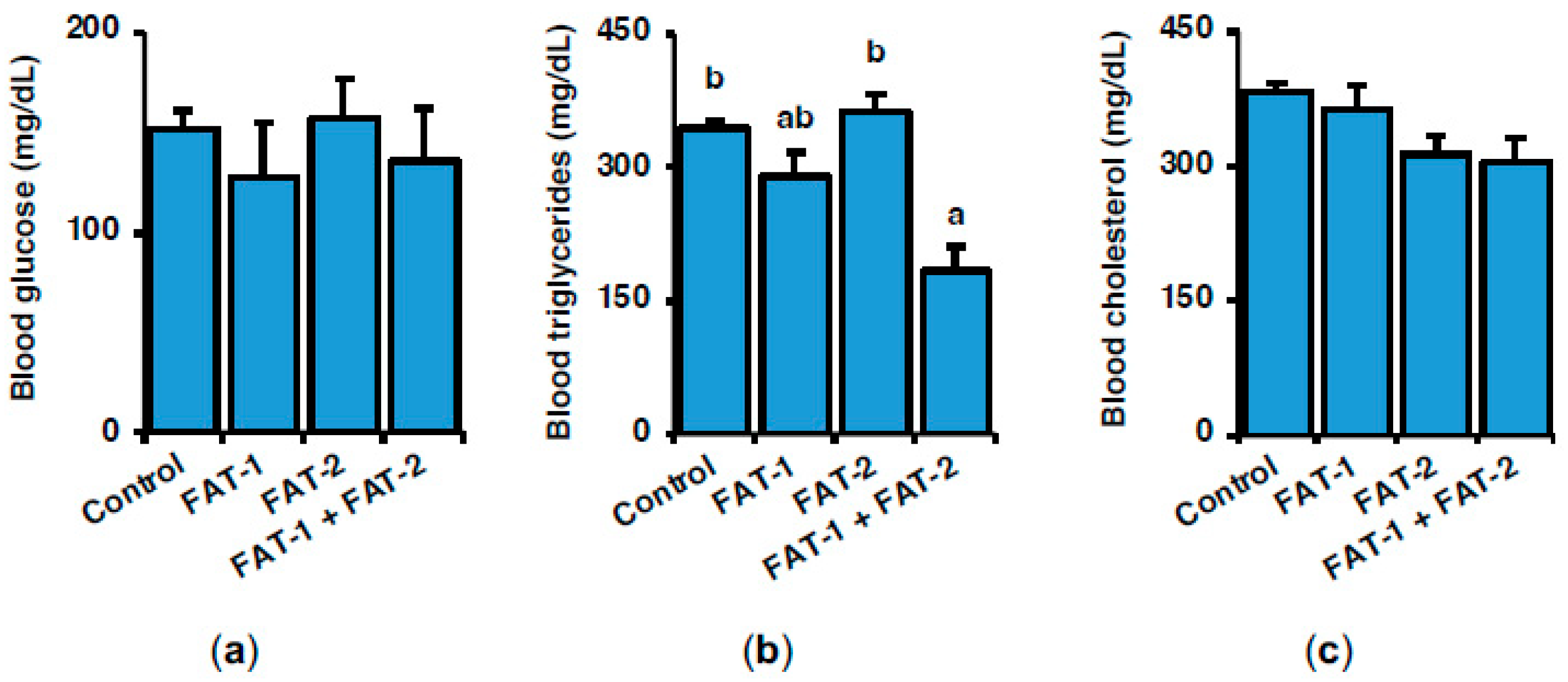
Figure 2 shows the effect of hepatic expression of fish codon-optimized fat-1 and fat-2 on serum metabolites, including glucose, triglycerides, and cholesterol, 72 h after intraperitoneal administration of a single dose of chitosan-TPP-DNA nanoparticles. Blood glucose and cholesterol were not significantly affected by any of the treatments (Figure 2a–c), while co-expression of fat-1 and fat-2 significantly decreased triglycerides to 53.4% of control levels (Figure 2b).
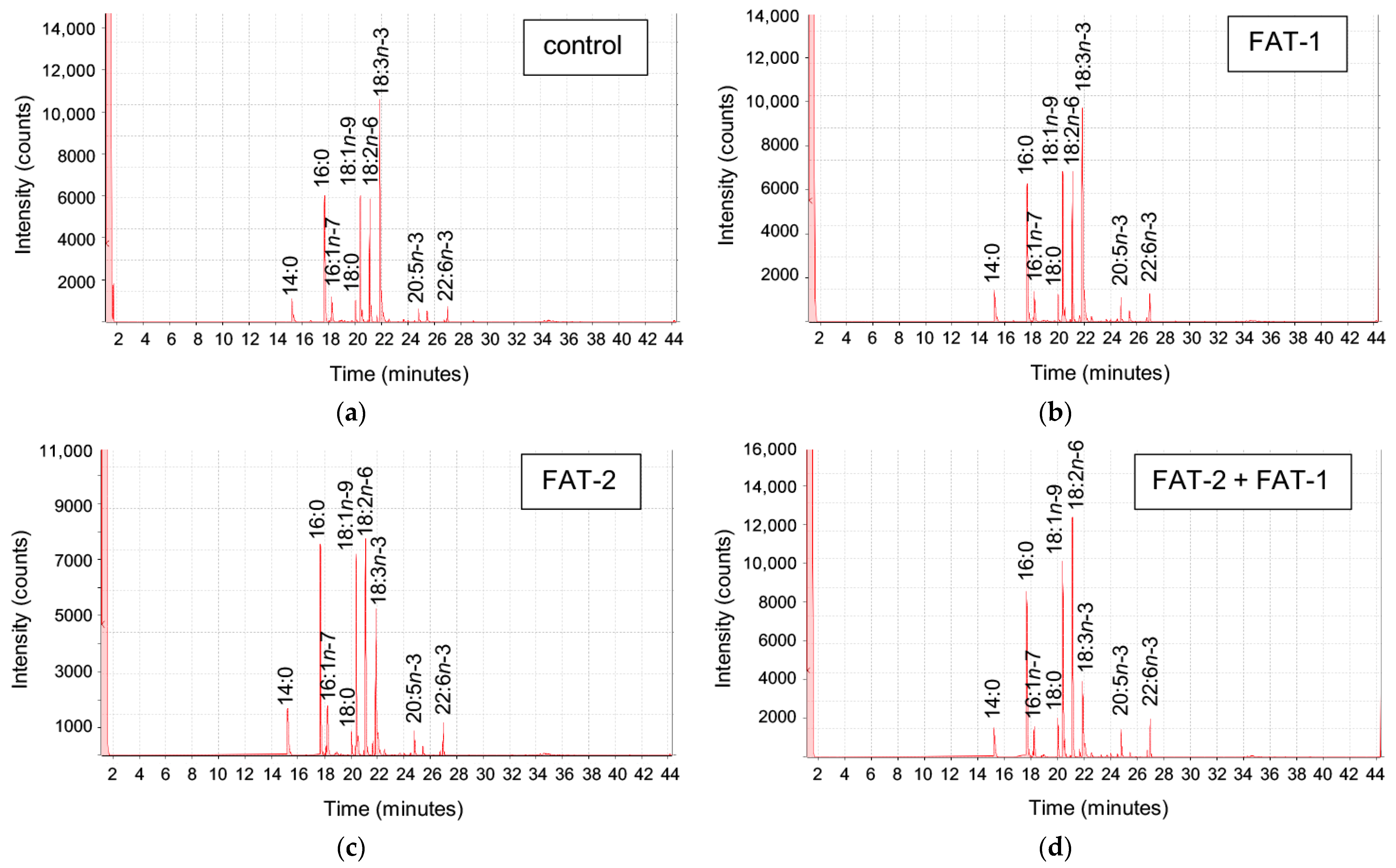
The effect of chitosan-TPP nanoparticles carrying pSG5 (control), pSG5-FAT-1, pSG5-FAT-2, and pSG5-FAT-1 + pSG5-FAT-2 on the fatty acid profile of S. aurata liver was analyzed 72 h post-administration (Figure 3).
Among fatty acids identified (30) and those representing higher than 1%, myristic acid (14:0), palmitoleic acid (16:1n-7), LA (18:2n-6c), EPA (20:5n-3), DHA (22:6n-3), PUFA, and total n-6 and n-3 fatty acids were significantly affected by the treatments. Specifically, fat-1 expression increased palmitoleic acid 1.6-fold and EPA, DHA, and total n-3 fatty acids 1.5-fold, while fat-2 increased palmitoleic acid (2.0-fold), DHA (1.6-fold), and total n-3 fatty acids (1.5-fold). Compared to the control fish, co-expression of fat-1 and fat-2 increased palmitoleic acid (1.8-fold), LA (1.5-fold), EPA (1.6-fold), DHA (2.0-fold), PUFA (1.5-fold), total n-6 fatty acids (1.5-fold), and total n-3 fatty acids (1.7-fold) (Table 1).

2.3. Effect of Chitosan-Mediated Expression of fat-1 and fat-2 on Key Genes Related to Lipid and Glucose Metabolism

To shed light on the short-term effects on lipogenic genes resulting from the hepatic expression of fish codon-optimized fat-1 and fat-2, the expression of key enzymes in fatty acid and LC-PUFA synthesis was assayed by RT-qPCR in S. aurata liver. Compared to controls, treatment with fat-1, fat-2 and fat-1 + fat-2 significantly downregulated lipogenic genes, including acetyl-CoA carboxylase 1 (acaca; to 6.8%, 3.4%, and 4.9% of control values, respectively), acetyl-CoA carboxylase 2 (acacb; to 57.7%, 68.7%, and 53.0%), fatty acid synthase (fasn; to 27.4%, 23.3%, and 21.5%), stearoyl-CoA desaturase-1a (scd1a; to 17.8%, 12.7%, and 22.9%), and acyl-CoA 6-desaturase (fads2; to 38.9%, 11.9%, and 36.8%) (Figure 4a–e). A similar non-significant trend was found for elongation of very long chain fatty acids protein 4a (elovl4a) and elongation of very long chain fatty acids protein 4b (elovl4b) (Figure 4f,g). No significant effects were observed in elongation of very long chain fatty acids protein 5 (elovl5) mRNA levels (Figure 4h). Although not statistically significant for fat-1 + fat-2, a trend toward decreased 3-hydroxy-3-methylglutaryl-coenzyme A reductase (hmgcr) expression to 20.1%, 43.4%, and 59.7% of the control values, respectively, was observed (Figure 4i).
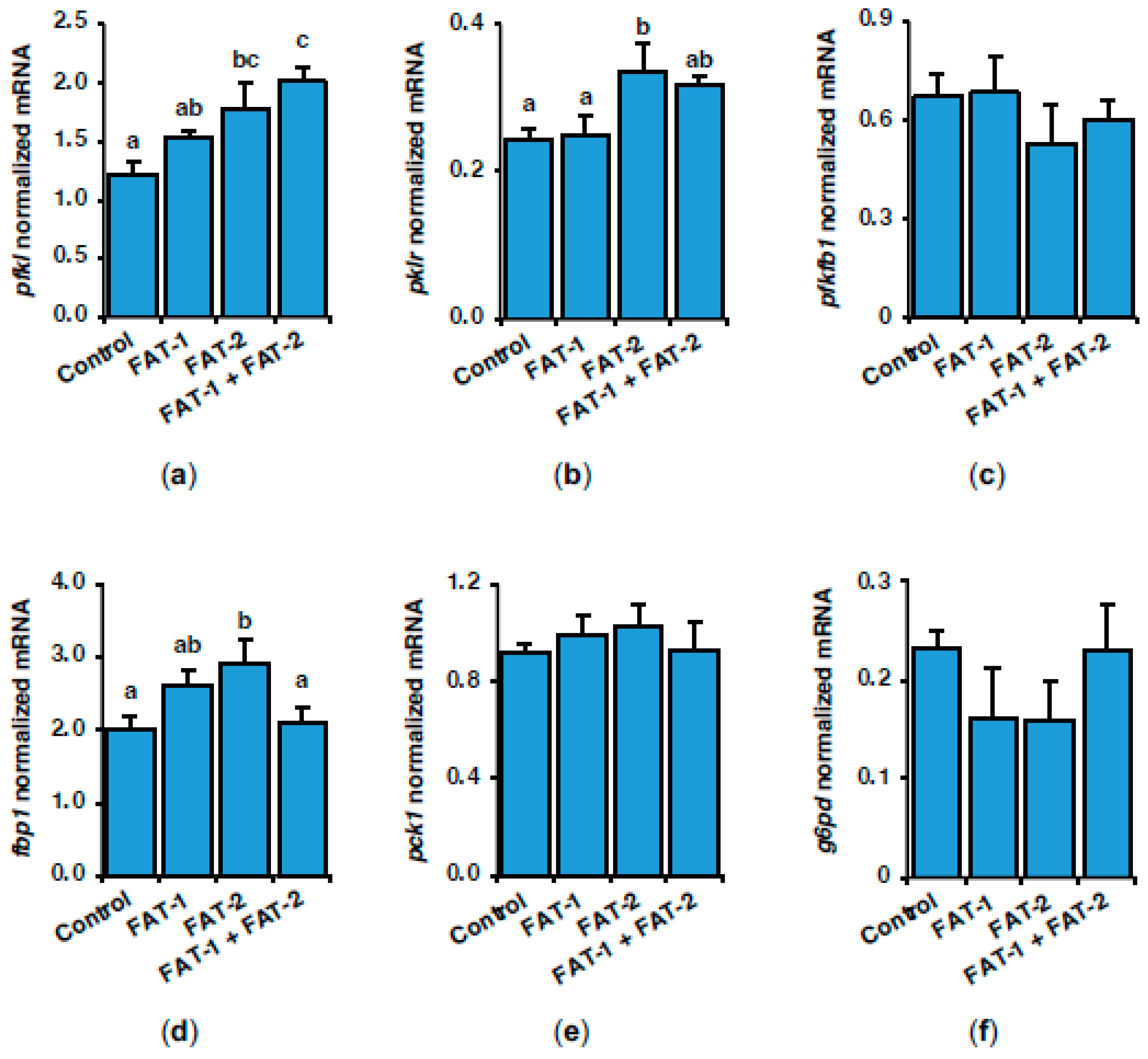
Given that hepatic lipogenesis is closely related to glucose metabolism, we also analyzed the expression of genes encoding key enzymes in glycolysis–gluconeogenesis and the pentose phosphate pathway (PPP) in response to the hepatic expression of fat-1 and fat-2 in S. aurata. Figure 5 shows the effect of fat-1 and fat-2 expression on liver mRNA levels of rate-limiting enzymes in glycolysis-gluconeogenesis and PPP, 72 h following nanoparticle administration. Compared to control values, the hepatic expression of fat-2 upregulated 6-phosphofructo-1-kinase (pfkl; 1.5-fold), pyruvate kinase (pklr; 1.4-fold), and fructose-1,6-bisphosphatase (fbp1; 1.5-fold). Co-expression of fat-1 and fat-2 also induced the mRNA abundance of pfkl (1.7-fold) (Figure 5a,b,d). No significant effects were observed in 6-phosphofructo-2-kinase/fructose-2,6-bisphosphatase (pfkfb1), phosphoenolpyruvate carboxykinase (pck1), and glucose-6-phosphate dehydrogenase (g6pd) expression (Figure 5c,e,f).
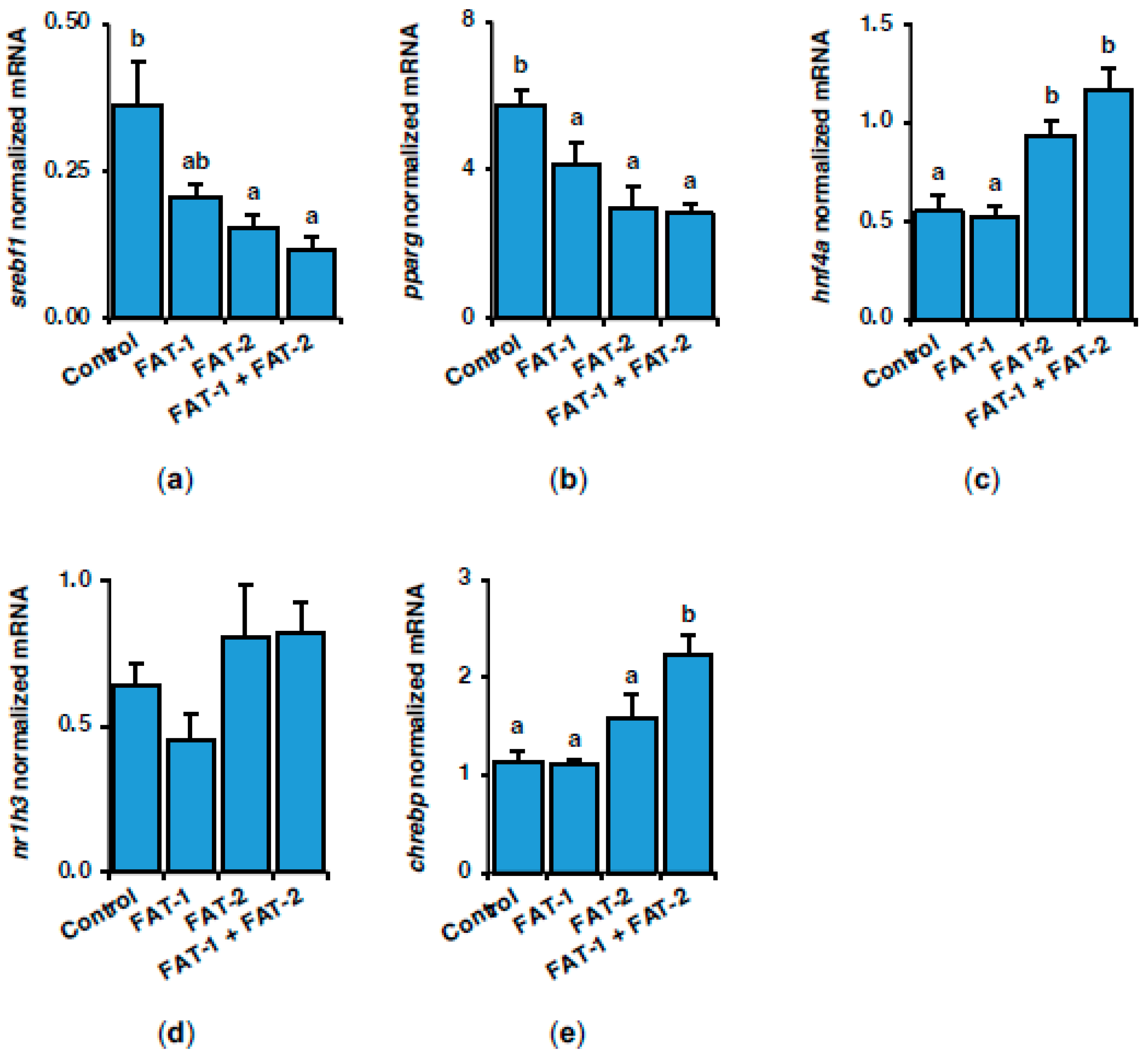
To further address the short-term effects resulting from expressing fish codon-optimized fat-1 and fat-2 in the liver of S. aurata, the mRNA levels of key transcription factors involved in controlling the expression of genes related to lipogenesis and glucose metabolism were also evaluated. Expression of fat-1, fat-2 and fat-1 and fat-2 significantly downregulated peroxisome proliferator-activated receptor gamma (pparg) to 72.3%, 52.0% and 49.0% of control values, respectively, while treatment with fat-2 and fat-1 + fat-2 also downregulated hepatic sterol regulatory element-binding protein 1 (srebf1) to 42.1% and 32.3% of control values (Figure 6a,b). Treatment with fat-2 and fat-1 + fat-2 increased the mRNA levels of hepatocyte nuclear factor 4 alpha (hnf4a) (1.7-fold and 2.1-fold of the control values, respectively) and caused similar non-significant effects on oxysterols receptor LXR-alpha (nr1h3) expression (Figure 6c,d). Co-expression of fat-1 and fat-2 upregulated 1.9-fold the expression levels of carbohydrate-responsive element-binding protein (chrebp) (Figure 6e).

2.4. Effect of Chitosan-Mediated Expression of fat-1 and fat-2 on Genes Related to Inflammation

Previous studies demonstrated that n-3 LC-PUFA inhibits the activation of immune cells. Hence, in the present study, the expression of genes related to inflammation was also addressed in the liver of S. aurata. Seventy-two hours following treatment with chitosan-TPP-DNA nanoparticles, a significant decrease in the mRNA levels of nuclear factor NF-kappa-B p105 subunit (nfkb1) was found in the liver of fish administered with fat-1, fat-2, or fat-1 + fat-2 (to 49.9%, 17.8%, and 27.7% of control values, respectively), while the expression of tumor necrosis factor (tnfa) also decreased after treatment with fat-2 and fat-1 + fat-2 nanoparticles (to 51.1% and 41.4% of control values, respectively) (Figure 7a,b). Although not significant, a similar trend was observed in regard to the expression of nuclear factor NF-kappa-B p100 subunit (nfkb2), interleukin-6 (il6), and myeloid differentiation primary response protein MyD88 (myd88), while no significant differences were noticed for interleukin-1 beta (il1b) (Figure 7c–f).

3. Discussion

To better understand the molecular events triggered by hepatic expression of C. elegans fat-1 and fat-2 in S. aurata, the effects of a single intraperitoneal administration of chitosan-TPP nanoparticles loaded with expression plasmids carrying fish codon-optimized C. elegans fat-1 and fat-2 were investigated on S. aurata intermediary metabolism and inflammation. Seventy-two hours post-treatment, a rapid enhancement of the expression levels of C. elegans fat-1 (Δ15/n-3 desaturase) and fat-2 (Δ12/n-6 desaturase) as well as the n-3 LC-PUFA content, particularly EPA and DHA, was observed in the liver of S. aurata. Studies examining the effects of n-3 LC-PUFAs on fish metabolism and immunity have primarily relied on dietary supplementation with varying levels of n-3 LC-PUFAs. However, this approach may underestimate differences in fatty acid oxidation, as n-3 LC-PUFAs are more prone to oxidation than unsaturated fatty acids. In contrast, the genetic approach used in this study prevented interferences due to fatty acid oxidation during diet manipulation and therefore avoided its putative negative effects on fish health and inflammation [15,16]. Moreover, unravelling the effects of n-3 LC-PUFAs on hepatic energy metabolism is essential for understanding and addressing growth retardation associated with decreased use of fish oil in aquafeeds [2].
It is well known that increased n-3 LC-PUFA values are associated with decreased plasma triglycerides and reduced cardiovascular risk in vertebrates [17]. Consistently, herein we showed that chitosan-mediated co-expression of fat-1 and fat-2 for 72 h in the fish liver caused an important reduction in blood triglycerides. Consistent with our results, fat-1 transgenic models in mice [18], pigs [19], cattle [20], and zebrafish [10], also showed reduced circulating triglycerides and n-3 LC-PUFA accumulation. The triglyceride-lowering effect of fat-1 and fat-2 could be attributed to decreased production of very low-density lipoprotein (VLDL)-loaded triglycerides and enhanced triglyceride clearance, processes that may be facilitated by elevated n-3 LC-PUFA levels, as previously described in mice [17]. The fat-1 transgenic models are also associated with reduced blood cholesterol [10,18,20]. Although not significant, a similar trend was found in fish expressing fat-2 and fat-1 + fat-2. Indeed, n-3 LC-PUFA was shown to decrease total cholesterol and low-density lipoprotein (LDL)-loaded cholesterol, while increasing high-density lipoprotein (HDL)-loaded cholesterol in mammals [21]. In the present study, glycemia was not significantly affected by any of the treatments assayed. However, the effect of n-3 LC-PUFA on blood glucose levels may depend on the fish species. Similarly to S. aurata, orange-spotted grouper (Epinephelus coioides) fed a soybean oil-based diet showed only marginal differences in blood glucose levels compared to fish fed fish oil-based diets [22]. In contrast, large yellow croaker (Larimichthys crocea) receiving 100% soybean oil as dietary lipid source showed higher blood glucose and lower hepatic glycogen levels than fish fed a fish oil diet [23].
Mutations in the C. elegans fat-1 gene were shown to decrease 18:3n-3, 20:4n-3, and 20:5n-3 and increase 18:2n-6, 20:3n-6, and 20:4n-6 proportions [24], indicating its activity at Δ15 and Δ17 fatty acid substrates. Regarding fat-2, heterologous expression revealed its ability to sequentially insert double bonds at the Δ12 and Δ15 positions of fatty acids when a Δ9 double bond preexisted [25]. In line with these observations, herein the expression of fat-1 increased the proportions of EPA, DHA, and total n-3 LC-PUFA, while fat-2 also increased DHA and total n-3 LC-PUFA. Co-expression of fat-1 and fat-2 elevated EPA, DHA, total PUFA, and total n-3 and n-6 LC-PUFA, while a decreasing trend was found for the n-6 to n-3 ratio in S. aurata liver. Consistently, fat-1 and fat-2 transgenesis also enhances n-3 LC-PUFA synthesis in mammals and zebrafish [1,12,18,20,26].
To address the short-term effects of C. elegans fat-1 and fat-2 on transcription factors playing a key role in controlling the expression levels of rate-limiting enzymes in lipid and carbohydrate intermediary metabolism, the mRNA abundance of srebf1, pparg, hnf4a, nr1h3, and chrebp was investigated in the liver of S. aurata treated with chitosan-TPP-DNA nanoparticles. Similarly, as we reported after long-term sustained expression of C. elegans fat-1 and fat-2 in S. aurata [14] 72 h after a single nanoparticle administration to induce fat-2 and fat-1 + fat-2 expression, a significant downregulation of hepatic srebf1 was observed, which exerts a pivotal role in the transcriptional activation of lipogenic genes [27,28]. In agreement with our findings, transgenic models expressing fat-1 and fat-2 led to downregulation of srebf1 in the liver [1,10,12]. Bearing in mind that DHA downregulates srebf1 expression and promotes its protein degradation [29], increased levels of DHA in the liver of S. aurata expressing C. elegans fat-1 and fat-2 seem to be mainly responsible for downregulating srebf1. Concomitant to the alteration of liver fatty acid profile and decreased expression of srebf1, the hepatic expression of fat-1, fat-2, and fat-1 + fat-2 for 72 h downregulated genes related to de novo synthesis of fatty acids (acaca, acacb, fasn, and scd1a) and LC-PUFA (fads2) in S. aurata. Activation of Pparg has been shown to be involved in the regulation of lipogenesis in mice through the upregulation of genes such as acaca, scd1, and fasn [30]. Consistently, the short-term effects of C. elegans fat-1 and fat-2 on liver srebf1 and lipogenic genes were reinforced by the downregulation of pparg. In support of this hypothesis, an increased dietary n-3/n-6 LC-PUFA ratio was also associated with reduced pparg levels in the liver of grass carp and large yellow croaker [31,32], while studies on rabbitfish (Siganus canaliculatus) hepatocytes showed that agonist-activation of Pparg upregulates the expression of pparg, elovl5, and srebf1 [33]. Furthermore, fat-1 transgenic zebrafish showed downregulation of hepatic pparg [10].
In transgenic animals, the expression levels of lipogenic genes were not only affected by fat-1 and fat-2 but also depended on the species and dietary lipid composition. For instance, fat-1 transgenic zebrafish fed a low-fat diet presented higher hepatic expression of fasn, acaca, and scd1 than fish fed with a high-fat diet [10]. Indeed, similar effects were reported in mice [34,35]. Although the differences were not statistically significant, we also observed lower mRNA levels of elovl4a and elovl4b in the liver of fish co-expressing fat-1 and fat-2. However, we could not observe differences in elovl5 expression, which may be due to the relatively short duration of the treatment. Previous reports in mice showed that EPA-enriched diets suppress the hepatic expression of hmgcr, a rate-limiting enzyme for cholesterol synthesis [36]. Accordingly, 72 h following nanoparticle administration, the expression of fat-1 and fat-2 downregulated hmgcr in S. aurata liver, while co-expression of fat-1 + fat-2 induced a non-significant trend toward decreasing blood cholesterol. Given that long-term sustained expression of C. elegans fat-1 and fat-2 significantly decreased both hepatic hmgr gene expression and blood cholesterol in S. aurata submitted to regular administration of chitosan-TPP-DNA nanoparticles [14], prolonged expression of fat-1 and fat-2 seems required to produce significant changes in the circulating levels of cholesterol.
As a carnivore, S. aurata exhibits low ability to metabolize dietary carbohydrates, which in turn leads to prolonged hyperglycemia both after a glucose load or when feeding high-carbohydrate diets [37]. However, we previously showed that long-term sustained expression of C. elegans fat-2 (and fat-1 + fat-2) improved glucose homeostasis in the liver of S. aurata through enhancing the activity of key enzymes in glycolysis and the PPP [14]. Consistently, 72 h following nanoparticle administration, the short-term effects of fat-2 and fat-1 + fat-2 expression in fish liver included the early upregulation of pfkl and pklr expression. In contrast, our findings suggest that a stronger stimulus (e.g., long-term sustained expression of fat-2) would be required to produce significant upregulation of pfkfb1 expression and the subsequent increase in fructose-2,6-bisphosphate levels, leading to allosteric activation of pfkl and inhibition of fbp1 [38]. This would ultimately enhance the glycolytic flux, as well as g6pd expression and PPP in the liver of S. aurata.
Hnf4a is a master regulator of liver metabolism through transcriptional regulation of target genes involved in glucose and lipid metabolism [39]. Consistent with previous reports indicating a major role of hnf4a and nr1h3 in the long-term effects of sustained fat-2 and fat-1 + fat-2 overexpression on key hepatic glycolytic enzymes in S. aurata [14], the present study showed that 72 h post-treatment with nanoparticles, C. elegans fat-2 and fat-1 + fat-2, also upregulated hnf4a expression at short-term in the liver of S. aurata. In agreement with our findings, dietary DHA supplement upregulated hnf4 in mice liver [40]. Consistent with upregulation of hnf4a and enhancement of pfkl and pklr by fat-2 and fat-1 + fat-2 in the present study, hnf4a expression decreased in S. aurata under conditions favoring gluconeogenesis versus glycolysis, such as treatment with streptozotocin and fasting [41]. Hnf4a regulates glucose and lipid metabolism mainly via chrebp and nr1h3 [42]. While nr1h3 expression showed a comparable but non-significant trend relative to hnf4a, co-expression of fat-1 and fat-2 also upregulated chrebp in the S. aurata liver. Hepatic activation of Chrebp is known to promote the glycolytic flux via increasing pklr expression, a mechanism that may contribute to pklr upregulation in the liver of S. aurata treated with fat-1 and fat-2 nanoparticles.
Based on the increased weight gain observed in S. aurata subjected to regular administration of chitosan-TPP-DNA nanoparticles to induce long-term sustained expression of C. elegans fat-1 and fat-2 [14], and considering that fat-1 transgenesis prevents liver steatosis, glucose intolerance, and insulin resistance while exerting protective vascular effects through reduced inflammation [10,18,19,20,43], we hypothesized that elevated n-3 LC-PUFA levels and a decreased n-6/n-3 fatty acid ratio may contribute to improved fish health and enhanced immune status. Indeed, n-3 LC-PUFA play significant roles in the resolution of inflammation via downstream lipid mediators that can bind specific receptors and modulate the immune responses [44]. Tnfa, a cytokine secreted by macrophages, plays a pivotal role in diverse functions, including inflammatory responses, cell death, survival, proliferation, differentiation, and migration [45]. In the present study, a noteworthy reduction in tnfa mRNA levels was observed in S. aurata 72 h after the administration of fat-2 and fat-1 + fat-2 nanoparticles, suggesting a potential attenuation of inflammatory reactions and an overall improvement of health condition. Consistently, in transgenic models expressing C. elegans fat-1, a similar trend was reported for the mRNA abundance and serum protein levels of this cytokine [46,47]. Changes in the proportions of certain fatty acids may result in a differential expression of tnfa. In this regard, resolvins derived from n-3 LC-PUFAs have been shown to resolve inflammation by inhibiting proinflammatory cytokines like tnfa during the inflammatory response [48]. Conversely, saturated fatty acids, such as stearic acid, correlate with increased expression of proinflammatory genes [49].
S. aurata treated with fat-1, fat-2, and fat-1 + fat-2 nanoparticles also presented a reduced hepatic expression of nfkb1 (and a similar trend was found for nfkb2). Nfkb is a transcription factor involved in the transcriptional regulation of various genes related to the inflammatory response, including il1, il6, and tnfa [50]. Therefore, downregulation of nfkb1 may possibly explain the reduced expression levels of il6 and tnfa in the liver of fish co-expressing fat-1 and fat-2 for 72 h. In agreement with our findings, decreased nucleus phosphorylation and activity of Nfkb was observed in hepatic T cells of fat-1 transgenic mice [47]. Furthermore, DHA and EPA were shown to decrease the expression levels and phosphorylation of Nfkb in the mammalian liver [51,52,53], while saturated fatty acids caused the opposite effects [54]. Conceivably, a lower inflammation status in fish expressing fat-1 and fat-2 may benefit growth performance. Hence, a recent study in grass carp (Ctenopharyngodon idellus) confirmed cytokine-mediated inhibition of growth hormone-induced actions [55], which is consistent with the elevated expression of these cytokines in fish fed diets unfavorable for growth [56,57]. In addition to the negative regulation of n-3 LC-PUFA on nfkb expression, we cannot rule out that fat-1- and fat-2-dependent downregulation of nfkb1 in S. aurata may also result from molecular crosstalk with other affected transcription factors, notably the decreased expression of srebf1 and the upregulation of hnf4a. In this regard, studies in cow hepatocytes have shown that reactive oxygen species produced as a result of srebp1c overexpression activate the Nfkb inflammatory pathway [58], while disruption of the Nfkb complex has been shown to decrease srebp1c protein levels in the mouse liver [59]. Furthermore, Hnf4a expression suppressed both basal and Tnfa-stimulated Nfkb activity in mammalian hepatic cells [60]. Indeed, Tnfa-mediated Nfkb activation was shown to interfere with the transactivation activity of Hnf4a in HepG2 cells [61].

4. Materials and Methods

4.1. Chitosan-TPP-DNA Nanoparticles

Fish codon-optimized C. elegans fat-1 and fat-2 DNA sequences were synthesized and ligated into the pSG5 plasmid (Agilent Technologies, Santa Clara, CA, USA). Chitosan-TPP nanoparticles complexed with empty pSG5 (used as a control), pSG5-FAT-1, or pSG5-FAT-2 were prepared by ionic gelation, as previously described [14]. Low-molecular-weight chitosan (Sigma-Aldrich, St. Louis, MO, USA) was dissolved at room temperature in acetate buffer solution (2 mg/mL) under magnetic stirring and filtered. After mixing 1 mL of 1 mg/mL pSG5, pSG5-FAT-1, or pSG5-FAT-2 with 4 mL of 0.84 mg/mL TPP (Sigma-Aldrich, St. Louis, MO, USA), the TPP-DNA solutions were added dropwise to 10 mL of the chitosan-acetate buffer solution to form chitosan-TPP-DNA nanoparticles. Nanoparticles were obtained by centrifugation at 36,000× g at 15 °C for 20 min and resuspended in 2 mL of 2% w/v mannitol, which was used as a cryoprotectant during subsequent lyophilization. After a freeze-drying cycle at −47 °C for 48 h, chitosan-TPP-DNA nanoparticles were stored at −20 °C until use. The particle size and Z potential were analyzed by dynamic light scattering and laser Doppler electrophoresis, respectively, employing a Zetasizer Nano ZS instrument (Malvern Instruments, Malvern, UK) with a 633 nm laser source. The nanoparticles were resuspended in 0.9% NaCl before being administered to S. aurata.

4.2. Animals

S. aurata juveniles with an average weight of 7.5 g (Avramar, Burriana, Spain) were transported to the aquatic animal facility at CCiTUB, where they were maintained at 23.0 °C in marine water 250 L tanks. Fish were maintained in a closed-circuit water system equipped with pump filters (eXperience 250, EHEIM, Deizisau, Germany), UV sterilizers (reeflexUV 800, EHEIM, Deizisau, Germany), and air pumps (air100, EHEIM, Deizisau, Germany). The photoperiod was 12 h light/dark. During acclimatization, a commercial diet (Microbaq 165, Dibaq, Segovia, Spain) was provided at a daily ratio of 5% body weight (BW), supplied at 9:00 and 17:00. For 2 weeks before beginning the experimental procedures and until the end of the process, the daily ration was kept at 3% of BW. Every 14 days, fish were weighed to readjust the feeding amount. Before handling and sampling, fish were anesthetized with 70 mg/L tricaine methansulfonate (MS-222, Thermo Fisher Scientific, Waltham, MA, USA) in marine water. Chitosan-TPP-DNA nanoparticles were injected intraperitoneally at a concentration of 10 μg DNA/g of BW and according to the following treatments: (1) chitosan-TPP-pSG5 (control with empty plasmid); (2) chitosan-TPP-pSG5-FAT-1; (3) chitosan-TPP-pSG5-FAT-2; (4) chitosan-TPP-pSG5-FAT-1 + chitosan-TPP-pSG5-FAT-2. Seventy-two hours later, fish were anesthetized and sacrificed by cervical section. The blood was collected and the liver was dissected out, immediately frozen in liquid nitrogen and kept at −80 °C until use. Ethical treatment of fish followed the guidelines of the Animal Welfare Committee of the University of Barcelona (proceeding #10811, Generalitat de Catalunya), in compliance with local (RD 53/2013) and EU (2010/63/EU) regulations.

4.3. Serum Metabolites

Serum glucose (Linear Chemicals, Montgat, Spain), triglycerides (Linear Chemicals, Montgat, Spain), and cholesterol (Spinreact, Sant Esteve de Bas, Spain) were measured according to the protocols of the corresponding commercial kit. Spectrophotometric determinations were performed in a Varioskan LUX multimode microplate reader (Thermo Fisher Scientific, Waltham, MA, USA).

4.4. Reverse Transcriptase-Coupled Quantitative Real-Time PCR (RT-qPCR)

Liver RNA from S. aurata was isolated with HigherPurity™ Tissue Total RNA Purification Kit (Canvax, Valladolid, Spain), and reverse-transcribed with Moloney murine leukaemia virus reverse transcriptase (Canvax, Valladolid, Spain) following manufacturer instructions. The mRNA levels of the genes listed in Table 2 were assayed with the QuantStudio™ 3 Real-Time PCR System (Thermo Fisher Scientific, Waltham, MA, USA) in 10 μL-reaction mixtures containing 0.4 μM of each primer (Table 2), 5 μL of SYBR Green (Thermo Fisher Scientific, Waltham, MA, USA), and 0.8 μL of diluted cDNA and sterilized milli-Q water. The amplification cycle was set at 95 °C for 10 min, followed by 40 cycles at 95 °C for 15 s and 62 °C for 1 min. Dissociation curves were performed to confirm single product amplification, while standard curves for determining the efficiency of the amplification reaction for each gene were generated by serial dilution of control cDNA. The geometric mean of S. aurata ribosomal subunit 18s (18s), β-actin (actb), and eukaryotic translation elongation factor 1 alpha (eef1a) was used to normalize gene expression levels. The standard ΔΔCT method was used to calculate variations in gene expression [62].

4.5. Fatty Acid Methyl Ester (FAME) Analysis

In total, 50 mg of liver and skeletal muscle samples were vortexed for 2 min in the presence of 1.5 mL of ice-cold methanol:chloroform solution 2:1 (v/v) in glass tubes. This step was followed by the addition of 0.5 mL of ice-cold chloroform and 0.5 mL of ice-cold milli-Q water, vortexing for 30 s after each addition. After centrifuging at 1000× g and 4 °C for 15 min, the middle layer was transferred to opaque vials and submitted to solvent volatilization at 25 °C under gentle nitrogen flux until only oil residue was present. The residue was dissolved by vortexing in 0.5 mL of n-hexane, followed by the addition of 0.2 mL of 2 M potassium hydroxide in methanol solution, vortexing for 30 sec, incubation at 25 °C for 3 min, and centrifugation for 5 min at 2000× g and 4 °C. The upper phase was transferred to gas chromatography vials and kept at −20 °C until gas chromatography was performed.
Fatty acid profiles in liver and skeletal muscle samples were analyzed by gas chromatography with flame ionization detection using GC-2025 (Shimadzu, Kyoto, Japan) with capillary column BPX70, 30 m × 0.25 mm × 0.25 mm (Trajan Scientific and Medical, Ringwood, Australia). The oven temperature was held for 1 min at 60 °C and then raised to 260 °C at a rate of 6 °C/min. Injector (AOC-20i, Shimadzu, Kyoto, Japan) and detector temperatures were set at 260 °C and 280 °C, respectively. One microliter of the sample was injected with helium as carrier gas and a split ratio of 1:20. Supelco 37 Component FAME Mix (Sigma-Aldrich, St. Louis, MO, USA) was included as a reference for fatty acid identification. Gas chromatography data were expressed as percentage of content, calculated with OpenchromV13 (Lablicate, Hamburg, Germany).

4.6. Statistics

A one-way ANOVA was employed to determine significant differences among fish treated with pSG5 (control), FAT-1, FAT-2, and FAT-1 + FAT-2. When the ANOVA was significant (p < 0.05), the Duncan post hoc test was applied to perform pairwise statistical comparisons between the experimental groups and identify homogeneous subsets of means (p < 0.05). Statistical analyses were conducted using SPSS software Version 26 (IBM, Armonk, NY, USA).

5. Conclusions

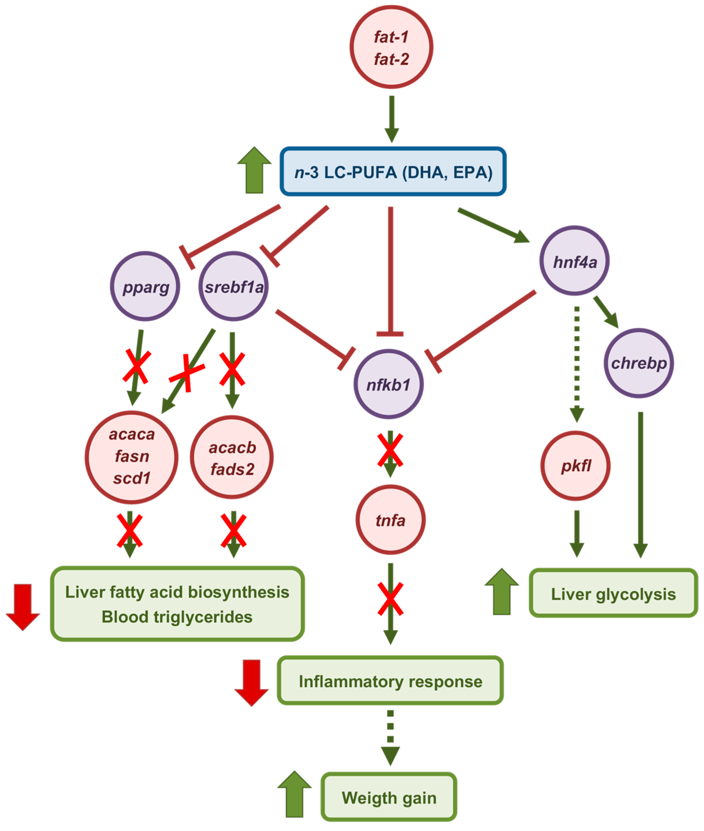
The present study discloses the short-term molecular events triggered by chitosan-TPP-DNA nanoparticles expressing fish codon-optimized C. elegans fat-1 and fat-2 in the liver of S. aurata. Co-expression of both enzymes resulted in the most pronounced effects across the majority of the parameters analyzed (Figure 8). In addition to elevating n-3 LC-PUFA such as EPA and DHA in the liver, 72 h post-treatment with fat-1 and fat-2 nanoparticles suppressed the hepatic expression of srebf1, pparg, and genes encoding key enzymes in de novo lipogenesis and LC-PUFA synthesis, while fat-2 and fat-1 + fat-2 upregulated hnf4a, chrebp, and rate-limiting enzymes in glycolysis. Notably, our data show that C. elegans fat-1 and fat-2 exerted rapid effects, within less than 72 h, by enhancing EPA and DHA levels in the liver of S. aurata, which in turn may have determined downregulation of proinflammatory factors such as tnfa and nfkb1. Altogether, our findings suggest that in addition to enabling the production of cultured fish rich in n-3 LC-PUFAs for human consumption, fat-1 and fat-2 expression may impact the health status and growth of fish. Future studies will be required to assess the extent to which the observed changes could benefit the growth performance of farmed fish.

Author Contributions

Conceptualization, I.M.; methodology, Y.W. and A.F.; validation, M.P.A. and I.M.; formal analysis, Y.W., A.R., W.D., M.P.A. and I.M.; investigation, Y.W., A.R., W.D., M.P.A. and I.M.; resources, I.M.; writing—original draft preparation, Y.W. and I.M.; writing—review and editing, Y.W., A.R., W.D., A.F., M.P.A. and I.M.; supervision, I.M.; project administration, I.M.; funding acquisition, I.M. All authors have read and agreed to the published version of the manuscript.

Funding

This research was funded by MICIU/AEI/10.13309/501100011033 and by “ERDF A way of making Europe” (Ministerio de Ciencia, Innovación y Universidades/Agencia Estatal de Investigación, Spain), grant number PID2021-125642OB-I00. Y.W. and W.D. were the recipients of China Scholarship Council (P.R. China) predoctoral fellowships 201908510154 and 202308110060, respectively. A.R. was the recipient of the PREDOCS-UB predoctoral fellowship (Universitat de Barcelona, Spain).

Institutional Review Board Statement

The animal study protocol was approved by the Animal Welfare Committee of the University of Barcelona and Generalitat de Catalunya (proceeding #10811; date of approval: 14 April 2020).

Data Availability Statement

Data will be made available from the corresponding author upon reasonable request.

Acknowledgments

The authors thank Piscicultura Marina Mediterranea (AVRAMAR Group, Burriana, Spain) for providing S. aurata juveniles and Eurocoyal S.L. (Sant Cugat del Vallès, Spain) for supplying dietary components. The manuscript is partially based on a thesis by the authors [63].

Conflicts of Interest

The authors declare no conflicts of interest. The funders had no role in the design of the study; in the collection, analyses, or interpretation of data; in the writing of the manuscript; or in the decision to publish the results.

Abbreviations

The following abbreviations are used in this manuscript:
ALAα-Linolenic acid
DHADocosahexaenoic acid
EPAEicosapentaenoic acid
LALinoleic acid
OAOleic acid
BWBody weight
TPPTripolyphosphate
n-3 LC-PUFAomega-3 long-chain polyunsaturated fatty acids

References

  1. Pang, S.-C.; Wang, H.-P.; Li, K.-Y.; Zhu, Z.-Y.; Kang, J.X.; Sun, Y.-H. Double Transgenesis of humanized fat1 and fat2 genes promotes omega-3 polyunsaturated fatty acids synthesis in a zebrafish model. Mar. Biotechnol. 2014, 16, 580–593. [Google Scholar] [CrossRef]
  2. Wu, Y.; Rashidpour, A.; Metón, I. Bayesian meta-analysis: Impacts of eating habits and habitats on omega-3 long-chain polyunsaturated fatty acid composition and growth in cultured fish. Animals 2024, 14, 2118. [Google Scholar] [CrossRef]
  3. Rivero-Ramírez, F.; Torrecillas, S.; Betancor, M.B.; Izquierdo, M.S.; Caballero, M.J.; Montero, D. Effects of dietary arachidonic acid in European sea bass (Dicentrarchus labrax) distal intestine lipid classes and gut health. Fish Physiol. Biochem. 2020, 46, 681–697. [Google Scholar] [CrossRef]
  4. Castro, L.F.C.; Tocher, D.R.; Monroig, O. Long-chain polyunsaturated fatty acid biosynthesis in chordates: Insights into the evolution of Fads and Elovl gene repertoire. Prog. Lipid Res. 2016, 62, 25–40. [Google Scholar] [CrossRef]
  5. Zhang, J.Y.; Kothapalli, K.S.D.; Brenna, J.T. Desaturase and elongase-limiting endogenous long-chain polyunsaturated fatty acid biosynthesis. Curr. Opin. Clin. Nutr. Metab. Care 2016, 19, 103–110. [Google Scholar] [CrossRef]
  6. Tu, W.C.; Cook-Johnson, R.J.; James, M.J.; Mühlhäusler, B.S.; Gibson, R.A. Omega-3 long chain fatty acid synthesis is regulated more by substrate levels than gene expression. Prostaglandins Leukot. Essent. Fat. Acids 2010, 83, 61–68. [Google Scholar] [CrossRef]
  7. Hattori, T.; Obinata, H.; Ogawa, A.; Kishi, M.; Tatei, K.; Ishikawa, O.; Izumi, T. G2A plays proinflammatory roles in human keratinocytes under oxidative stress as a receptor for 9-hydroxyoctadecadienoic acid. J. Investig. Dermatol. 2008, 128, 1123–1133. [Google Scholar] [CrossRef]
  8. Tan, P.; Ding, Y.; Li, X.; Dong, X.; Mai, K.; Ai, Q. Nrf2 pathway in vegetable oil-induced inflammation of large yellow croaker (Larimichthys crocea). Fish Shellfish Immunol. 2022, 127, 778–787. [Google Scholar] [CrossRef]
  9. Yan, X.-B.; Dong, X.-H.; Tan, B.-P.; Zhang, S.; Chi, S.-Y.; Yang, Q.-H.; Liu, H.-Y.; Yang, Y.-Z. Influence of different oil sources on growth, disease resistance, immune response and immune-related gene expression on the hybrid grouper (♀ Epinephelus fuscoguttatus × ♂ E. lanceolatu), to Vibrio parahaemolyticus challenge. Fish Shellfish Immunol. 2020, 99, 310–321. [Google Scholar] [CrossRef]
  10. Sun, S.; Castro, F.; Monroig, Ó.; Cao, X.; Gao, J. Fat-1 transgenic zebrafish are protected from abnormal lipid deposition induced by high-vegetable oil feeding. Appl. Microbiol. Biotechnol. 2020, 104, 7355–7365. [Google Scholar] [CrossRef]
  11. Zhang, X.; Pang, S.; Liu, C.; Wang, H.; Ye, D.; Zhu, Z.; Sun, Y. A Novel Dietary Source of EPA and DHA: Metabolic engineering of an important freshwater species-common carp by fat1-transgenesis. Mar. Biotechnol. 2018, 21, 171–185. [Google Scholar] [CrossRef]
  12. Tang, F.; Yang, X.; Liu, D.; Zhang, X.; Huang, X.; He, X.; Shi, J.; Li, Z.; Wu, Z. Co-expression of fat1 and fat2 in transgenic pigs promotes synthesis of polyunsaturated fatty acids. Transgenic Res. 2019, 28, 369–379. [Google Scholar] [CrossRef] [PubMed]
  13. Wu, Y.; Rashidpour, A.; Almajano, M.P.; Metón, I. Chitosan-based drug delivery system: Applications in fish biotechnology. Polymers 2020, 12, 1177. [Google Scholar] [CrossRef] [PubMed]
  14. Wu, Y.; Rashidpour, A.; Fàbregas, A.; Almajano, M.P.; Metón, I. Chitosan-based delivery of fish codon-optimised Caenorhabditis elegans FAT-1 and FAT-2 boosts EPA and DHA biosynthesis in Sparus aurata. Rev. Fish Biol. Fish. 2024, 34, 995–1016. [Google Scholar] [CrossRef]
  15. Liu, D.; Chi, S.; Tan, B.; Dong, X.; Yang, Q.; Liu, H.; Zhang, S.; Han, F.; He, Y. Effects of fish oil with difference oxidation degree on growth performance and expression abundance of antioxidant and fat metabolism genes in orange spotted grouper, Epinephelus coioides. Aquac. Res. 2019, 50, 188–197. [Google Scholar] [CrossRef]
  16. Song, C.; Liu, B.; Xu, P.; Xie, J.; Ge, X.; Zhou, Q.; Sun, C.; Zhang, H.; Shan, F.; Yang, Z. Oxidized fish oil injury stress in Megalobrama amblycephala: Evaluated by growth, intestinal physiology, and transcriptome-based PI3K-Akt/NF-κB/TCR inflammatory signaling. Fish Shellfish Immunol. 2018, 81, 446–455. [Google Scholar] [CrossRef]
  17. Shearer, G.C.; Savinova, O.V.; Harris, W.S. Fish oil—How does it reduce plasma triglycerides? Biochim. Biophys. Acta—Mol. Cell Biol. Lipids 2012, 1821, 843–851. [Google Scholar] [CrossRef]
  18. Romanatto, T.; Fiamoncini, J.; Wang, B.; Curi, R.; Kang, J.X. Elevated tissue omega-3 fatty acid status prevents age-related glucose intolerance in fat-1 transgenic mice. Biochim. Biophys. Acta—Mol. Basis Dis. 2014, 1842, 186–191. [Google Scholar] [CrossRef]
  19. Liu, X.; Pang, D.; Yuan, T.; Li, Z.; Li, Z.; Zhang, M.; Ren, W.; Ouyang, H.; Tang, X. N-3 polyunsaturated fatty acids attenuates triglyceride and inflammatory factors level in hfat-1 transgenic pigs. Lipids Health Dis. 2016, 15, 89. [Google Scholar] [CrossRef]
  20. Liu, X.; Wei, Z.; Bai, C.; Ding, X.; Li, X.; Su, G.; Cheng, L.; Zhang, L.; Guo, H.; Li, G. Insights into the function of n-3 PUFAs in fat-1 transgenic cattle. J. Lipid Res. 2017, 58, 1524–1535. [Google Scholar] [CrossRef]
  21. Lorente-Cebrián, S.; Costa, A.G.V.; Navas-Carretero, S.; Zabala, M.; Martínez, J.A.; Moreno-Aliaga, M.J. Role of omega-3 fatty acids in obesity, metabolic syndrome, and cardiovascular diseases: A review of the evidence. J. Physiol. Biochem. 2013, 69, 633–651. [Google Scholar] [CrossRef]
  22. He, L.; Qin, Y.; Wang, Y.; Li, D.; Chen, W.; Ye, J. Effects of dietary replacement of fish oil with soybean oil on the growth performance, plasma components, fatty acid composition and lipid metabolism of groupers Epinephelus coioides. Aquac. Nutr. 2021, 27, 1494–1511. [Google Scholar] [CrossRef]
  23. Gu, Z.; Mu, H.; Shen, H.; Deng, K.; Liu, D.; Yang, M.; Zhang, Y.; Zhang, W.; Mai, K. High level of dietary soybean oil affects the glucose and lipid metabolism in large yellow croaker Larimichthys crocea through the insulin-mediated PI3K/AKT signaling pathway. Comp. Biochem. Physiol. Part B Biochem. Mol. Biol. 2019, 231, 34–41. [Google Scholar] [CrossRef] [PubMed]
  24. Watts, J.L.; Browse, J. Genetic dissection of polyunsaturated fatty acid synthesis in Caenorhabditis elegans. Proc. Natl. Acad. Sci. USA 2002, 99, 5854–5859. [Google Scholar] [CrossRef] [PubMed]
  25. Zhou, X.R.; Green, A.G.; Singh, S.P. Caenorhabditis elegans Δ12-desaturase FAT-2 is a bifunctional desaturase able to desaturate a diverse range of fatty acid substrates at the Δ12 and Δ15 positions. J. Biol. Chem. 2011, 286, 43644–43650. [Google Scholar] [CrossRef]
  26. Lai, L.; Kang, J.X.; Li, R.; Wang, J.; Witt, W.T.; Yong, H.Y.; Hao, Y.; Wax, D.M.; Murphy, C.N.; Rieke, A.; et al. Generation of cloned transgenic pigs rich in omega-3 fatty acids. Nat. Biotechnol. 2006, 24, 435–436. [Google Scholar] [CrossRef]
  27. Carmona-Antoñanzas, G.; Tocher, D.R.; Martinez-Rubio, L.; Leaver, M.J. Conservation of lipid metabolic gene transcriptional regulatory networks in fish and mammals. Gene 2014, 534, 1–9. [Google Scholar] [CrossRef]
  28. Rashidpour, A.; Wu, Y.; Almajano, M.P.; Fàbregas, A.; Metón, I. Chitosan-based sustained expression of sterol regulatory element-binding protein 1a stimulates hepatic glucose oxidation and growth in Sparus aurata. Mar. Drugs 2023, 21, 562. [Google Scholar] [CrossRef] [PubMed]
  29. Jump, D.B. N-3 polyunsaturated fatty acid regulation of hepatic gene transcription. Curr. Opin. Lipidol. 2008, 19, 242–247. [Google Scholar] [CrossRef]
  30. Wang, Y.; Nakajima, T.; Gonzalez, F.J.; Tanaka, N. PPARs as metabolic regulators in the liver: Lessons from liver-specific PPAR-null mice. Int. J. Mol. Sci. 2020, 21, 2061. [Google Scholar] [CrossRef]
  31. Tian, J.; Ji, H.; Oku, H.; Zhou, J. Effects of dietary arachidonic acid (ARA) on lipid metabolism and health status of juvenile grass carp, Ctenopharyngodon idellus. Aquaculture 2014, 430, 57–65. [Google Scholar] [CrossRef]
  32. Wang, X.-X.; Li, Y.-J.; Hou, C.-L.; Gao, Y.; Wang, Y.-Z. Influence of different dietary lipid sources on the growth, tissue fatty acid composition, histological changes and peroxisome proliferator-activated receptor γ gene expression in large yellow croaker (Pseudosciaena crocea R.). Aquac. Res. 2012, 43, 281–291. [Google Scholar] [CrossRef]
  33. You, C.; Jiang, D.; Zhang, Q.; Xie, D.; Wang, S.; Dong, Y.; Li, Y. Cloning and expression characterization of peroxisome proliferator-activated receptors (PPARs) with their agonists, dietary lipids, and ambient salinity in rabbitfish Siganus canaliculatus. Comp. Biochem. Physiol. Part B Biochem. Mol. Biol. 2017, 206, 54–64. [Google Scholar] [CrossRef] [PubMed]
  34. Echeverría, F.; Valenzuela, R.; Bustamante, A.; Álvarez, D.; Ortiz, M.; Espinosa, A.; Illesca, P.; Gonzalez-Mañan, D.; Videla, L.A. High-fat diet induces mouse liver steatosis with a concomitant decline in energy metabolism: Attenuation by eicosapentaenoic acid (EPA) or hydroxytyrosol (HT) supplementation and the additive effects upon EPA and HT co-administration. Food Funct. 2019, 10, 6170–6183. [Google Scholar] [CrossRef]
  35. Wang, C.; Liu, W.; Yao, L.; Zhang, X.; Zhang, X.; Ye, C.; Jiang, H.; He, J.; Zhu, Y.; Ai, D. Hydroxyeicosapentaenoic acids and epoxyeicosatetraenoic acids attenuate early occurrence of nonalcoholic fatty liver disease. Br. J. Pharmacol. 2017, 174, 2358–2372. [Google Scholar] [CrossRef]
  36. Chang, M.; Zhang, T.; Han, X.; Tang, Q.; Yanagita, T.; Xu, J.; Xue, C.; Wang, Y. Comparative analysis of EPA/DHA-PL forage and liposomes in orotic acid-induced nonalcoholic fatty liver rats and their related mechanisms. J. Agric. Food Chem. 2018, 66, 1408–1418. [Google Scholar] [CrossRef]
  37. Polakof, S.; Panserat, S.; Soengas, J.L.; Moon, T.W. Glucose metabolism in fish: A review. J. Comp. Physiol. B 2012, 182, 1015–1045. [Google Scholar] [CrossRef]
  38. Okar, D.A.; Wu, C.; Lange, A.J. Regulation of the regulatory enzyme, 6-phosphofructo-2-kinase/fructose-2,6-bisphosphatase. Adv. Enzyme Regul. 2004, 44, 123–154. [Google Scholar] [CrossRef]
  39. Meng, J.; Feng, M.; Dong, W.; Zhu, Y.; Li, Y.; Zhang, P.; Wu, L.; Li, M.; Lu, Y.; Chen, H.; et al. Identification of HNF-4α as a key transcription factor to promote ChREBP expression in response to glucose. Sci. Rep. 2016, 6, 23944. [Google Scholar] [CrossRef]
  40. Zhuang, P.; Li, H.; Jia, W.; Shou, Q.; Zhu, Y.; Mao, L.; Wang, W.; Wu, F.; Chen, X.; Wan, X.; et al. Eicosapentaenoic and docosahexaenoic acids attenuate hyperglycemia through the microbiome-gut-organs axis in db/db mice. Microbiome 2021, 9, 185. [Google Scholar] [CrossRef] [PubMed]
  41. Salgado, M.C.; Metón, I.; Anemaet, I.G.; González, J.D.; Fernández, F.; Baanante, I.V. Hepatocyte nuclear factor 4alpha transactivates the mitochondrial alanine aminotransferase gene in the kidney of Sparus aurata. Mar. Biotechnol. 2012, 14, 46–62. [Google Scholar] [CrossRef]
  42. Lu, H.; Lei, X.; Winkler, R.; John, S.; Kumar, D.; Li, W.; Alnouti, Y. Crosstalk of hepatocyte nuclear factor 4a and glucocorticoid receptor in the regulation of lipid metabolism in mice fed a high-fat-high-sugar diet. Lipids Health Dis. 2022, 21, 46. [Google Scholar] [CrossRef]
  43. Boyle, K.E.; Magill-Collins, M.J.; Newsom, S.A.; Janssen, R.C.; Friedman, J.E. Maternal fat-1 transgene protects offspring from excess weight gain, oxidative stress, and reduced fatty acid oxidation in response to high-fat diet. Nutrients 2020, 12, 767. [Google Scholar] [CrossRef] [PubMed]
  44. López-Vicario, C.; Rius, B.; Alcaraz-Quiles, J.; García-Alonso, V.; Lopategi, A.; Titos, E.; Clària, J. Pro-resolving mediators produced from EPA and DHA: Overview of the pathways involved and their mechanisms in metabolic syndrome and related liver diseases. Eur. J. Pharmacol. 2016, 785, 133–143. [Google Scholar] [CrossRef] [PubMed]
  45. Bradley, J. TNF-mediated inflammatory disease. J. Pathol. 2008, 214, 149–160. [Google Scholar] [CrossRef]
  46. Guo, X.F.; Gao, J.L.; Li, J.M.; Li, D. Fat-1 mice prevent high-fat plus high-sugar diet-induced non-alcoholic fatty liver disease. Food Funct. 2017, 8, 4053–4061. [Google Scholar] [CrossRef]
  47. Li, Y.; Tang, Y.; Wang, S.; Zhou, J.; Zhou, J.; Lu, X.; Bai, X.; Wang, X.-Y.; Chen, Z.; Zuo, D. Endogenous n-3 polyunsaturated fatty acids attenuate T cell-mediated hepatitis via autophagy activation. Front. Immunol. 2016, 7, 350. [Google Scholar] [CrossRef] [PubMed]
  48. Abdolmaleki, F.; Kovanen, P.T.; Mardani, R.; Gheibi-hayat, S.M.; Bo, S.; Sahebkar, A. Resolvins: Emerging players in autoimmune and inflammatory diseases. Clin. Rev. Allergy Immunol. 2020, 58, 82–91. [Google Scholar] [CrossRef]
  49. Anderson, E.K.; Hill, A.A.; Hasty, A.H. Stearic acid accumulation in macrophages induces toll-like receptor 4/2-independent inflammation leading to endoplasmic reticulum stress-mediated apoptosis. Arterioscler. Thromb. Vasc. Biol. 2012, 32, 1687–1695. [Google Scholar] [CrossRef]
  50. Liu, T.; Zhang, L.; Joo, D.; Sun, S.C. NF-κB signaling in inflammation. Signal Transduct. Target. Ther. 2017, 2, 17023. [Google Scholar] [CrossRef]
  51. Hernández-Rodas, M.C.; Valenzuela, R.; Echeverría, F.; Rincón-Cervera, M.Á.; Espinosa, A.; Illesca, P.; Muñoz, P.; Corbari, A.; Romero, N.; Gonzalez-Mañan, D.; et al. Supplementation with docosahexaenoic acid and extra virgin olive oil prevents liver steatosis induced by a high-fat diet in mice through PPAR-α and Nrf2 upregulation with concomitant SREBP-1c and NF-kB downregulation. Mol. Nutr. Food Res. 2017, 61, 1700479. [Google Scholar] [CrossRef] [PubMed]
  52. Yang, Y.C.; Lii, C.K.; Wei, Y.L.; Li, C.C.; Lu, C.Y.; Liu, K.L.; Chen, H.W. Docosahexaenoic acid inhibition of inflammation is partially via cross-talk between Nrf2/heme oxygenase 1 and IKK/NF-κB pathways. J. Nutr. Biochem. 2013, 24, 204–212. [Google Scholar] [CrossRef]
  53. Echeverría, F.; Valenzuela, R.; Espinosa, A.; Bustamante, A.; Álvarez, D.; Gonzalez-Mañan, D.; Ortiz, M.; Soto-Alarcon, S.A.; Videla, L.A. Reduction of high-fat diet-induced liver proinflammatory state by eicosapentaenoic acid plus hydroxytyrosol supplementation: Involvement of resolvins RvE1/2 and RvD1/2. J. Nutr. Biochem. 2019, 63, 35–43. [Google Scholar] [CrossRef]
  54. Yang, W.; Liu, R.; Xia, C.; Chen, Y.; Dong, Z.; Huang, B.; Li, R.; Li, M.; Xu, C. Effects of different fatty acids on BRL3A rat liver cell damage. J. Cell. Physiol. 2020, 235, 6246–6256. [Google Scholar] [CrossRef]
  55. Jiang, X.; He, M.; Bai, J.; Chan, C.B.; Wong, A.O.L. Signal Transduction for TNFα-induced type II SOCS expression and its functional implication in growth hormone resistance in carp hepatocytes. Front. Endocrinol. 2020, 11, 20. [Google Scholar] [CrossRef]
  56. Ren, M.; Habte-Tsion, H.M.; Liu, B.; Miao, L.; Ge, X.; Xie, J.; Liang, H.; Zhou, Q.; Pan, L. Dietary leucine level affects growth performance, whole body composition, plasma parameters and relative expression of TOR and TNF-α in juvenile blunt snout bream, Megalobrama amblycephala. Aquaculture 2015, 448, 162–168. [Google Scholar] [CrossRef]
  57. Mu, H.; Wei, C.; Xu, W.; Gao, W.; Zhang, W.; Mai, K. Effects of replacement of dietary fish oil by rapeseed oil on growth performance, anti-oxidative capacity and inflammatory response in large yellow croaker Larimichthys crocea. Aquac. Rep. 2020, 16, 100251. [Google Scholar] [CrossRef]
  58. Li, X.; Huang, W.; Gu, J.; Du, X.; Lei, L.; Yuan, X.; Sun, G.; Wang, Z.; Li, X.; Liu, G. SREBP-1c overactivates ROS-mediated hepatic NF-κB inflammatory pathway in dairy cows with fatty liver. Cell. Signal. 2015, 27, 2099–2109. [Google Scholar] [CrossRef]
  59. Guo, Y.; Zhang, X.; Zhao, Z.; Lu, H.; Ke, B.; Ye, X.; Wu, B.; Ye, J. NF- B/HDAC1/SREBP1c pathway mediates the inflammation signal in progression of hepatic steatosis. Acta Pharm. Sin. B 2020, 10, 825–836. [Google Scholar] [CrossRef] [PubMed]
  60. Ning, B.-F.; Ding, J.; Liu, J.; Yin, C.; Xu, W.-P.; Cong, W.-M.; Zhang, Q.; Chen, F.; Han, T.; Deng, X.; et al. Hepatocyte nuclear factor 4α-nuclear factor-κB feedback circuit modulates liver cancer progression. Hepatology 2014, 60, 1607–1619. [Google Scholar] [CrossRef] [PubMed]
  61. Nikolaidou-Neokosmidou, V.; Zannis, V.I.; Kardassis, D. Inhibition of hepatocyte nuclear factor 4 transcriptional activity by the nuclear factor κB pathway. Biochem. J. 2006, 398, 439–450. [Google Scholar] [CrossRef] [PubMed]
  62. Livak, K.J.; Schmittgen, T.D. Analysis of relative gene expression data using real-time quantitative PCR and the 2−ΔΔCT method. Methods 2001, 25, 402–408. [Google Scholar] [CrossRef] [PubMed]
  63. Wu, Y. Administration of Chitosan-Tripolyphosphate-DNA Nanoparticles Overexpressing Key Enzymes to Improve Omega-3 Long-Chain Polyunsaturated Fatty Acid Synthesis in Gilthead Sea Bream (Sparus aurata). Ph.D. Thesis, Universitat de Barcelona, Barcelona, Spain, 2023. [Google Scholar]
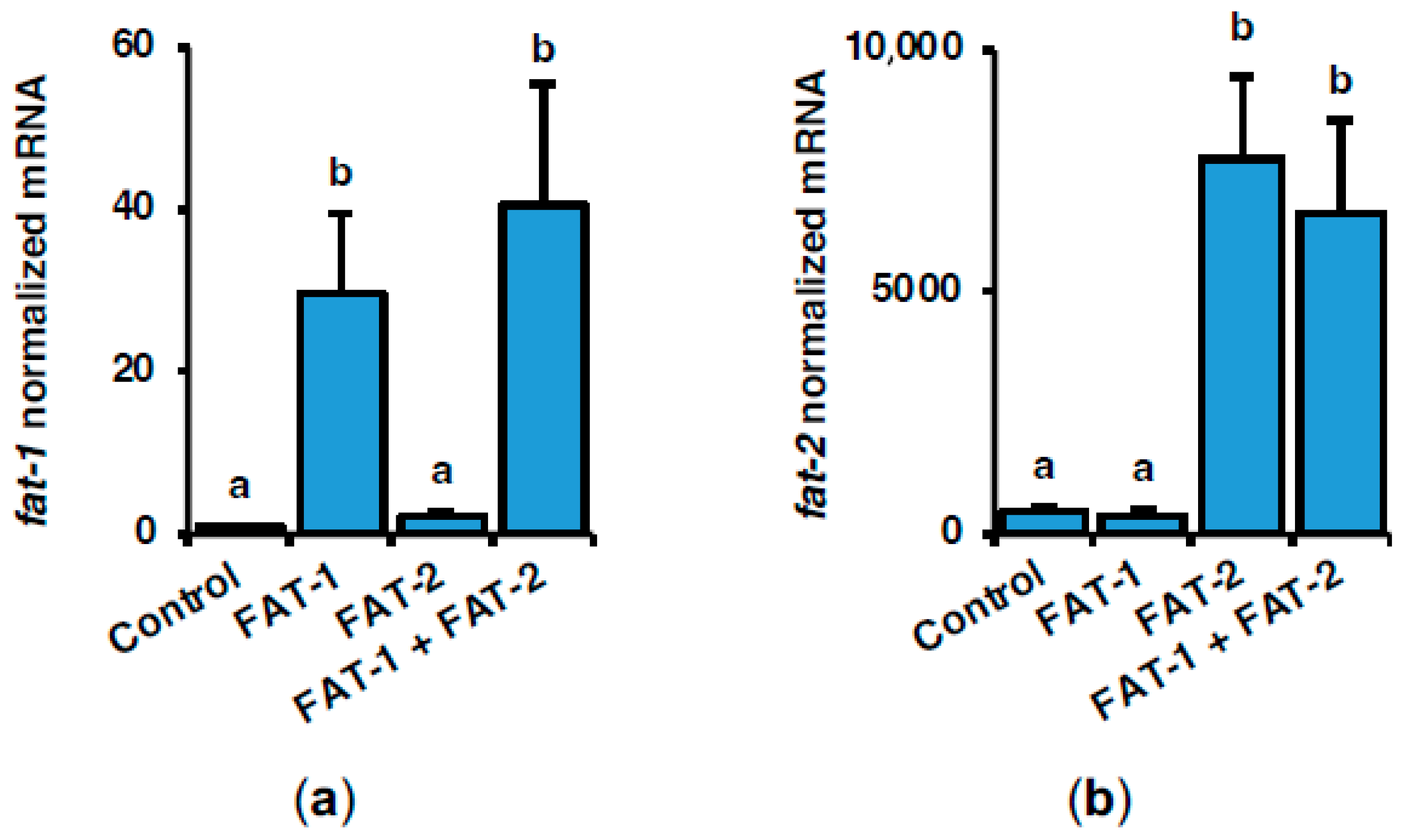
Figure 1. Short-term effects of intraperitoneal administration of chitosan-TPP nanoparticles encapsulating pSG5 (control), pSG5-FAT-1, pSG5-FAT-2, and pSG5-FAT-1 + pSG5-FAT-2 on the mRNA levels of fish-codon optimized C. elegans fat-1 (a) and fat-2 (b) in S. aurata liver. Seventy-two hours post-treatment, fish were sacrificed, and the liver was collected. Following RNA isolation from liver samples, exogenous fat-1 and fat-2 expression was assayed by RT-qPCR, normalized to the geometric mean of S. aurata 18s, actb, and eef1a mRNA levels, and expressed as mean ± SEM (n = 5). Different letters indicate distinct homogeneous subsets determined by the post hoc test (p < 0.05).
Marinedrugs 23 00434 g001
Figure 2. Short-term effects of intraperitoneal administration of chitosan-TPP nanoparticles encapsulating pSG5 (control), pSG5-FAT-1, pSG5-FAT-2, and pSG5-FAT-1 + pSG5-FAT-2 on serum (a) glucose, (b) triglycerides, and (c) cholesterol in S. aurata. Seventy-two hours post-treatment, fish were sacrificed, and blood was collected. Values are represented as mean ± SEM (n = 4–5). Different letters indicate distinct homogeneous subsets determined by the post hoc test (p < 0.05).
Marinedrugs 23 00434 g002
Figure 3. Fatty acid methyl ester analysis of liver samples from S. aurata. Representative chromatograms obtained by gas chromatography performed on fish treated with chitosan-TPP nanoparticles complexed with (a) pSG5 (control), (b) pSG5-FAT-1, (c) pSG5-FAT-2, and (d) pSG5-FAT-1 + pSG5-FAT-2. Fatty acids with a composition higher than 1% are indicated.
Marinedrugs 23 00434 g003
Figure 4. Short-term effects of intraperitoneal administration of chitosan-TPP nanoparticles encapsulating pSG5 (control), pSG5-FAT-1, pSG5-FAT-2, and pSG5-FAT-1 + pSG5-FAT-2 on the expression of key enzymes in de novo lipogenesis in the liver of S. aurata. Seventy-two hours post-treatment, fish were sacrificed, and the liver was collected. (ai) Following RNA isolation from liver samples, gene expression was assayed by RT-qPCR, normalized to the geometric mean of S. aurata 18s, actb, and eef1a mRNA levels, and expressed as mean ± SEM (n = 5). Different letters indicate distinct homogeneous subsets determined by the post hoc test (p < 0.05).
Marinedrugs 23 00434 g004
Figure 5. Short-term effects of intraperitoneal administration of chitosan-TPP nanoparticles encapsulating pSG5 (control), pSG5-FAT-1, pSG5-FAT-2, and pSG5-FAT-1 + pSG5-FAT-2 on the expression of key enzymes in glycolysis-gluconeogenesis and the PPP in the liver of S. aurata. Seventy-two hours post-treatment, fish were sacrificed, and the liver was collected. (af) Following RNA isolation from liver samples, gene expression was assayed by RT-qPCR and normalized to the geometric mean of S. aurata 18s, actb, and eef1a mRNA levels. Expression levels and enzyme activity are presented as mean ± SEM (n = 5). Different letters indicate distinct homogeneous subsets determined by the post hoc test (p < 0.05).
Marinedrugs 23 00434 g005
Figure 6. Short-term effects of intraperitoneal administration of chitosan-TPP nanoparticles encapsulating pSG5 (control), pSG5-FAT-1, pSG5-FAT-2, and pSG5-FAT-1 + pSG5-FAT-2 on the expression of key transcription factors in lipogenesis and carbohydrate metabolism in the liver of S. aurata. Seventy-two hours post-treatment, fish were sacrificed, and the liver was collected. (ae) Following RNA isolation from liver samples, gene expression was assayed by RT-qPCR, normalized to the geometric mean of S. aurata 18s, actb, and eef1a mRNA levels, and expressed as mean ± SEM (n = 5). Different letters indicate distinct homogeneous subsets determined by the post hoc test (p < 0.05).
Marinedrugs 23 00434 g006
Figure 7. Short-term effects of intraperitoneal administration of chitosan-TPP nanoparticles encapsulating pSG5 (control), pSG5-FAT-1, pSG5-FAT-2, and pSG5-FAT-1 + pSG5-FAT-2 on the expression of pro-inflammatory factors in the liver of S. aurata. Seventy-two hours post-treatment, fish were sacrificed, and the liver was collected. (af) Following RNA isolation from liver samples, gene expression was assayed by RT-qPCR, normalized to the geometric mean of S. aurata 18s, actb, and eef1a mRNA levels, and expressed as mean ± SEM (n = 5). Different letters indicate distinct homogeneous subsets determined by the post hoc test (p < 0.05).
Marinedrugs 23 00434 g007
Figure 8. Schematic representation of the short-term molecular events triggered by co-expressing fish codon-optimized C. elegans fat-1 and fat-2 in the liver of S. aurata. Seventy-two h after the administration of chitosan-TPP-DNA nanoparticles, the expression of fish codon-optimized C. elegans fat-1 and fat-2 stimulated the hepatic synthesis of n-3 LC-PUFA, particularly EPA and DHA. This event upregulated transcription factors and key enzymes involved in glycolysis, while downregulating those associated with fatty acid biosynthesis and proinflammatory factors, thereby potentially improving growth performance. Red crossed arrows indicate loss of activating effect. Dotted arrows indicate potential interactions.
Marinedrugs 23 00434 g008
Table 1. Effect of chitosan-TPP nanoparticles complexed with pSG5 (control), pSG5-FAT-1, pSG5-FAT-2, and pSG5-FAT-1 + pSG5-FAT-2 on the fatty acid composition of S. aurata liver.
Fatty AcidpSG5FAT-1FAT-2FAT-1 + FAT-2
14:05.7 a ± 0.97.2 ab ± 0.18.7 b ± 0.77.4 ab ± 0.7
16:019.0 ± 2.920.3 ± 0.922.3 ± 0.524.4 ± 1.4
18:03.0 ± 0.73.2 ± 0.62.6 ± 0.34.0 ± 0.4
16:1n-72.6 a ± 0.74.2 b ± 0.35.4 b ± 0.24.9 b ± 0.3
18:1n-914.4 ± 2.716.1 ± 1.817.6 ± 1.418.8 ± 0.7
18:2n-616.0 a ± 3.318.2 ab ± 1.220.2 ab ± 1.424.0 b ± 1.1
18:3n-30.8 ± 0.31.3 ± 0.41.1 ± 0.21.0 ± 0.2
20:5n-31.6 a ± 0.42.4 b ± 0.12.3 ab ± 0.12.5 b ± 0.2
22:6n-31.7 a ± 0.22.6 b ± 0.22.6 bc ± 0.2 3.2 c ± 0.2
SFA28.0 ± 4.630.9 ± 1.433.7 ± 0.736.1 ± 1.8
MUFA17.7 ± 2.721.1 ± 2.223.8 ± 1.323.8 ± 0.7
PUFA20.6 a ± 3.725.2 ab ± 1.527.0 ab ± 1.931.4 b ± 1.3
n-34.1 a ± 0.66.2 b ± 0.46.0 b ± 0.56.9 b ± 0.3
n-616.5 a ± 3.218.8 ab ± 1.220.7 ab ± 1.424.3 b ± 1.1
n-914.8 ± 2.716.7 ± 1.918.3 ± 1.319.0 ± 0.6
n-6/n-34.0 ± 0.53.0 ± 0.23.5 ± 0.23.5 ± 0.1
Data are expressed as a percentage of total fatty acids and represented as the mean ± SEM (n = 4). Only fatty acids exceeding 1% are shown. Different letters indicate distinct homogeneous subsets determined by the post hoc test (p < 0.05).
Table 2. Primer sequences used for RT-qPCR in the present study.
GeneForward Sequences (5′ to 3′)Reverse Sequences (5′ to 3′)GenBank
Accession
acacaCCCAACTTCTTCTACCACAGGAACTGGAACTCTACTACACJX073712
acacbTGACATGAGTCCTGTGCTGGGCCTCAGTTCGTATGATGGTJX073714
actbCTGGCATCACACCTTCTACAACGAGGCGGGGGTGTTGAAGGTCTCX89920
chrebpCTTCGACACGGTGAACAGGAAGGGACATGCAGTCGAACAGNC044199
eef1aCCCGCCTCTGTTGCCTTCGCAGCAGTGTGGTTCCGTTAGCAF184170
elovl4aAAGAACAGAGAGCCCTTCCAGTGCCACCCTGACTTCATTGMK610320
elovl4bTCTACACAGGCTGCCCATTCCGAAGAGGATGATGAAGGTGACMK610321
elovl5GGGATGGCTACTGCTCGACACAGGAGAGTGAGGCCCAGATAY660879
fads2CACTATGCTGGAGAGGATGCCTATTTCGGTCCTGGCTGGGCAY055749
fasnGTAGAGGACACGCCCATCGATTGCGTATGACCTCTTGGTGTGCTJQ277708
fat-1TTCAACCCCATTCCTTTCAGCGTAGGCGCACACGCAGCAGCAON374024
fat-2AAGAGGACTACAACAACAGAACCGCCACGAACAGTCTGCTCCAAGGCCAAON374025
fbp1CAGATGGTGAGCCGTGTGAGAAGGATGGCCGTACAGAGCGTAACCAGCTGCCAF427867
g6pdTGATGATCCAACAGTTCCTAGCTCGTTCCTGACACACTGAJX073711
hmgcrACTGATGGCTGCTCTGGCTGGGGACTGAGGGATGACGCACMN047456
hnf4aGTGGACAAAGACAAGCGAAATCGCATTGATGGATGGTAAACTGCFJ360721
il1bCGTCATCGCCATGGAGAGGTTGAGCTGGTTTTGCAGTGCGAJ277166
il6CGGAGCAGCATCGTCACTTTTTCTGCATGGCACACACAATEU244588
myd88GCGACGCCTGTGACTTTCAGGGGTGTAGTCGCAGAGGGTGXM_030399037
nfkb1GCTGCTGCTCGGGATCAAACGTCCACACTGAGCCACTGGAXM_030396749
nfkb2GTGTGTTGCTGCCGTGTGACGTCTGTCCGTCTGCTCCCTCXM_030441891
nr1h3GCATCTGGACGAGGCTGAATACACTTAGTGTGCGAAGGCTCACCFJ502320
pck1CAGCGATGGAGGAGTGTGGTGGGAGCCCATCCCAATTCCCGCTTCTGTGCTCCGGCTGGTCAGTGTAF427868
pfkfb1TGCTGATGGTGGGACTGCCGCTCGGCGTTGTCGGCTCTGAAGU84724
pfk1TGCTGGGGACAAAACGAACTCTTCCAAACCCTCCGACTACAAGCAGAGCTKF857580
pklrCAAAGTGGAAAGCCGGCAAGGGGTCGCCCCTGGCAACCATAACKF857579
ppargTGCGAGGGCTGTAAGGGTTTCGTTTCTCCTTCTCCGCCTGGGAY590304
srebf1CAGCAGCCCGAACACCTACATTGTGGTCAGCCCTTGGAGTTGJQ277709
scd1aTCCCTTCCGCATCTCCTTTGTTGTGGTGAACCCTGTGGTCTCJQ277703
tnfaGTCTTCCGCCCCTCAGATCCGAAAGCCGAAGCATGAGCCCAJ413189
18sTTACGCCCATGTTGTCCTGAGAGGATTCTGCATGATGGTCACCAM490061
Disclaimer/Publisher’s Note: The statements, opinions and data contained in all publications are solely those of the individual author(s) and contributor(s) and not of MDPI and/or the editor(s). MDPI and/or the editor(s) disclaim responsibility for any injury to people or property resulting from any ideas, methods, instructions or products referred to in the content.