Article

5Z-7-Oxozeanol Isolated from the Fungus Curvularia sp. MDCW-1060 Inhibits the Proliferation of MDA-MB-231 Cells via the PI3K-Akt and MAPK Pathways

1
Institute of Marine Drugs/Guangxi Key Laboratory of Marine Drugs/Guangxi University Engineering Research Center of High-Efficient Utilization of Marine Traditional Chinese Medicine Resources, Guangxi University of Chinese Medicine, Nanning 530200, China
2
Key Laboratory of Chemistry and Engineering of Forest Products/State Ethnic Affairs Commission; Guangxi Key Laboratory of Chemistry and Engineering of Forest Products/Engineering Research Center of Low-Carbon and High-Quality Utilization of Forest Biomass, University of Guangxi/School of Chemistry and Chemical Engineering/Guangxi Minzu University, Nanning 530006, China
*
Authors to whom correspondence should be addressed.
Hong Zhang and Jianjian Wang contributed equally to this work.
Mar. Drugs 2025, 23(11), 414; https://doi.org/10.3390/md23110414
Submission received: 30 September 2025 / Revised: 21 October 2025 / Accepted: 22 October 2025 / Published: 23 October 2025
(This article belongs to the Special Issue Marine Compounds as Inhibitors)

Abstract

The discovery of novel marine natural products and their sustainable application continue to be vital focuses in marine biological research. The aim of this study is to investigate the inhibitory effect of the compound 5Z-7-Oxozeaenol isolated from the fungus Curvularia sp. MDCW-1060 on the proliferation of MDA-MB-231 cells and its molecular mechanism. A series of functional assays, including 3-(4,5-Dimethylthiazol-2-yl)-2,5-diphenyltetrazolium bromide (MTT), flow cytometry, Transwell migration, and colony formation, were employed to evaluate the effects of 5Z-7-Oxozeaenol on cellular viability, apoptosis, migration, and clonogenicity. The RNA sequencing (RNA-seq) coupled with bioinformatic analysis was conducted to identify affected differentiated gene expression and signaling pathways. The molecular docking was performed to predict potential protein targets, and Western blot was used to validate expression and phosphorylation levels of key signaling molecules. The results demonstrated that 5Z-7-Oxozeaenol significantly suppressed proliferation and migration while promoting apoptosis in MDA-MB-231 cells. The transcriptomic analysis indicated enrichment in pathways related to cancer, cytokine–cytokine receptor interaction, MAPK and PI3K-Akt signaling, and cell adhesion molecules. The molecular docking suggested a high binding affinity between 5Z-7-Oxozeaenol and PTPRN. While Western blot analysis confirmed the downregulation of phosphorylated FAK, PI3K, Akt, and MAPK, along with reduced cyclin D1 expression. Additionally, 5Z-7-Oxozeaenol upregulated the pro-apoptotic proteins p53 and cleaved caspase-3. In conclusion, 5Z-7-Oxozeaenol exerts potent antitumor effects on MDA-MB-231 cells through multi-pathway inhibition and induction of apoptosis, highlighting its potential as a marine-derived therapeutic candidate for breast cancer treatment.

1. Introduction

Breast cancer remains the most frequently diagnosed malignancy and the leading cause of cancer-related mortality among women worldwide [1]. In recent years, both the incidence and detection of breast tumors have continued to rise, with the disease increasingly affecting younger populations, thus posing a major threat to women’s health [2]. According to the statistical analysis and release of the results of the International Agency for Research on Cancer (IARC) GLOBOCAN database in 2022, the incidence of breast cancer is expected to reach 11.6%, which is the highest among female malignant tumors worldwide, with a fatality rate of 6.9% [3]. Epidemiological surveys have shown that the disease burden of breast cancer is not optimistic in China, with the incidence of breast cancer being higher in urban areas than in rural areas [4]. Despite advances in medical standards around the world, the comprehensive treatment of breast cancer based on surgery has greatly extended the survival of breast cancer patients [1]; however, it is still an important public health problem worldwide because of its rapid progress and high mortality rate [5].
Unlike other breast cancer subtypes, triple-negative breast cancer (TNBC) lacks the expression of estrogen receptor (ER), progesterone receptor (PR), human epidermal growth factor receptor 2 (HER-2), making it unresponsive to hormonal and HER2-targeted therapies [6,7,8]. TNBC accounts for approximately 15–20% of all breast cancer cases and is associated with poor prognosis due to its aggressive nature and limited treatment options [2]. The heterogeneity of TNBC poses significant challenges in treatment, necessitating a broader screening of chemotherapeutic drugs [9,10,11,12].
5Z-7-Oxozeaenol has been reported to induce caspase-3-mediated apoptosis in HeLa cells and inhibit TGF-β-activated kinase 1 (TAK1) activity [13,14]. However, its biological activities and underlying mechanisms remain incompletely understood. In this study, we isolated 5Z-7-Oxozeaenol from the marine-derived fungus Curvularia sp. MDCW-1060, collected from the Beibu Gulf in Guangxi, China. We then performed a series of in vitro assays to evaluate its effects on breast cancer cells. Transcriptomic and bioinformatic analyses, combined with molecular biology approaches, were employed to elucidate the signaling pathways and molecular mechanisms through which 5Z-7-Oxozeaenol inhibits breast cancer cell proliferation. These findings provide a foundation for the development of novel therapeutic agents against breast tumors.

2. Results

2.1. 5Z-7-Oxozeaenol Structure

Here, we report the isolation of 5Z-7-Oxozeaenol (C19H22O7), a fungal metabolite obtained from Curvularia sp. MDCW-1060—originally isolated from a crab specimen collected in the Beibu Gulf, Guangxi, China (Figure 1). The compound was obtained as a colorless powder, and its structure was elucidated through analysis of MS, 1H and 13C NMR spectral data (Figures S1–S3). The spectroscopic data were consistent with those previously reported for 5Z-7-Oxozeaenol [14], confirming its identity.

2.2. Antiproliferative Efficacy of 5Z-7-Oxozeaenol on Breast Cancer Cells

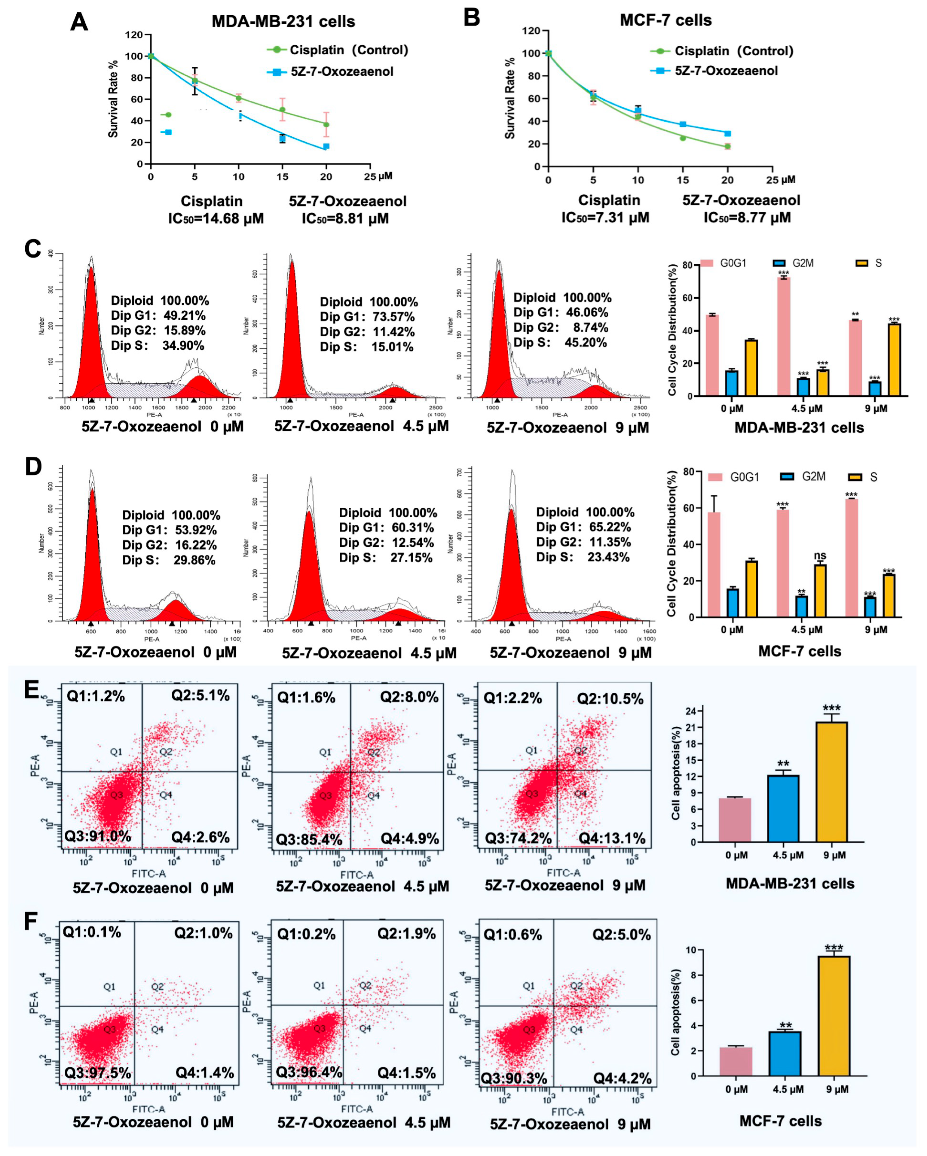
The antiproliferative activity of 5Z-7-Oxozeaenol was evaluated in MDA-MB-231 and MCF-7 breast cancer cells using the MTT assay, with cisplatin serving as a positive control. The results demonstrated that 5Z-7-Oxozeaenol suppressed cell proliferation in a dose-dependent manner. After 48 h of treatment with cisplatin and 5Z-7-Oxozeaenol in MDA-MB-231 cells, the half-maximal inhibitory concentration (IC50) values were 14.68 and 8.81, respectively. In the MCF-7 cells, the IC50 values were 7.30 and 8.77, correspondingly (Figure 2A).
Given the potent cytotoxicity of 5Z-7-Oxozeaenol, we further investigated its effects on cell cycle progression and apoptosis. Treatment of MDA-MB-231 and MCF-7 cells with increasing concentrations of 5Z-7-Oxozeaenol (0, 4.5, and 9 µM) for 48 h resulted in a concentration-dependent decrease in the proportion of cells in G2 phase, accompanied by an accumulation of cells in S and G1 phases (Figure 2B). These findings indicate that 5Z-7-Oxozeaenol induces cell cycle arrest primarily in S and G1 phases.
Apoptosis analysis revealed that 5Z-7-Oxozeaenol treatment significantly increased the total apoptotic cell population (including both early and late apoptotic cells) in MDA-MB-231 cells in a dose-dependent manner. The apoptotic rate increased from 7.7% (0 µM) to 12.9% (4.5 µM; p < 0.001) and further to 23.6% (9 µM; p < 0.001) (Figure 2B). Similarly, MCF-7 cells exhibited a concentration-dependent increase in apoptosis following 5Z-7-Oxozeaenol treatment (Figure 2C). Comparative analysis demonstrated that MDA-MB-231 cells showed significantly greater sensitivity to 5Z-7-Oxozeaenol-induced apoptosis compared to MCF-7 cells (Figure 2C). Notably, 5Z-7-Oxozeaenol exhibited superior efficacy in both suppressing proliferation and inducing apoptosis in MDA-MB-231 cells when compared to the conventional chemotherapeutic agent cisplatin. Based on these findings, subsequent mechanistic studies were conducted using MDA-MB-231 cells.

2.3. 5Z-7-Oxozeaenol Inhibited MDA-MB-231 Cells Migration and Growth

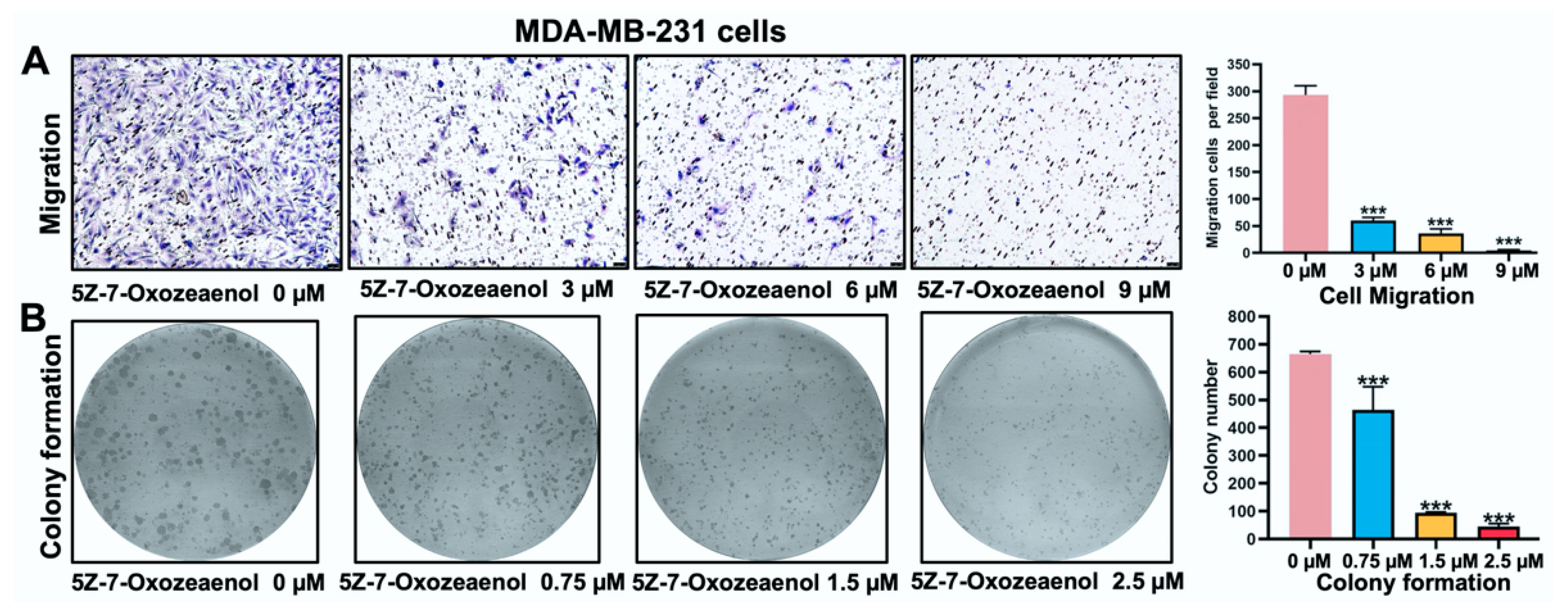
Transwell assays were used to examine the effects of 5Z-7-Oxozeaenol on cell migration ability. Treatment with 5Z-7-Oxozeaenol resulted in a significant reduction in the number of migrated cells compared to the control group, demonstrating its potent inhibitory effect on MDA-MB-231 cell migration (Figure 3A). Furthermore, colony formation assays revealed that 5Z-7-Oxozeaenol suppressed cell growth in a concentration-dependent manner, confirming its strong anti-proliferative activity (Figure 3B).

2.4. RNA-Seq Analysis of MDA-MB-231 Cells Treated with 5Z-7-Oxozeaenol

To elucidate the mechanism of action of 5Z-7-Oxozeaenol on MDA-MB-231 cells, RNA-seq was performed. Transcriptomic analysis following treatment with 5Z-7-Oxozeaenol (9.0 μM) for 48 h revealed distinct clustering of gene expression profiles between treated and control groups, as visualized in the heatmap (Figure 4A).
The RNA-seq identified 1966 differentially expressed genes (DEGs), comprising 1063 significantly upregulated and 903 downregulated genes (Figure 4A). A total of 20 down-regulated genes with |log2(FoldChange)| > 2.0 were identified from the DEGs: CSF3, IL11, TNC, MMP3, ADGRG1, CXCL8, IL24, LAPTM5, IL1B, LINC02009, TGM2, MMP1, KRT81, SCD, ETV1, IL1A, NT5E, CDH4, COL13A1, and GRAMD1B (indicated in the green bar on the right side of Figure 4B). Additionally, 11 up-regulated genes that met the same fold change threshold were selected: ID3, PSG4, CLIC3, CLU, C4BPB, CYP1A1, DPP4, RARRES2, ANGPTL4, SEMA5A, and ALPP (shown in the red bar on the right side of Figure 4B). These genes were subsequently used to construct a protein–protein interaction (PPI) network using the STRING database. PPI network analysis revealed that 5Z-7-Oxozeaenol markedly altered the expression of extracellular matrix (ECM)-related genes, including MMP subfamily members (MMP1, MMP3), fibronectin (FN) and its regulatory genes (NT5E, TNC, CLU, TGM2), CXC chemokine genes (CXCL2, CXCL8, CXCL10), and immunoregulatory genes (IL1A, IL1B, IL11, IL6R, IL9, IL24), all of which were significantly downregulated after 5Z-7-Oxozeaenol treatment (Figure 4B).
Gene Ontology (GO) enrichment analysis highlighted the overall functional characteristics of DEGs, which are significantly enriched genes related to core biological functions, including biological process (BP), molecular function (MF), and cellular component (CC). The enrichment of the results indicated that the response to stimulation, multicellular organism processes, developmental processes, positive regulation of biological processes, and anatomical structure development were clustered in the top five positions of the enriched BP terms. The cell periphery, plasma membrane, endomembrane system, extracellular region and vesicle were clustered in the top five positions of the CC terms. Binding, protein binding, molecular function regulator activity, enzyme binding and signaling receptor binding were clustered in the top five positions of the MF terms (Figure 4C). The Gene Set Enrichment Analysis (GSEA) further supported these findings, indicating significant enrichment in protein tyrosine kinase binding, regulation of immune system processes, peptidase activity, xenobiotic metabolic process, and progesterone metabolic process (Figure S4).
The Kyoto Encyclopedia of Genes and Genomes (KEGG) pathway analysis was conducted to identify the pathways involved. The enrichment results for the DEGs related to functional pathways indicated that 5Z-7-Oxozeaenol had a significant effect on human diseases, organismal systems, and environmental information processing (Figure 4D). Multiple pathways with a high number of DEGs included pathways related to cancer, cytokine–cytokine receptor interaction, MAPK signaling pathway, PI3K-Akt signaling pathway, and cell adhesion molecules. These pathways form an interactive network, with cancer pathways exhibiting notable crosstalk with both PI3K-Akt and MAPK signaling cascades, collectively regulating critical cellular behaviors such as proliferation and apoptosis.

2.5. Molecular Docking for 5Z-7-Oxozeaenol with the Protein Tyrosine Phosphatase Receptor Type N (PTPRN)

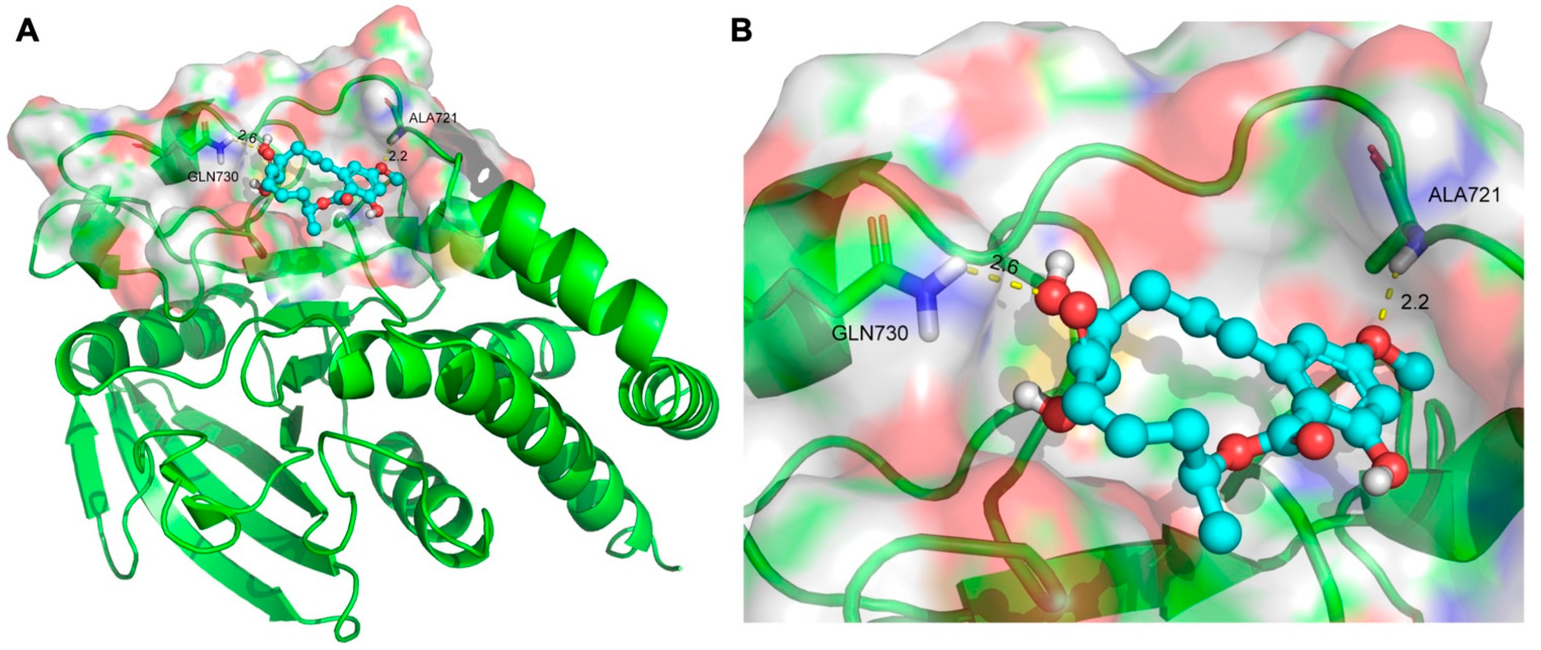
To assess the binding affinity of 5Z-7-Oxozeaenol with potential targets, molecular docking analysis was performed, identifying the receptor-type protein tyrosine phosphatase PTPRN (PDB: 2I1Y) as the top-ranking candidate. The docking results demonstrated that 5Z-7-Oxozeaenol forms hydrogen bonds with key residues ALA721 and GLN730 in the active site of PTPRN, accompanied by a favorable binding energy of −7.6 kcal/mol, indicating a highly stable interaction (Figure 5A,B). These findings suggest that 5Z-7-Oxozeaenol may exert its biological effects by binding to and modulating the activity of PTPRN. Functionally, PTPRN acts as a phosphatase that dephosphorylates tyrosine residues on downstream signaling proteins—including receptor tyrosine kinases (RTKs) and focal adhesion kinase (FAK)—thereby attenuating proliferation, survival, and migration signals mediated by tyrosine phosphorylation.

2.6. Western Blot Analysis of Relative FAK, PI3K-Akt, MAPK Protein Expression

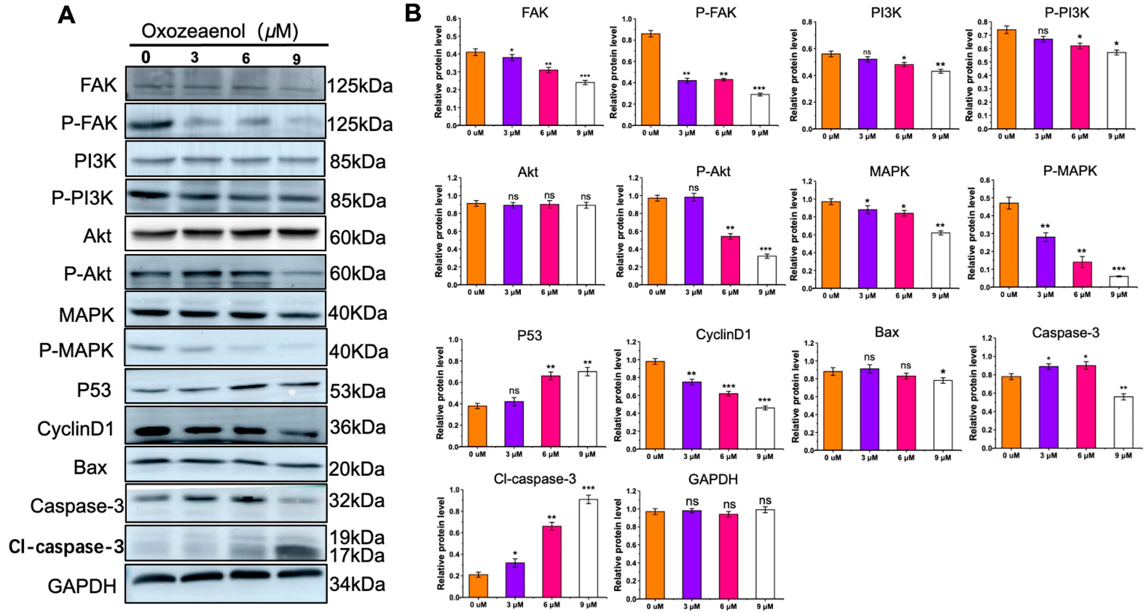
To further validate the KEGG and molecular docking predictions, we examined the expression and phosphorylation levels of key signaling molecules—FAK, PI3K-Akt, and MAPK—implicated in cell proliferation via Western blotting. Treatment of MDA-MB-231 cells with increasing concentrations of 5Z-7-Oxozeaenol for 6 h resulted in a dose-dependent reduction in the phosphorylation levels of FAK, PI3K-Akt, and MAPK (Figure 6A,B). Concurrently, the protein expression of the tumor suppressor p53 increased, while levels of the downstream effector Cyclin D1 decreased in a dose-dependently. Furthermore, a marked increase in cleaved caspase-3 (Cl-caspase-3) was observed following treatment. These results indicate that 5Z-7-Oxozeaenol suppresses cell proliferation and induces apoptosis likely through inhibition of FAK and its downstream PI3K-Akt and MAPK signaling pathways.

3. Discussion

The Beibu Gulf, a semi-enclosed bay situated in the northwestern South China Sea [15], is recognized as a region of global biodiversity significance [16]. It hosts representative marine ecosystems—such as coral reefs, mangroves, seagrass beds, and coastal wetlands—that support rich microbial and broader biological diversity [17]. Between November 2003 and September 2022, scientific studies have reported the discovery of 477 marine natural products from this region, among which 206 are marine terpenoids [18]. Of these terpenoids, 72 have demonstrated notable bioactivities, including cytotoxicity (24 compounds), antibacterial effects (19 compounds), enzyme inhibition (13 compounds), anti-inflammatory properties (10 compounds), among others [19]. Marine natural products play a vital role in the discovery and development of novel therapeutics [18]. Given their significant biological activities and therapeutic potential against various human diseases, these compounds have attracted growing interest among biochemists and pharmacologists [20,21].
5Z-7-Oxozeaenol, originally isolated from a Phoma-related fungal strain (MSX 63935; Mycosynthetix library), was initially identified as a fungal metabolite with limited bioactivity. However, it has since been recognized as a highly selective and potent kinase inhibitor, exhibiting notable antiproliferative and anti-inflammatory properties [14]. Previous studies demonstrated that combining 5Z-7-Oxozeaenol with hyperthermia (HT) enhanced cell death in KRAS-mutant A549 lung cancer cells in a dose-dependent manner [22]. This effect was accompanied by increased reactive oxygen species (ROS) production, loss of mitochondrial membrane potential, and elevated expression of caspase-3 and -7 in HeLa cells, compared to paclitaxel treatment [23]. Additionally, 5Z-7-Oxozeaenol acts as an effective inhibitor of the NF-κB pathway and induces caspase-3 and -7 expression in both HeLa and HT-29 cancer cells [24,25].
In the present study, we investigated the molecular mechanisms underlying the inhibitory effects of 5Z-7-Oxozeaenol in triple-negative breast cancer MDA-MB-231 cells. Our results demonstrate that treatment led to a significant G1 phase cell cycle arrest, accompanied by concentration-dependent suppression of migration and colony formation.
KEGG pathway analysis suggested that the antiproliferative effects of 5Z-7-Oxozeaenol involve multiple interconnected signaling pathways, including FAK, PI3K-Akt, and MAPK, which collectively regulate cellular behaviors. The FAK pathway exhibits extensive crosstalk with both PI3K-Akt and MAPK cascades [26]. For instance, the FAK–Src complex directly phosphorylates and activates PI3K [27], leading to PIP3 production and subsequent Akt activation—a key mechanism promoting cell survival, proliferation, and migration [28]. Furthermore, FAK–Src activates Ras, initiating the Raf–MEK–ERK cascade to drive proliferation and differentiation [29]. FAK also upregulates matrix metalloproteinases (MMPs) via MAPK/ERK or PI3K/AKT signaling, facilitating cell invasion [30,31,32,33]. The PI3K-Akt and MAPK signaling pathways engage in extensive crosstalk, forming a complex, interconnected network that co-regulates essential cellular processes such as proliferation, survival, differentiation, and metabolism. Additionally, Akt can phosphorylate and modulate components of the MAPK pathway, including RAF and MEK, thereby fine-tuning MAPK signaling activity. Conversely, RSK, a downstream effector of MAPK, can phosphorylate and inhibit upstream elements of the PI3K-Akt pathway, creating negative feedback loops. Such reciprocal regulation ensures signal specificity, amplification, and homeostasis [34].
The Western blot analysis confirmed that 5Z-7-Oxozeaenol dose-dependently reduced phosphorylation levels of FAK, PI3K, Akt, and MAPK. Since Akt activation suppresses apoptosis by phosphorylating caspase-9 and inhibiting caspase-3 cleavage, as well as downregulating p53 and Bax, our findings that 5Z-7-Oxozeaenol decreased p-Akt levels while increasing p53, Bax, and cleaved caspase-3 are consistent with Akt pathway inhibition and subsequent induction of MDA-MB-231 cells apoptosis and cell cycle arrest.
Moreover, PPI analysis indicated that 5Z-7-Oxozeaenol downregulated expression of several extracellular matrix (ECM)-related genes (NT5E, TNC, CLU, TGM2) and MMPs (MMP1, MMP3), likely resulting from suppressed FAK phosphorylation.
Cyclin D1, a critical regulator of G1–S transition, is transcriptionally regulated by both MAPK and PI3K/Akt pathways [35,36,37]. Conversely, p53 activation induces CDK inhibitors such as p21, thereby inhibiting Cyclin D1–CDK4/6 activity [34]. In line with this, 5Z-7-Oxozeaenol treatment significantly reduced Cyclin D1 expression in MDA-MB-231 cells, providing a mechanistic basis for the observed G1 arrest.
Through target screening, PTPRN emerged as a top candidate target of 5Z-7-Oxozeaenol. PTPRN, a receptor-type protein tyrosine phosphatase, plays a regulatory role in both the MEK/ERK and PI3K/AKT signaling pathways, contributing to the complex cross-talk between these critical cascades [38]. PTPRN was upregulated in lung adenocarcinoma (LUAD) and promotes metastasis and poor prognosis [39]. In colorectal cancer, PTPRN deletion impairs invasiveness and metastatic potential through dysregulated epithelial–mesenchymal transition (EMT) and insulin receptor signaling [40]. Based on molecular docking and Western blot results, we propose that 5Z-7-Oxozeaenol binds to PTPRN, perturbing its catalytic function and modulating downstream FAK and PI3K-Akt pathways, ultimately impairing cell proliferation, survival, and migration. Nonetheless, the involvement of additional signaling pathways cannot be excluded and warrants further investigation.
In summary, 5Z-7-Oxozeaenol exerts significant anti-tumor effects on MDA-MB-231 cells by inducing G1 phase cell cycle arrest, suppressing migration and colony formation, and promoting apoptosis. Mechanistically, it inhibits phosphorylation of key signaling nodes within the FAK/PI3K-Akt/MAPK network, leading to reduced Akt activation, increased expression of pro-apoptotic proteins (p53, Bax, and cleaved caspase-3), and downregulation of Cyclin D1. Additionally, PPI revealed that 5Z-7-Oxozeaenol downregulated extracellular matrix-related genes and MMPs likely through FAK suppression. Preliminary target screening suggests PTPRN as a potential binding partner, through which it may modulate downstream pathways to impair malignant phenotypes, though further validation is required to elucidate full mechanistic breadth.

4. Materials and Methods

4.1. Fungal Materials

Curvularia sp. MDCW-1060 was isolated from a crab sample collected at the Guangxi Beibu Gulf and identified by the 18S rDNA gene sequence (GenBank accession no. PV355094).

4.2. Fermentation and Extraction

Curvularia sp. MDCW-1060 was cultured at 28 °C for 4 days on PDA medium. Spores were cultivated in 60 × 1000 mL Erlenmeyer flasks (120 mL seawater, 80 g rice). The flasks were cultured under static conditions for 30 days at room temperature. The culture was extracted with EtOAc to yield 52.0 g.

4.3. Isolation

The extracts were further extracted by 90% MeOH–H2O and petroleum ether, yielding 41.5 g of petroleum ether extract and 9.4 g of MeOH–H2O extract. The methanol layer extract was fractionated into 8 fractions (Fr.1–Fr.8) by VLC and eluting with petroleum and ether–EtOAc. Fr.7 (420.2 mg) was purified by HPLC on an ODS column (from 50:50 to 85:15 MeOH/H2O) to yield compound 5Z-7-Oxozeaenol (29.0 mg, tR = 11.6 min). 5Z-7-Oxozeaenol is a colorless powder and was isolated, and the following experimental procedures were performed: Preparative HPLC was performed on an LC3000 system using a preparative C18 column (5 µm, 20 × 250 mm, 10 mL/min Cosmosil, Kyoto, Japan). Analytical HPLC was performed on a Shimadzu LC-10AD VP system using an analytical HPLC C18 column (5 µm, 4.6 × 250 mm, 1 mL/min Cosmosil, Kyoto, Japan). Optical rotation was measured on SGWzz-1 digital polarimeter, HRESIMS spectra were obtained using a Mariner API-TOF, and NMR spectra were recorded on a Bruker Avance 400 spectrometer (Bruker, Fallanden, Switzerland). The optical rotation for 5Z-7-Oxozeaenol is [ α ] D   25 = −148.3 (c = 0.12, MeOH). The structure of 5Z-7-oxozeaenol was determined by its MS, 1H and 13C NMR data (Table 1).

4.4. Cell Culture and Reagents

The human cell lines MDA-MB-231 and MCF-7 were purchased from the Institute of Biochemistry and Cell Biology, Chinese Academy of Sciences (Shanghai, China). Cells were cultured in Dulbecco’s Modified Eagle Medium (DMEM) supplemented with 10% fetal bovine serum (FBS) and 1% penicillin–streptomycin in a humidified incubator at 37 °C with 5% CO2. Stock solutions of 5Z-7-Oxozeaenol were dissolved in dimethylsulfoxide (DMSO), and all treated concentrations were adjusted in the culture medium.

4.5. Cell Proliferation Assays

An MTT assay was used to assess cell viability following exposure to 5Z-7-Oxozeaenol. Cells were initially seeded at a density of 1 × 10⁴ per well in 96-well plates and cultured overnight. In the MTT experiment, five distinct groups of cells were treated with 5Z-7-Oxozeaenol (0, 5, 10, 15, and 20 µM) at 37 °C for 48 h, with each concentration having four replicates. The plates were then added with 0.5 mg/mL MTT solution and incubated at 37 °C for another 4 h. The supernatant was separated, and the formazan crystals were dissolved in 1 mL of DMSO. An aliquot of the DMSO lysed solution (200 μL) was obtained from the 24-well plates and transmitted to 96-well reader plates. The absorbance was measured at a wavelength of 490 nm using a microplate reader (Bio Tek Instruments, Bad Friedrichshall, Germany). The IC50 values were calculated using GraphPad Prism 9.5.

4.6. Cell Cycle Assay

MDA-MB-231 cells were cultured in a 6-well plate at a density of 2 × 10⁵ cells per well and treated with 5Z-7-Oxozeaenol at concentrations of 0, 4.5, and 9 µM for a duration of 48 h. Following treatment, the MDA-MB-231 cells and MCF-7 cells were harvested and fixed overnight using ice-cold ethanol (75%). A working solution containing propidium iodide (PI) at a concentration of 100 μg/mL and RNase A at the same concentration was subsequently added to each sample (500 μL). The samples were then incubated in the dark for 30 min. The cell cycle distribution was analyzed via flow cytometry using an LSRFortessa instrument from BD Biosciences (Canton, MA, USA). For data analysis related to cell cycle progression, ModFit LT version 5.0 software was employed. This experiment was conducted in triplicate to ensure reproducibility.

4.7. Apoptosis Assays

To assess apoptosis, MDA-MB-231 cells and MCF-7 cells were seeded at a density of 3 × 10⁵ cells per well in 6-well culture plates and incubated for 24 h. Following this incubation period, the cells were treated with different concentrations 5Z-7-Oxozeaenol. After an additional two days of incubation, the cells were harvested and resuspended in 1× Annexin-binding buffer. They were subsequently stained with FITC-conjugated Annexin V and propidium iodide (PI). Data acquisition and analysis were performed using a flow cytometer, ensuring that data from a minimum of 10,000 cells per sample were collected for accurate results.

4.8. Cell Migration Assays

The experimental protocol for assessing cellular migration was conducted as below: First, MDA-MB-231 cells were subjected to serum starvation for 12 h to achieve cell cycle synchronization, followed by trypsinization and resuspension in serum-deprived medium. For the cell migration assay, 1 × 10⁵ cells were seeded into upper chambers with serum-free medium, while the lower chambers contained medium with 20% FBS as a chemoattractant in Transwell system equipped with an 8-μm porous membrane (Labselect, Hangzhou, China). After cells were cultured at 37 °C with 5% CO2 for 48 h, the migrated cells adhering to the underside of the membrane were fixed using 4% paraformaldehyde (Biosharp, Shanghai, China) for 30 min and subsequently visualized with 0.5% crystal violet staining for 15 min. Nonmigratory cells remaining on the upper membrane surface were removed using cotton swabs. Quantitative analysis was performed by counting violet-stained cells in three randomly chosen microscopic fields at 100× magnification, with representative images documented using an inverted phase-contrast microscope.

4.9. Cell Colony Formation Assay

In the colony formation assay, MDA-MB-231 cells were seeded in 6-well plates at a density of 500 cells per well and cultured for 24 h. Then, after treating the cells with 5Z-7-oxozeaenol, cells were continuously cultured for 8 days. The growth medium was replaced every 72 h. When colony formation was clearly observed under a microscope, the culture medium was aspirated, and the cells were fixed with 4% paraformaldehyde solution for 15 min. The plate was rinsed three times with phosphate-buffered saline (PBS), and then stained with 0.5% crystal violet solution for 15 min. Subsequently, the plate was washed with PBS to remove unbound dye, ensuring optimal visualization and quantification of colonies.

4.10. RNA-Seq

Total RNA was extracted from cells using a TRIzol reagent kit (Invitrogen, Carlsbad, CA, USA) and assessed for quality using an Agilent 2100 Bioanalyzer system (Agilent Technologies, Palo Alto, CA, USA). Library preparation for sequencing was subsequently conducted using the VAHTS Universal V6 RNA-seq Library Prep Kit for MGI (Vazyme, Nanjing, China) and sequencing was performed on the DNBSEQ T7 platform (Benagen, Wuhan, China). The obtained clean data were processed by eliminating reads containing adapters and low-quality sequences from the raw data. Alignments of the clean reads with the assembled genome of GRCh38_Ensembl_114 were performed using STAR (2.7.9a). The FPKM value of each gene in each sample was determined using RSEM (v 1.3.3). Genes with a p-value of <0.05 and a fold change of >2 were considered to be significantly differentially expressed.

4.11. Construction of the Protein—Protein Interaction (PPI) Network

A protein—protein interaction (PPI) network was constructed using the STRING (https://cn.string-db.org) database, with screening parameters set to a confidence score of 0.7. The cytoHubba plug-in within the Cytoscape 3.10.4 software was used to identify the core modules and the key gene targets with the top 20 scores within the PPI network.

4.12. GO and KEGG Pathway Enrichment Analysis

An enrichment analysis of GO and KEGG pathway terms was subsequently conducted with specific parameters (enrichment conditions: overlap ≥ 3, p value cut-off ≤ 0.05). For the GO enrichment analysis, the 10 pathways with the highest count values and p < 0.01 were selected, encompassing cellular components, molecular functions, and biological processes. Visualization and analysis were carried out using a bioinformatics platform (http://www.bioinformatics.com.cn/, accessed on 16 August 2025) to generate Sankey diagrams and histograms.

4.13. Molecular Docking

A computer simulation approach was employed to evaluate 5Z-7-Oxozeaenol through molecular docking with target proteins. The Maestro program in Glide software version 8.1 was used for docking. The human target protein human PTPRN (ID:2I1Y) was obtained from the PDB protein database (15 September 2025), while the structures of the 5Z-7-oxozeaenol compounds were downloaded from the PubChem database (15 September 2025) and obtained from the PDB database (http://www.rcsb.org, accessed on 15 September 2025).
Prior to docking, water molecules were eliminated from the 3D conformation, and the proteins were hydrogenated. The 3D structures of the target compounds were subsequently energetically minimized for optimization. Molecular docking analysis was conducted to assess the binding interactions between the ligands and receptors, and the potential active compounds were evaluated on the basis of the -CDOCKER ENERGY score.

4.14. Western Blot Analysis

MDA-MB-231 cells were planted in 6 cm dishes at a density of 5 × 10⁵ cells/dish for 24 h and then cultured with various drugs as explained in figure legends. Cells were collected as programmed after treatment under various conditions. Determine protein concentration using the BCA method. Equivalent total proteins (20 μg) of cell lysates were separated through 10% SDS-PAGE and then transferred onto a PVDF membrane for 50–75 min. The PVDF membranes were maintained with 5% nonfat milk in PBST buffer for 1 h to block nonspecific binding. After blocking, the PVDF membranes were incubated with the following primary antibodies at 4 °C overnight. Afterwards, the HRP-conjugated goat anti-mouse (1:3000) or -rabbit secondary antibody (1:3000) was added, and the samples were incubated at room temperature for 2 h. Immunoreactive bands were analyzed using a chemiluminescence Western blot detection system, and quantification was performed using ImageJ 1.8.0 software. All primary antibodies were purchased from Cell Signaling Technology (“CST”, Danvers, MA, USA) (Table 2), and secondary antibodies were purchased from Affinity Biosciences.

4.15. Statistical Analysis

We utilized Prism GraphPad 9.5 software to perform all the statistical analyses, including Student’s t-test, one-way ANOVA, and two-way ANOVA; * p <0.05, ** p < 0.01, *** p < 0.001, **** p < 0.0001.

Supplementary Materials

The following supporting information can be downloaded at: https://www.mdpi.com/article/10.3390/md23110414/s1, Figure S1: 1H NMR (400 MHz) spectrum of compound 1 in DMSO-d6. Figure S2: 13C NMR (100 MHz) spectrum of compound 1 in DMSO-d6. Figure S3: HR-ESI-MS spectrum of compound 1. Figure S4: GSEA was conducted using GO pathways’ biological process branch as the gene sets of interest.

Author Contributions

Conceptualization, X.Q., Y.L. and C.W.; methodology, H.Z., K.L. and. J.W.; software, J.W. and K.L.; validation, J.W., K.L., H.Z., C.X. and J.X.; formal analysis, J.W. and H.Z.; investigation, J.W., Z.H. and H.Z.; resources, C.W. and X.Q.; data curation, J.W., C.X. and Z.H.; writing—original draft preparation, J.W., C.W., X.Q. and H.Z.; writing—review and editing, C.W. and X.Q.; visualization, H.Z.; supervision, C.W. and X.Q.; project administration, C.W., Y.L. and X.Q.; funding acquisition, C.W. and X.Q. All authors have read and agreed to the published version of the manuscript.

Funding

This research was funded by the National Natural Science Foundation of China. (No. 82204276), the Natural Science Foundation of Guangxi (No. 2024GXNSFAA010459, No. 2025GXNSFAA069557) and the Research Launching Fund Project from Guangxi University of Chinese Medicine Introduced Doctoral (2022BS021).

Institutional Review Board Statement

Not applicable.

Data Availability Statement

The data presented in this study are available upon request from the corresponding author.

Conflicts of Interest

The authors declare no conflicts of interest.

Abbreviations

The following abbreviations are used in this manuscript:
IARCInternational agency for research on cancer
PIPropidium iodide
PTPRNProtein tyrosine phosphatase receptor type N
MTTMethylthiazolyldiphenyl-tetrazolium bromide
DEGsDifferentially expressed genes
PPIProtein–Protein interactions
GOGene ontology
KEGGKyoto encyclopedia of genes and genomes
GSEAGene set enrichment analysis
BPBiological Processes
MFMolecular functions
CCCellular components
MMPMatrix metallopeptidase
RTKsReceptor tyrosine kinases
FAKFocal adhesion kinase
ECMExtracellular Matrix
HTHyperthermia
ROSReactive oxygen species
PFsPurkinje Fibers
PTPProtein tyrosine phosphatase
LUADLung adenocarcinoma
CRCColorectal cancer

References

  1. Bashey, S.Z.; Kordon, A.; Ositelu, K.; Rao, A.; Akhter, N. The role of statins in breast cancer survivors. Breast Cancer Res. Tr. 2025, 210, 1–10. [Google Scholar] [CrossRef]
  2. Dee, E.C.; Laversanne, M.; Bhoo-Pathy, N.; Ho, F.D.; Feliciano, E.J.G.; Eala, M.A.B.; Ting, F.I.L.; Ginsburg, O.; Moraes, F.Y.; Gyawali, B.; et al. Cancer incidence and mortality estimates in 2022 in southeast Asia: A comparative analysis. Lancet Oncol. 2025, 26, 516–528. [Google Scholar] [CrossRef] [PubMed]
  3. Bray, F.; Laversanne, M.; Sung, H.Y.A.; Ferlay, J.; Siegel, R.L.; Soerjomataram, I.; Jemal, A. Global cancer statistics 2022: GLOBOCAN estimates of incidence and mortality worldwide for 36 cancers in 185 countries. CA-Cancer J. Clin. 2024, 74, 229–263. [Google Scholar] [CrossRef] [PubMed]
  4. Lei, S.Y.; Zheng, R.S.; Zhang, S.W.; Chen, R.; Wang, S.M.; Sun, K.X.; Zeng, H.M.; Wei, W.Q.; He, J. Breast cancer incidence and mortality in women in China: Temporal trends and projections to 2030. Cancer Biol. Med. 2021, 18, 900–909. [Google Scholar] [CrossRef]
  5. Jing, Z.J.; Huang, W.Q.; Mei, J.; Bhushan, S.; Wu, X.H.; Yan, C.B.; Zheng, H.M.; Yang, Y.L. Advances in novel cell death mechanisms in breast cancer: Intersecting perspectives on ferroptosis, cuproptosis, disulfidptosis, and pyroptosis. Mol. Cancer 2025, 24, 224. [Google Scholar] [CrossRef]
  6. Zeng, C.; Lin, M.X.; Jin, Y.Z.; Zhang, J. Identification of Key Genes Associated with Brain Metastasis from Breast Cancer: A Bioinformatics Analysis. Med. Sci. Monit. 2022, 28, e935071-1–e935071-9. [Google Scholar] [CrossRef]
  7. Chen, Y.C.; Lien, W.C.; Su, S.Y.; Jhuang, J.R.; Chiang, C.J.; Yang, Y.W.; Lee, W.C. Birth Cohort Effects in Breast Cancer Incidence: Global Patterns and Trends. Am. J. Epidemiol. 2022, 191, 1990–2001. [Google Scholar] [CrossRef]
  8. Puklin, L.S.; Ferrucci, L.M.; Harrigan, M. Improving Lifestyle Behaviors During Chemotherapy for Breast Cancer: The Lifestyle, Exercise, and Nutrition Early After Diagnosis (LEANer) Trial; American Cancer Society: Atlanta, GA, USA, 2024; Volume 130, pp. 2440–2452. [Google Scholar]
  9. Radwan, R.M.; Schuster, A.L.R.; Hertz, D.L.; Lustberg, M.B.; Vachhani, H.R.; Zacholski, E.H.; Sheppard, V.B.; Bridges, J.F.P.; Salgado, T.M. Tolerance for chemotherapy-induced peripheral neuropathy among women with metastatic breast cancer: A discrete-choice experiment. Breast Cancer Res. Treat. 2025, 212, 149–159. [Google Scholar] [CrossRef]
  10. Liu, X.X.; Zhuang, C.Y.; Liu, L.; Xiong, L.; Xie, X.; He, P.; Li, J.J.; Wei, B.; Yan, X.; Tian, T.L.; et al. Exploratory phase II trial of an anti-PD-1 antibody camrelizumab combined with a VEGFR-2 inhibitor apatinib and chemotherapy as a neoadjuvant therapy for triple-negative breast cancer (NeoPanDa03): Efficacy, safety and biomarker analysis. Signal Transduct. Target. Ther. 2025, 10, 237. [Google Scholar] [CrossRef]
  11. Xiong, X.; Zheng, L.W.; Ding, Y.; Chen, Y.F.; Cai, Y.W.; Wang, L.P.; Huang, L.; Liu, C.C.; Shao, Z.M.; Yu, K.D. Breast cancer: Pathogenesis and treatments. Signal Transduct. Target. Ther. 2025, 10, 49. [Google Scholar] [CrossRef]
  12. Ma, C.; Xu, A.S.; Zuo, L.P.; Li, Q.; Fan, F.J.; Hu, Y.; Sun, C.Y. Methionine Dependency and Restriction in Cancer: Exploring the Pathogenic Function and Therapeutic Potential. Pharmaceuticals 2025, 18, 640. [Google Scholar] [CrossRef]
  13. Acuña, U.M.; Wittwer, J.; Ayers, S.; Pearce, C.J.; Oberlies, N.H.; De Blanco, E.J.C. Effects of (5Z)-7-Oxozeaenol on the Oxidative Pathway of Cancer Cells. Anticancer Res. 2012, 32, 2665–2671. [Google Scholar]
  14. Ayers, S.; Graf, T.N.; Adcock, A.F.; Kroll, D.J.; Matthew, S.; de Blanco, E.J.C.; Shen, Q.; Swanson, S.M.; Wani, M.C.; Pearce, C.J.; et al. Resorcylic Acid Lactones with Cytotoxic and NF-κB Inhibitory Activities and Their Structure-Activity Relationships. J. Nat. Prod. 2011, 74, 1126–1131. [Google Scholar] [CrossRef]
  15. Yuan, W.H.; Yu, R.C.; Sun, W. Unique climate features of the rainfall center along the northern coast of the Beibu Gulf. Clim. Dyn. 2025, 63, 169. [Google Scholar] [CrossRef]
  16. Chen, B.Y.; Lu, F.T.; Zhang, X.L.; Wu, C.W.; Lü, L.G.; Jefferson, T.A. The largest known population of Eden’s whale aggregates in the Beibu Gulf, southern China. Mar. Mammal Sci. 2025, 41, e13226. [Google Scholar] [CrossRef]
  17. Che, W.X.; Zhao, H.; Man, Y.; Tan, X. Spatial characteristics of microbial communities and their functions in sediments of subtropical Beibu Gulf, China. Mar. Environ. Res. 2025, 207, 107077. [Google Scholar] [CrossRef] [PubMed]
  18. Wang, J.M.; Qin, Y.N.; Lin, M.P.; Song, Y.Y.; Lu, H.M.; Xu, X.Y.; Liu, Y.H.; Zhou, X.F.; Gao, C.H.; Luo, X.W. Marine Natural Products from the Beibu Gulf: Sources, Chemistry, and Bioactivities. Mar. Drugs 2023, 21, 63. [Google Scholar] [CrossRef] [PubMed]
  19. Carroll, A.R.; Copp, B.R.; Grkovic, T.; Keyzers, R.A.; Prinsep, M.R. Marine natural products. Nat. Prod. Rep. 2025, 42, 257–297. [Google Scholar] [CrossRef]
  20. Brito, M.F.; Fagundes, T.S.F.; Jimenez, P.C.; Valverde, A.L. Marine Natural Product as a Source of New Drugs: 15 Years and One Pandemic Later. Quim. Nova 2025, 48, e20250167. [Google Scholar] [CrossRef]
  21. Alves, C.; Diederich, M. Marine Natural Products as Anticancer Agents 3.0. Mar. Drugs 2025, 23, 243. [Google Scholar] [CrossRef]
  22. Acuña, U.M.; Wittwer, J.; Ayers, S.; Pearce, C.J.; Oberlies, N.H.; de Blanco, E.J.C. Effects of (5Z)-7-Oxozeaenol on MDA-MB-231 Breast Cancer Cells. Anticancer Res. 2012, 32, 2415–2421. [Google Scholar] [PubMed]
  23. Li, P.; Zhao, Q.L.; Jawaid, P.; Rehman, M.U.; Sakurai, H.; Kondo, T. Enhancement of hyperthermia-induced apoptosis by 5Z-7-oxozeaenol, a TAK1 inhibitor, in A549 cells. Cell Stress Chaperon. 2016, 21, 873–881. [Google Scholar] [CrossRef] [PubMed]
  24. Chien, C.Y.; Chien, C.T.; Wang, S.S. Progressive thermopreconditioning attenuates rat cardiac ischemia/reperfusion injury by mitochondria-mediated antioxidant and antiapoptotic mechanisms. J. Thorac. Cardiov. Sur. 2014, 148, 705–713. [Google Scholar] [CrossRef] [PubMed]
  25. Fan, Y.H.; Cheng, J.; Vasudevan, S.A.; Patel, R.H.; Liang, L.; Xu, X.; Zhao, Y.L.; Jia, W.; Lu, F.M.; Zhang, H.; et al. TAK1 inhibitor 5Z-7-oxozeaenol sensitizes neuroblastoma to chemotherapy. Apoptosis 2013, 18, 1224–1234. [Google Scholar] [CrossRef]
  26. Yang, Z.Z.; Liu, H.T.; Song, Y.S.; Gao, N.; Gao, P.; Hui, Y.R.; Li, Y.H.; Fan, T.L. Luteolin enhances drug chemosensitivity by downregulating the FAK/PI3K/AKT pathway in paclitaxel-resistant esophageal squamous cell carcinoma. Int. J. Mol. Med. 2024, 54, 77. [Google Scholar] [CrossRef]
  27. Liu, J.Y.; Liu, T.; Qian, X.; Ma, D.Y.; Chen, L.L.; Li, A.L.; Liang, X.R.; Hu, Z.; Qi, C.X.; Qi, W. Tuina Inhibits Chondrocyte Apoptosis by Regulating the FAK/PI3K/Akt Pathway in Rabbits with IVDD. Comb. Chem. High Throughput Screen. 2025, 28, 1746–1753. [Google Scholar] [CrossRef]
  28. Sun, L.H.; Chen, W.; Yuan, W.X.; Huang, Q.W.; Yang, H.; Zhang, W.; Tang, J.J.; Hu, P. Ginkgetin inhibits the proliferation and migration of lung cancer cells via FAK/STAT3/AKT pathway. Mol. Biol. Rep. 2025, 52, 458. [Google Scholar] [CrossRef]
  29. Tan, J.C.; Lin, G.Q.; Zhang, R.; Wen, Y.T.; Luo, C.Y.; Wang, R.; Wang, F.Y.; Peng, S.J.; Zhang, J.G. Bufotalin Induces Oxidative Stress-Mediated Apoptosis by Blocking the ITGB4/FAK/ERK Pathway in Glioblastoma. Antioxidants 2024, 13, 1179. [Google Scholar] [CrossRef]
  30. Guo, J.; Santiago-O’Farrill, J.M.; Orellana, V.; Ozyurt, R.; Yang, H.L.; Pina, M.; Bildik, G.; Mao, W.Q.; Bast, R.C.; Lu, Z. DIRAS3 Inhibits Ovarian Cancer Cell Growth by Blocking the Fibronectin-Mediated Integrin β1/FAK/AKT Signaling Pathway. Cells 2025, 14, 1250. [Google Scholar] [CrossRef]
  31. Zhou, Q.W.; You, Y.; Zhao, Y.Y.; Xiao, S.X.; Song, Z.Q.; Huang, C.X.; Qian, J.L.; Lu, W.Q.; Tong, H.X.; Zhang, Y.; et al. TRPV4 drives the progression of leiomyosarcoma by promoting ECM1 generation and co-activating the FAK/PI3K/AKT/GSK3β pathway. Cell. Oncol. 2025, 48, 455–470. [Google Scholar] [CrossRef]
  32. Wu, T.J.; Lin, C.Y.; Tsai, C.H.; Huang, Y.L.; Tang, C.H. Glucose suppresses IL-1β-induced MMP-1 expression through the FAK, MEK, ERK, and AP-1 signaling pathways. Environ. Toxicol. 2018, 33, 1061–1068. [Google Scholar] [CrossRef] [PubMed]
  33. Gupta, N.; Nafees, S.; Rahman, A.U.; Akhtar, J.; Khan, A.A.; Sharma, A. Itrifal-e-Aftimoon potentiates imatinib-induced anti-leukemic effect by influencing FAK/STAT/Akt/ERK signalling pathways against chronic myeloid leukaemia in vitro. J. Pharm. Pharmacol. 2022, 74, 1330–1341. [Google Scholar] [CrossRef] [PubMed]
  34. Chang, W.L.; Yang, K.C.; Peng, J.Y.; Hong, C.L.; Li, P.C.; Chye, S.M.; Lu, F.J.; Shih, C.W.; Chen, C.H. Parecoxib Enhances Resveratrol against Human Colorectal Cancer Cells through Akt and TXNDC5 Inhibition and MAPK Regulation. Nutrients 2024, 16, 3020. [Google Scholar] [CrossRef] [PubMed]
  35. Skopek, R.; Özcan, S.G.; Chmiel, P.; Morgner, S.; Schütt, J.; Zadegan, F.G.; Stanko, C.; Palusinska, M.; Maslinska-Gromadka, K.; Sbirkov, Y.; et al. Synergistic potential of CDK4/6 inhibitors and ATRA in non-APL AML. Br. J. Haematol. 2025, 207, 1279–1288. [Google Scholar] [CrossRef]
  36. Cardona-Benavides, I.J.; Cristobal-Vargas, S.; De Ramón, C.; Rojas, E.A.; Misiewicz-Krzeminska, I.; Isidro, I.; Pérez, J.J.; Paiva, B.; Puig, N.; Alcoceba, M.; et al. Exploring the Role of Cyclin D1 in the Pathogenesis of Multiple Myeloma Beyond Cell Cycle Regulation; Molecular Oncology: Heidelberg, Germany, 2025. [Google Scholar]
  37. Suzuki, Y.; Usuki, S.; Nishizawa, M.; Tanaka, N.; Suhara, Y.; Yajima, I. Acyclic Retinoid Overcomes Vemurafenib Resistance in Melanoma Cells via Dual Inhibition of MAPK and PI3K/AKT/mTOR Pathways. Anticancer Res. 2025, 45, 2265–2278. [Google Scholar] [CrossRef]
  38. Kambaru, A.; Chaudhary, N. Role of Protein Tyrosine Phosphatase in Regulation of Cell Signaling Cascades Affecting Tumor Cell Growth: A Future Perspective as Anti-Cancer Drug Target. Curr. Pharm. Biotechnol. 2022, 23, 920–931. [Google Scholar] [CrossRef]
  39. Song, X.Y.; Jiao, X.; Yan, H.; Yu, L.F.; Jiang, L.Y.; Zhang, M.; Chen, L.Z.; Ju, M.Y.; Wang, L.; Wei, Q.; et al. Overexpression of PTPRN Promotes Metastasis of Lung Adenocarcinoma and Suppresses NK Cell Cytotoxicity. Front. Cell Dev. Biol. 2021, 9, 622018. [Google Scholar] [CrossRef]
  40. Garranzo-Asensio, M.; Solís-Fernández, G.; Montero-Calle, A.; García-Martínez, J.M.; Fiuza, M.C.; Pallares, P.; Palacios-Garcia, N.; García-Jiménez, C.; Guzman-Aranguez, A.; Barderas, R. Seroreactivity Against Tyrosine Phosphatase PTPRN Links Type 2 Diabetes and Colorectal Cancer and Identifies a Potential Diagnostic and Therapeutic Target. Diabetes 2022, 71, 497–510. [Google Scholar] [CrossRef]
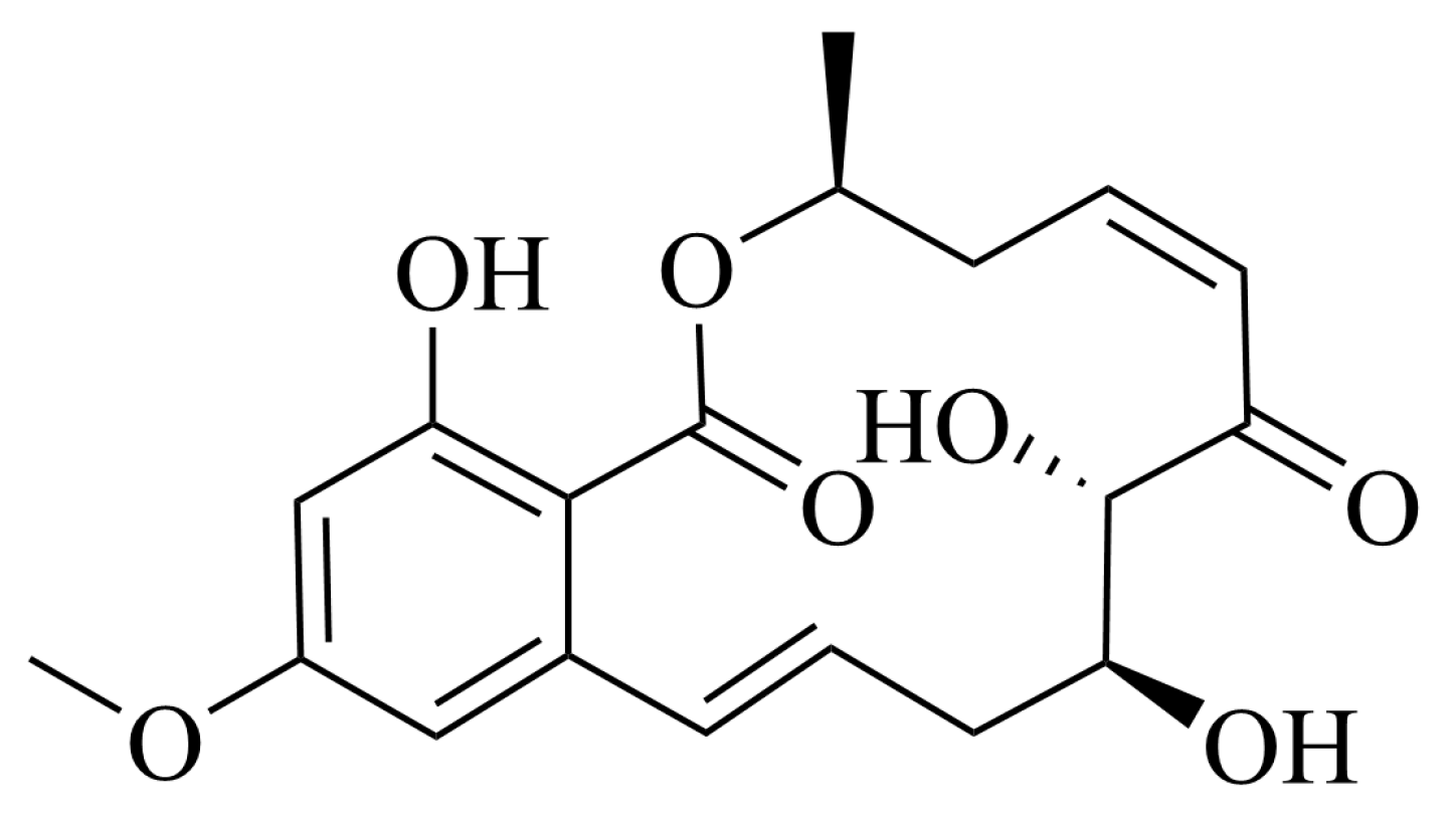
Figure 1. The structure of 5Z-7-Oxozeaenol.
Marinedrugs 23 00414 g001
Figure 2. Antiproliferative efficacy of 5Z-7-Oxozeaenol on breast cancer cells: (A) The cell viability was determined by an MTT assay in MDA-MB-231 cells. (B) The cell viability was determined by a MTT assay in MCF-7 cells. (C) The cell cycle distribution was assessed by flow cytometric analysis in MDA-MB-231 cells. (D) The cell cycle distribution was assessed by flow cytometric analysis in MCF-7 cells. (E) The cell apoptosis rate was assessed by flow cytometric analysis in MDA-MB-231 cells. (F) The cell apoptosis rate was assessed by flow cytometric analysis in MCF-7 cells; ns, not significant, ** p < 0.01, *** p < 0.001, compared with the control group.
Marinedrugs 23 00414 g002
Figure 3. 5Z-7-Oxozeaenol inhibited MDA-MB-231 cells migration and growth. (A) Transwell assays evaluating the migration ability of MDA-MB-231 cells treated with the indicated concentrations of 5Z-7-Oxozeaenol for 48 h. (B) MDA-MB-231 cells were treated with the indicated concentrations of 5Z-7-Oxozeaenol for 8 days, after which colony formation was assessed; *** p < 0.001, compared with the control group.
Marinedrugs 23 00414 g003
Figure 4. Transcriptome analysis of MDA-MB-231 cells treated with compound 5Z-7-Oxozeaenol vs. the control: (A) Volcanic map showing the differential gene expression distribution. The scattered dots in the figure represent individual genes, with grey dots indicating genes whose expression did not with not significantly differ, red dots representing upregulated genes whose expression did not significantly increased, and blue dots representing downregulated genes whose expression significantly decreased. (B) Construction of the PPI network for the identification of hub genes. Colored nodes: query proteins and first shell of interactors. White nodes: second shell of interactors. The red bars on the right side of the PPI chart represent: 11 up-regulated genes with |log2(FoldChange)| > 2.0 were selected from the DEGs. The green bars on the right side of the PPI chart represent: 20 down-regulated genes with |log2(FoldChange)| > 2.0 were identified from the DEGs. (C) GO functional enrichment analysis of the DEGs. (D) KEGG pathway enrichment of DEGs.
Marinedrugs 23 00414 g004
Figure 5. The binding mode of 5Z-7-Oxozeaenol to its targets, determined by molecular docking: (A) Three-dimensional structures of the binding pockets were constructed for the binding mode of 5Z-7-Oxozeaenol to PTPRN (PDB: 2I1Y): (B) 5Z-7-Oxozeaenol docking with the active site of the target protein PTPRN.
Marinedrugs 23 00414 g005
Figure 6. Effects of 5Z-7-Oxozeaenol on FAK, PI3K-Akt and MAPK pathway-related protein expression in MDA-MB-231 cells. (A) FAK/p-FAK, PI3K/p-PI3K, Akt/p-Akt, MAPK/P-MAPK, p53, Cyclin D1, Bax, Caspase-3 and Cl-caspase-3 protein levels were analyzed by Western blotting. The analysis utilized actin as a reference for quantifying relative protein expression levels. (B) Relative quantification of protein expression compared with that in the control group. ns, not significant, * p < 0.05, ** p < 0.01, and *** p < 0.001, compared with the control group.
Marinedrugs 23 00414 g006
Table 1. 1H-NMR (400 MHz) and 13C-NMR (100 MHz) data of 5Z-7-Oxozeaenol in DMSO-d6.
No.δC, TypeδH, Mult. (J in Hz)
1170.4 (C)
373.8 (CH)5.20, qdd (12.0, 5.9, 1.9)
436.3 (CH2)2.58, m
3.20, m
5142.4 (CH)6.19, ddd (13.6, 10.0, 3.5)
6126.7 (CH)6.43, dd (11.3, 2.6)
7200.9 (C)
881.7 (CH)4.38, m
972.7 (CH)3.85, m
1036.0 (CH2)2.02, m
11132.4 (CH)6.06, ddd (14.9, 9.2, 5.2)
12131.0 (CH)6.74, dd (15.3)
13143.9 (C)
14106.1 (CH)6.42, d (2.5)
15163.4 (C)
16100.2 (CH)6.38, d (2.5)
17163.7 (C)
18104.5 (C)
1920.1 (CH3)1.37, d (6.1)
2055.5 (CH3)3.78, s
8-OH 4.98, d (4.9)
9-OH 5.06, d (6.1)
17-OH 11.77, s
Table 2. The primary antibody product used in this article.
No.Primary AntibodyDilutionIsotypeSource
1FAK1:1000Rabbit IgG (#71433)Cell Signaling Technology
3P-FAK1:1000Rabbit IgG (#8556)Cell Signaling Technology
4PI3K1:1000Rabbit IgG (#4257)Cell Signaling Technology
5P-PI3K1:1000Tyr458/199 (#4228)Cell Signaling Technology
6Akt1:1000Rabbit IgG (#4691)Cell Signaling Technology
7P-Akt1:2000Rabbit IgG (#4060)Cell Signaling Technology
8MAPK1:1000Rabbit IgG (#8690)Cell Signaling Technology
9P-MAPK1:1000Rabbit IgG (#4377)Cell Signaling Technology
10P531:1000Rabbit IgG (#2527)Cell Signaling Technology
11CyclinD11:1000Rabbit IgG (#55506)Cell Signaling Technology
12Bax1:1000Rabbit IgG (#5023)Cell Signaling Technology
13Caspase-31:1000Rabbit IgG (#14220)Cell Signaling Technology
14Cleaved-caspase-31:1000Rabbit IgG (#9664)Cell Signaling Technology
15GAPDH1:1000Mouse IgG (#T0004)Cell Signaling Technology
Disclaimer/Publisher’s Note: The statements, opinions and data contained in all publications are solely those of the individual author(s) and contributor(s) and not of MDPI and/or the editor(s). MDPI and/or the editor(s) disclaim responsibility for any injury to people or property resulting from any ideas, methods, instructions or products referred to in the content.