Article

Tracing the Impact of Domestic Storage Conditions on Antioxidant Activity and Lipid Profiles in the Edible Microalgae Chlorella vulgaris and Tetraselmis chui

1
Centre for Environmental and Marine Studies (CESAM), Department of Chemistry, Campus Universitário de Santiago, University of Aveiro, 3810-193 Aveiro, Portugal
2
Mass Spectrometry Centre & Associated Laboratory for Green Chemistry of the Network of Chemistry and Technology (LAQV-REQUIMTE), Department of Chemistry, Campus Universitário de Santiago, University of Aveiro, 3810-193 Aveiro, Portugal
*
Author to whom correspondence should be addressed.
Mar. Drugs 2024, 22(6), 254; https://doi.org/10.3390/md22060254
Submission received: 29 April 2024 / Revised: 21 May 2024 / Accepted: 24 May 2024 / Published: 30 May 2024

Abstract

:
The microalgae Chlorella vulgaris and Tetraselmis chui are valued for their nutrient-rich content, including lipids and polyunsaturated fatty acids (PUFA). However, little is known about how storage and processing affect their lipid quality. This study aimed to assess the impact of domestic storage and cooking practices in dried biomass of C. vulgaris and T. chui. Four conditions were tested: control (newly opened package), light (storage at room temperature and daily light regimen for three weeks), frozen (storage in the freezer at −20 °C for three weeks), and heated (three cycles of 90 min at 100 °C). Lipid extracts were analyzed by GC-MS and LC-MS, and antioxidant activity through DPPH and ABTS radical scavenging assays. Tested storage conditions promoted a decrease in fatty acid content and in diacyl/lyso lipid species ratios of phospholipid (PC/LPC, PE/LPE) and betaine lipids (DGTS/MGTS). Lipid extracts from light treatment showed the lowest antioxidant activity in C. vulgaris (ABTS, IC40: 104.9; DPPH, IC20: 187.9 ± 15.0), while heat affected the antioxidant activity of T. chui (ABTS, IC40: 88.5 ± 2.8; DPPH, IC20 209.4 ± 10.9). These findings underscore the impact of managing storage and processing conditions to optimize the nutritional and functional benefits of C. vulgaris and T. chui in food and feed applications.

1. Introduction

Current consumer demand for natural-origin and functional food is increasing the research of matrices with health-promoting properties. Within this realm, microalgae have emerged as prominent candidates, boasting a diverse array of high-value nutritional molecules and demonstrating potential as feedstock for the production of bioactive compounds [1,2,3,4].
Chlorella vulgaris and Tetraselmis chui from the Chlorophyta phylum have garnered significant attention for their incorporation into functional food and feed formulations [5]. Chlorella vulgaris is a freshwater green microalga approved for human nutrition, esteemed for its high protein content, and a source of nutritional lipids [6]. This microalga has high photosynthetic ability and adaptability to various growth conditions, including autotrophic, mixotrophic, and heterotrophic modes, making it well-suited for large-scale cultivation and commercial production [7]. On the other hand, T. chui is a flagellated marine green microalga boasting rapid growth, a non-toxic nature, and remarkable nutritional value composed mainly of proteins with all essential amino acids, carbohydrates, and fats [1,8]. Due to its antioxidant properties and nutritional significance, it is extensive used as efficient feed in the aquaculture industry [9,10,11]. Moreover, dried biomass of T. chui has been authorized in the EU as novel food and food supplement (Commission Implementing Regulation (EU) 2017/2470) [12,13,14].
The significance of these microalgae species lies in their potential as sources of beneficial compounds, particularly lipids and polyunsaturated fatty acids (PUFA), including essential omega-3 (n-3) and omega-6 (n-6) PUFA-like alpha-linolenic acid (18:3 n-3, ALA) and linoleic acid (18:2 n-6, LA), respectively. Given that ALA and LA are not synthesized in mammalian cells, their intake through dietary sources is crucial for the proper metabolism of these organisms [15]. Furthermore, in vitro assays have demonstrated that lipid extracts of C. vulgaris exhibits antioxidant and anti-inflammatory properties [16,17], underscoring the functional significance of these compounds.
However, the biochemical profile of food matrices is significantly influenced by storage conditions [18,19]. Regarding lipids, during the food manufacturing and storage, they are susceptible to degradation when exposed to excess humidity, heat, and light. These factors can prompt the hydrolysis of esters bonds, leading to the release of fatty acids (FA) chains and formation of lyso forms (e.g., lysophosphatidylcholines) [20,21]. Furthermore, lipids are prone to oxidation, which can compromise both sensory attributes and health-promoting qualities, resulting in the production of low molecular weight off-flavor compounds, depletion of antioxidants, and accumulation of free radicals [22]. The stability of lipids against oxidation is primarily influenced by oxygen, temperature, and light [21]. Moreover, household practices like freezing and cooking can also tiger lipid oxidation, leading in loss of quality [23].
In this regard, assessing the stability of the biochemical composition of algae biomass under various storage scenarios becomes important. Despite the promising attributes of microalgae-derived lipids, their vulnerability to degradation poses a critical challenge to maintaining the integrity and efficacy of the biomass [24]. Storage conditions play a pivotal role in preserving the pigment, FA, and lipid profiles, as well as antioxidant activities inherent to microalgae lipid extracts. Understanding the changes induced by different conditions is crucial for optimizing strategies to preserve microalgae biomass, ensuring the high-quality lipid extracts for commercial applications.
This study aims to evaluate the impact of simulated domestic storage and cooking practices of dried biomass of C. vulgaris and T. chui on their lipid extract, focusing on alteration in the lipid composition and antioxidant activity. Four conditions were selected for analysis: condition 1—control: biomass stored at room temperature under vacuum, protected from light and opened for the first time on the day of analysis; condition 2—light: biomass stored at room temperature in an open container exposed to light and oxygen for 3 weeks; condition 3—frozen: biomass stored in the freezer at −20 °C for three weeks; and condition 4—heated: biomass heated at 100 °C for 90 min during three cycles. Lipids were extracted from the biomass subject to different conditions, and their FA and lipid profiles were analyzed using gas chromatography–mass spectrometry (GC-MS) and liquid chromatography (LC-MS), respectively. Additionally, the antioxidant activity of the lipid extracts was evaluated by 2,20-Azino-bis-3-Ethylbenzothiazoline-6-Sulfonic Acid (ABTS) and 2-Diphenyl-1-Picrylhydrazyl (DPPH) radical scavenging assays.

2. Results

2.1. Fatty Acids Profile and Lipid Quality Indices

The lipid yield of C. vulgaris under control, light, frozen, and heated conditions corresponded to 4.6 ± 0.7%, 5.7 ± 0.8%, 5.0 ± 0.3%, and 6.6 ± 0.6% of dry weight (DW), respectively. For T. chui, this yield corresponds to a 3.9 ± 0.8%, 5.7 ± 2.0%, 6.3 ± 0.4%, and 6.5 ± 0.9% DW, respectively.
The FA profile of C. vulgaris and T. chui exposed to distinct storage and analyzed by gas chromatography–mass spectrometry (GC-MS) allowed for identifying a total of eight FA in C. vulgaris (Table 1) and eleven in T. chui (Table 2). Chlorella vulgaris profile showed the FA 16:0, linoleic acid (LA, 18:2n-6), and the alpha linolenic acid (ALA, 18:3n-3) as the most abundant ones (Table 1). In the T. chui profile, 16:0, 16:4n-3, and ALA were the predominant FA (Table 2).
In C. vulgaris, the FA analysis revealed a decrease of each fatty acid across all conditions compared to the control condition, which was most pronounced under light and frozen conditions. Specifically, the highest reduction in light condition was observed in the most unsaturated FA, such as 16:3n-3 and 18:3n-3, which exhibited significant differences compared to the other conditions (Table 1). Additionally, the n-6/n-3 ratio remained lower than 1 across all conditions, with PUFA exhibiting the lowest value under light condition.
Lipid quality indices, atherogenicity index (AI) and thrombogenicity index (TI), showed no differences in all conditions (AI and TI equal to 0.2 ± 0.0), with exception of ice conditions (TI equal to 0.1 ± 0.0). The hypo/hypercholesterolemic ratio (h/H) was lower in control and frozen conditions but significative differences were not recorded between storages conditions (Table 1).
In the FA profiles of T. chui, the most pronounced differences were observed in the unsaturated FA 16:4n-3 and 18:3n-3, which exhibited reductions under light conditions. However, no significant differences were recorded in FA composition between storage conditions (Table 2). Similarly to C. vulgaris, T. chui also exhibited a consistently low n-6/n-3 ratio (0.2) across all conditions, with n-3 PUFA displaying the lowest value under light conditions. Lipid quality indices showed no significative differences between conditions. The AI was equal to 0.3 ± 0.0 with exception of control conditions (0.4 ± 0.0), while the TI was equal to 0.2 ± 0.0 in control and light conditions and lower in frozen and heated conditions (0.1 ± 0.0). The hypo/hypercholesterolemic ratio was lower in control condition although with no significative differences.

2.2. Lipid Ratios

The analysis of lipidomics data using the MS Dial software allowed for the identification of lyso species of phospholipids phosphatidylcholine (PC) and phosphatidylethanolamine (PE) and betaine lipids diacylglyceryltrimethylhomoserine (DGTS). Our analysis was focused on semi-quantities of both diacyl (PC, PE, DGTS) and the lyso forms lysophosphatidylcholine, lysophosphatidylethanolamine, and monoacylglyceryltrimethylhomoserine (LPC, LPE, and MGTS). Subsequently, the following ratios were calculated: PC/LPC, PE/LPE, and DGTS/MGTS, for both C. vulgaris and T. chui (Figure 1).
The analysis of lipid ratios between the different storage conditions revealed that the light condition exhibited the lowest ratios across the different lipid groups in both microalgae species, C. vulgaris and T. chui. These results revealed an increment in the abundance of lyso lipid species in the light condition compared to the other conditions in both phospholipids and betaine lipids. In C. vulgaris, the PC/LPC ratio was significant different between light and heated treatment, while PE/LPE e DGTS/MGTS ratios showed significant differences between light and frozen treatments (Figure 1A). In T. chui, the three calculated ratios were significant differences between control and light treatments (Figure 1B).
Interestingly, lipid species of oxidized phospholipids were only identified in C. vulgaris. Six lipid species of oxidized PC and four oxidized PE were identified, and the non-oxidized/oxidized species ratio was calculated (Table 3). The ratio corresponding to light-exposed samples exhibited lower values, indicating an increment of oxidized species in this treatment.
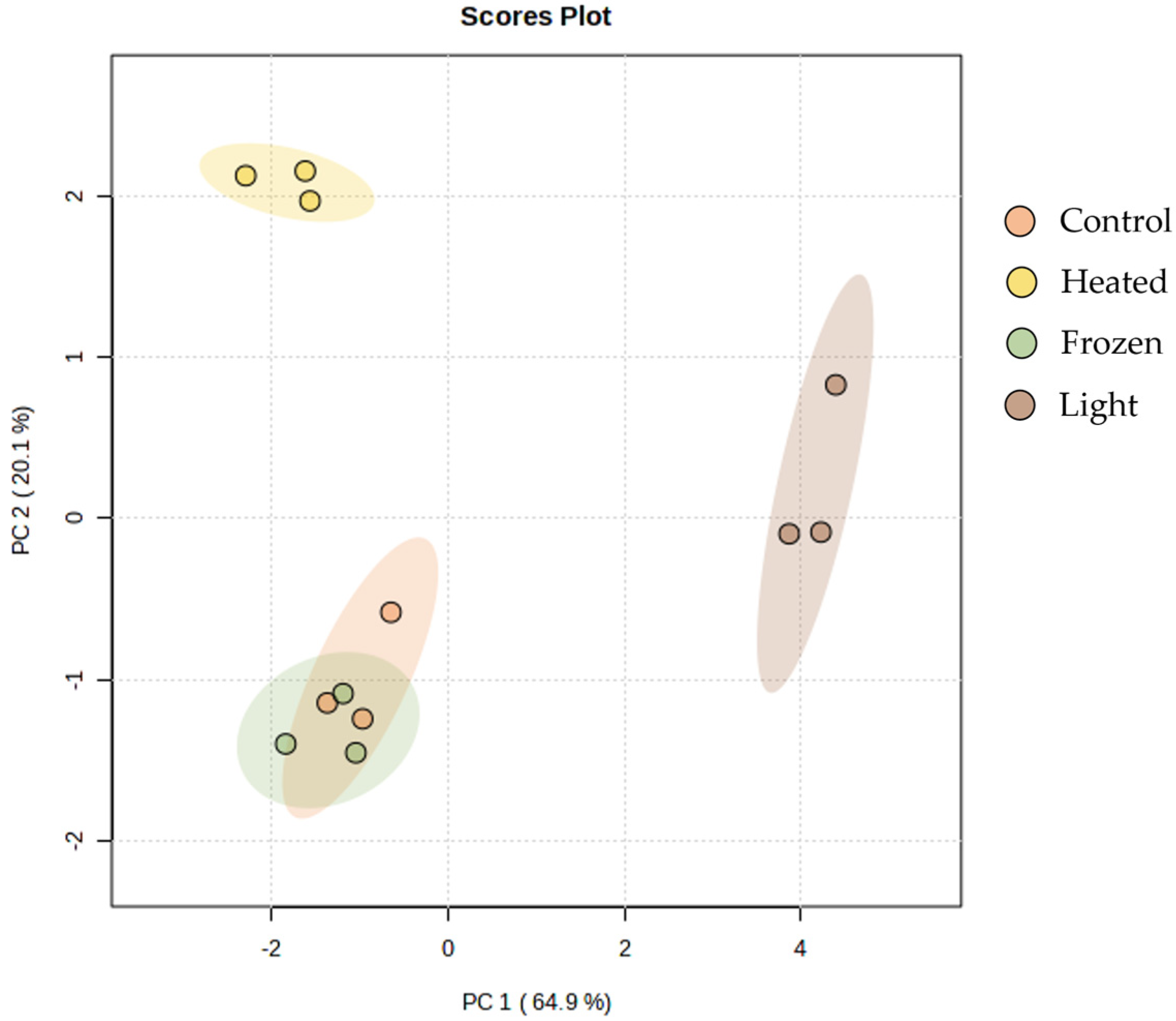
The PCA calculated using the normalized extracted ion chromatogram (XIC) areas of non-oxidized and oxidized lipids species showed that its two principal components explained 85% of total variance, with principal component 1 (PC 1) and principal component 2 (PC 2) axes explaining 64.9% and 20.1%, respectively (Figure 2). A clear discrimination can be observed between samples from different storage conditions; while control and frozen samples overlapped, light-exposed samples were separated from this group along PC 1, and heated-exposed samples along PC 2 (Figure 2).

2.3. Antioxidant Activity

The in chemico antioxidant activity of lipid extracts of C. vulgaris and T. chui exposed to different conditions was determined by 2,20-Azino-bis-3-Ethylbenzothiazoline-6-Sulfonic Acid (ABTS) and 2-Diphenyl-1-Picrylhydrazyl (DPPH) scavenging assays. The concentration of lipid extract required to achieve 40% inhibition of ABTS radicals (IC40) and 20% inhibition of DPPH radicals (IC20) was determined.
An IC40 of 32 ± 11.0 μg mL−1 and IC20 of 97.8 ± 27.2 μg mL−1 were obtained in ABTS and DPPH assays, respectively, for C. vulgaris under control conditions (Table 4). Remarkably, in both assays, the control condition showcased the highest antioxidant activity, while the light condition demonstrated the lowest antioxidant activity.
Regarding the antioxidant activity of lipid extracts of T. chui exposed to different storage conditions (Table 5), the results of control condition showed different tendencies in ABTS and DPPH assays. In ABTS assay, the best antioxidant activity was achieved in the frozen condition, with an IC40 of 29 ± 5.4 μg mL−1, whereas the lowest activity was obtained for heated samples with an IC40 of 88.5 ± 2.8 μg mL−1. On the other hand, in the DPPH assay, the control condition exhibited the best antioxidant activity with an IC20 of 70.6 ± 23.1 μg mL−1, while the heated condition again displayed the lowest antioxidant activity with an IC20 of 209.4 ± 10.9 μg mL−1.

3. Discussion

The microalgae species C. vulgaris and T. chui are highly valued in functional food and feed applications, due to their abundance in beneficial lipids, particularly omega-3 PUFA and their documented bioactive lipid species, as well as other intriguing compounds [9,17].
In this study a slight increase in lipid yield was verified under heated conditions for both C. vulgaris and T. chui. This phenomenon could be attributed to protein denaturation at higher temperatures, which enhances the accessibility of extraction solvents to membranes [25]. Additionally, the disruption of membrane structures induced by both low (frozen condition) and high (heated condition) temperatures may enhance lipid accessibility, thereby facilitating their extraction. Interestingly, a similar increase in lipid extractability was observed in T. chui stored on freezer. This phenomenon of increased lipid extractability with different temperature has been previously reported for other food matrices [26].
The FA profiles of both microalgae were analyzed using basic catalysis transmethylation, focusing on esterified FA, which are of particular interest in this study due to their nutritional relevance in food and feed applications. It is important to note that this method does not methylate free fatty acids (FFA), and therefore these FFA were not included in the FA analysis. The results are consistent with previous studies, with 16:0, linoleic acid (LA, 18:2n-6), and alpha-linolenic acid (ALA, 18:3n-3) being the most abundant in the lipid extracts of C. vulgaris [17,27], and 16:0, 16:4n-3, and ALA being predominant for T. chui [28,29]. Linoleic acid and ALA are essential n-6 and n-3 FA, respectively, crucial for human health as they cannot be synthesized by the body and must be obtained through diet. They serve as precursors of long-chain essential PUFA such as arachidonic acid (AA, 20:4n-6), eicosapentaenoic acid (EPA, 20:5n-3), and docosahexaenoic acid (22:6n-3), which play a relevant role in cognitive and immune functions. The FA profile of C. vulgaris and T. chui, although without significant differences, showed a decrease in PUFA in light conditions compared to control conditions. This observation aligns with the known susceptibility of PUFA to chemical modifications, such as autoxidation or metal-catalyzed oxidation, attributed to their multiple double bonds that render them vulnerable to electrophilic attack [30]. Moreover, both frozen and heated conditions also led to a decrease in PUFA in C. vulgaris, although this effect was less pronounced than under light conditions. Interestingly, the SFA of both C. vulgaris and T. chui also showed a decrease under light, frozen, and heated conditions compared to the control condition. This reduction was already observed in other food matrices during storage [31]. The decrease in SFA as well as other FA could potentially be linked to the action of lipases. Lipases are enzymes widely distributed in nature, exhibiting high specificity and selectivity for their substrates. They hydrolyze lipids, releasing free fatty acids, and demonstrate selectivity towards specific FA at particular regions bonded to specific carbons of glycerol. They are considered stable and versatile, able to catalyze reactions under extremes of temperature, pH, and organic solvents [32,33]. Given that the analyzed FA in this work reflects esterified FA, the observed decrease in FA under different storage conditions could indeed be associated with membrane damage, allowing lipases greater access to lipids and subsequently reducing esterified FA levels. The n-6/n-3 ratio for a healthy diet should be 6:1 [34,35]. Reducing the intake of n-6/n-3 ratio can enhance blood lipids and quality of life, and is crucial for preventing cardiovascular disease [36]. The lack of differences in the n-6/n-3 ratio between treatments suggests that the loss of FA was proportional in PUFA regardless of the double bond position. Although the different conditions tested, the amount of n-3 PUFA was always higher than n-6 PUFA, resulting in consistently low n-6/n-3 ratio across all conditions for both C. vulgaris and T. chui. This result indicates that despite the exposed conditions, these microalgae maintain a nutritionally beneficial ratio. The AI and TI indices assess the potential for stimulating platelet aggregation, as well as the development of blood clots, atheroma, and thrombus formation [37,38]. Consequently, smaller IA and IT values suggest greater protective potential against cardiovascular diseases [39]. This study revealed that these indices were not significantly affected by the tested storage conditions, with both C. vulgaris and T. chui exhibiting very similar values (lower than 1 in both species). The indices observed in C. vulgaris and T. chui are consistent with those previous reported [17,40]. The h/H index evaluates the ratio between hypocholesterolemic and hypercholesterolemic FA, reflecting their association with cholesterol metabolism. High h/H values are considered more favorable for human health [41,42]. In this context, C. vulgaris displayed higher h/H indices compared to T. chui, without significative differences between storage conditions for both microalgae. The h/H values of both C. vulgaris and T. chui were higher than those of fish lipids (ranging from 0.6 to 2.5) [34,42,43], with fish traditionally considered a source of healthy lipids. This highlights these microalgae as a promising alternative source of healthy lipids.
The detailed lipid characterization of C. vulgaris and Tetraselmis sp. were already reported in previous studies [16,44,45]. Therefore, this study aimed to elucidate lipid alterations under different storage conditions by identifying lipid species indicative of degradation, particularly lyso forms and oxidized species. The analysis revealed a higher abundance of lyso species in the PC, PE, and DGTS classes. Consequently, this study was focused on these lipid classes, investigating the PC/LPC, PE/LPE, and DGTS/MGTS ratios to gain insights into lipid degradation processes. Curiously, for T. chui, both frozen and heated conditions affected the PE and DGTS classes in a very similar manner. The authors speculate that these differences may be attributed to lipid localization within cellular membranes. Given that phospholipids and betaine lipids constitute extraplastidial membranes, with potential variations between inner and outer membranes, lipids more impacted in C. vulgaris and T. chui could potentially reside in the outer membrane, rendering them more susceptible to modifications. These findings highlight the potential impact of varying storage conditions on lipid composition, which appears to be species-dependent among microalgae. Previous studies investigating the storage of bivalve mollusks have revealed that exposure to low temperatures result in an increase in lyso phospholipids, including LPC and LPE [46,47]. This increase serves as a marker of prolonged refrigeration. Interestingly, a similar elevation in lyso phospholipids is observed when bivalves are stored in non-refrigerated seawater with temperatures exceeding 25 °C.
In C. vulgaris, oxidized lipid species were identified in PC and PE classes, with the non-oxidized/oxidized ratio consistently lower in samples exposed to light storage conditions. Conversely, in other storage conditions, this ratio closely resembled the control ratio. These results suggest that these species might naturally occur in the biomass of C. vulgaris; however, they experience an increase under prolonged exposure to natural light, which was particularly high in PE36:1;O species. PCA analysis reveals that the control and frozen conditions exhibit closer proximity than the light and heated conditions, suggesting distinct oxidation patterns influenced by the specific environmental conditions they experienced. Lipid peroxidation involves the attack of oxidants, such as free radicals or nonradical species, on lipids containing carbon–carbon double bonds, particularly PUFA. This process results in the abstraction of hydrogen from a carbon atom, followed by oxygen insertion, leading to the formation of lipid peroxyl radicals and hydroperoxides [48,49]. Phospholipids are common targets of peroxidative modification. Additionally, lipids can be oxidized by enzymes like lipoxygenases that are quite active in plants and algae [50]. The oxidation of lipids can give rise to harmful compounds and thus being detrimental to human health [51]. Intriguingly, in T. chui were not detected oxidized PC and PE species, suggesting that the microalga’s cell structure integrity may offer enhanced protection against lipid oxidation during storage conditions or that they are present in a lower concentration than in C. vulgaris.
In terms of antioxidant activity, in C. vulgaris, both the ABTS and DPPH assays consistently demonstrate lower antioxidant activity in the lipid extracts from light-exposed samples. The values of the control condition of C. vulgaris align with previously reported results for C. vulgaris cultured under autotrophic conditions [16,17]. However, in T. chui, lipid extract from heated-exposed samples displayed the lowest antioxidant activity. This variance in antioxidant activity may be linked to PUFA, suggesting that the reduction observed in light-exposed C. vulgaris samples could stem from higher degradation of these FA. However, this phenomenon was not observed in T. chui. It is worth noting that besides lipids, other compounds such as photosynthetic pigments and lipid-soluble compounds (e.g., phenolic compounds) also contribute significantly to antioxidant activity. These compounds could potentially have a greater influence on the decreased antioxidant activity observed in heat-exposed T. chui.

4. Materials and Methods

4.1. Microalgae Samples

The C. vulgaris and T. chui biomasses were provided by Allmicroalgae Natural Products S.A. (Pataias, Portugal). The species were grown autotrophically using Guillard’s F2 culture medium adapted to local water. T. chui was supplemented with a magnesium mixture (Necton, Olhão, Portugal) and NaCl (Salexpor, Coimbra, Portugal) at a salinity of 30 g·L−1. The microalgae cultures were cultivated in 5 L flask reactors under continuous 700 μmol photons·m2 s−1 light exposure for 7 to 15 days. Five 5 L flask reactors were utilized to inoculate one 0.1 m3 outdoor flat panel (FP) reactor, which was subsequently scaled up to 1 m3 FPs. Of the five FPs, four were used as inoculum for a 10 m3 tubular photobioreactor (PBR). The PBR was exposed to ambient light and temperature conditions until reaching the stationary phase. A sprinkler-like irrigation system was employed to maintain the temperature of the PBR below the maximum limit, while pulse injections of CO2 were utilized to stabilize the pH. Temperature and pH conditions in the 10 m3 PBRs were operated as described in [40].
The biomass was recovered through centrifugation and subsequently dried. Microalgae at approximately 50 g·L−1 were dried by atomization in a spray dryer with an evaporation capacity of 150 kg water·h−1. Drying was rapidly achieved using an air stream at 215 ± 5 °C, with the outlet air temperature with the biomass powder measured at 92 ± 3 °C. The resulting powder was collected via a cyclone and stored under protection from light and humidity.

4.2. Evaluated Conditions

The C. vulgaris and T. chuii biomass was analyzed from sealed commercial packages stored at room temperature under vacuum, protected from light, and opened for the first time on the day of analysis, serving as the baseline control condition (condition 1—control). Alongside, biomass was separated from the same packages to explore three additional conditions: condition 2—light; condition 3—frozen; and condition 4—heated. For the second condition, the biomass was stored at room temperature in an open container exposed to light and oxygen for 3 weeks (condition 2—light). For the third condition, the biomass was stored involved in a freezer at −20 °C for 3 weeks (condition 3—frozen), while the fourth condition the biomass was heated at 100 °C during 270 min—three cycles of 90 min (condition 4—heated).

4.3. Lipid Extraction

Folch’s method [52] was used to extract total lipid content of C. vulgaris and T. chui (Allmicroalgae Natural Products S.A) samples under the four different experimental conditions. The extraction was executed individually for each condition and three replicates were made. A volume of 2 mL of DCM:MeOH (2:1, v/v) was added to 25 mg of microalga powder in glass tube and vortexed for 2 min, followed by incubation on ice for 30 min in an orbital shaker (Stuart SSL2 reciprocating shaker). Afterwards, the mixture was centrifuged at 2000 rpm for 10 min (Centurion Scientific limited prO-analytical centrifuge). This step was repeated three more times for each replicate. The organic phase was completely dried under a nitrogen stream. To remove non-lipid compounds, the nitrogen-dried extract was dissolved in 3 mL DCM:MeOH (2:1, v/v), and 0.75 mL of Milli-Q water was added. The mixture was vortexed for 2 min followed by centrifugation at 2000 rpm for 10 min to induce phase separation. This was followed by collection of the organic phase (lower phase), and re-extraction of the aqueous phase by addition of 2 mL of DCM, then vortexed for 1 min and centrifuged for 5 min at 2000 rpm (two more times). The combined organic phases were filtered to a new glass tube using a paper filter (WhatmanTM, Kent, UK) to remove eventual suspended particles and dried under nitrogen stream. The lipid extracts were then transferred to amber vials and stored at −20 °C until further analysis.

4.4. Analysis of Esterified Fatty Acid Analysis by Gas Chromatography–Mass Spectrometry

Fatty acid methyl esters (FAME) were prepared from the by base-catalyzed transmethylation of the C. vulgaris and T. chui lipid extracts. The dried lipid extracts were mixed with 1 mL of an internal standard solution (1.30 µg mL−1 of methyl nonadecanoate in n-hexane) followed by addition of 200 μL of KOH solution (2.0 M in MeOH) and vortexed for 2 min. Afterwards, 2 mL of saturated NaCl solution (10 mg mL−1 in water) was added, followed by centrifugation at 2000 rpm for 5 min. The organic phase (600 µg of upper phase) was collected into a new glass tube and dried under a nitrogen gas steam. To prepare the sample for GC-MS analysis, the dried FAMEs were dissolved in 100 μL of 99% n-hexane and 2 µL of this solution were injected in a GC-MS (Agilent Technologies 6890N Network, Santa Clara, CA, USA), equipped with a DB-1 column 30 m in length, 0.25 mm internal diameter, and 0.1 m film thickness (J&W Scientific, Folsom, CA, USA). The GC-MS system was coupled with an Agilent 5973 Network Mass Selective Detector (Agilent Technologies) operating with an electron impact mode at 70 eV and scanning a range of 40–500 m/z in a 1 s cycle in full-scan mode acquisition.
The temperature of the oven was programmed to start at 58 °C (hold time 2 min), followed by a linear increase to 160 °C at a rate of 25 °C per min, then a linear increase of 2 °C per min to 210 °C, and finally a 20 °C per min increase until reaching 225 °C (hold time 15 min). The injector and detector temperatures were set at 220 °C and 230 °C, respectively. Helium gas served as the carrier at flow rate of 1.7 mL min−1. Identification of the FAMEs was conducted by comparing the retention time and mass spectrum of each FAME with a mix of FAME standards (C6–C24, Supelco 37 Component FAMEs Mix). Confirmation was achieved through comparison with the Wiley chemical database and the spectral library, the “NIST Lipid Library”. Calculation of FA amounts was carried out following peak integration and normalization using the amount of internal standard methyl nonadecanoate.
The lipid quality indices, namely, the atherogenicity (AI) and thrombogenicity (TI) indices as well as the hypocholesterolemic/hypercholesterolemic (h/H) ratio, were obtained from the formulas described below [53]:
A I = 12 : 0 + 4 × 14 : 0 + 16 : 0 M U F A + P U F A   n 6 + P U F A   n 3
T I = 14 : 0 + 16 : 0 + 18 : 0 ( 0.5 × M U F A ) + ( 0.5 × P U F A   n 6 ) + ( 3 × P U F A   n 3 ) + P U F A   n 3 P U F A   n 6
h / H = c i s   18 : 1 + P U F A 12 : 0 + 14 : 0 + 16 : 0

4.5. Analysis of Lipid by C18-MS

The lipidome analysis was conducted via liquid chromatography C18, utilizing an Ascentis Express 90 Å C18 HPLC column (15 cm × 2.1 mm; 2.7 μm, Supelco, Sigma-Aldrich, Bellefonte, PA, USA), integrated into an HPLC system (Ultimate 3000 Dionex, Thermo Fisher Scientific, Bremen, Germany) with an autosampler linked to the Q-Exactive® hybrid quadrupole Orbitrap mass spectrometer (Thermo Fisher, Scientific, Bremen, Germany). A volume of 5 μL of each sample, equivalent to 10 μg of lipid extract (in 10 µL of dichloromethane), 82 µL of a solvent system comprising 50% isopropanol/50% methanol, and 8 µL of phospholipid standards mixture was injected into the HPLC column at a flow rate of 260 μL/min. The column oven temperature was maintained at 50 °C. A 33 min gradient was employed, starting with 32% mobile phase B at 0 min, 45% B at 1.5 min, 52% B at 4 min, 58% B at 5 min, 66% B at 8 min, 70% B at 11 min, 85% B at 14 min, 97% B at 18 min, 97% B at 25 min, 32% B at 25.01 min, and 32% B at 33 min. Mass spectrometry was conducted in positive and negative ionization modes over an m/z range of 200–2000 with a resolution of 70,000. Tandem MS (MS/MS) experiments were carried out using a top 10 data-dependent acquisition method, with the most abundant precursor ions selected for fragmentation via higher energy dissociation (HCD). The MS/MS spectra were obtained with a resolution of 17,500, and lipid species were identified using mass spectrometry–data-independent analysis (MS DIAL) v4.70 software.
PC, LPC, PE, LPE, DGTS, and MGTS lipid species were identified in positive ionization modes using the raw files acquired in MS/MS mode and converted by the ABF converter against the lipid database provided by MS-DIAL 4 software. Tolerances for MS and MS/MS search were set at 0.01 Da and 0.05 Da, respectively. Following MS-DIAL identifications, peaks were automatically integrated in MZmine 3.9.0 for LC–MS data normalization. Integrated peak areas of extracted ion chromatograms (XIC) were exported from MZmine 3.9.0 software, and each specie’ peak area was normalized against the peak area of the lipid standard with the closest retention time. The resulting matrix (.xls) with normalized areas of all species facilitated the study of origin-driven variations at the lipid species level.

4.6. Evaluation of Antioxidant Activity of Lipid Extracts by DPPH and ABTS Radical Scavenging Activity

The antioxidant activity of C. vulgaris and T. chui lipid extracts was evaluated by measuring its capacity to scavenge DPPH [54] and ABTS radicals [55]. For the DPPH assay, a stock solution of DPPH in ethanol (250 µmol L−1) was prepared and diluted to prepare a solution with an absorbance value of approximately 0.9, measured at 517 nm using a UV–vis spectrophotometer (Multiskan GO 1.00.38, Thermo Scientific, Hudson, NH, USA). To evaluate the lipid extract scavenging potential, a volume of 150 µL of C. vulgaris and T. chui lipid extracts (12.5, 62.5, 125, and 250 µg mL−1 in ethanol) and 150 µL of Trolox standard solution (5, 12.5, 25, and 37.5 µmol L−1 in ethanol) were placed in each well followed by addition of 150 µL of DPPH-diluted solution, followed by an incubation period of 120 min, with absorbance measured at 517 nm every 5 min [56].
For ABTS assay, a solution of ABTS (3.5 mmol L−1) was prepared by combining 10 mL of ABTS stock solution (7 mmol L−1 in water) with 10 mL of potassium persulfate, K2S2O8 (2.45 mmol L−1 in water). This mixture was left for 12 h at room temperature and then diluted in ethanol to obtain an absorbance value of ~0.9 measured at 734 nm using a UV–vis spectrophotometer (Multiskan GO 1.00.38, Thermo Scientific, Hudson, NH, USA). To assess the scavenging potential of extracts, 150 µL of C. vulgaris and T. chui lipid extracts (12.5, 62.5, 125, and 250 µg mL−1 in ethanol) or 150 µL of Trolox standard solution (5, 12.5, 25, and 37.5 µmol L−1 in ethanol) were added to each well, followed by the addition of 150 µL of ABTS•+ diluted solution, and incubated for 120 min, with absorbance measurements at 734 nm every 5 min [57]. Radical reduction was monitored by measuring the decrease in absorbance during the reaction, thereby quantifying radical scavenging, which is accompanied by a radical color change.
The free radical scavenging activity of samples was determined as the percentage of inhibition of DPPH and ABTS radicals according to Equation (1).
I n h i b i t i o n % = A b s   r a d i c a l A b s   S a m p l e A b s   C o n t r o l A b s   r a d i c a l × 100
The concentration of samples capable of reducing 20% and 40% of DPPH and ABTS radical, respectively, after 120 min (IC20 and IC40, respectively) were calculated by linear regression using the concentration of samples and the percentage of the inhibition curve. The antioxidant activity was also expressed as Trolox equivalents (TE, µmol Trolox.g−1 of lipid extract), according to Equation (2)
T E ( μ m o l   T r o l o x . g 1   o f   l i p i d   e x t r a c t ) = I C 20   o r   40   T r o l o x   ( μ m o l   g 1 ) I C 20   o r   40   S a m p l e   ( μ g   m L 1 ) × 1000

4.7. Statistical Analysis

The non-parametric Kruskal–Wallis test was used to identify significant differences between microalga storage conditions in fatty acid content and PC/LPC, PE/LPE, and DGTS/MGTS ratios. Post-hoc comparisons were performed using Dunn’s tests. p-values were corrected for multiple testing using the Benjamin–Hochberg method (q values). Differences with q-value < 0.05 were considered statistically significant. These statistical analyses were performed using R version 4.1.0 (R Core Team, 2021) in rStudio 2023.6.1.524 (Posit Team 2023). Non-oxidized and oxidized ratios of phosphatidilcholine and phosphatidylethanolamine lipid species identified in Chlorella vulgaris (Figure S1) were calculated using the normalized extracted ion chromatograms (XIC). Ratio data were log-transformed and auto-scaled and principal component analysis (PCA) was performed to assess the clustering of the four storage treatments in Metaboanalyst 6.0. software.

5. Conclusions

The utilization of functional ingredients to achieve beneficial effects through diet is currently expanding in human and animal feeding. However, storage conditions can significantly impact the molecular composition of food ingredients, namely, FA and lipid profiles.
This study demonstrated a decrease in FA content across all tested conditions, with the most pronounced reduction in PUFA under light exposure treatment. Among the microalgae species studied, C. vulgaris exhibited the highest susceptibility to this reduction. The alterations in diacyl species/lyso species ratio of phospholipid (PC and PE) and betaine lipids (DGTS) suggest a degradation in the quality of these lipids with the treatments. Additionally, this study also demonstrated how exposition to light and heat reduce the antioxidant potential of both C. vulgaris and T. chui. These results emphasize the need for the proper preservation of microalgae biomass to realize their full potential in nutritional and functional food and feed applications. Future research should focus on evaluating the stability of biomass under different preservation methods to optimize storage and conservation techniques, ensuring the maintenance of biomass quality. Such advancements are crucial for the efficient use of microalgae in various applications.

Supplementary Materials

The following supporting information can be downloaded at: https://www.mdpi.com/article/10.3390/md22060254/s1, Figure S1:Total ion chromatograms of lipid extract of Chlorella vulgaris under control condition in positive (A) and negative (B) ionization modes by C18-LC-MS; Figure S2: Total ion chromatograms of lipid extract of Tetraselmis chui under control condition in positive (A) and negative (B) ionization modes by C18-LC-MS; Figure S3: LC-MS/MS spectrum in negative mode of PE 32:1 specie at m/z 688.5 corresponding to PE 16:0; 16:1 identified in Chlorella vulgaris under control condition (A) and PE 32:1;O at m/z 704.5 corresponding to PE 16:0; 16:1; O identified in Chlorella vulgaris under light condition (B); Figure S4: GC-MS chromatogram of Chlorella vulgaris under control condition highlighting the identified esterified fatty acids; Figure S5: GC-MS chromatogram of Tetraselmis chui under control condition highlighting the identified esterified fatty acids.

Author Contributions

Conceptualization, D.L. and F.R.; methodology, D.L. and F.R.; software, D.L., F.R. and T.M.; validation, F.R. and R.D.; formal analysis, A.G., L.D., M.P. and J.P.; investigation, A.G. and L.D.; data curation, D.L. and F.R.; writing—original draft preparation, D.L.; writing—review and editing, F.R., T.M. and R.D.; supervision, R.D. All authors have read and agreed to the published version of the manuscript.

Funding

This research was funded by Fundação para a Ciência e Tecnologia through CESAM (UIDP/50017/2020 + UIDB/50017/2020 + LA/P/0094/2020). This work was financially supported by “Pacto da Bioeconomia azul” (Project No. 16 with the application No. C644915664-00000026) within the WP5 Algae Vertical, funded by Next Generation EU European Fund and the Portuguese Recovery and Resilience Plan (PRR), under the scope of the incentive line “Agendas for Business Innovation” through the funding scheme C5—Capitalization and Business Innovation. Felisa Rey and Tânia Melo thank their Junior Researcher contracts in the scope of the Individual Call to Scientific Employment Stimulus (CEECIND/00580/2017 to Felisa Rey (https://doi.org/10.54499/CEECIND/00580/2017/CP1459/CT0005) and CEECIND/01578/2020 to Tânia Melo (https://doi.org/10.54499/2020.01578.CEECIND/CP1589/CT0010)). Marisa Pinho was funded by national funds through the FCT-Foundation for Science and Technology, I.P., under the PhD granted via CESAM (UI/BD/153346/2022). This work is a contribution of the Marine Lipidomics laboratory.

Institutional Review Board Statement

Not applicable.

Data Availability Statement

Dataset available on request from the authors.

Acknowledgments

Thanks to ALLMICROALGAE—Natural Products SA, for providing microalgae biomass, and to LAQV/REQUIMTE (UIDB/50006/2020).

Conflicts of Interest

The authors declare no conflicts of interest.

References

  1. Spolaore, P.; Joannis-Cassan, C.; Duran, E.; Isambert, A. Commercial Applications of Microalgae. J. Biosci. Bioeng. 2006, 101, 87–96. [Google Scholar] [CrossRef] [PubMed]
  2. Torres-Tiji, Y.; Fields, F.J.; Mayfield, S.P. Microalgae as a Future Food Source. Biotechnol. Adv. 2020, 41, 107536. [Google Scholar] [CrossRef] [PubMed]
  3. de Jesus Raposo, M.F.; de Morais, R.M.S.C.; de Morais, A.M.M.B. Health Applications of Bioactive Compounds from Marine Microalgae. Life Sci. 2013, 93, 479–486. [Google Scholar] [CrossRef] [PubMed]
  4. Draaisma, R.B.; Wijffels, R.H.; Slegers, P.E.; Brentner, L.B.; Roy, A.; Barbosa, M.J. Food Commodities from Microalgae. Curr. Opin. Biotechnol. 2013, 24, 169–177. [Google Scholar] [CrossRef]
  5. Paterson, S.; Gómez-Cortés, P.; de la Fuente, M.A.; Hernández-Ledesma, B. Bioactivity and Digestibility of Microalgae Tetraselmis sp. and Nannochloropsis sp. as Basis of Their Potential as Novel Functional Foods. Nutrients 2023, 15, 477. [Google Scholar] [CrossRef] [PubMed]
  6. Canelli, G.; Tarnutzer, C.; Carpine, R.; Neutsch, L.; Bolten, C.J.; Dionisi, F.; Mathys, A. Biochemical and Nutritional Evaluation of Chlorella and Auxenochlorella Biomasses Relevant for Food Application. Front. Nutr. 2020, 7, 565996. [Google Scholar] [CrossRef] [PubMed]
  7. Vidotti, A.D.; Riaño-Pachón, D.M.; Mattiello, L.; Giraldi, L.A.; Winck, F.V.; Franco, T.T. Analysis of Autotrophic, Mixotrophic and Heterotrophic Phenotypes in the Microalgae Chlorella vulgaris Using Time-Resolved Proteomics and Transcriptomics Approaches. Algal Res. 2020, 51, 102060. [Google Scholar] [CrossRef]
  8. Becker, E.W. Micro-Algae as a Source of Protein. Biotechnol. Adv. 2007, 25, 207–210. [Google Scholar] [CrossRef]
  9. Rahman, N.A.; Khatoon, H.; Yusuf, N.; Banerjee, S.; Haris, N.A.; Lananan, F.; Tomoyo, K. Tetraselmis chuii Biomass as a Potential Feed Additive to Improve Survival and Oxidative Stress Status of Pacific White-Leg Shrimp Litopenaeus Vannamei Postlarvae. Int. Aquat. Res. 2017, 9, 235–247. [Google Scholar] [CrossRef]
  10. Roy, S.S.; Pal, R. Microalgae in Aquaculture: A Review with Special References to Nutritional Value and Fish Dietetics; Springer: Berlin/Heidelberg, Germany, 2015; Volume 68, pp. 1–8. [Google Scholar]
  11. Shah, M.R.; Lutzu, G.A.; Alam, A.; Sarker, P.; Kabir Chowdhury, M.; Parsaeimehr, A.; Liang, Y.; Daroch, M. Microalgae in Aquafeeds for a Sustainable Aquaculture Industry. J. Appl. Phycol. 2018, 30, 197–213. [Google Scholar] [CrossRef]
  12. EFSA Panel on Biological Hazards (BIOHAZ); Koutsoumanis, K.; Allende, A.; Alvarez-Ordóñez, A.; Bolton, D.; Bover-Cid, S.; Chemaly, M.; Davies, R.; De Cesare, A.; Hilbert, F. Update of the List of QPS-recommended Biological Agents Intentionally Added to Food or Feed as Notified to EFSA 15: Suitability of Taxonomic Units Notified to EFSA until September 2021. EFSA J. 2022, 20, e07045. [Google Scholar] [PubMed]
  13. EFSA Panel on Biological Hazards (BIOHAZ); Koutsoumanis, K.; Allende, A.; Alvarez-Ordóñez, A.; Bolton, D.; Bover-Cid, S.; Chemaly, M.; Davies, R.; De Cesare, A.; Hilbert, F. Scientific Opinion on the Update of the List of QPS-recommended Biological Agents Intentionally Added to Food or Feed as Notified to EFSA (2017–2019). EFSA J. 2020, 18, e05966. [Google Scholar] [PubMed]
  14. European Comission Commission Implementing Regulation (EU) 2017/2470 Establishing the Union List of Novel Foods in Accordance with Regulation (EU) 2015/2283 of the European Parliament and of the Council on Novel Foods. OJ L 351. 30 December 2017, pp. 72–201. Available online: https://eur-lex.europa.eu/eli/reg_impl/2017/2470/oj (accessed on 1 November 2023).
  15. Abedi, E.; Sahari, M.A. Long-chain Polyunsaturated Fatty Acid Sources and Evaluation of Their Nutritional and Functional Properties. Food Sci. Nutr. 2014, 2, 443–463. [Google Scholar] [CrossRef] [PubMed]
  16. Couto, D.; Melo, T.; Conde, T.A.; Costa, M.; Silva, J.; Domingues, M.R.M.; Domingues, P. Chemoplasticity of the Polar Lipid Profile of the Microalgae Chlorella vulgaris Grown under Heterotrophic and Autotrophic Conditions. Algal Res. 2021, 53, 102128. [Google Scholar] [CrossRef]
  17. Maurício, T.; Couto, D.; Lopes, D.; Conde, T.; Pais, R.; Batista, J.; Melo, T.; Pinho, M.; Moreira, A.S.; Trovão, M. Differences and Similarities in Lipid Composition, Nutritional Value, and Bioactive Potential of Four Edible Chlorella vulgaris Strains. Foods 2023, 12, 1625. [Google Scholar] [CrossRef] [PubMed]
  18. Meléndez-Martínez, A.J.; Esquivel, P.; Rodriguez-Amaya, D.B. Comprehensive Review on Carotenoid Composition: Transformations during Processing and Storage of Foods. Food Res. Int. 2023, 169, 112773. [Google Scholar] [CrossRef] [PubMed]
  19. Sharma, S.; Kaur, A.; Bansal, A.; Gill, B.S. Positional Effects on Soybean Seed Composition during Storage. J. Food Sci. Technol. 2013, 50, 353–359. [Google Scholar] [CrossRef] [PubMed]
  20. Siriwardane, D.A.; Wang, C.; Jiang, W.; Mudalige, T. Quantification of Phospholipid Degradation Products in Liposomal Pharmaceutical Formulations by Ultra Performance Liquid Chromatography-Mass Spectrometry (UPLC-MS). Int. J. Pharm. 2020, 578, 119077. [Google Scholar] [CrossRef] [PubMed]
  21. Capriotti, A.L.; Cerrato, A.; Aita, S.E.; Montone, C.M.; Piovesana, S.; Laganà, A.; Cavaliere, C. Degradation of the Polar Lipid and Fatty Acid Molecular Species in Extra Virgin Olive Oil during Storage Based on Shotgun Lipidomics. J. Chromatogr. A 2021, 1639, 461881. [Google Scholar] [CrossRef]
  22. Esposto, S.; Taticchi, A.; Urbani, S.; Selvaggini, R.; Veneziani, G.; Di Maio, I.; Sordini, B.; Servili, M. Effect of Light Exposure on the Quality of Extra Virgin Olive Oils According to Their Chemical Composition. Food Chem. 2017, 229, 726–733. [Google Scholar] [CrossRef]
  23. Jiménez-Colmenero, F.; Cofrades, S.; Herrero, A.M.; Ruiz-Capillas, C. Implications of Domestic Food Practices for the Presence of Bioactive Components in Meats with Special Reference to Meat-Based Functional Foods. Crit. Rev. Food Sci. Nutr. 2018, 58, 2334–2345. [Google Scholar] [CrossRef] [PubMed]
  24. Geng, L.; Liu, K.; Zhang, H. Lipid Oxidation in Foods and Its Implications on Proteins. Front. Nutr. 2023, 10, 1192199. [Google Scholar] [CrossRef]
  25. Zhang, L.; Zhou, R.; Zhang, J.; Zhou, P. Heat-Induced Denaturation and Bioactivity Changes of Whey Proteins. Int. Dairy J. 2021, 123, 105175. [Google Scholar] [CrossRef]
  26. Wazir, H.; Chay, S.Y.; Zarei, M.; Hussin, F.S.; Mustapha, N.A.; Wan Ibadullah, W.Z.; Saari, N. Effects of Storage Time and Temperature on Lipid Oxidation and Protein Co-Oxidation of Low-Moisture Shredded Meat Products. Antioxidants 2019, 8, 486. [Google Scholar] [CrossRef]
  27. Couto, D.; Melo, T.; Conde, T.A.; Moreira, A.S.; Ferreira, P.; Costa, M.; Silva, J.; Domingues, R.; Domingues, P. Food Grade Extraction of Chlorella vulgaris Polar Lipids: A Comparative Lipidomic Study. Food Chem. 2022, 375, 131685. [Google Scholar] [CrossRef]
  28. Conlon, T.; Parkes, R.; Fierli, D.; Touzet, N. Comparative Pigment and Fatty Acid Profiling of Marine Species within the Chlorophyte Genus Tetraselmis. Food Biosci. 2024, 58, 103660. [Google Scholar] [CrossRef]
  29. Moser, G.A.O.; Barrera-Alba, J.J.; Ortega, M.J.; Alves-de-Souza, C.; Bartual, A. Comparative Characterization of Three Tetraselmis chui (Chlorophyta) Strains as Sources of Nutraceuticals. J. Appl. Phycol. 2022, 34, 1–15. [Google Scholar] [CrossRef]
  30. Saini, R.K.; Prasad, P.; Sreedhar, R.V.; Akhilender Naidu, K.; Shang, X.; Keum, Y.-S. Omega-3 Polyunsaturated Fatty Acids (PUFAs): Emerging Plant and Microbial Sources, Oxidative Stability, Bioavailability, and Health Benefits—A Review. Antioxidants 2021, 10, 1627. [Google Scholar] [CrossRef] [PubMed]
  31. Chaula, D.; Jacobsen, C.; Laswai, H.S.; Chove, B.E.; Dalsgaard, A.; Mdegela, R.; Hyldig, G. Changes in Fatty Acids during Storage of Artisanal-processed Freshwater Sardines (Rastrineobola argentea). Food Sci. Nutr. 2023, 11, 3040–3047. [Google Scholar] [CrossRef]
  32. Kumar, R.R.; Bhargava, D.; Pandit, K.; Goswami, S.; Shankar, S.M.; Singh, S.P.; Rai, G.K.; Satyavathi, C.T.; Praveen, S. Lipase–The Fascinating Dynamics of Enzyme in Seed Storage and Germination—A Real Challenge to Pearl Millet. Food Chem. 2021, 361, 130031. [Google Scholar] [CrossRef]
  33. Reyes-Reyes, A.L.; Valero Barranco, F.; Sandoval, G. Recent Advances in Lipases and Their Applications in the Food and Nutraceutical Industry. Catalysts 2022, 12, 960. [Google Scholar] [CrossRef]
  34. Rincón-Cervera, M.Á.; González-Barriga, V.; Romero, J.; Rojas, R.; López-Arana, S. Quantification and Distribution of Omega-3 Fatty Acids in South Pacific Fish and Shellfish Species. Foods 2020, 9, 233. [Google Scholar] [CrossRef] [PubMed]
  35. Wijendran, V.; Hayes, K. Dietary N-6 and n-3 Fatty Acid Balance and Cardiovascular Health. Annu. Rev. Nutr. 2004, 24, 597–615. [Google Scholar] [CrossRef] [PubMed]
  36. Yang, Y.; Xia, Y.; Zhang, B.; Li, D.; Yan, J.; Yang, J.; Sun, J.; Cao, H.; Wang, Y.; Zhang, F. Effects of Different n-6/n-3 Polyunsaturated Fatty Acids Ratios on Lipid Metabolism in Patients with Hyperlipidemia: A Randomized Controlled Clinical Trial. Front. Nutr. 2023, 10, 1166702. [Google Scholar] [CrossRef] [PubMed]
  37. Manuela, A.G.; Robert, V.-A.; Yoannis, N.; Elvira, L.; Rossana, R.; Roberta, M.; Giacomo, D.; Daniele, G. Fatty Acids Profile, Atherogenic (IA) and Thrombogenic (IT) Health Lipid Indices, of Raw Roe of Blue Fin Tuna (Thunnus thynnus L.) and Their Salted Product “Bottarga”. Food Nutr. Sci. 2011, 2011, 7235. [Google Scholar]
  38. Khalili Tilami, S.; Kouřimská, L. Assessment of the Nutritional Quality of Plant Lipids Using Atherogenicity and Thrombogenicity Indices. Nutrients 2022, 14, 3795. [Google Scholar] [CrossRef] [PubMed]
  39. Turan, H.; Sönmez, G.; Kaya, Y. Fatty Acid Profile and Proximate Composition of the Thornback Ray (Raja clavata, L. 1758) from the Sinop Coast in the Black Sea. J. Fish. Sci. 2007, 1, 97–103. [Google Scholar] [CrossRef]
  40. Conde, T.A.; Neves, B.F.; Couto, D.; Melo, T.; Neves, B.; Costa, M.; Silva, J.; Domingues, P.; Domingues, M.R. Microalgae as Sustainable Bio-Factories of Healthy Lipids: Evaluating Fatty Acid Content and Antioxidant Activity. Mar. Drugs 2021, 19, 357. [Google Scholar] [CrossRef]
  41. Testi, S.; Bonaldo, A.; Gatta, P.P.; Badiani, A. Nutritional Traits of Dorsal and Ventral Fillets from Three Farmed Fish Species. Food Chem. 2006, 98, 104–111. [Google Scholar] [CrossRef]
  42. Fernandes, C.E.; da Silva Vasconcelos, M.A.; de Almeida Ribeiro, M.; Sarubbo, L.A.; Andrade, S.A.C.; de Melo Filho, A.B. Nutritional and Lipid Profiles in Marine Fish Species from Brazil. Food Chem. 2014, 160, 67–71. [Google Scholar] [CrossRef]
  43. Zhang, X.; Ning, X.; He, X.; Sun, X.; Yu, X.; Cheng, Y.; Yu, R.-Q.; Wu, Y. Fatty Acid Composition Analyses of Commercially Important Fish Species from the Pearl River Estuary, China. PLoS ONE 2020, 15, e0228276. [Google Scholar] [CrossRef] [PubMed]
  44. Conde, T.; Aveiro, S.; Melo, T.; Santos, T.; Neves, B.; Domingues, P.; Varela, J.; Pereira, H.; Domingues, M.R. Cross-Stress Lipid Response of Tetraselmis striata CTP4 to Temperature and Salinity Variation. Algal Res. 2023, 74, 103218. [Google Scholar] [CrossRef]
  45. Couto, D.; Conde, T.A.; Melo, T.; Neves, B.; Costa, M.; Silva, J.; Domingues, R.; Domingues, P. The Chemodiversity of Polar Lipidomes of Microalgae from Different Taxa. Algal Res. 2023, 70, 103006. [Google Scholar] [CrossRef]
  46. Facchini, L.; Losito, I.; Cianci, C.; Cataldi, T.R.; Palmisano, F. Structural Characterization and Profiling of Lyso-phospholipids in Fresh and in Thermally Stressed Mussels by Hydrophilic Interaction Liquid Chromatography—Electrospray Ionization—Fourier Transform Mass Spectrometry. Electrophoresis 2016, 37, 1823–1838. [Google Scholar] [CrossRef] [PubMed]
  47. Losito, I.; Facchini, L.; Catucci, R.; Calvano, C.D.; Cataldi, T.R.; Palmisano, F. Tracing the Thermal History of Seafood Products through Lysophospholipid Analysis by Hydrophilic Interaction Liquid Chromatography–Electrospray Ionization Fourier Transform Mass Spectrometry. Molecules 2018, 23, 2212. [Google Scholar] [CrossRef] [PubMed]
  48. Yin, H.; Xu, L.; Porter, N.A. Free Radical Lipid Peroxidation: Mechanisms and Analysis. Chem. Rev. 2011, 111, 5944–5972. [Google Scholar] [CrossRef]
  49. Ayala, A.; Muñoz, M.F.; Argüelles, S. Lipid Peroxidation: Production, Metabolism, and Signaling Mechanisms of Malondialdehyde and 4-Hydroxy-2-Nonenal. Oxidative Med. Cell. Longev. 2014, 2014, 360438. [Google Scholar] [CrossRef] [PubMed]
  50. Viswanath, K.K.; Varakumar, P.; Pamuru, R.R.; Basha, S.J.; Mehta, S.; Rao, A.D. Plant Lipoxygenases and Their Role in Plant Physiology. J. Plant Biol. 2020, 63, 83–95. [Google Scholar] [CrossRef]
  51. Abeyrathne, E.D.N.S.; Nam, K.; Ahn, D.U. Analytical Methods for Lipid Oxidation and Antioxidant Capacity in Food Systems. Antioxidants 2021, 10, 1587. [Google Scholar] [CrossRef]
  52. Folch, J.; Lees, M.; Sloane Stanley, G.H. A Simple Method for the Isolation and Purification of Total Lipids from Animal Tissues. J. Biol. Chem. 1957, 226, 497–509. [Google Scholar] [CrossRef]
  53. Chen, J.; Liu, H. Nutritional Indices for Assessing Fatty Acids: A Mini-Review. Int. J. Mol. Sci. 2020, 21, 5695. [Google Scholar] [CrossRef] [PubMed]
  54. Brand-Williams, W.; Cuvelier, M.-E.; Berset, C. Use of a Free Radical Method to Evaluate Antioxidant Activity. LWT-Food Sci. Technol. 1995, 28, 25–30. [Google Scholar] [CrossRef]
  55. Rice-Evans, C.A.; Miller, N.J.; Paganga, G. Structure-Antioxidant Activity Relationships of Flavonoids and Phenolic Acids. Free Radic. Biol. Med. 1996, 20, 933–956. [Google Scholar] [CrossRef] [PubMed]
  56. Magalhães, L.M.; Segundo, M.A.; Reis, S.; Lima, J.L. Automatic Method for Determination of Total Antioxidant Capacity Using 2, 2-Diphenyl-1-Picrylhydrazyl Assay. Anal. Chim. Acta 2006, 558, 310–318. [Google Scholar] [CrossRef]
  57. Magalhães, L.M.; Barreiros, L.; Maia, M.A.; Reis, S.; Segundo, M.A. Rapid Assessment of Endpoint Antioxidant Capacity of Red Wines through Microchemical Methods Using a Kinetic Matching Approach. Talanta 2012, 97, 473–483. [Google Scholar] [CrossRef]
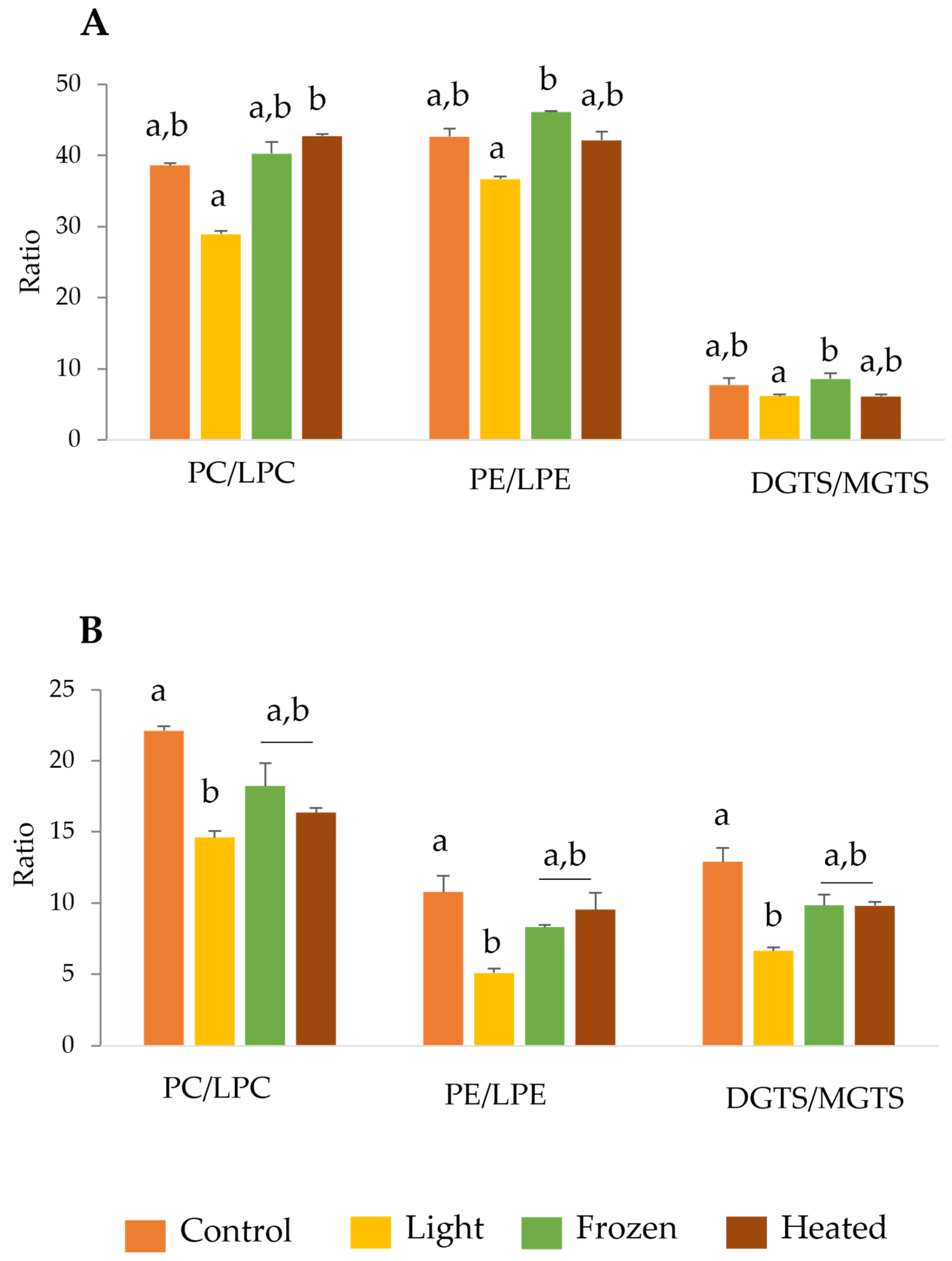
Figure 1. Polar lipid ratios between diacyl and lyso forms of phosphatidylcholine (PC) and lyso-phosphatidylcholine (LPC); phosphatidylethanolamine (PE) and lyso-phosphatidylethanolamine (LPE); diacylglyceryltrimethylhomoserine (DGTS) and monoacylglyceryltrimethylhomoserine (MGTS) for Chlorella vulgaris (A) and Tetraselmis chui (B) exposed to different storages conditions (control, light, frozen, and heated). Values are means of the normalized extracted ion chromatograms (XIC) areas of three replicates (n = 3) ± standard deviation. Different letters represent significant differences among conditions (q < 0.05, Kruskal–Wallis test followed by Dunn’s post-hoc comparisons).
Marinedrugs 22 00254 g001
Figure 2. Principal component analysis (PCA) of log-transformed normalized extracted-ion chromatogram (XIC) areas of non-oxidized and oxidized phosphatidilcholines and phosphatidylethanolamines lipid species identified in Chlorella vulgaris exposed to different conditions (control, light, frozen, and heated). Three (n = 3) samples by condition were analyzed.
Marinedrugs 22 00254 g002
Table 1. Fatty acid profiles and lipid quality indices of Chlorella vulgaris exposed to different storages conditions (control, light, frozen, and heated), determined by gas chromatography–mass spectrometry (GC–MS) and expressed in μg mg−1 of lipid extract. Values are presented as average of three replicates (n = 3) ± standard deviation. The fatty acids 16:1 and 18:1 correspond to the sum of two peaks for which the exact position of unsaturation within the chain have not been clearly assigned. Different letters in the same line represent significant differences among conditions (q < 0.05, Kruskal–Wallis test followed by Dunn’s post-hoc comparisons).
Fatty AcidsControlLightFrozenHeated
16:052.3 ± 4.835.1 ± 6.237.5 ± 2.946.2 ± 3.2
16:19.3 ± 0.76.2 ± 1.17.3 ± 0.99.2 ± 0.7
16:2n-621.4 ± 1.514.2 ± 2.416.9 ± 2.320.8 ± 1.2
16:3n-338.7 ± 2.7 a24.1 ± 4.0 b30.8 ± 4.7 a,b35.2 ± 2.0 a,b
18:013.4 ± 5.38.4 ± 1.66.3 ±0.810.6 ± 0.4
18:126.9 ± 1.719.3 ± 3.021.2 ± 1.828.0 ± 2.3
18:2n-661.9 ± 3.546.3 ± 7.349.9 ± 5.862.7 ± 4.7
18:3n-372.3 ± 4.2 a45.9 ± 7.3 b56.7 ± 7.4 a,b65.5 ± 4.1 a,b
n-3111.0 ± 6.970.0 ± 11.287.5 ± 12.1100.7 ± 6.1
n-683.3 ± 4.960.6 ± 9.766.8 ± 8.183.5 ± 5.8
n-6/n-30.8 ± 0.00.9 ± 0.00.8 ± 0.00.8 ± 0.0
SFA65.7 ± 9.943.7 ± 6.943.8 ± 3.556.8 ± 2.8
MUFA36.2 ± 2.425.5 ± 4.128.5 ± 2.737.2 ± 2.9
PUFA230.5 ± 14.3156.0 ± 25.0182.8 ± 22.8221.4 ± 14.8
AI0.2 ± 0.00.2 ± 0.00.2 ± 0.00.2 ± 0.0
TI0.2 ± 0.00.2 ± 0.00.1 ± 0.00.2 ± 0.0
h/H4.3 ± 0.44.3 ± 0.14.7 ± 0.24.6 ± 0.0
Abbreviations: SFA: saturated fatty acids; MUFA: monounsaturated fatty acids; PUFA: polyunsaturated fatty acids; AI: atherogenicity index; TI: thrombogenicity index; h/H: hypo/hypercholesterolemic ratio.
Table 2. Fatty acid profiles and lipid quality indices of Tetraselmis chui exposed to different storages conditions (control, light, frozen, and heated), determined by gas chromatography–mass spectrometry (GC–MS) and expressed in μg mg−1 of lipid extract. Values are presented as average of three replicates (n = 3) ± standard deviation. The fatty acids 16:1 and 18:1 correspond to the sum of two peaks for which the exact position of unsaturation within the chain have not been clearly assigned.
Fatty AcidsControlLightFrozenHeated
16:067.6 ± 9.654.4 ± 5.651.9 ± 5.453.0 ± 3.6
16:16.6 ± 0.86.4 ± 0.87.1 ± 0.87.1 ± 0.5
16:3n-33.8 ± 0.43.7 ± 0.54.3 ± 0.64.4 ± 0.3
16:4n-337.0 ± 4.835.3 ± 4.641.5 ± 4.540.9 ± 4.1
18:021.5 ± 5.75.0 ± 2.23.8 ± 0.34.8 ± 1.2
18:135.6 ± 3.734.1 ± 4.235.6 ±3.537.8 ± 2.2
18:2n-618.9 ± 2.116.8 ± 2.119.3 ± 2.020.0 ± 1.3
18:3n-69.0 ± 1.18.3 ± 1.110 ± 1.19.8 ± 0.9
18:3n-347.5 ± 4.843.7 ± 3.851.1 ± 8.451.6 ± 6.6
18:4n-313.9 ± 1.613.4 ± 1.415.9 ± 2.716.1 ± 2.6
20:5n-313.4 ± 1.512.2 ± 1.316.0 ± 2.815.6 ± 2.2
n-3115.6 ± 11.9108.3 ± 11.5128.8 ± 26.7128.5 ± 15.8
n-627.9 ± 3.125.1 ± 3.229.2 ± 4.429.9 ± 2.2
n-6/n-30.2 ± 0.00.2 ± 0.00.2 ± 0.00.2 ± 0.0
SFA89.1 ± 15.359.5 ± 7.355.7 ± 8.157.8 ± 3.0
MUFA42.3 ± 4.540.5 ± 5.142.7 ± 6.244.9 ± 2.7
PUFA185.8 ± 19.5173.9 ± 19.8200.7 ± 37.3203.3 ± 20.7
AI0.4 ± 0.00.3 ± 0.00.3 ± 0.00.3 ± 0.0
TI0.2 ± 0.00.2 ± 0.00.1 ± 0.00.1 ± 0.0
h/H2.7 ± 0.23.1 ± 0.03.7 ± 0.13.7 ± 0.1
Abbreviations: SFA: saturated fatty acids; MUFA: monounsaturated fatty acids; PUFA: polyunsaturated fatty acids; AI: atherogenicity index; TI; thrombogenicity index; h/H; hypo/hypercholesterolemic ratio.
Table 3. Ratio of non-oxidized and oxidized phosphatidylcholine (PC) and phosphatidylethanolamine (PE) lipid species identified in Chlorella vulgaris exposed to different storage conditions (control, light, frozen, and heated). Values are means of the normalized extracted ion chromatograms (XIC) areas of three replicates (n = 3) ± standard deviation.
Lipid Species RatioControlLightFrozenHeated
PC 34:2/PC 34:2;O70.6 ± 2.626.8 ± 1.348.4 ± 16.772.2 ± 17.4
PC 34:3/PC 34:3;O84.2 ± 3.641.0 ± 2.984.9 ± 5.278.7 ± 6.7
PC 34:5/PC 34:5;O30.7 ± 6.123.8 ± 0.543.9 ± 1.745.5 ± 2.8
PC 36:4/PC 36:4;O61.3 ± 2.040.9 ± 2.463.5 ± 7.064.0 ± 3.4
PC 36:5/PC 36:5;O56.9 ± 10.338.3 ± 8.264.9 ± 2.741.4 ± 0.8
PC 36:6/PC 36:6;O36.0 ± 4.929.2 ± 6.850.6 ± 2.256.0 ± 2.0
PE 32:1/PE 32:1;O809.3 ± 5.223.0 ± 2.9822.6 ± 65.6685.4 ± 187.3
PE 34:2/PE 34:2;O89.8 ± 2.536.5 ± 2.577.6 ± 4.0116 ± 46.8
PE 34:3/PE 34:3;O58.3 ± 5.633.4 ± 2.748.1 ± 2.7105.0 ± 9.7
PE 36:3/PE 36:3;O11.1 ± 0.36.5 ± 0.611.2 ± 0.75.1 ± 0.2
Table 4. Inhibition concentration (IC) of lipid extracts (μg mL−1) providing 40% and 20% of inhibition (IC40, IC20) of ABTS●+ and DPPH radicals, respectively, after 120 min of reaction, and the corresponding Trolox-equivalent units (TE) (μmol of Trolox g−1 lipid) in samples of Chlorella vulgaris exposed to different storage conditions (control, light, frozen, and heated). Results are averages of three assays (n = 3) ± standard deviation.
ControlLightFrozenHeated
ABTS
IC4032.0 ± 11.0104.9 ± 6.291.1 ± 15.349.4 ± 7.0
TE402.8 ± 173.4111.1 ± 6.3131.7 ± 24.1238.1 ± 31.3
DPPH
IC2097.8 ± 27.2187.9 ± 15.0103.6 ± 24.6 132.1 ± 2.9
TE107.1 ± 35.552.6 ± 4.298.3 ± 21.074.5 ± 1.6
Table 5. Inhibitory concentration (IC) of lipid extracts (μg mL−1) providing 40% and 20% of inhibition (IC40, IC20) of ABTS●+ and DPPH radicals, respectively, after 120 min of reaction, and the corresponding Trolox equivalent units (TE) (μmol of Trolox g−1 lipid) in samples of Tetraselmis chui exposed to different conditions (control, light, frozen, and heated). Results are averages of three assays (n = 3) ± standard deviation.
ControlLightFrozenHeated
ABTS
IC4041.4 ± 1.041.1 ± 0.629.0 ± 5.488.5 ± 2.8
TE281.0 ± 6.8283.0 ± 3.9411.1 ± 85.2131.4 ± 4.3
DPPH
IC2070.6 ± 23.1109.4 ± 10.790.5 ± 7.0209.4 ± 10.9
TE152.5 ± 58.390.9 ± 8.4109.6 ± 8.147.3 ± 2.4
Disclaimer/Publisher’s Note: The statements, opinions and data contained in all publications are solely those of the individual author(s) and contributor(s) and not of MDPI and/or the editor(s). MDPI and/or the editor(s) disclaim responsibility for any injury to people or property resulting from any ideas, methods, instructions or products referred to in the content.