

Surf Redfish-Based ZnO-NPs and Their Biological Activity with Reference to Their Non-Target Toxicity

 ,
,  , , , ,
Abstract
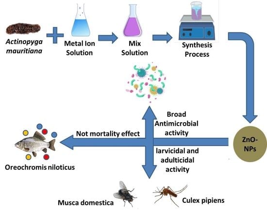
1. Introduction
2. Results and Discussion
2.1. Identification and Specification of Collected Samples
2.2. Characterization of Biosynthesized ZnO-NPs
2.3. Insecticidal Activity
2.4. Antibacterial Activity of Biosynthesized ZnO-NPs
2.5. Non-Target Effects
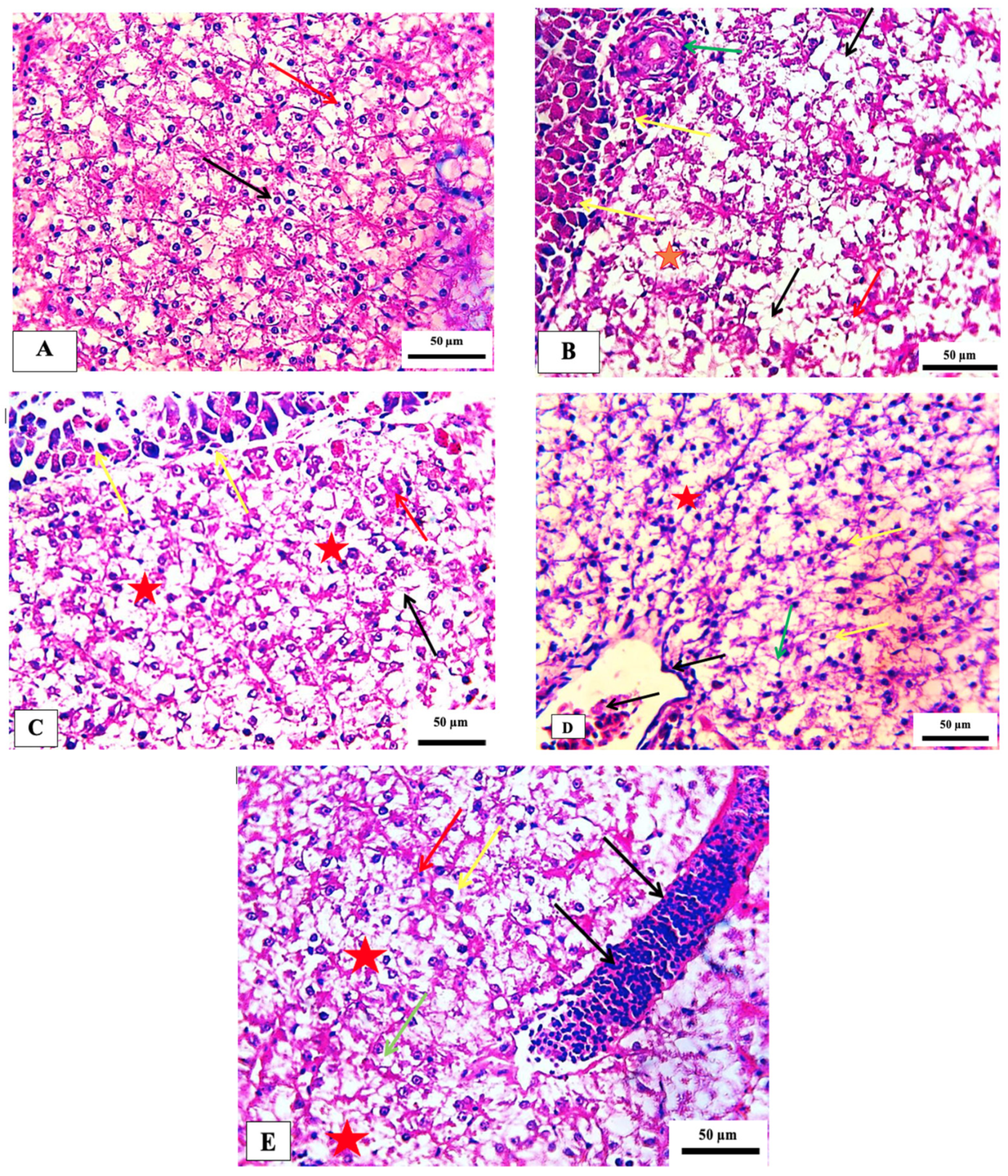
2.5.1. Gill Histopathology
2.5.2. Liver Histopathology
2.5.3. Hematological Parameters
3. Conclusions
4. Materials and Methods
4.1. Sampling, Preservation and Identification of Sponge Specimen
4.2. Preparation of the Aqueous Extract
4.3. Biosynthesis of ZnO-NPs Using Extract of Surf Redfish
4.4. Characterization of Biosynthesized ZnO-NPs
4.5. Insecticidal Bioassays
4.5.1. Insects
4.5.2. Toxicity on Culex pipiens Larvae
4.5.3. Toxicity on Musca Domestica Adults
4.6. Antibacterial Activity of Biosynthesized ZnO-NPs Using Surf Redfish
4.7. Non-Target Effects
4.7.1. Water Quality Parameters
4.7.2. Experimental Diet
4.7.3. Histology of Non-Targeted Organisms
4.7.4. Hematological Assays
Blood Sampling and Hematological Analysis
Serum Biochemical Analysis
Antioxidants and Other Activities
4.8. Statistical Analysis
Author Contributions
Funding
Institutional Review Board Statement
Data Availability Statement
Acknowledgments
Conflicts of Interest
References
- Czyżowska, A.; Barbasz, A. A review: Zinc oxide nanoparticles–friends or enemies? Int. J. Environ. Health Res. 2022, 32, 885–901. [Google Scholar]
- Bai, Z.; Yan, X.; Chen, X.; Liu, H.; Shen, Y.; Zhang, Y. ZnO nanowire array ultraviolet photodetectors with self-powered properties. Curr. Appl. Phys. 2013, 13, 165–169. [Google Scholar] [CrossRef]
- Khan, Z.U.H.; Sadiq, H.M.; Shah, N.S.; Khan, A.U.; Muhammad, N.; Hassan, S.U.; Tahir, K.; Khan, F.U.; Imran, M.; Ahmad, N.; et al. Greener synthesis of zinc oxide nanoparticles using Trianthema portulacastrum extract and evaluation of its photocatalytic and biological applications. J. Photochem. Photobiol. B Biol. 2019, 192, 147–157. [Google Scholar] [CrossRef]
- Faizan, M.; Hayat, S.; Pichtel, J. Effects of zinc oxide nanoparticles on crop plants: A perspective analysis. In Sustainable Agriculture Reviews 41; Springer: Cham, Switzerland, 2020; pp. 83–99. [Google Scholar]
- Hasaballah, A.I.; El Naggar, H.A.; Abdelbary, S.; Bashar, M.A.E.; Selim, T.A. Eco friendly Synthesis of Zinc Oxide Nanoparticles by Marine Sponge, Spongia offcinalis: Antimicrobial and Insecticidal Activities against the Mosquito Vectors, Culex pipiens and Anopheles pharoensis. BioNanoScience 2021, 12, 89–104. [Google Scholar] [CrossRef]
- Kołodziejczak-Radzimska, A.; Jesionowski, T. Zinc oxide—From synthesis to application: A review. Materials 2014, 7, 2833–2881. [Google Scholar] [CrossRef] [PubMed]
- Mona, M.H.; El-Naggar, H.A.; El-Gayar, E.E.; Masood, M.F.; Mohamed, E.N.E. Effect of human activities on biodiversity in Nabq Protected Area, South Sinai, Egypt. Egypt. J. Aquat. Res. 2019, 45, 33–43. [Google Scholar] [CrossRef]
- El-Naggar, H.A.; Salem, E.-S.S.; El-Kafrawy, S.B.; Bashar, M.A.; Shaban, W.M.; El-Gayar, E.E.; Ahmed, H.O.; Ashour, M.; Abou-Mahmoud, M.E. An integrated field data and remote sensing approach for impact assessment of human activities on epifauna macrobenthos biodiversity along the western coast of Aqaba Gulf. Ecohydrology 2022, 15, e2400. [Google Scholar] [CrossRef]
- EL-Naggar, H.A.; Bashar, M.A.E.; Rady, I.; El-Wetidy, M.S.; Suleiman, W.B.; Al-Otibi, F.O.; Al-Rashed, S.A.; Abd El-Maoula, L.M.; Salem, E.S.; Attia, E.M.H.; et al. Two Red Sea Sponge Extracts (Negombata magnifica and Callyspongia siphonella) Induced Anticancer and Antimicrobial Activity. Appl. Sci. 2022, 12, 1400. [Google Scholar] [CrossRef]
- Hasaballah, A.I.; Gobaara, I.M.M.; El Naggar, H.A. Larvicidal activity and ultrastructural abnormalities in the ovaries of the housefly “Musca domestica” induced by the soft coral “Ovabunda macrospiculata” synthesized ZnO nanoparticles. Egypt. J. Aquat. Biol. Fish. 2021, 25, 721–738. [Google Scholar] [CrossRef]
- Metwally, A.S.; El-Naggar, H.A.; El-Damhougy, K.A.; Bashar, M.A.E.; Ashour, M.; Abo-Taleb, H.A.H. GC-MS analysis of bioactive components in six different crude extracts from the Soft Coral (Sinularia maxim) collected from Ras Mohamed, Aqaba Gulf, Red Sea, Egypt. Egypt. J. Aquat. Biol. Fish. 2020, 24, 425–434. [Google Scholar] [CrossRef]
- Attia, M.S.; El-Naggar, H.A.; Abdel-Daim, M.M.; El-Sayyad, G.S. The potential impact of Octopus cyanea extracts to improve eggplant resistance against Fusarium-wilt disease: In vivo and in vitro studies. Environ. Sci. Pollut. Res. 2021, 27, 35854–35869. [Google Scholar] [CrossRef] [PubMed]
- Arlyza, I.S. Teripang dan bahan aktifnya. Oseana 2009, 34, 1–2. [Google Scholar]
- Cui, H.; Bashar, M.A.E.; Rady, I.; El-Naggar, H.A.; Abd El-Maoula, L.M.; Mehany, A.B.M. Antiproliferative activity, proapoptotic effect, and cell cycle arrest in human cancer cells of some marine natural product extract. Oxid. Med. Cell. Longev. 2020, 2020, 7948705. [Google Scholar] [CrossRef] [PubMed]
- Vaidya, H.; Cheema, S.K. Sea cucumber and blue mussel: New sources of phospholipid enriched omega-3 fatty acids with a potential role in 3T3-L1 adipocyte metabolism. Food Funct. 2014, 5, 3287–3295. [Google Scholar] [CrossRef] [PubMed]
- Haug, T.; Kjuul, A.K.; Styrvold, O.B.; Sandsdalen, E.; Olsen, O.M.; Stensvåg, K. Antibacterial activity in Strongylocentrotus droebachiensis (Echinoidea), Cucumaria frondosa (Holothuroidea), and Asterias rubens (Asteroidea). J. Invertebr. Pathol. 2002, 8, 94–102. [Google Scholar] [CrossRef]
- Kumar, R.; Chaturvedi, A.K.; Shuklab, P.K.; Lakshmia, V. Antifungal activity in triterpene glycosides from the sea cucumber Actinopyga lecanora. Bioorg. Med. Chem. Lett. 2007, 17, 4387–4391. [Google Scholar] [CrossRef]
- Weissenböck, H.; Hubálek, Z.; Bakonyi, T.; Nowotny, N. Zoonotic mosquito-borne flaviviruses: Worldwide presence of agents with proven pathogenicity and potential candidates of future emerging diseases. Vet. Microbiol. 2010, 140, 271–280. [Google Scholar] [CrossRef]
- Sattler, M.; Mtasiwa, D.; Kiama, M.; Premji, Z.; Tanner, M.; Killeen, G. Habitat characterization and spatial distribution of Anopheles sp. mosquito larvae in Dar es Salaam (Tanzania) during an extended dry period. Malar. J. 2005, 4, 4–10. [Google Scholar] [CrossRef][Green Version]
- Fotedar, R.; Banerjee, U.; Singh, S.; Shriniwas, A.; Verma, A.K. The house fly Musca domestica as a carrier of pathogenic microorganisms in a Hospital Environment. J. Hosp. Infect. 1992, 20, 209–215. [Google Scholar] [CrossRef]
- Kettle, D.S. Muscidae (Houseflies, Stableflies) Medical and Veterinary Entomology; Kettle, D.S., Ed.; CAB International: Wallingford, UK, 1990; pp. 223–240. [Google Scholar]
- Grubel, P.; Hoffman, J.S.; Chong, F.K.; Burstein, N.A.; Mepani, C.; Cave, D. Vector potential of houseflies (Musca domestica) for Helicobacter pylori. J. Clin. Microbiol. 1997, 35, 1300–1303. [Google Scholar] [CrossRef]
- Ehsan, S.; Sajjad, M. Bioinspired synthesis of zinc oxide nanoparticle and its combined efficacy with different antibiotics against multidrug resistant bacteria. J. Biomater. Nanobiotechnol. 2017, 8, 159–175. [Google Scholar] [CrossRef][Green Version]
- Mohamed, A.A.; Fouda, A.; Abdel-Rahman, M.A.; Hassan, S.E.D.; El-Gamal, M.S.; Salem, S.S.; Shaheen, T.I. Fungal strain impacts the shape, bioactivity and multifunctional properties of green synthesized zinc oxide nanoparticles. Biocatal. Agric. Biotechnol. 2019, 19, 101103. [Google Scholar] [CrossRef]
- Yusof, H.; Rahman, A.; Mohamad, R.; Zaidan, U.H.; Samsudin, A.A. Biosynthesis of zinc oxide nanoparticles by cell-biomass and supernatant of Lactobacillus plantarum TA4 and its antibacterial and biocompatibility properties. Sci. Rep. 2020, 10, 19996. [Google Scholar] [CrossRef] [PubMed]
- El-Belely, E.F.; Farag, M.; Said, H.A.; Amin, A.S.; Azab, E.; Gobouri, A.A.; Fouda, A. Green synthesis of zinc oxide nanoparticles (ZnO NPs) using Arthrospira platensis (Class: Cyanophyceae) and evaluation of their biomedical activities. Nanomaterials 2021, 11, 95. [Google Scholar] [CrossRef]
- Chaudhuri, S.K.; Malodia, L. Biosynthesis of zinc oxide nanoparticles using leaf extract of Calotropis gigantea: Characterization and its evaluation on tree seedling growth in nursery stage. Appl. Nanosci. 2017, 7, 501–512. [Google Scholar] [CrossRef]
- Shehabeldine, A.M.; Elbahnasawy, M.A.; Hasaballah, A.I. Green phytosynthesis of silver nanoparticles using Echinochloa stagnina extract with reference to their antibacterial, cytotoxic, and larvicidal activities. BioNanoScience 2021, 11, 526–538. [Google Scholar] [CrossRef]
- Elbahnasawy, M.A.; Shehabeldine, A.M.; Khattab, A.M.; Amin, B.H.; Hashem, A.H. Green biosynthesis of silver nanoparticles using novel endophytic Rothia endophytica: Characterization and anticandidal activity. J. Drug Deliv. Sci. Technol. 2021, 62, 102401. [Google Scholar] [CrossRef]
- Mohammadian, M.; Es’haghi, Z.; Hooshmand, S. Green and chemical synthesis of zinc oxide nanoparticles and size evaluation by UV–vis spectroscopy. J. Nanomed. Res. 2018, 7, 00175. [Google Scholar]
- Jayarambabu, N.; Kumari, B.S.; Rao, K.V.; Prabhu, Y.T. Germination and growth characteristics of mungbean seeds (Vigna radiata L.) affected by synthesized zinc oxide nanoparticles. Int. J. Curr. Eng. Technol. 2014, 4, 2347–5161. [Google Scholar]
- Vinotha, V.; Yazhiniprabha, M.; Raj, D.S.; Mahboob, S.; Al-Ghanim, K.A.; Al-Misned, F.; Vaseeharan, B. Biogenic synthesis of aromatic cardamom-wrapped zinc oxide nanoparticles and their potential antibacterial and mosquito larvicidal activity: An effective eco-friendly approach. J. Environ. Chem. Eng. 2020, 8, 104466. [Google Scholar] [CrossRef]
- Raghunath, A.; Perumal, E. Metal oxide nanoparticles as antimicrobial agents: A promise for the future. Int. J. Antimicrob. Agents 2017, 49, 137–152. [Google Scholar] [CrossRef]
- Wagner, G.; Korenkov, V.; Judy, J.D.; Bertsch, P.M. Nanoparticles composed of Zn and ZnO inhibit Peronospora tabacina spore germination in vitro and P. tabacina infectivity on tobacco leaves. Nanomaterials 2016, 6, 50. [Google Scholar] [CrossRef] [PubMed]
- Sirelkhatim, A.; Mahmud, S.; Seeni, A.; Kaus, N.H.M.; Ann, L.C.; Bakhori, S.K.M.; Mohamad, D. Review on zinc oxide nanoparticles: Antibacterial activity and toxicity mechanism. Nano-Micro Lett. 2015, 7, 219–242. [Google Scholar] [CrossRef] [PubMed]
- Tiwari, V.; Mishra, N.; Gadani, K.; Solanki, P.S.; Shah, N.A.; Tiwari, M. Mechanism of anti-bacterial activity of zinc oxide nanoparticle against carbapenem-resistant Acinetobacter baumannii. Front. Microbiol. 2018, 9, 1218. [Google Scholar] [CrossRef]
- Jothinarendiran, N. Effect of dimethoate pesticide on oxygen consumption and gill histology of the fish, Channa punctatus. Curr. Biotica 2012, 5, 500–507. [Google Scholar]
- Pal, S.; Kokushi, E.; Koyama, J.; Uno, S.; Ghosh, A.R. Histopathological alterations in gill, liver and kidney of common carp exposed to chlorpyrifos. J. Environ. Sci. Health A 2012, 47, 180–195. [Google Scholar] [CrossRef]
- Mustafa, S.A.; Al-Faragi, J.K.; Salman, N.M.; Al-Rudainy, A.J. Histopathological Alterations in Gills, Liver and Kidney of Common Carp, Cyprinus Carpio L. Exposed to Lead Acetate. Adv. Anim. Vet. Sci. 2017, 5, 371–376. [Google Scholar]
- Dawood, M.A.; Abd-El-Kader, M.F.; Moustafa, E.M.; Gewaily, M.S.; Abdo, S.E. Growth performance and hemato-immunological responses of Nile tilapia (Oreochromis niloticus) exposed to deltamethrin and fed immunobiotics. Environ. Sci. Pollut. Res. 2020, 27, 11608–11617. [Google Scholar] [CrossRef]
- Ghayyur, S.; Khan, M.F.; Munawar, S.T.; Ahmad, S. A comparative study on the effects of selected pesticides on hemato-biochemistry and tissue histology of freshwater fish Cirrhinus mrigala (Hamilton, 1822). Saudi J. Biol. Sci. 2021, 28, 603–611. [Google Scholar] [CrossRef]
- Zhu, X.; Zhu, L.; Duan, Z.; Qi, R.; Li, Y.; Lang, Y. Comparative toxicity of several metal oxide nanoparticle aqueous suspensions to zebra fish (Danio rerio) early developmental stage. J. Environ. Sci. Health A 2008, 43, 278–284. [Google Scholar] [CrossRef]
- Zhu, X.; Wang, J.; Zhang, X.; Chang, Y.; Chen, Y. The impact of ZnO nanoparticle aggregates on the embryonic development of zebrafish (Danio rerio). Nanotechnology 2009, 20, 195103. [Google Scholar] [CrossRef] [PubMed]
- Elbahnasawy, M.A.; El-Naggar, H.A.; Kalaba, M.H.; Moghannem, S.A.; Abd-El Rahman, I.E.; Abdelzaher, O.F.; Mabrouk, M.M.; Gewida, A.G.A.; AbdEl-Kader, M.F.; Hasaballah, A.I. Biosynthesized ZnO-NPs using sea cucumber, Holothuria impatiens: Antimicrobial potential, Insecticidal activity and toxicity to the non-target Nile Tilapia fish, Oreochromis niloticus. Separations 2023, 10, 173. [Google Scholar] [CrossRef]
- Campbell, A.C. Echinoderms of the Red Sea. In Red Sea (Key Environments); Edwards, A.J., Head, S.M., Eds.; Pergamon Press: Oxford, UK, 1987; pp. 215–232. [Google Scholar]
- Cherbonnier, G. Echinodermes: Holothuries. Faune de Madagascar, 70; ORSTOM: Paris, France, 1988; p. 292. [Google Scholar]
- Erwin, D.G.; Picton, B.E. Guide to Inshore Marine Life; Immel Publishing: Petaluma, CA, USA, 1990; p. 219. [Google Scholar]
- Lieske, E.; Myers, R.F. Coral Reef Guide Red Sea: The Definitive Guide to Over 1200 Species of Underwater Life; London Harper Collins: Glasgow, UK, 2004; p. 211. [Google Scholar]
- Ballantine, D.L.; Gerwick, W.H.; Velez, S.M.; Alexander, E.; Guevara, P. Antibiotic activity of lipid-soluble extracts from Caribbean marine algae. Hydrobiologia 1987, 151, 463–469. [Google Scholar] [CrossRef]
- Kalaba, M.H.; Moghannem, S.A.; El-Hawary, A.S.; Radwan, A.A.; Sharaf, M.H.; Shaban, A.S. Green synthesized ZnO nanoparticles mediated by Streptomyces plicatus: Characterizations, antimicrobial and nematicidal activities and cytogenetic effects. Plants 2021, 10, 1760. [Google Scholar] [CrossRef] [PubMed]
- Selim, T.A.; Abd-El Rahman, I.E.; Mahran, H.A.; Adam, H.A.M.; Imieje, V.; Zaki, A.A.; Bashar, M.A.E.; Hwihy, H.; Hamed, A.; Elhenawy, A.A.; et al. Mosquitocidal Activity of the Methanolic Extract of Annickia chlorantha and Its Isolated Compounds against Culex pipiens, and Their Impact on the Non-Target Organism Zebrafish, Danio Rerio. Insects 2022, 13, 676. [Google Scholar] [CrossRef] [PubMed]
- Hasaballah, A.I.; Selim, T.A.; Tanani, M.A.; Nasr, E.E. Lethality and Vitality Efficiency of Different Extracts of Salix safsaf Leaves against the House Fly, Musca domestica L. (Diptera: Muscidae). Afr. Entomol. 2021, 29, 479–490. [Google Scholar] [CrossRef]
- World Health Organization. Guidelines for Laboratory and Field Testing of Mosquito Larvicides; World Health Organization: Geneva, Switzerland, 2005; pp. 1–39. [Google Scholar]
- Wright, J.W. The WHO Programme for the Evaluation and Testing of New Insecticides. Bull. World Health Org. 1971, 44, 11–12. [Google Scholar]
- Sharaf, M.H.; Abdelaziz, A.M.; Kalaba, M.H.; Radwan, A.A.; Hashem, A.H. Antimicrobial, Antioxidant, Cytotoxic Activities and Phytochemical Analysis of Fungal Endophytes Isolated from Ocimum Basilicum. Appl. Biochem. Biotechnol. 2021, 194, 1271–1289. [Google Scholar] [CrossRef]
- Mount, D.I. Methods for Aquatic Toxicity Identification Evaluations: Phase I: Toxicity Characterization Procedures; US Environmental Protection Agency, Environmental Research Laboratory, National Effluent Toxicity Assessment Center United States: Denver, CO, USA, 1988; Volume 88.
- Al-Motabagani, M.A. Histological and histochemical studies on the effects of methotrexate on the liver of adult male albino rat. Int. J. Morphol. 2006, 24, 417–422. [Google Scholar] [CrossRef]


| Concentrations (ppm) | n | Larval mortality % ± (SE) | LC50 (LCL–UCL) (ppm) | LC90 (LCL–UCL) (ppm) | χ2 (d.f. = 4) |
|---|---|---|---|---|---|
| Control | 125 | 0.8 ± 0.8 a | 15.412 (13.679–17.580) | 52.745 (42.286–70.192) | 11.065 n.s. |
| Temephos | 125 | 100 ± 0.0 f | |||
| 2 | 125 | 3.2 ± 0.8 ab | |||
| 4 | 125 | 8.8 ± 1.49 b | |||
| 8 | 125 | 20.8 ± 1.49 c | |||
| 16 | 125 | 47.2 ± 1.96 d | |||
| 32 | 125 | 82.4 ± 1.6 e |
| Concentrations (μg/Adult) | n | Adult Mortality % ± (SE) | LD50 (LCL–UCL) (ppm) | LD90 (LCL–UCL) (ppm) | χ2 (d.f. = 4) |
|---|---|---|---|---|---|
| Control | 50 | 0.0 ± 0.0 a | 21.132 (16.884–28.518) | 84.930 (54.883–172.908) | 13.048 n.s. |
| Cypermethrin | 50 | 100 ± 0.0 f | |||
| 2 | 50 | 2.0 ± 2.0 a | |||
| 4 | 50 | 10.0 ± 1.96 ab | |||
| 8 | 50 | 14.0 ± 2.45 b | |||
| 16 | 50 | 32.0 ± 2.0 d | |||
| 32 | 50 | 72.0 ± 4.89 e |
| Bacteria | Mean ± SD of Inhibition Zone Diameter (mm) of the Tested Compounds | |||
|---|---|---|---|---|
| ZnO-NPs (NPs) | Surf Redfish Extract (F) | Zinc Acetate (S) | Antibiotic (C) | |
| Pseudomonas aeruginosa | 31 ± 3.05 | 0 | 0 | 0 |
| Aeromonas hydrophila | 49 ± 2.08 | 0 | 0 | 27 ± 0.57 |
| Type of Gill Change | Control | Treatments (ppm/mL) | |||
|---|---|---|---|---|---|
| 75 | 150 | 300 | 600 | ||
| Lamellar fusion | − | + | + | + | ++ |
| Epithelium lifting | − | − | + | + | + |
| Vasodilatation | − | + | − | − | − |
| Aneurisms | − | + | − | − | − |
| Edema | − | − | + | + | ++ |
| Necrosis | − | − | + | − | + |
| Cytoplasmic vacuolation | − | + | + | − | − |
| Type of Liver Change | Control | Treatments (ppm/mL) | |||
|---|---|---|---|---|---|
| 75 | 150 | 300 | 600 | ||
| Cytoplasmic vacuolation | − | + | + | + | + |
| Pancreatic degeneration | − | + | + | − | − |
| Pyknotic nuclei | − | + | + | + | + |
| Necrosis | − | + | + | + | ++ |
| Parameters | Treatments (ppm) | ||||
|---|---|---|---|---|---|
| 0 | 75 | 150 | 300 | 600 | |
| RBCS (×10/mm³) | 4.12 | 3.18 | 3.31 | 2.79 | 2.82 |
| HBg/100ml | 12.6 | 9.6 | 10 | 8.45 | 8.59 |
| PCV% | 40 | 31 | 33 | 27 | 28 |
| MCV(μm³/cell) | 97.09 | 97.48 | 99.7 | 96.77 | 99.29 |
| MCH (pg/cell) | 30.58 | 30.19 | 30.21 | 30.29 | 30.46 |
| MCHC | 31.5 | 30.97 | 30.3 | 31.3 | 30.68 |
| WBCs (×10³/mm³) | 10.76 | 10.19 | 9.98 | 10.32 | 10.2 |
| Heterophil (×10³/mm³) | 2.15 | 2.75 | 2.2 | 2.48 | 1.94 |
| Lymphocyte (×10³/mm³) | 7.53 | 6.42 | 6.59 | 6.91 | 7.34 |
| Monocyte (×10³/mm³) | 0.86 | 0.71 | 0.7 | 0.72 | 0.71 |
| Basophil (×10³/mm³) | 0.11 | 0.1 | 0.2 | 0.10 | 0.1 |
| Esinophil (×10³/mm³) | 0.11 | 0.2 | 0.3 | 0.10 | 0.1 |
| Parameters | Treatments (ppm) | ||||
|---|---|---|---|---|---|
| 0 | 75 | 150 | 300 | 600 | |
| Lysozyme (µg/mL) | 6.29 | 6.05 | 4.85 | 3.99 | 5.16 |
| Phagocytic index (%) | 1.3 | 1.2 | 1.22 | 1.09 | 0.99 |
| Phagocytic activity (%) | 12.24 | 8.15 | 9.23 | 10.06 | 11.64 |
| IgM (µg/mL) | 5.26 | 3.99 | 2.89 | 4.86 | 5.01 |
| Parameters | Treatments (ppm) | ||||
|---|---|---|---|---|---|
| 0 | 75 | 150 | 300 | 600 | |
| Glucose (mg/dL) | 10.25 | 14.86 | 15.67 | 14.66 | 15.95 |
| Cholesterol (mg/dL) | 69.86 | 79.86 | 85.36 | 90.01 | 79.64 |
| Triglyceride (mg/dL) | 103.15 | 82.65 | 94.33 | 99.87 | 100 |
| Total protein (g/dL) | 4.19 | 3.69 | 3.51 | 3.78 | 3.71 |
| Albumin (g/dL) | 1.66 | 1.41 | 1.5 | 1.62 | 1.53 |
| Globulin (g/dL) | 2.53 | 2.28 | 2.01 | 2.16 | 2.18 |
| AST (U/L) | 30.11 | 40.19 | 38.25 | 41.26 | 29.86 |
| ALT (U/L) | 30.16 | 40.12 | 27.98 | 30.18 | 31.45 |
| Urea (mg/dL) | 2.19 | 2.64 | 2.41 | 2.38 | 2.27 |
| Creatinine (mg/dL) | 0.4 | 0.52 | 0.43 | 0.44 | 0.49 |
| Lipase (U/L) | 29.86 | 29.15 | 28.75 | 29.98 | 27.02 |
| Amylase (U/L) | 61.01 | 44.98 | 51.32 | 39.86 | 40.58 |
| Parameters | Treatments (ppm) | ||||
|---|---|---|---|---|---|
| 0 | 75 | 150 | 300 | 600 | |
| MDA nmol/g | 16.54 | 26.53 | 17.64 | 29.32 | 23.02 |
| CAT U/gm | 18.17 | 13.85 | 17.64 | 15.02 | 16.88 |
| SOD U/gm | 23.58 | 16.94 | 15.89 | 18.98 | 20.15 |
| Diet ingredients% | |
| Fish meal | 18.0 |
| soybean meal | 29.0 |
| yellow corn | 20.0 |
| wheat bran | 15.0 |
| alfalfa hay | 12.0 |
| sunflower oil | 3.0 |
| minerals mixture | 1.0 |
| vitamin mixture | 1.0 |
| carboxymethyl cellulose | 1.0 |
| Total | 100 |
| Chemical composition (g/kg) | |
| crude protein | 30.11 |
| ether extract | 12.35 |
| Ash | 14.34 |
| NFE 1 | 43.20 |
| GE 2 | 4600 Kcal/kg |
Disclaimer/Publisher’s Note: The statements, opinions and data contained in all publications are solely those of the individual author(s) and contributor(s) and not of MDPI and/or the editor(s). MDPI and/or the editor(s) disclaim responsibility for any injury to people or property resulting from any ideas, methods, instructions or products referred to in the content. |
© 2023 by the authors. Licensee MDPI, Basel, Switzerland. This article is an open access article distributed under the terms and conditions of the Creative Commons Attribution (CC BY) license (https://creativecommons.org/licenses/by/4.0/).
Share and Cite
Hasaballah, A.I.; El-Naggar, H.A.; Abd-El Rahman, I.E.; Al-Otibi, F.; Alahmadi, R.M.; Abdelzaher, O.F.; Kalaba, M.H.; Amin, B.H.; Mabrouk, M.M.; Gewida, A.G.A.; et al. Surf Redfish-Based ZnO-NPs and Their Biological Activity with Reference to Their Non-Target Toxicity. Mar. Drugs 2023, 21, 437. https://doi.org/10.3390/md21080437
Hasaballah AI, El-Naggar HA, Abd-El Rahman IE, Al-Otibi F, Alahmadi RM, Abdelzaher OF, Kalaba MH, Amin BH, Mabrouk MM, Gewida AGA, et al. Surf Redfish-Based ZnO-NPs and Their Biological Activity with Reference to Their Non-Target Toxicity. Marine Drugs. 2023; 21(8):437. https://doi.org/10.3390/md21080437
Chicago/Turabian StyleHasaballah, Ahmed I., Hussein A. El-Naggar, Ibrahim E. Abd-El Rahman, Fatimah Al-Otibi, Reham M. Alahmadi, Othman F. Abdelzaher, Mohamed H. Kalaba, Basma H. Amin, Mohamed M. Mabrouk, Ahmed G. A. Gewida, and et al. 2023. "Surf Redfish-Based ZnO-NPs and Their Biological Activity with Reference to Their Non-Target Toxicity" Marine Drugs 21, no. 8: 437. https://doi.org/10.3390/md21080437
APA StyleHasaballah, A. I., El-Naggar, H. A., Abd-El Rahman, I. E., Al-Otibi, F., Alahmadi, R. M., Abdelzaher, O. F., Kalaba, M. H., Amin, B. H., Mabrouk, M. M., Gewida, A. G. A., Abd El-Kader, M. F., & Elbahnasawy, M. A. (2023). Surf Redfish-Based ZnO-NPs and Their Biological Activity with Reference to Their Non-Target Toxicity. Marine Drugs, 21(8), 437. https://doi.org/10.3390/md21080437