Article

Connection of Isolated Stereoclusters by Combining 13C-RCSA, RDC, and J-Based Configurational Analyses and Structural Revision of a Tetraprenyltoluquinol Chromane Meroterpenoid from Sargassum muticum

by
Juan Carlos C. Fuentes-Monteverde
1,2,
Nilamoni Nath
3,
Abel M. Forero
1,
Elena M. Balboa
4,
Armando Navarro-Vázquez
5,
Christian Griesinger
2,*,
Carlos Jiménez
1,* and
Jaime Rodríguez
1,*
1
Departamento de Química e Centro de Investigacións Científicas Avanzadas (CICA), Universidade da Coruña, 15071 A Coruña, Spain
2
NMR Based Structural Biology, MPI for Multidisciplinary Sciences, Am Fassberg 11, 37077 Göttingen, Germany
3
Department of Chemistry, Gauhati University, Gopinath Bardoloi Nagar, Guwahati 781014, India
4
Department of Chemical Engineering, Faculty of Science, Campus Ourense, University of Vigo, As Lagoas s/n, 32004 Ourense, Spain
5
Departamento de Química Fundamental, CCEN, Universidade Federal de Pernambuco, Cidade Universitária, Recife 50740-550, Brazil
*
Authors to whom correspondence should be addressed.
Mar. Drugs 2022, 20(7), 462; https://doi.org/10.3390/md20070462
Submission received: 21 June 2022 / Revised: 6 July 2022 / Accepted: 15 July 2022 / Published: 18 July 2022
(This article belongs to the Special Issue Marine Drugs Research in Spain)

Abstract

:
The seaweed Sargassum muticum, collected on the southern coast of Galicia, yielded a tetraprenyltoluquinol chromane meroditerpene compound known as 1b, whose structure is revised. The relative configuration of 1b was determined by J-based configurational methodology combined with an iJ/DP4 statistical analysis and further confirmed by measuring two anisotropic properties: carbon residual chemical shift anisotropies (13C-RCSAs) and one-bond 1H-13C residual dipolar couplings (1DCH-RDCs). The absolute configuration of 1b was deduced by ECD/OR/TD-DFT methods and established as 3R,7S,11R.

1. Introduction

Sargassum muticum (SM), also known as Japanese wireweed, is a brown alga, first described and classified by Yendo in 1907 as Sargassum kjellmanianum form muticum, based on morphological and ecological differences [1]. In 1955, Fensholt reconsidered this form as sufficiently differentiated from S. kjellmanianum and separated it into two species of their own, until it was finally named Sargassum muticum (Yendo) Fensholt [2,3].
This species has been the object of a multitude of studies. In 2017, Balboa et al. reported the use of SM as a potential antioxidant agent and as a preservative in the preparation of cosmetics [4]. Another work, conducted by Park, investigated the feasibility of using SM as a source of bioactive compounds [5]. This study concluded that adding SM pills to the normal diet showed liver benefits in addition to decreasing fatigue and stress. These effects were related to bioactive compounds compatible with meroterpenes or fucoxanthines [6].
Meroditerpenoids of the chromene, where the polyprenyl moiety is bonded to a hydroquinone or similar aromatic ring [7], constitute a very important family of compounds isolated from Sargassum. Almost a hundred meroditerpenes have been reported from marine organisms such as fish, macroalgae, sponges, coelenterates, and tunicates [7,8,9,10]. These compounds have been used in various cancer therapies based on their ability to protect against oxidative damage [11,12].
In 1993, Praud et al. isolated C3-epimeric meroditerpenoids 1a and 1b from Cystoseira baccata [13]. They proposed a cis-fused chromene ring structure, although the configuration at C3 could not be established at that point. Rapid epimer interconversion was observed in CDCl3. Later, Varela et al. also reported antileishmanial activity in those compounds [14]. In 2015, we isolated tetraprenyltoluquinol chromane meroterpenoids 1a and 1b as an epimeric mixture at position C3 from the methanolic extract of the alga (See Figure 1). Moreover, we described their use as photoprotective agents used to mitigate skin damage caused by UV exposure [10].
Considering that obtaining the 1a/1b mixture was probably due to the use of CDCl3 [10,13,15], we repeated the isolation procedure recording the NMR analysis in CD2Cl2. Additionally, the relative configuration of two well-separated stereoclusters (C3 and C7/C11) in 1a1b was determined by J-based configurational analysis along with 13C-residual chemical shift anisotropies (13C-RCSA) [16] and residual dipolar couplings (RDCs) [17].

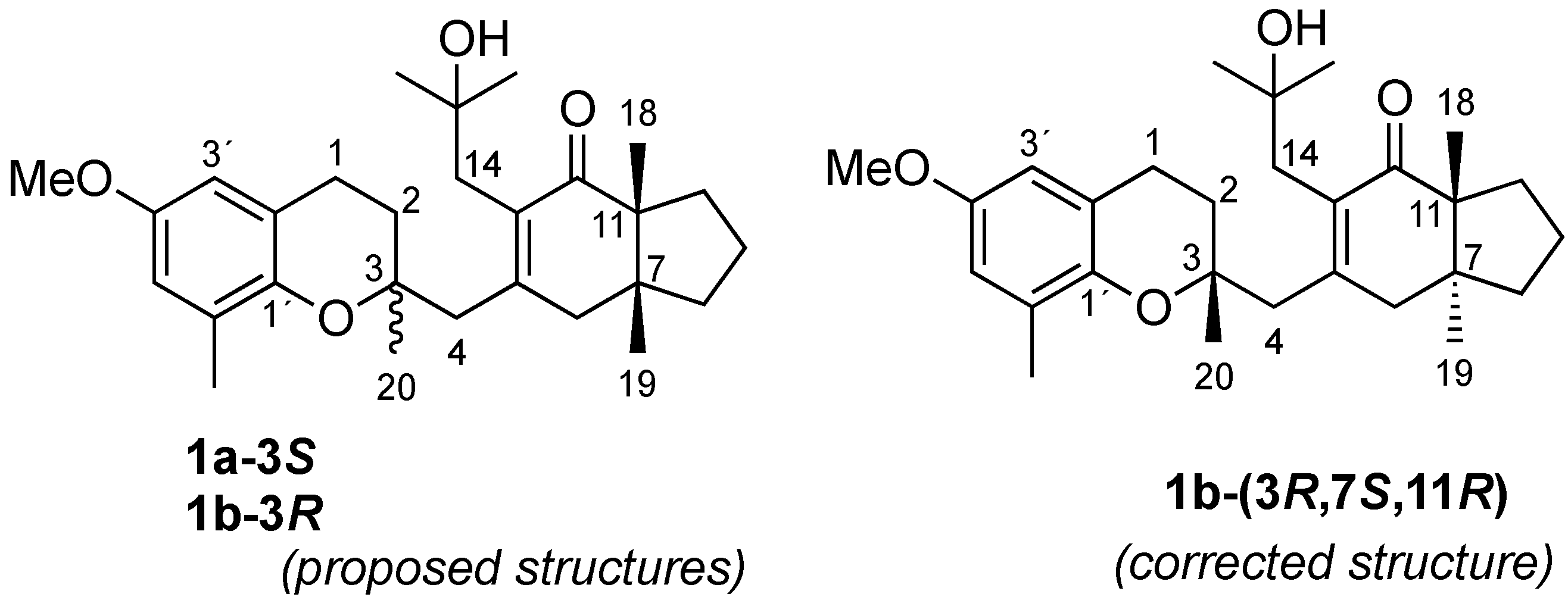
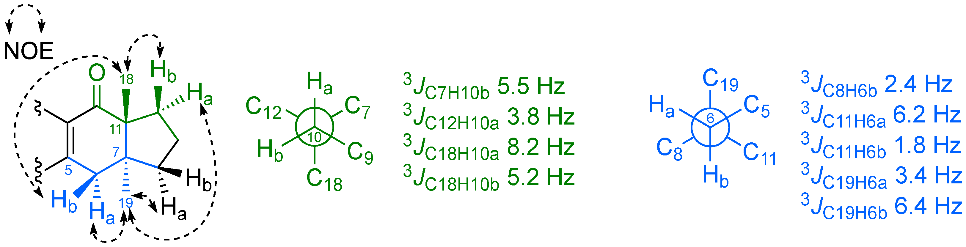
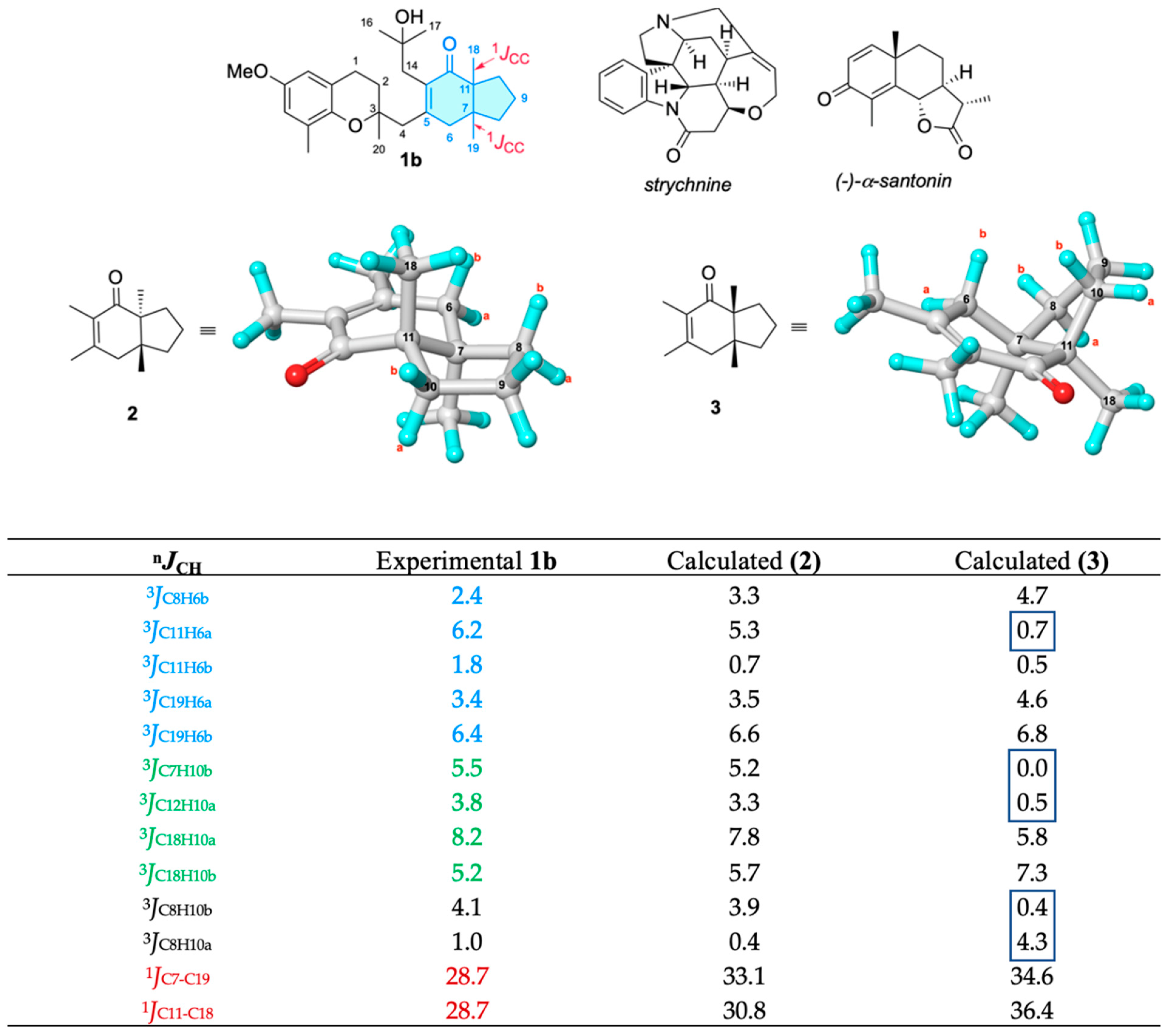
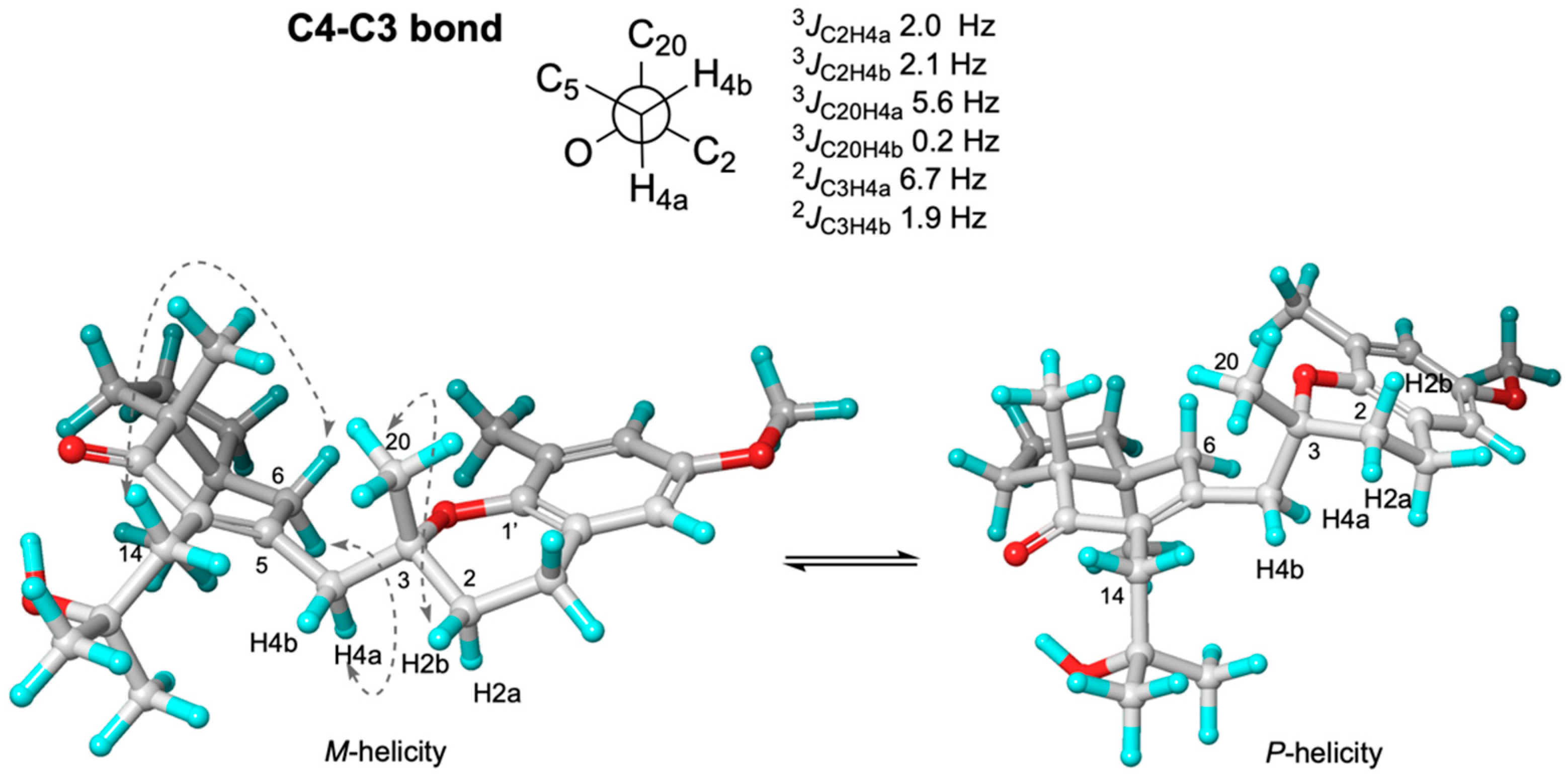
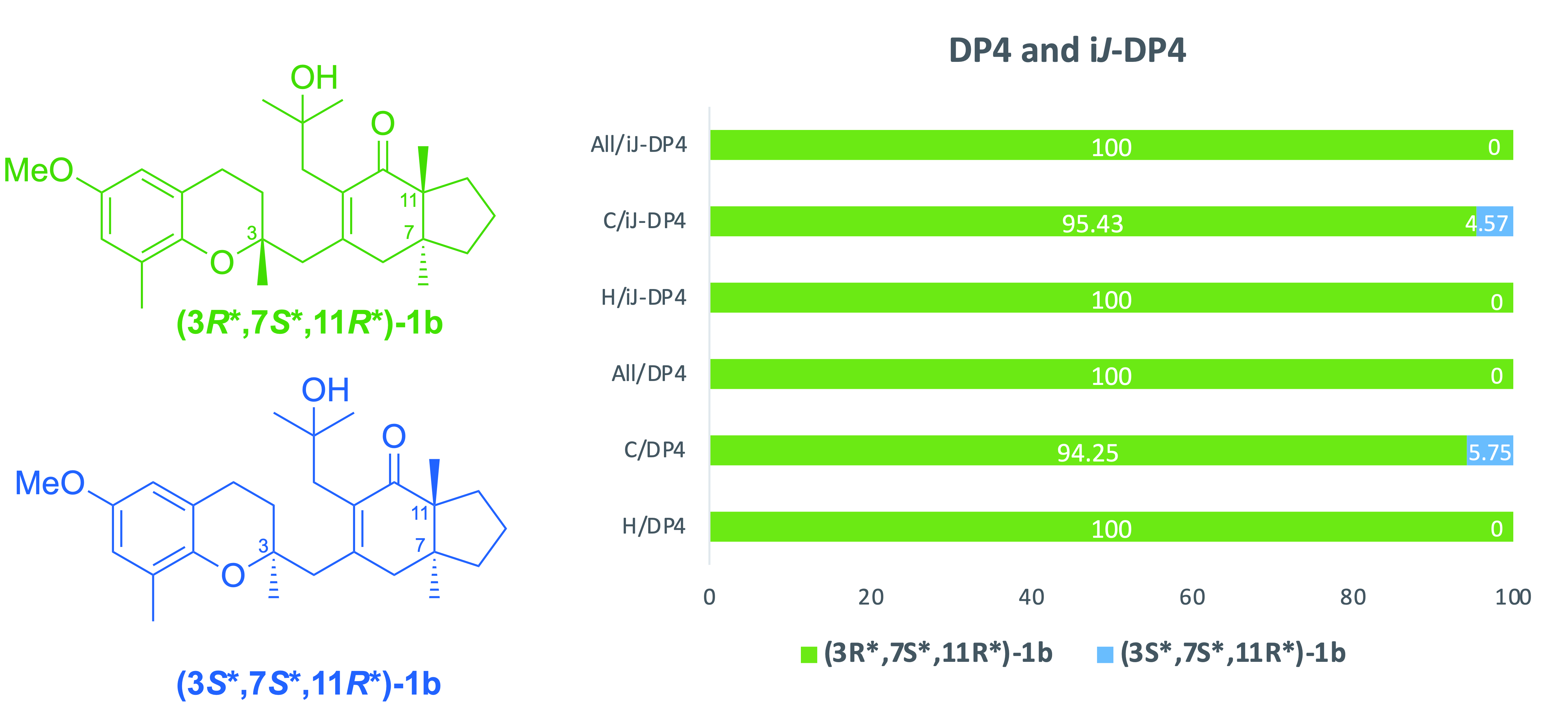
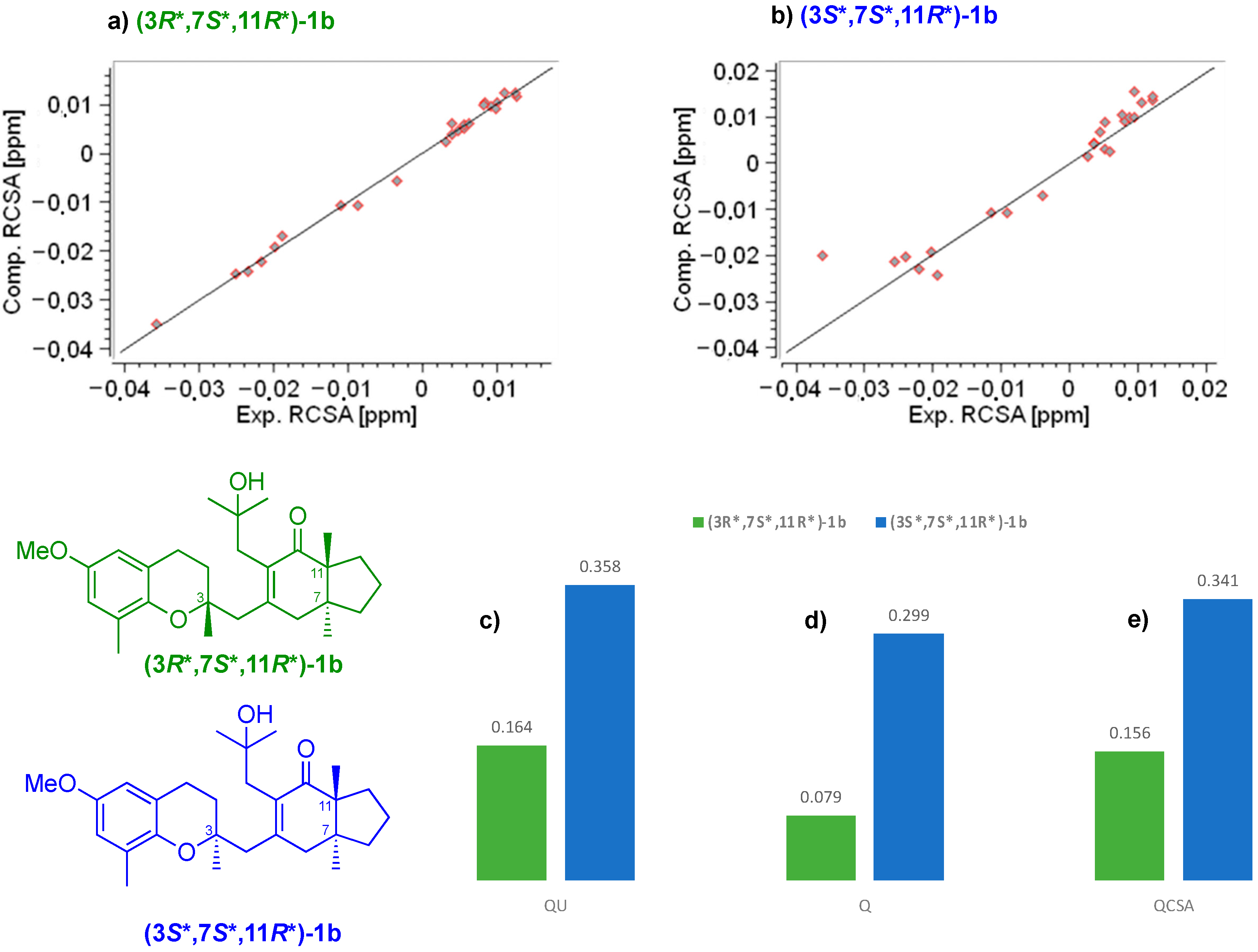
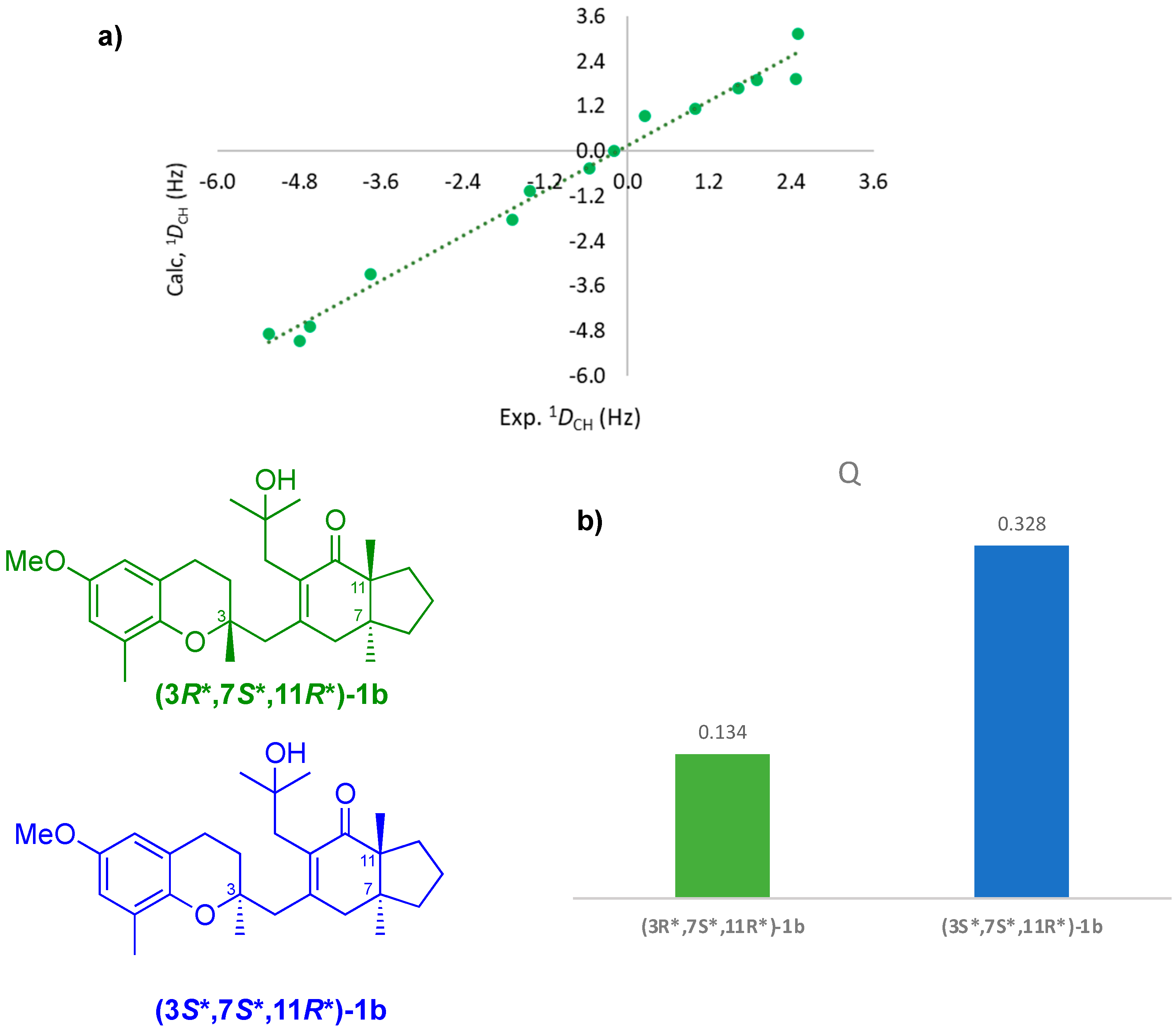
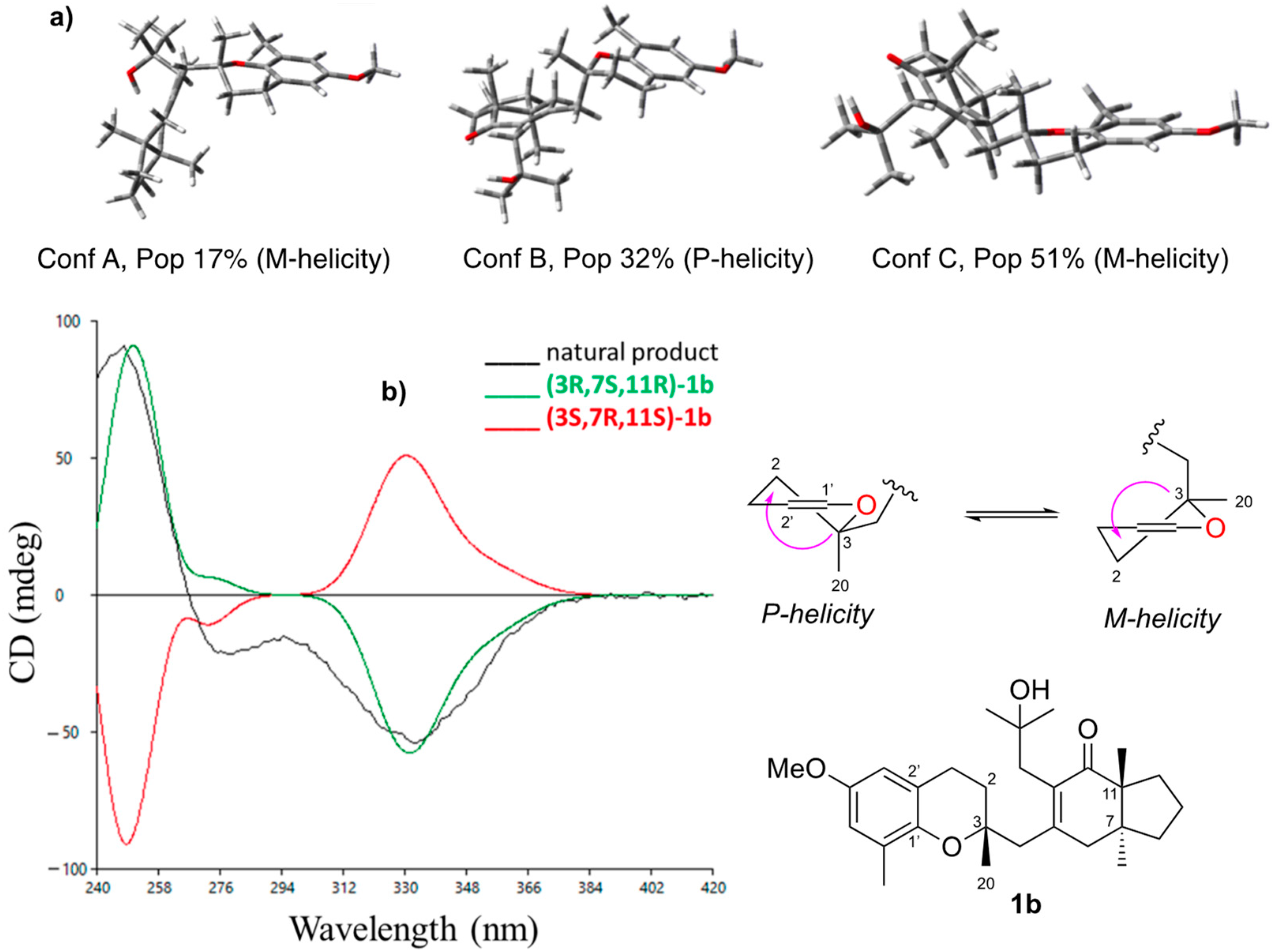
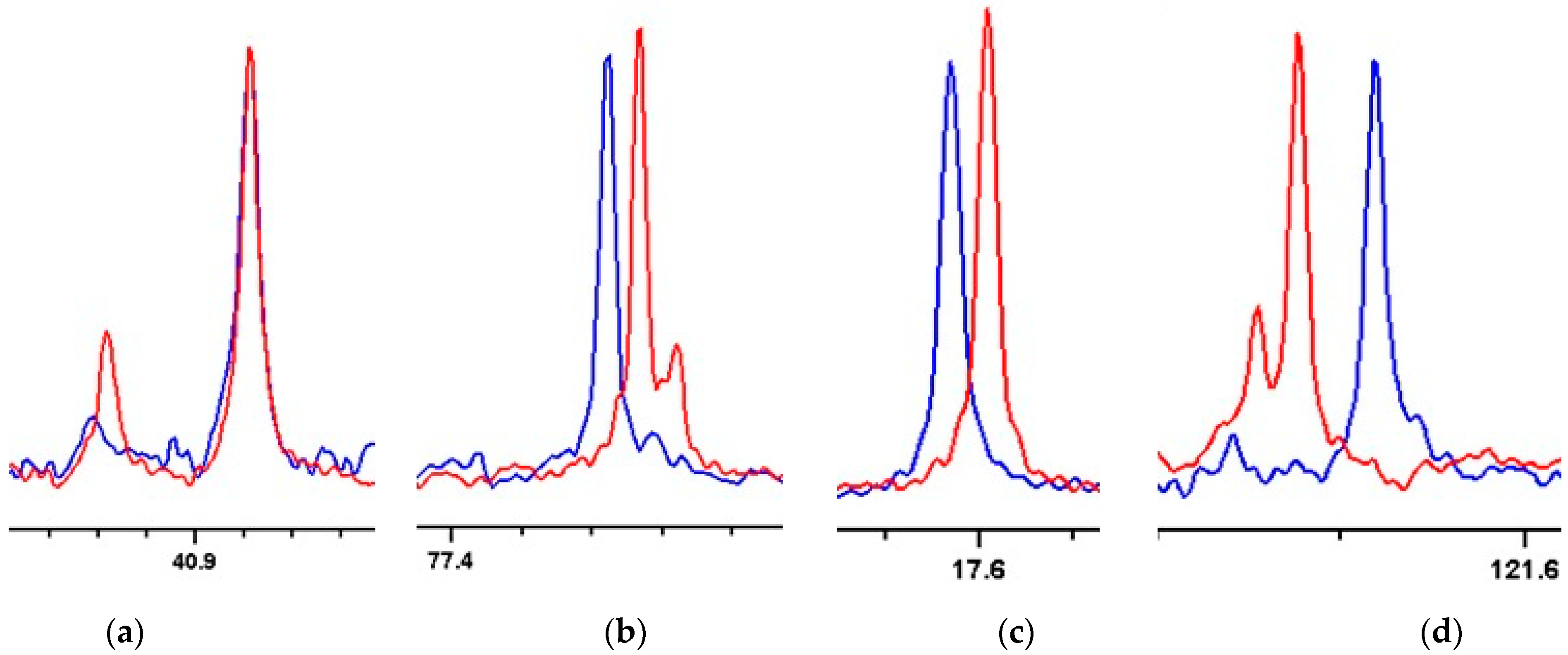
2. Results and Discussion

From the methanolic extract of SM, the n-hexane soluble fraction was separated by flash column chromatography and then purified by HPLC to obtain one of the epimers at position 3. The 950 MHz NMR spectra collected in CD2Cl2 did not show any evidence of epimerization for compound 1b. We observed the epimerization of the pure compound in CDCl3 after 25 min. The high resolution mass spectrum showed peaks at m/z 441.2969 ((M + H)+), 463.2811 ((M + Na)+), and 903.5741 ((2M + Na)+), thus confirming the molecular formula C28H40O4. The 1H NMR spectrum of 1b (Table 1) was then assigned in CD2Cl2, showing a singlet signal at δH 3.701, corresponding to the methoxy group attached to the aromatic nucleus. Two doublets, at δH 6.563 and 6.450, were assigned to aromatic protons, a harmonious relationship then being revealed between the small coupling constant (J = 3.0 Hz) and the 1′,2′,4′,6′-substituents meta-arranged around the benzene ring. A spin system formed by two contiguous methylene groups was also assigned by observing a 1H-1H COSY correlation between the protons at δH 1.805 (J = 13.5 Hz and 6.9 Hz) and at δH 2.774 (J = 6.9 Hz) disclosing the remaining chromane moiety. The five singlet signals belonging to methyl groups on sp3 quaternary carbons were observed at δH 0.804, 1.041, 1.105, 1.225, and 1.235, along with the three isolated methylene systems at δH 2.705/2.514 (J = 13.7 Hz), 3.028/2.237 (J = 18.7 Hz), and 2.570/2.506 (J = 14.3 Hz). These three pairs were easily assigned to non-equivalent protons located at C4, C6, and C14. The remaining structure of 1b was completed by 1H and 13C data, 2D NMR (1H–1H COSY, HSQC, HMBC), and mass spectral data (see Figures S3–S13 and Table 1) confirmed the structure of the epimers previously isolated from Sargassum muticum and from a macroalgae belonging to the Cystoseira genus [13,14].
Configurational analysis of 1b. Surprisingly, when we tried to confirm the reported cis relationship of the two methyl groups at C7 and C11 in the hydrindane skeleton [10,13,14], we found an unexpected lack of NOE contact between Me18 and Me19 when both were selectively irradiated in a 1D-NOE experiment. A deeper NOE study (see Figure 2), along with a J-based configurational analysis [18,19,20,21] was performed through both 3JCH and 2JCH coupling constants obtained from IPAP-HSQMBC spectra and 1JCC from a 2D J-modulated ADEQUATE spectrum (see Figure 3) to deduce the bicyclo[4.3.0]nonane fusion present in 1b (see Figures S14–S22 and Table S4).
The relative configuration of the C6 prochiral protons could be established just by the observation of NOE contacts between H19 and H6a and between H10a and H6a. The NOE correlations observed between H6b/H18 and H10b/H18, and between H6a/H19 and H8a/H10a, suggested that the relative configuration at C7 and C11 must be trans-7S*,11R* rather than cis. The high 3JCH values measured between H10a and C18 of 8.2 Hz, and between H6b and C19 of 6.4 Hz in an IPAP-HSQMBC experiment (Figures S14–S18) were in harmony with a trans disposition between both methyl groups (see Figure 2).
To discriminate between cis and trans dimethyl configurations, 2JCH and 3JCH couplings were computed on in silico models 2 (trans) and 3 (cis) (see Figure 3) by using DFT calculations and compared with the experimental values of 1b. We tested different combinations of functional/basis set/gas phase, and solvent models to obtain heteronuclear couplings as accurate as possible, by using (-)- α -santonin and strychnine as test systems [22,23,24], (see Figure 3). To further improve accuracy, a scaling factor was used in the computations as described in the Supplementary Information (SI).
Overall, 46 DFT methods were examined using an empirical scaling factor to improve the accuracy of the computations (see SI) [25,26,27,28,29,30,31,32,33,34,35]. Heteronuclear 2JCH and 3JCH for (-)-α-santonin and strychnine were measured in DMSO-d6 from IPAP-HSQMBC and HECADE-HSQC experiments. Among all the methodologies tested, the combination GIAO/OLYP/Def2TZV gave the best performance/computation time ratio. Afterwards, the standard deviation (σ) and the coefficient of determination (R2) were computed (2JCH: σ = 1.5 Hz, R2 = 0.967, 3JCH: σ = 2.3 Hz, R2 = 0.978; see all details in SI). The good performance of OLYP is notable considering it is a pure GGA functional that does not include exact exchange. The DFT-calculated heteronuclear coupling constant values involving C7 and C11 in the trans-model 2 were in much better agreement with those of 1b in relation to the theoretical data computed for the cis-model 3 (see Figure 3). The trans fusion was also confirmed for this hydrindane by measuring the one-bond carbon–carbon scalar coupling constants (1JCC) between C7–C19 and C11–C18 in 1b from the 2D J-modulated ADEQUATE spectrum and comparing them to those theoretically obtained by DFT computation for models 2 and 3 at the same OLYP/Def2TZV level of theory. Both 1JC7C19 and 1JC11C18 of 28.7 Hz betokened the dimethyl trans configuration (DFT-calculated 1JC7C19 and 1JC11C18 are 33.1 and 30.8 Hz, respectively).
Once the relative configuration of 7S*,11R* for 1b was unequivocally assigned, we allocated its relation to C3. Unfortunately, J-based configurational analyses and NOE measurements were not able to discriminate the possible configurations for the two stereoclusters, whether (3R*,7S*,11R*) or (3S*,7S*,11R*) of 1b (Figure 4 only shows the analysis of 3R*,7S*,11R*; for 3S*,7S*,11R*, see Figure S31).
A conformational equilibrium around the C1′-O-C3-C2 dihedral, leading to two possible helicities, M- (C1′-O-C3-C2 < 0°) and P- (C1′-O-C3-C2 > 0°), was deduced from the observed NOE correlations observed between H2b/H20 (strong) and H2a/H20 (weak), and from the 3JHH, 3JCH, and 2JCH coupling constants.
This conformational flip/flop is characteristic of this type of chromane conformer on the dihydropyran ring (see Figure 4, Figures S24 and S25), as it was deduced from the isochronic chemical shifts observed in both H1 and the averaged coupling constant measured for 3JH1H2a = 6.7 Hz, 2JC3H2a = 3.4 Hz, and 2JC3H2b = 2.8 Hz (see Figure S21). From the observed coupling constants, we were able to deduce an equilibrium between P-helicity and M-helicity conformers in a 3:2 ratio.
Trying to establish the relative configuration at C3, we followed the iJ/DP4 protocol of Daranas, Sarotti et al., involving a conformational search within a 5-kcal/mol interval, and then calculated the theoretical chemical shifts at the B3LYP/6-31G(d,p) level [36,37,38]. Contrasting calculated vs. experimental chemical shifts gave rise to a better DP4 score for the (3R*,7S*,11R*)-1b diastereoisomer (C/DP4: 94.25%, H/DP4: 100%). Next, 3JC2H4a = 2.0 Hz, 3JC2H4b = 2.1 Hz, 3JC20H4a = 5.6 Hz, 3JC20H4b = 0.2 Hz, ǀ2JC3H4aǀ = 6.7 Hz, and ǀ2JC3H4bǀ = 1.9 Hz from IPAP-HSQMBC experiments (see Newman projection on Figure 4) were read into the application of the iJ/DP4, implying four dihedral angle restrictions around the C3–C4 bond, to wit: dihedral O-C3-C4-H4b (180 ° ± 15 ° ), dihedral O-C3-C4-H4a (±60 ° ± 15 ° ), dihedral C20-C3-C4-H4a (180 ° ± 15 ° ), and dihedral C20-C3-C4-H4b (±60 ° ± 15 ° ).
Using iJ/DP4, we fully predicted the (3R*,7S*,11R*)-1b isomer, a slight 95.43% improvement being gained at 13C and a complete one at 1H (see Figure 5). Most populated conformers in both plausible configurations in the DP4 ensembles, generated by an unrestricted conformational search, satisfy the main constraints of chromane helicities, prochiral protons at C4 and C14, and weak hydrogen bonding between OH at C15 and C12 (C=O).
Similarly, the forementioned set of conformers of plausible configurations of 1b was studied by applying the Computer-Assisted 3D Structure Elucidation (CASE-3D) strategy, as implemented in MNova StereoFitter [39,40], which makes no a priori assumptions about the conformational space. Only isotropic NMR data were utilized to elucidate the relative configuration of 1b. Both constitutions were ranked according to chemical shift predictions at the DFT level (GIAO/B3LYP/6-31G(d,p)) and 3JCH coupling computed by Karplus-like equations. The CASE-3D analysis scored the (3R*,7S*,11R*)-1b configuration slightly better (See Figure S42).
Anisotropic measurements of 1b: 13C-RCSA and 1DCH-RDC. To revalidate the structure of 1b deduced from chemical shift-based configurational analyses, anisotropic parameters such as carbon residual chemical shift anisotropy (13C-RCSA) were utilized to relate both stereoclusters, separated from each other by four bonds. The RCSAs are manifested by the change in the chemical shift when the analyte is placed in a weak alignment medium such as a gel or a liquid crystal. The carbon RCSAs provide the relative orientations of the structural moieties, including those of non-protonated carbons C3, C7, and C11 as in our case study [16,41,42].
After an unrestricted geometry search, we then subjected all conformers to gas phase geometry optimization at B3LYP/6-31G+(d,p) for plausible configurations (3R*,7S*,11R*)-1b and (3S*,7S*,11R*)-1b. Subsequently, from DFT atomic coordinates, we selected those optimized conformers fitting short-range NMR data (J-couplings, chemical shifts, nuclear Overhauser effects (NOEs)) [43].
The conformational ensemble used for this analysis is shown for the correct configuration in Figure 6. Twenty-eight 13C-RCSA values were measured using an in-house made 3 mm compression device and compressible PMMA-d8 gel. Before applying the former to the RCSA data collection of 1b, its performance was tested using estrone as a standard sample (see Section S6 in SI). RCSAs were induced although small isotropic chemical shift changes also ensued because of the change in the solvent to gel ratio under compressed conditions (See Figure S32) [16]. These ΔRCSA values, not corrected for isotropic contributions, ranged between −7.4 and 5.5 Hz (see Table S7). Aromatic and carbonyl carbons exhibited larger ΔRCSAs than aliphatic carbons, reflecting the respective sizes of the CSA tensors [16,40]. A higher error in the fitting curve was noticed in the (3S*,7S*,11R*)-1b diastereoisomer relative to the other plausible configuration, even though automatic isotropic shift correction was not applied (Figure 6c and Figure S38).
When we corrected for isotropic shifts, a better agreement was achieved between the experimental and back-calculated 13C-RCSAs for (3R*,7S*,11R*)-1b (Q(QCSA) 0.079 (0.156)) as compared to (3S*,7S*,11R*)-1b (Q(QCSA) 0.299 (0.341)) (Figure 6a,d,e). Furthermore, a relative difference in Q between both diastereomers was increased (ΔQu = 0.19; ΔQ = 0.22) (Figure 6c,d).
Consequently, we were able to differentiate between both plausible diastereomers by 13C RCSA and to establish the relative configuration between the stereocenter at C3 and the stereocluster C7–C11 located four bonds away.
Data discrimination was tested by two resampling methods, with and without replacement, to wit: Monte Carlo bootstrapping [16,22] and leave-one-out cross-validation (Jackknife) [44]. Both bell curves derived from bootstrapping and resampled influence bar-plotted from Jackknife clearly indicate that 13C-RCSA data unambiguously confirmed (3R*,7S*,11R*)-1b as the correct relative configuration (Figures S39 and S40).
One-bond 13C-1H residual dipolar couplings (1DCH-RDC) measurements were also conducted to confirm the relative configuration of 1b. Thus, a CLIP/CLAP-HSQC, spurred by non-uniform sampling [45,46] with a 1.6 mg sample, and using a deuterated PMMA-d8 (70/0.25) in a low viscosity solvent (CD2Cl2), was measured to obtain RDC values. The accuracy of the 1DCH values was enhanced as there was almost no interference from deuterated polymer background signals.
Seventeen RDCs were meticulously measured thanks to the narrow spectral line, resulting in 1DCH values within a range of between 3.0 and −6.7 Hz. RDC data were fitted on an experimentally constrained ensemble of conformers for both plausible configurations as in the RCSA analysis (See Figure 7).
Fitting the 1DCH data, determining and aligning the tensor, and calculating Cornilescu’s quality factor Q were carried out through single-tensor approximation on the MSpin-RDC software. Configuration (3R*,7S*,11R*)-1b showed a Q of 0.134, while (3S*,7S*,11R*)-1b had a Q of 0.328, the discrimination presenting a degree of linearity between experimental and back-calculated values (see Figure 7b). By using 1DCH-RDC, configuration (3S*,7S*,11R*)-1b was removed from consideration with ease, showing the efficacy of the RDC methodology for establishing relative orientations of two distant stereoclusters of meroditerpene 1b.
Absolute configuration of 1b. Finally, we resorted to chiroptical methods (ECD/OR) to assign the absolute configuration (AC) of 1b. We used the empirical chromane helicity rule [47,48] and compared the experimental chiroptical data with predicted ECD/ORD from ab initio time-dependent DFT (TD-DFT). Currently, chiroptical methodologies associated with quantum chemical calculations are some of the most powerful tools for elucidating stereochemistry and examining even minute changes in the geometry of chiral molecules [49,50,51]. Determining the AC of this framework is also possible by following a chromane helicity rule [47,48]. This rule, established by Crabbé and related to 1Lb excitation of the chromophore, relates the sign of the ECD band with the helicity of the trisubstituted dihydropyran ring [48] at around 270–290 nm. An extra requirement for the application of the rule is that the largest substituent at the C3 carbon atom should favorably occupy an equatorial position. This rule was exhaustively studied in 2014 by Górecki and Frelek, who revised both the scope and limitations of comparing the experimental ECD spectra with those simulated by TD-DFT, and should, therefore, be used with great caution [52,53]. In our case, following Crabbé’s rule, the ECD curve strongly depends on the population ratio of 1b conformers with the methyl group at C3 either in axial or equatorial positions.
Both the experimental and calculated ECD spectra as well as the OR value were applied to establish the AC of the stereoisomeric pairs exhibiting opposite ECD and OR. Two different levels of theory were chosen to calculate ECD curves: PBE0/Def2TVZ/W06 and HSE06/6-311+G(2d,p)/DGA1 basis sets, with 50 and 38 transition states, respectively, and COSMO-IEFPCM as solvent models (CH2Cl2). Populations were calculated through Q factor minimization by NMR anisotropy constraints [54,55,56]. Calculations at two levels of theory clearly reproduced the 240–290 nm range of the ECD of 1b, demonstrating an equilibrium between conformers with M- (68%) and P- helicities (32%) with an excellent agreement towards the (3R,7S,11R) AC for 1b (see Figure 8). Figure 8a depicts the conformers found for the correct configuration of 1b along with their populations, derived from the analysis of NMR anisotropic parameters.
The specific optical rotation ( [ α ] D 25 ) of 1b was experimentally measured, −11.67 (c 0.12, CH2Cl2), and then computed at CAM-B3LYP/6-311++G(2d,2p)/DGA1 DFT level. The IEFPCM (CH2Cl2) solvation effect model and the conformer population were the same as with the ECD calculation (Figure 8). After comparing the experimental results with the DFT computed values, the sign was well reproduced, although the [ α ] D 25 was overestimated for (3R,7S,11R)-1b −49.4 (see Table S8). The absolute signs indicate that our approach is suitable for correlating the absolute configuration of molecules with rather restricted flexibility to the sign of the [ α ] D 25 . Both the ECD and the optical rotation approaches agree with the same identifications of 1b as 3R,7S,11R [57].

3. Materials and Methods

3.1. General

ECD and UV spectra were recorded on a JASCO J-815 CD spectrometer. The ECD spectrum of 1b was recorded in the region from 225 to 470 nm at a concentration of 0.2 mg/mL (2.88 × 10⁻⁴ M) in CH2Cl2 in a 1 mm cell, totaling five accumulations at a scan rate of 20 nm/min and a temperature of 25 °C. Specific optical rotation (OR) of 1b was recorded on a Jasco DIP-1000 polarimeter at 589 nm (Na lamp) at a concentration of 0.12 g/100 mL in CH2Cl2 in a 1 dm/SiO2 cuvette (25 °C). NMR spectra were recorded on either a 950, 900, 800, or a 700 MHz Bruker, all the signals being referenced to 13C (54.00 ppm) and 1H (5.320 ppm) signals of CD2Cl2. HR-MS were obtained on a Thermo Scientific LTQ Orbitrap XL mass spectrometer (Thermo Fisher Scientific, Waltham, MA, USA). Semipreparative HPLC was performed on an Agilent column (RP-C18 column 10 × 100 mm; 4.6 mL/min). TLC was performed on silica gel (Merck, Kieselgel 60 F254) plates; the spots were visualized by exposure to UV light (254 nm). Column chromatography was carried on silica gel (Merck, Kieselgel 60).

3.2. Data Processing

Experimental and calculated ECD and UV data were handled with SpecDis V 1.7.1 developed by T. Bruhn [58]. NMR data were processed and analyzed by Bruker TopSpin Software V 4.1.1. Graphics were carried out on Microsoft Excel 365. Geometrical optimization and TDSCF calculation were carried out with the Gaussian 16 Suite. Molecular structures were drawn in ChemDraw V 12.0 and Avogadro V 1.0 [59]. Orbitrap files were processed using Xcalibur™ V 3.0 Software—Thermo Fisher Scientific—and its chemical molecular generator module was employed to provide elemental formulas. Conformer weighing by isotropic constraints was handled by the StereoFitter module nested in MNova Suite [39,40]. Anisotropic NMR and J-coupling data processing were performed using MSpin-RDC V 2.6.1.

3.3. Raw Material

The seaweed Sargassum muticum (SM) was collected on a rocky shore on the southern coast of Galicia, Praia da Mourisca (Alcabre, Spain) during the summer of 2011. Specimens were washed carefully with water and then oven-dried at 50 °C for 72 h to preserve them until use. Dried algae were milled to facilitate sample handling and provide higher extraction yields by increasing the contact surface. First, it was milled in a cutting mill up to a particle size of 1–2 cm length and then further milled to obtain a coarse powder.

3.4. Extraction and Isolation

The raw methanolic extract of SM (4.9 g) was dissolved in methanol/water (1:10) and subsequently partitioned with: n-hexane (FH), dichloromethane (FD), n-butanol (WB), and water (WW) [60]. Fractions were concentrated under reduced pressure (temp: 32 °C); yielded FH 1.1 g, FD 0.2 g, WB 0.3 g, and WW 2.1 g. The FH (1.1 g) was subjected to NMR-guided fractioning through flash silica gel column chromatography (25 × 2 cm; 80 mL/min) using a stepwise gradient of hexane/Et2O and Et2O/AcOEt to produce 30 fractions, which were grouped by TLC. Fraction 19 was eluted with a 17:8 Hex/Et2O solution.

Purification of 1b

Fraction 19 (Fr. 19) showed doublet (d) belonging to two meta-coupled aromatic protons (protons 3′ and 5′) in an aromatic ring part of the bicycling system (6.412–6.533 ppm; J = 3.0 Hz) characteristic of the meroditerpenes. F.19 (103 mg) was further purified by semipreparative HPLC (Atlantis RP-C18 column 10 × 100 mm; 4.6 mL/min) equipped with a variable wavelength detector (VWD) at 320 nm, an Agilent pump supplying the following solvent profile: 2.2 min isocratic step (80 v/v% ACN/H2O) and 12.3 min gradient step (from 80 to 100 v/v% ACN/H2O) to afford compound 1b (27.4 mg; Rt = 5.17 min).

3.5. Computational Section

Conformational searches for J-DP4 and CASE-3D analyses were performed by employing MAESTRO software, using an energy window of 5 kcal/mol. Energy cut-off (5 kcal/mol) for CASE-3D was based on calculations at B3LYP/6-31G(d,p), solvent model = IEFPCM (CH2Cl2). Conformational searches for NMR anisotropy analyses were carried out on relevant rotatable bounds ρ1, ρ2, ρ3, and ρ4 (See Figures S21–S27) using the grid search module in PCModel V10. Both P- and M- helicities were considered. Conformers used on NMR anisotropy analyses were optimized with DFT calculations at B3LYP/6-31+G(d) level, with vibrational frequency calculations confirming the presence of minima, using the Gaussian16 program. Chemical shielding tensors (CST) were computed at the GIAO/MPW1PW9/6-311+G(2d,p) (in gas phase). J-couplings were computed either at DFT level or by using Karplus-like equations [61,62]. All chiroptical properties were calculated as Boltzmann averages, weighted with conformer population factors obtained from NMR anisotropic Q factor minimization. Time-dependent DFT calculations were performed for each configuration using the combinations PBE0/Def2TZV/W06, solvent model: COSMO, 50 excited states and HSE06/6-311+G(2d,p)/DGA1; solvent model: IEFPCM, 38 excited states. In both, solvent CH2Cl2 parameters were used. ECD spectra were generated using the program SpecDis by applying a Gaussian band shape with a 0.18 eV width and 25 blue shifts to facilitate comparison to the experimental data. OR values were computed, for both configurations, at CAM-B3LYP/6-311++G(2d,2p)/DGA1 (IEFPCM = CH2Cl2) level of theory at 589 nm.

J-Coupling Calculation

(-)-(α)-santonin and strychnine were purchased at Sigma-Aldrich and both molecular models were optimized based on RCSA/RDC and NOE, respectively, as described before [22]. For strychnine, two conformers were taken into account as found by Butts et al. [24] for 2,3JCH benchmarking at different levels of theory. J-coupling data were handled using the CST module nested in MSpin (See SI).

3.6. 13C-RCSA Measurements

A first attempt to establish the relative configuration of meroditerpene 1b by 13C RCSA involved the use of a 4 mg sample in protonated PMMA swollen in CD2Cl2 and using a conventional 5 mm outer diameter compression device. A number of resonances were obscured by the polymer background signal (see Figures S35–S43 and Table S6). Therefore, 2.2 mg of 1b was dissolved in CD2Cl2 and swollen into a deuterated chemically cross-linked poly(methylmethacrylate) (PMMA-d8) gel in an in-house made 3 mm compression device. RCSA were extracted as the difference in the referenced chemical shift between signals at maximum and minimum compression (13C-RCSAi = Δδi,max − Δδi,min) [63]. Due to the inherent properties of the compression device, the gel was surrounded by a layer of isotropic solution; carbon signals were phenomenologically observed from within (Δδi,min) and without (Δδi,iso) the gel (asterisk marked in Figure 9) [16]. After compressing the gel, the analyte concentration inside the gel increased, changing the isotropic contribution of the chemical shifts, and hindering the precise extraction of RCSAs without compensating for these isotropic shift contributions [16]. Therefore, an isotropic shift contribution of gel, expressed as free parameter c, was computed by applying an automatic linear correction [64] (Equation (1)). A quality factor computed without using this means is considered as uncorrected (Qu); it tends to have less discriminating power or even give ambiguous results. Δδi represents the referenced chemical shift with respect to a selected resonance at the corresponding conditions. Whenever possible, a nucleus with low associated anisotropy, a methylene group, for instance, was taken (Equation (2)). Data were fitted in units of ppm to obtain reasonable weight [63].
13C-RCSAi = Δδi,max − Δδi,min – c × (Δδi,min − Δδi,iso)
Δδi,max = δi,max − δref,max
The quality of the fitting is scored in terms of quality factor Q. When carbon chemical shift variations due to analyte concentration changes are not considered, the uncorrected quality factor is named as QU. To yield minimum Q factor [65], free parameter c was minimized simultaneously along with the alignment tensor during the fitting procedure, and RCSA was fitted by singular value decomposition [63,66]. Conformers were fitted using a multiconformer single tensor approach and distances among the heavy atoms were minimized. The unscaled quality factor (Q) and the chemical shift anisotropic weighted quality factor (QCSA) were computed using Equations (3) and (4), respectively [16].
Q = ( i = 1 n ( C 13   R C S A e x p , i C 13   R C S A c a l c , i ) 2 i = 1 n C 13   R C S A 2 i ) 1 2
Q C S A = ( i = 1 n ( [ C 13   R C S A e x p , i C 13   R C S A c a l c , i ] / C S A i , a x ) 2 i = 1 n ( C 13   R C S A i / C S A i , a x ) 2 ) 1 2
Data robustness was addressed by either bootstrapping analysis or Jackknife resampling as implemented in MSpin software and Excel [67].
Some significant variations in carbon chemical shifts in the oriented media were observed (see Figure 9). A certain amount of isotropic analyte was always detected because the gel in the compression device did not completely fill the entire sample space, even under maximum compression [68]. As before, the isotropic signals were easily distinguishable from their anisotropic counterparts, as their intensities diminished upon compression.

4. Conclusions

We demonstrated that the use of an integrated approach combining isotropic and anisotropic NMR means and incorporating chiroptical methods is a perfect toolbox to deduce the three-dimensional structure containing stereoclusters that cannot be connected through local correlations.
We also detected, through 3JHH, 2JCH, 3JCH, coupling constants and RDC, flip-flop conformational changes around the C1′-O-C3-C2 dihedral. Using this approach, the structure of the tetraprenyltoluquinol chromane meroterpenoids 1b isolated from Sargassum muticum was corrected to the trans fusion dimethylbicyclo [4.3.0]nonane system; and the distant stereocenter at C3 was related to C7 and C11 with the use of DP4, iJ-DP4, CASE-3D, 1DCH-RDC, and 13C-RCSA methodologies. Finally, a set of chiroptical methods (ECD and OR) and of DFT calculations allows the absolute configuration of 1b to be assigned as (3R,7S,11R). This is the first time this kind of terpenoids has been well studied by anisotropic NMR methodology, which unambiguously confirms the relative configuration of two distant stereoclusters separated by four covalent bonds.

Supplementary Materials

The following supporting information can be downloaded at: https://www.mdpi.com/article/10.3390/md20070462/s1.

Author Contributions

J.C.C.F.-M., J.R., C.G. and C.J. conceived the project. J.C.C.F.-M., J.R. and N.N. performed the measurements, the DFT calculations, and the NMR anisotropy data evaluations. J.C.C.F.-M. designed the 3 mm compression device. Hilgenberg GmbH manufactured the 3 mm tube. The semi micro compression apparatus was manufactured in the mechanical workshop of the Max Planck Institute for Multidisciplinary Sciences. E.M.B. collected the seaweed samples. J.C.C.F.-M., A.M.F., J.R. and C.J. isolated the meroditerpene; conducted the J-based and NOE quantitative analyses; took NMR measurements; carried out the DFT calculations for constitution analyses; and discussed the data. C.G., J.C.C.F.-M. and A.M.F. measured the ECD spectrum and OR value. J.C.C.F.-M. formulated and prepared the PMMA-d8. A.N.-V. provided the tailored MSpin-RDC version and all codes for automated data handling and recommendations from NMR data analysis. J.C.C.F.-M. and J.R. wrote the paper. All authors contributed to discussions on the written paper. All authors have read and agreed to the published version of the manuscript.

Funding

This work was funded by grants RTI2018-093634-B-C22 from the State Agency for Research (AEI) of Spain, both co-funded by the European Regional Development Fund (ERDF), BLUEBIOLAB (0474_BLUEBIOLAB_1_E), Programme INTERREG V A of Spain–Portugal (POCTEP) and GRC2018/039 and Agrupación Estratégica CICA-INIBIC ED431E 2018/03 from Xunta de Galicia. This work was also supported by the Max Planck Society and grew out of a collaboration in the context of the Forschergruppe (FOR 934), continued now by the DFG (Gr1211/19–1 and Re1007/9–1)/CAPES 418729698 Project. N.N. gratefully acknowledges the financial support by SERB, New Delhi for ECR Grant with File No.: ECR/2017/001811.

Institutional Review Board Statement

Not applicable.

Informed Consent Statement

Not applicable.

Data Availability Statement

All data can be obtained from JR or CJ at CICA at Universidad da Coruña.

Acknowledgments

C.J. and J.R. acknowledge CESGA for the computational support. J.C.C.F.-M. acknowledges predoctoral research stay grant Inditex-UDC. A.N.-V. thanks CNPq for a research fellowship and financial support M(426216/2018–0). J.C.C.F.-M. thanks both Roberto Gil, for providing protonated PMMA gel sticks for the initial analysis of compound 1b, and Christian F. Pantoja, for discussing long-range coupling NMR experiments. Thanks to Carlos Mota for the English and style corrections.

Conflicts of Interest

The authors declare no conflict of interest.

References

  1. Norton, T.A. The Growth and Development of Sargassum Muticum (Yendo) Fensholt. J. Exp. Mar. Biol. Ecol. 1977, 26, 41–53. [Google Scholar] [CrossRef]
  2. Critchley, A.T. Sargassum Muticum: A Taxonomic History Including World-Wide and Western Pacific Distributions. J. Mar. Biol. Assoc. U. K. 1983, 63, 617–625. [Google Scholar] [CrossRef]
  3. Davison, D.M. Sargassum Muticum in Scotland, 2008: A Review of Information, Issues and Implications; Commissioned Report No. 324; Scottish Natural Heritage: Perth, Scotland, 2009. [Google Scholar]
  4. Balboa, E.; Conde, E.; Constenla, A.; Falqué, E.; Domínguez, H. Sensory Evaluation and Oxidative Stability of a Suncream Formulated with Thermal Spring Waters from Ourense (NW Spain) and Sargassum Muticum Extracts. Cosmetics 2017, 4, 19. [Google Scholar] [CrossRef]
  5. Park, S.Y.; Seo, I.S.; Lee, S.J.; Lee, S.P. Study on the Health Benefits of Brown Algae Sargassum Muticum in Volunteers. J. Food Nutr. Res. 2015, 3, 126–130. [Google Scholar] [CrossRef] [Green Version]
  6. Heo, S.J.; Jeon, Y.J. Protective Effect of Fucoxanthin Isolated from Sargassum Siliquastrum on UV-B Induced Cell Damage. J. Photochem. Photobiol. B Biol. 2009, 95, 101–107. [Google Scholar] [CrossRef] [PubMed]
  7. Nazir, M.; Saleem, M.; Tousif, M.I.; Anwar, M.A.; Surup, F.; Ali, I.; Wang, D.; Mamadalieva, N.Z.; Alshammari, E.; Ashour, M.L.; et al. Meroterpenoids: A Comprehensive Update Insight on Structural Diversity and Biology. Biomolecules 2021, 11, 957. [Google Scholar] [CrossRef]
  8. Kim, J.A.; Ahn, B.N.; Kong, C.S.; Kim, S.K. The Chromene Sargachromanol e Inhibits Ultraviolet A-Induced Ageing of Skin in Human Dermal Fibroblasts. Br. J. Dermatol. 2013, 168, 968–976. [Google Scholar] [CrossRef]
  9. Kang, H.S.; Kim, J.P. New Chromene Derivatives with Radical Scavenging Activities from the Brown Alga Sargassum Siliquastrum. J. Chem. Res. 2017, 41, 116–119. [Google Scholar] [CrossRef]
  10. Balboa, E.M.; Li, Y.-X.; Ahn, B.-N.; Eom, S.-H.; Domínguez, H.; Jiménez, C.; Rodríguez, J. Photodamage Attenuation Effect by a Tetraprenyltoluquinol Chromane Meroterpenoid Isolated from Sargassum Muticum. J. Photochem. Photobiol. B: Biol. 2015, 148, 51–58. [Google Scholar] [CrossRef]
  11. Ferdous, U.T.; Yusof, Z.N.B. Algal Terpenoids: A Potential Source of Antioxidants for Cancer Therapy. In Terpenes and Terpenoids; Perveen, S., Al-Taweel, A.M., Eds.; IntechOpen: Rijeka, Croatia, 2021. [Google Scholar]
  12. Fisch, K.M.; Böhm, V.; Wright, A.D.; König, G.M. Antioxidative Meroterpenoids from the Brown Alga Cystoseira Crinita. J. Nat. Prod. 2003, 66, 968–975. [Google Scholar] [CrossRef]
  13. Valls, R.; Piovetti, L.; Banaigs, B.; Praud, A. Secondary Metabolites from Morocco Brown Algae of the Genus Cystoseira. Phytochemistry 1993, 32, 961–966. [Google Scholar] [CrossRef]
  14. de Sousa, C.B.; Gangadhar, K.N.; Morais, T.R.; Conserva, G.A.A.; Vizetto-Duarte, C.; Pereira, H.; Laurenti, M.D.; Campino, L.; Levy, D.; Uemi, M.; et al. Antileishmanial Activity of Meroditerpenoids from the Macroalgae Cystoseira Baccata. Exp. Parasitol. 2017, 174, 1–9. [Google Scholar] [CrossRef] [PubMed]
  15. Amico, V.; Cunsolo, F.; Oriente, G.; Piattelli, M.; Ruberto, G. Cystoketal, a New Metabolite From the Brown Alga Cystoseira Balearica. J. Nat. Prod. 1984, 47, 947–952. [Google Scholar] [CrossRef]
  16. Nath, N.; Schmidt, M.; Gil, R.R.; Williamson, R.T.; Martin, G.E.; Navarro-Vázquez, A.; Griesinger, C.; Liu, Y. Determination of Relative Configuration from Residual Chemical Shift Anisotropy. J. Am. Chem. Soc. 2016, 138, 9548–9556. [Google Scholar] [CrossRef]
  17. Gayathri, C.; Tsarevsky, N.V.; Gil, R.R. Residual Dipolar Couplings (RDCs) Analysis of Small Molecules Made Easy: Fast and Tuneable Alignment by Reversible Compression/Relaxation of Reusable PMMA Gels. Chem. –A Eur. J. 2010, 16, 3622–3626. [Google Scholar] [CrossRef]
  18. Matsumori, N.; Kaneno, D.; Murata, M.; Nakamura, H.; Tachibana, K. Stereochemical Determination of Acyclic Structures Based on Carbon-Proton Spin-Coupling Constants. A Method of Configuration Analysis for Natural Products. J. Org. Chem. 1999, 64, 866–876. [Google Scholar] [CrossRef]
  19. Matsumori, N.; Murata, M.; Tachibana, K. Conformational Analysis of Natural Products Using Long-Range Carbon-Proton Coupling Constants: Three-Dimensional Structure of Okadaic Acid in Solution. Tetrahedron 1995, 51, 12229–12238. [Google Scholar] [CrossRef]
  20. Pachler, K.G.R. Nuclear Magnetic Resonance Study of Some α-Amino Acids—II. Rotational Isomerism. Spectrochim. Acta 1964, 20, 581–587. [Google Scholar] [CrossRef]
  21. Pachler, K.G.R. Nuclear Magnetic Resonance Study of Some α-Amino Acids—I. Spectrochim. Acta 1963, 19, 2085–2092. [Google Scholar] [CrossRef]
  22. Nath, N.; Fuentes-Monteverde, J.C.; Pech-Puch, D.; Rodríguez, J.; Jiménez, C.; Noll, M.; Kreiter, A.; Reggelin, M.; Navarro-Vázquez, A.; Griesinger, C. Relative Configuration of Micrograms of Natural Compounds Using Proton Residual Chemical Shift Anisotropy. Nat. Commun. 2020, 11, 1–9. [Google Scholar] [CrossRef]
  23. Teles, R.R.; França, J.A.; Navarro-Vázquez, A.; Hallwass, F. Atribuição da Estereoquímica da α-santonina através das medidas do Acoplamento Dipolar Residual. Química Nova 2015, 38, 1345–1350. [Google Scholar]
  24. Butts, C.P.; Jones, C.R.; Harvey, J.N. High Precision NOEs as a Probe for Low Level Conformers—A Second Conformation of Strychnine. Chem. Commun. 2011, 47, 1193–1195. [Google Scholar] [CrossRef] [PubMed]
  25. Kim, K.; Jordan, K.D. Comparison of Density Functional and MP2 Calculations on the Water Monomer and Dimer. J. Phys. Chem. 1994, 98, 10089–10094. [Google Scholar] [CrossRef]
  26. Ernzerhof, M.; Scuseria, G.E. Assessment of the Perdew–Burke–Ernzerhof Exchange-Correlation Functional. J. Chem. Phys. 1999, 110, 5029–5036. [Google Scholar] [CrossRef] [Green Version]
  27. Adamo, C.; Barone, V. Exchange Functionals with Improved Long-Range Behavior and Adiabatic Connection Methods without Adjustable Parameters: The MPW and MPW1PW Models. J. Chem. Phys. 1998, 108, 664–675. [Google Scholar] [CrossRef]
  28. Adamo, C.; Barone, V. Toward Reliable Density Functional Methods without Adjustable Parameters: The PBE0 Model. J. Chem. Phys. 1999, 110, 6158–6170. [Google Scholar] [CrossRef]
  29. Heyd, J.; Scuseria, G.E.; Ernzerhof, M. Hybrid Functionals Based on a Screened Coulomb Potential. J. Chem. Phys. 2003, 118, 8207–8215. [Google Scholar] [CrossRef] [Green Version]
  30. Handy, N.C.; Cohen, A.J. Left-Right Correlation Energy. Mol. Phys. 2001, 99, 403–412. [Google Scholar] [CrossRef]
  31. Feller, D. The Role of Databases in Support of Computational Chemistry Calculations. J. Comput. Chem. 1996, 17, 1571–1586. [Google Scholar] [CrossRef]
  32. Weigend, F.; Ahlrichs, R. Balanced Basis Sets of Split Valence, Triple Zeta Valence and Quadruple Zeta Valence Quality for H to Rn: Design and Assessment of Accuracy. Phys. Chem. Chem. Phys. 2005, 7, 3297–3305. [Google Scholar] [CrossRef]
  33. Weigend, F. Accurate Coulomb-Fitting Basis Sets for H to Rn. Phys. Chem. Chem. Phys. 2006, 8, 1057–1065. [Google Scholar] [CrossRef] [PubMed]
  34. Klamt, A.; Schüürmann, G. COSMO: A New Approach to Dielectric Screening in Solvents with Explicit Expressions for the Screening Energy and Its Gradient. J. Chem. Soc. Perkin Trans. 2 1993, 2, 799–805. [Google Scholar] [CrossRef]
  35. Miertuš, S.; Scrocco, E.; Tomasi, J. Electrostatic Interaction of a Solute with a Continuum. A Direct Utilization of AB Initio Molecular Potentials for the Prevision of Solvent Effects. Chem. Phys. 1981, 55, 117–129. [Google Scholar] [CrossRef]
  36. Grimblat, N.; Gavín, J.A.; Hernández Daranas, A.; Sarotti, A.M. Combining the Power of J Coupling and DP4 Analysis on Stereochemical Assignments: The J-DP4 Methods. Org. Lett. 2019, 21, 4003–4007. [Google Scholar] [CrossRef] [PubMed]
  37. Tarazona, G.; Fernández, R.; Pérez, M.; Millán, R.E.; Jiménez, C.; Rodríguez, J.; Cuevas, C. Enigmazole C: A Cytotoxic Macrocyclic Lactone and Its Ring-Opened Derivatives from a New Species of Homophymia Sponge. J. Nat. Prod. 2022, 85, 1059–1066. [Google Scholar] [CrossRef]
  38. Smith, S.G.; Goodman, J.M. Assigning Stereochemistry to Single Diastereoisomers by GIAO NMR Calculation: The DP4 Probability. J. Am. Chem. Soc. 2010, 132, 12946–12959. [Google Scholar] [CrossRef]
  39. Troche-Pesqueira, E.; Anklin, C.; Gil, R.R.; Navarro-Vázquez, A. Computer-Assisted 3D Structure Elucidation of Natural Products Using Residual Dipolar Couplings. Angew. Chem. Int. Ed. 2017, 56, 3660–3664. [Google Scholar] [CrossRef]
  40. Navarro-Vázquez, A.; Gil, R.R.; Blinov, K. Computer-Assisted 3D Structure Elucidation (CASE-3D) of Natural Products Combining Isotropic and Anisotropic NMR Parameters. J. Nat. Prod. 2018, 81, 203–210. [Google Scholar] [CrossRef]
  41. Liu, Y.; Cohen, R.D.; Gustafson, K.R.; Martin, G.E.; Williamson, R.T. Enhanced Measurement of Residual Chemical Shift Anisotropy for Small Molecule Structure Elucidation. Chem. Commun. 2018, 54, 4254–4257. [Google Scholar] [CrossRef]
  42. Liu, Y.; Saurí, J.; Mevers, E.; Peczuh, M.W.; Hiemstra, H.; Clardy, J.; Martin, G.E.; Williamson, R.T. Unequivocal Determination of Complex Molecular Structures Using Anisotropic NMR Measurements. Science (1979) 2017, 356, eaam5349. [Google Scholar] [CrossRef] [Green Version]
  43. Schmidt, M.; Sun, H.; Rogne, P.; Scriba, G.K.E.; Griesinger, C.; Kuhn, L.T.; Reinscheid, U.M. Determining the Absolute Configuration of (+)−Mefloquine HCl, the Side-Effect-Reducing Enantiomer of the Antimalaria Drug Lariam. J. Am. Chem. Soc. 2012, 134, 3080–3083. [Google Scholar] [CrossRef] [PubMed] [Green Version]
  44. Tukey, J.W. Abstracts of Papers. Ann. Math. Stat. 1958, 29, 614–623. [Google Scholar] [CrossRef]
  45. Thiele, C.M.; Bermel, W. Speeding up the Measurement of One-Bond Scalar (1J) and Residual Dipolar Couplings (1D) by Using Non-Uniform Sampling (NUS). J. Magn. Reson. 2012, 216, 134–143. [Google Scholar] [CrossRef]
  46. Furrer, J.; John, M.; Kessler, H.; Luy, B. J-Spectroscopy in the Presence of Residual Dipolar Couplings: Determination of One-Bond Coupling Constants and Scalable Resolution. J. Biomol. NMR 2007, 37, 231–243. [Google Scholar] [CrossRef] [PubMed]
  47. Antus, S.; Snatzke, G.; Steinke, I. Circulardichroismus, LXXXI. Synthese Und Circulardichroismus von Steroiden Mit Isochromanon-Chromophor. Liebigs Ann. Der Chem. 1983, 1983, 2247–2261. [Google Scholar] [CrossRef]
  48. Nozoe, S.; Hirai, K.; Snatzke, F.; Snaizke, G. Circular Dichroism-LXIV, On The Chiroptical Properties Of Siccamn Derivatives And The Absolute Configuration Of Siccanochromene-A. Tetrahedron 1974, 30, 2773–2776. [Google Scholar] [CrossRef]
  49. Polavarapu, P.L.; Chakraborty, D.K. Absolute Stereochemistry of Chiral Molecules from Ab Initio Theoretical and Experimental Molecular Optical Rotations. J. Am. Chem. Soc. 1998, 120, 6160–6164. [Google Scholar] [CrossRef]
  50. Stephens, P.J.; McCann, D.M.; Cheeseman, J.R.; Frisch, M.J. Determination of Absolute Configurations of Chiral Molecules Using Ab Initio Time-Dependent Density Functional Theory Calculations of Optical Rotation: How Reliable Are Absolute Configurations Obtained for Molecules with Small Rotations? Chirality 2005, 17, S52–S64. [Google Scholar] [CrossRef]
  51. Polavarapu, P.L. Optical Rotation: Recent Advances in Determining the Absolute Configuration. Chirality 2002, 14, 768–781. [Google Scholar] [CrossRef]
  52. Górecki, M.; Suszczyńska, A.; Woźnica, M.; Baj, A.; Wolniak, M.; Cyrański, M.K.; Witkowski, S.; Frelek, J. Chromane Helicity Rule-Scope and Challenges Based on an ECD Study of Various Trolox Derivatives. Org. Biomol. Chem. 2014, 12, 2235–2254. [Google Scholar] [CrossRef]
  53. Batista, J.M.; Batista, A.N.L.; Rinaldo, D.; Vilegas, W.; Cass, Q.B.; Bolzani, V.S.; Kato, M.J.; López, S.N.; Furlan, M.; Nafie, L.A. Absolute Configuration Reassignment of Two Chromanes from Peperomia Obtusifolia (Piperaceae) Using VCD and DFT Calculations. Tetrahedron Asymmetry 2010, 21, 2402–2407. [Google Scholar] [CrossRef]
  54. Pescitelli, G.; Bruhn, T. Good Computational Practice in the Assignment of Absolute Configurations by TDDFT Calculations of ECD Spectra. Chirality 2016, 28, 466–474. [Google Scholar] [CrossRef] [PubMed] [Green Version]
  55. Melo Sousa, C.M.; Giordani, R.B.; Almeida, W.A.M.; Griesinger, C.; Gil, R.R.; Navarro-Vázquez, A.; Hallwass, F. Effect of the Solvent on the Conformation of Monocrotaline as Determined by Isotropic and Anisotropic NMR Parameters. Magn. Reson. Chem. 2021, 59, 561–568. [Google Scholar] [CrossRef] [PubMed] [Green Version]
  56. Kolmer, A.; Edwards, L.J.; Kuprov, I.; Thiele, C.M. Conformational Analysis of Small Organic Molecules Using NOE and RDC Data: A Discussion of Strychnine and α-Methylene-γ-Butyrolactone. J. Magn. Reson. 2015, 261, 101–109. [Google Scholar] [CrossRef] [PubMed] [Green Version]
  57. Stephens, P.J.; Devlin, F.J.; Cheeseman, J.R.; Frisch, M.J. Calculation of Optical Rotation Using Density Functional Theory. J. Phys. Chem. A 2001, 105, 5356–5371. [Google Scholar] [CrossRef]
  58. Bruhn, T.; Schaumlöffel, A.; Hemberger, Y.; Bringmann, G. SpecDis: Quantifying the Comparison of Calculated and Experimental Electronic Circular Dichroism Spectra. Chirality 2013, 25, 243–249. [Google Scholar] [CrossRef] [PubMed]
  59. Hanwell, M.D.; Curtis, D.E.; Lonie, D.C.; Vandermeersch, T.; Zurek, E.; Hutchison, G.R. Avogadro: An Advanced Semantic Chemical Editor, Visualization, and Analysis Platform. J. Cheminformatics 2012, 4, 1–17. [Google Scholar] [CrossRef] [Green Version]
  60. Anta, C.; González, N.; Rodríguez, J.; Jiménez, C. A New Secosterol from the Indonesian Octocoral Pachyclavularia Violacea. J. Nat. Prod. 2002, 65, 1357–1359. [Google Scholar] [CrossRef]
  61. Palermo, G.; Riccio, R.; Bifulco, G. Effect of Electronegative Substituents and Angular Dependence on the Heteronuclear Spin−Spin Coupling Constant 3 J C−H: An Empirical Prediction Equation Derived by Density Functional Theory Calculations. J. Org. Chem. 2010, 75, 1982–1991. [Google Scholar] [CrossRef]
  62. Haasnoot, C.A.G.; de Leeuw, F.A.A.M.; Altona, C. The Relationship between Proton-Proton NMR Coupling Constants and Substituent Electronegativities—I. Tetrahedron 1980, 36, 2783–2792. [Google Scholar] [CrossRef]
  63. Hallwass, F.; Schmidt, M.; Sun, H.; Mazur, A.; Kummerlöwe, G.; Luy, B.; Navarro-Vázquez, A.; Griesinger, C.; Reinscheid, U.M. Residual Chemical Shift Anisotropy (RCSA): A Tool for the Analysis of the Configuration of Small Molecules. Angew. Chem. Int. Ed. 2011, 50, 9487–9490. [Google Scholar] [CrossRef] [PubMed]
  64. Hallwass, F.; Teles, R.R.; Hellemann, E.; Griesinger, C.; Gil, R.R.; Navarro-Vázquez, A. Measurement of Residual Chemical Shift Anisotropies in Compressed Polymethylmethacrylate Gels. Automatic Compensation of Gel Isotropic Shift Contribution. Magn. Reson. Chem. 2018, 56, 321–328. [Google Scholar] [CrossRef] [PubMed]
  65. Cornilescu, G.; Marquardt, J.L.; Ottiger, M.; Bax, A. Validation of Protein Structure from Anisotropic Carbonyl Chemical Shifts in a Dilute Liquid Crystalline Phase. J. Am. Chem. Soc. 1998, 120, 6836–6837. [Google Scholar] [CrossRef]
  66. Losonczi, J.A.; Andrec, M.; Fischer, M.W.F.; Prestegard, J.H. Order Matrix Analysis of Residual Dipolar Couplings Using Singular Value Decomposition. J. Magn. Reson. 1999, 138, 334–342. [Google Scholar] [CrossRef] [Green Version]
  67. Navarro-Vázquez, A. MSpin-RDC. A Program for the Use of Residual Dipolar Couplings for Structure Elucidation of Small Molecules. Magn. Reson. Chem. 2012, 50, S73–S79. [Google Scholar] [CrossRef]
  68. Hellemann, E.; Teles, R.R.; Hallwass, F.; Barros, W.; Navarro-Vázquez, A.; Gil, R.R. Mechanical Behavior of Polymer Gels for RDCs and RCSAs Collection: NMR Imaging Study of Buckling Phenomena. Chem. –A Eur. J. 2016, 22, 16632–16635. [Google Scholar] [CrossRef]
Figure 1. Structures of the meroditerpenoids 1ab.
Marinedrugs 20 00462 g001
Figure 2. Experimental 3JCH and NOE correlations observed in the hydrindane skeleton of 1b.
Marinedrugs 20 00462 g002
Figure 3. Experimental 1JCC and 3JCH values in 1b extracted from an IPAP-HSQMBC used in the J-based configurational analysis vs. DFT-calculated 1JCC and 3JCH (GIAO/OLYP/Def2TZV//B3LYP/6-31G(d) gas phase) values in silico models 2 and 3. Several 1JCC were measured from a 2D J-modulated ADEQUATE experiment. Santonin and strychnine were used as tests. Strongly deviating couplings allowing the assignment of relative configuration are boxed. All vales are expressed in Hz.
Marinedrugs 20 00462 g003
Figure 4. Carbon–proton coupling constants from IPAP-HSQMBC experiments and NOE contacts to relate C3, C7, and C11 stereogenic centers of (3R*,7S*,11R*)-1b (see SI for the diastereoisomer (3S*,7S*,11R*)-1b). 2JCH couplings were measured as absolute value. See P- and M-helicities on Figure 8.
Marinedrugs 20 00462 g004
Figure 5. iJ-DP4 statistical analysis of (3R*,7S*,11R*)-1b and (3S*,7S*,11R*)-1b.
Marinedrugs 20 00462 g005
Figure 6. Anisotropic data (13C-RCSA) of meroditerpene 1b swollen in 70/0.25 PMMA-d8 gel (200 MHz, CD2Cl2. (a,b) Fitting for carbon residual chemical shift anisotropies of diastereoisomers (3R*,7S*,11R*)-1b (green) and (3S*,7S*,11R*)-1b (blue), respectively. (c) QU: quality factor of uncorrected RCSAs; (d) Q and (e) QCSA quality factors for the configurations.
Marinedrugs 20 00462 g006
Figure 7. RDC fitting curve of (3R*,7S*,11R*)-1b (a) and Q factor found for (3R*,7S*,11R*)-1b (green) and (3S*,7S*,11R*)-1b (blue) (b).
Marinedrugs 20 00462 g007
Figure 8. Assignment of the absolute configuration of compound 1b in equilibrium with P-helicity and M-helicity conformers (a), by comparison of the experimental CD curve in CH2Cl2 (black) with the theoretically predicted CD spectra (green and red) computed at HSE06/6-311G+(2d,p)/DGA1 level of theory (b).
Marinedrugs 20 00462 g008
Figure 9. (ad) shows resonances from the 13C-{1H} 200 MHz NMR spectra for meroditerpene 1b under minimum (Δδi,min; red) and maximum (Δδi,max, blue) compression. The C14 resonance shown in panel (a) was used as the reference resonance. Note the presence of both isotropic (marked with an asterisk) and anisotropic signals for some carbons. Spectra recorded with minimum alignment were recorded under complete relaxation of the PMMA-d8 gel. (∆HQ = 4.2 Hz).
Marinedrugs 20 00462 g009
Table 1. NMR spectroscopic data for compound 1b (CD2Cl2).
1b
PositionδC, mult. a,bδH, mult., J (in Hz) c
123.06 CH22.774 (t, 6.9)
234.04 CH2H2a: 1.859 (dt, 13.5, 6.9)
H2b: 1.805 (dt, 13.5, 6.9)
376.80 qC-
445.15 CH2H4b: 2.705 (d, 13.7)
H4a: 2.514 (d, 13.7)
5155.10 qC-
644.77 CH2H6b: 3.028 (d, 18.7)
H6a: 2.237 (d, 18.7)
745.25 qC-
835.38 CH2H8b: 1.754 (m)
H8a: 1.520 (m)
919.32 CH21.744 (m)
1030.03 CH2H10a: 1.944 (td, 12.0, 11.9, 6.8)
H10b: 1.441 (ddd, 13.1, 8.4, 3.1)
1155.42 qC-
12209.32 qC-
13133.45 qC-
1440.37 CH2H14b: 2.570 (d, 14.3)
H14a: 2.506 (d, 14.3)
1571.17 qC-
Me16-31.88 CH31.235 (s)
Me17-29.08 CH31.041 (s)
Me18-21.51 CH31.105 (s)
Me19-22.71 CH30.804 (s)
Me20-24.28 CH31.225 (s)
1′145.77 qC-
2′121.16 qC-
3′111.58 CH6.450 (d, 3.0)
4′153.13 qC-
5′115.64 CH6.563 (d, 3.0)
6′127.43 qC-
MeO-4′55.97 CH33.701 (s)
Me-6′17.08 CH32.165 (s)
OH-3.983 (br s)
a Multiplicities inferred from DEPT-135 and HSQC experiments. Solvent as internal standard s: singlet, d: doublet; dd: doublet of a doublet; t: triplet; m: multiplet. b Measured at 200 MHz. c Measured at 950 MHz.
Publisher’s Note: MDPI stays neutral with regard to jurisdictional claims in published maps and institutional affiliations.

