Article

Polyketide Derivatives, Guhypoxylonols A–D from a Mangrove Endophytic Fungus Aspergillus sp. GXNU-Y45 That Inhibit Nitric Oxide Production

1
State Key Laboratory for Chemistry and Molecular Engineering of Medicinal Resources, Collaborative Innovation Center for Guangxi Ethnic Medicine, College of Chemistry and Pharmaceutical Sciences, Guangxi Normal University, Guilin 541005, China
2
School of Chemical Engineering and Technology, Guangdong Industry Polytechnic, Guangzhou 510300, China
3
Guangxi Key Laboratory of Green Chemical Materials and Safety Technology, Beibu Gulf University, Qinzhou 535011, China
*
Authors to whom correspondence should be addressed.
Mar. Drugs 2022, 20(1), 5; https://doi.org/10.3390/md20010005
Submission received: 23 November 2021 / Revised: 17 December 2021 / Accepted: 20 December 2021 / Published: 21 December 2021
(This article belongs to the Special Issue Bioactive Compounds from Marine Angiosperms)

Abstract

:
Four undescribed compounds, guhypoxylonols A (1), B (2), C (3), and D (4), were isolated from the mangrove endophytic fungus Aspergillus sp. GXNU-Y45, together with seven previously reported metabolites. The structures of 14 were elucidated based on analysis of HRESIMS and NMR spectroscopic data. The absolute configurations of the stereogenic carbons in 13 were established through a combination of spectroscopic data and electronic circular dichroism (ECD). Compounds 111 were evaluated for their anti-inflammatory activity. Compounds 1, 3, 4, and 6 showed an inhibitory activity against the production of nitric oxide (NO), with the IC50 values of 14.42 ± 0.11, 18.03 ± 0.14, 16.66 ± 0.21, and 21.05 ± 0.13 μM, respectively.

1. Introduction

Marine-derived endophytic fungi have drawn considerable attention for drug discovery, and have been shown to produce various constituents, including sesquiterpenes, alkaloids, and polyketides [1]. Fungi are prolific producers of a variety of biologically active secondary metabolites, including anti-inflammatory, antibiotics, and cytotoxic compounds [1,2]. Lately, the investigation of the constituents of a fungus Pleosporales sp., isolated from diverse marine environments has led to the discovery of broad-spectrum cytotoxic secondary metabolites, such as dipleosporalones A and B [3]. In recent years, metabolites discovered from marine-derived fungi have been shown to display a broad range of promising biological activities [1,2,3,4,5,6]. Our group has reported a series of polyketides and structurally related polyketide derivatives from the culture of mangrove endophytic fungi [7,8,9,10].
As part of our ongoing project to discover anti-inflammatory polyketide derivatives from mangrove endophytic fungi, modifications of the composition of the culture medium were employed to reinvestigate the secondary metabolites of Aspergillus sp. GXNU-Y45, isolated from a fresh branch of the mangrove plant Acanthus ilicifolius L. Chemical investigation of its culture extracts resulted in the isolation of four undescribed polyketides, guhypoxylonols A (1), B (2), C (3), and D (4), together with seven previously reported metabolites (511) (Figure 1). Preliminarily screening of 111 in Supplementary Materials for their ability to prevent NO production of lipopolysaccharide (LPS)-stimulated RAW264.7 cells showed that 1, 3, 4, and 6 have significant inhibitory potency. Herein we report the details of isolation, structure elucidation, and anti-inflammatory activity evaluation of 1, 3, 4, and 6.

2. Results and Discussion

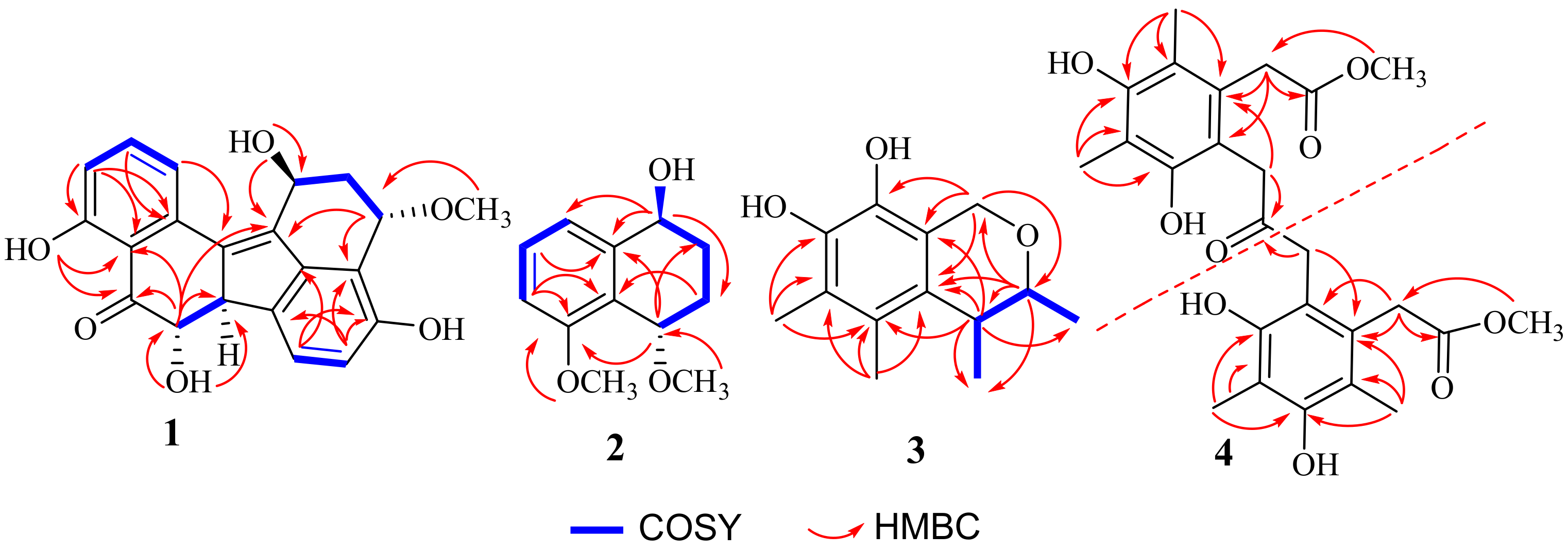
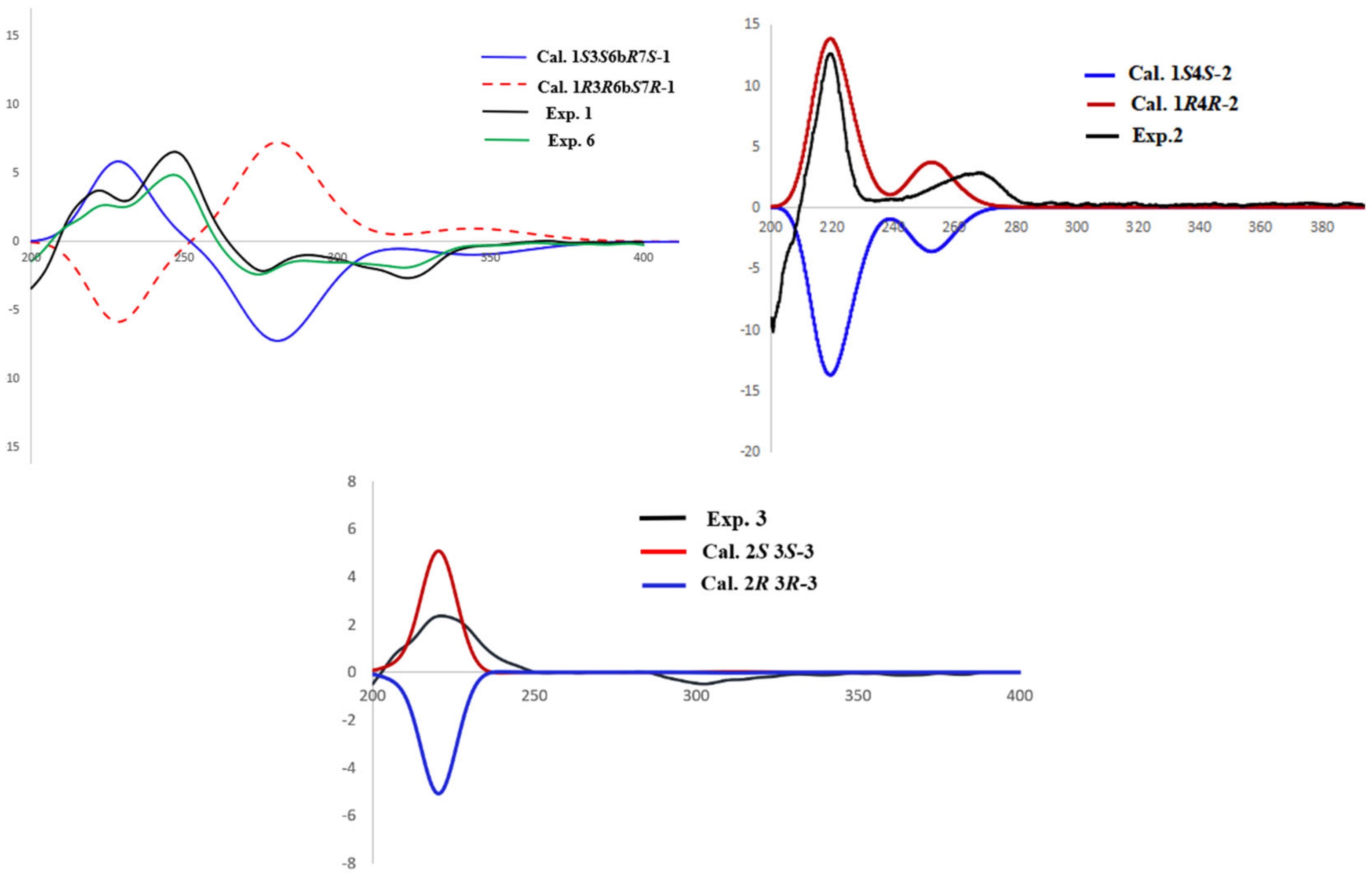
2.1. Structure Elucidation of the Compounds

Compound (1) was obtained as a brown oil. The molecular formula C21H18O6 was determined from the quasimolecular ion at m/z 389.1004 ([M + Na]+, calcd for C21H18O6Na, 389.1001) from a high resolution electrospray ionization mass spectrum (HRESIMS) and the 13C NMR spectrum (Table 1). The 1H NMR spectrum of 1 displayed two multiplets at δH 2.50 (1H, H-2α), and 1.68 (1H, H-2β), one multiplet at δH 5.22 (1H, H-1), one triplet at δH 4.74 (1H, H-3), two double doublets at δH 3.94 (1H, H-6b), and δH 3.78 (1H, H-7), five aromatic protons at δH6.71 (1H, H-5), 7.38 (1H, H-6), 6.84 (1H, H-10), 7.55 (1H, H-11), and 7.43 (1H, H-12), two phenolic hydroxyl protons at δH 9.54 (1H, H-4), and 12.32(1H, H-9). The 13C NMR spectrum (Table 1) exhibited 21 carbon signals including one ketone carbonyl at δC 206.5, one methoxyl at δC 55.9, one sp3 methylene at δC 39.7, four oxygenated methine sp3 at δC76.4, 70.4, 62.5, and 56.1, five protonated sp2 carbons at δC 136.1, 125.5, 121.5, 115.6, and 112.9, and eight non-protonated sp2 carbons at δC161.4, 154.4, 134.4, 117.8, 114.0, 138.2, 134.2, 140.0, and 144.9. Analysis of the 2D-NMR spectra (Figure 2) revealed that the structure of 1 resembled that of the previously reported 6 [11] except for the chemical shift value of C-7 which appeared at δC 76.4 CH, indicating that C-7 is oxygen-bearing.
The relative configuration of 1 was determined by the NOESY spectrum (Figure 3) analysis. The NOESY correlations between H-1 (δH 5.22) and OCH3-3 (δH 3.29), OCH3-3 and H-6b (δH 3.94), and H-6b and OH-7 (δH 6.17) determined the relative configuration of 1 as 1S*3S*6bR*7S*. The experimental ECD spectrum of 1 was recorded (Figure 4) and the calculated ECD spectrum of 1S3S6bR7S-1 fits well with the experimental ECD spectrum of 1, as shown in Figure 4. Since 1 has not been previously reported, it was named guhypoxylonol A.
Compound (2) was obtained as a colorless powder with a molecular formula of C12H16O3 as deduced from the HRESIMS m/z 231.0998 [M + Na]+ (cald 231.0997 for C12H16O3Na), indicating six degrees of unsaturation. The 1H-NMR (Table 2) showed two methoxyl singlets at δH 3.31 (3H, s, OCH3-4), and 3.75 (3H, s, OCH3-5), three aromatic protons at δH 7.24 (1H, d, J = 7.9 Hz, H-6), 7.14 (1H, d, J = 7.7 Hz, H-8), and 6.83 (1H, d, J = 8.1 Hz, H-7), two multiplets at δH 1.80 (2H, m, CH2-2), and 1.51, 2.09 (2H, m, CH2-3), and two multiplets at δH 4.41 (1H, m, H-1), and 4.35 (1H, m, H-4). The 13C NMR spectrum (Table 2) showed 12 carbon signals comprising six aromatic carbons of a benzene ring (δC 157.5 C, 143.1 C, 128.5 CH, 124.8 C, 118.7 CH and 108.8 CH), two methoxyls (δC 55.7 and 56.7), two methylene sp3 (δC 27.2 and 24.7), and two oxygenated methine sp3 (δC 69.8 and 67.8). The COSY spectrum (Table 2) of 2 displayed two isolated proton spin systems (H-1/H2-2/H2-3/H-4, and H-6/H-7/H-8). The HMBC spectrum showed correlations from the proton sinal at δH 4.41 (1H, m, H-1) to δC 24.7 (C-3), 118.7 (C-8), and 143.1 (C-8a), from δH 4.35 (1H, t, J = 2.8 Hz, H-4) to δC 157.5 (C-5), 27.2 (C-2), and 143.1 (C-8a). The 1H and 13C NMR spectra of 2 were very similar to those of nodulisporol [12]. The main difference between 2 and nodulisporol was the replacement of a hydroxyl group with a methoxy group at C-4.
The relative configuration of 2 was determined from its NOESY spectrum, which showed correlations from H-1/H-3α (δH 2.09), and H-4/H-3β (δH 1.51) suggesting that H-1 and H-4 were on the opposite face. To establish the absolute configuration of C-1 and C-4, the ECD spectra of two simplified isomers (1S4S, and 1R4R) of 2 were calculated at the Cam-B3LYP/6-31+G(d,p) level of theory in methanol, and these calculated spectra were compared with the experimental spectrum of 2. The experimental ECD spectrum of 2 showed an excellent fit with the calculated ECD spectrum of 1S4S-2 (Figure 4), establishing the absolute configurations of C-1 and C-4 as 1S4S. Since 2 has never been reported, it was named guhypoxylonol B.
Compound (3) was obtained as a colorless powder with a molecular formula of C13H18O3 as deduced from the HRESIMS m/z 223.1332 [M + H]+ (cald 223.1334 for C13H19O3), indicating five degrees of unsaturation. The 1H NMR (Table 3), in combination with DEPT and HSQC spectra, displayed two doublets of methylene group at δH 4.65 (J = 15.8 Hz, H-8) and 4.58 (J = 15.8 Hz, H-8), two multiplets of methine groups at δH 3.86 (J = 6.6, 2.6 Hz, H-2) and 2.63 (J = 6.8, 2.6 Hz, H-3), two methyl doublets at δH 1.18 (J = 6.8 Hz, H-11) and δH 1.19 (J = 6.6 Hz, H-12), and two methyl singlets at δH 2.10 (H-9, H-10). The 13C NMR (Table 3) spectrum, in combination with HMQC spectrum, of 3 revealed the presence of four methyl carbons at δC 21.0, 18.2, 9.1, and 11.1, one sp3 methylene carbon at δC 60.8, two sp3 methine carbons at δC 76.0 and 36.4, together with six non-protonated sp2 carbons at δC 153.3, 149.6, 134.8, 115.9, 114.4, and 111.3. The COSY (Figure 2) correlations from H-2 to H-3 and H3-11, and H-3 to H3-12 suggest the existence of -CH(CH3)CH(CH3)O-. The HMBC (Figure 2) correlations from H-2 to δC 21.0 (C-11), 134.8 (C-3a), 36.4 (C-3), and 60.8 (C-8), from H-3 to δC 134.8 (C-3a), 115.9 (C-4), 114.4 (C-7a), 18.2 (C-12), and 21.0 (C-11), suggests that C-3 is connected to C-3a. The HMBC correlations from H-9 (δH 2.10) to C-4, C-5 (δC 111.3), and C-3a, from H-10 (δH 2.10) to C-4, C-5, C-6 (δC 153.3), indicate that the two methyl groups were on C-4 and C-5, respectively. Finally, the HMBC correlations from H-8 to C-3a, C-7a, C-2 (δC 76.0), and C-7 (δC 149.6), indicated that the remaining substructure of 3 was established as shown in Figure 1.
A NOSEY correlation observed between H-2 and H-3, suggests that the relative configuration of 3 is either 2R*3R* or 2S*3S* (Figure 3). The absolute configurations of C-2 and C-3 were established by comparing the experimental and calculated ECD spectra of 2R3R, and 2S3S. The experimental ECD spectrum of 3 matched very well with the calculated 2S3S-3 ECD spectrum (Figure 4), calculated at the Cam-B3LYP/6-311+G (2d, p) level of theory in methanol. Therefore, the absolute configurations of C-2 and C-3 were determined to be 2S3S. Since 3 has never been reported, it was named guhypoxylonol C.
Compound (4) was obtained as a white powder and the molecular formula C25H30O9 was deduced from the HRESIMS m/z 473.1816 [M − H] (cald 473.1812 for C25H29O9), indicating 11 degrees of unsaturation. The 1H NMR (Table 4) spectrum of 4 displayed two methyl singlets at δH 2.10 (H-9) and 2.07 (H-10), one methoxyl singlet at δH 3.67 (-OCH3-8), and two singlets at δH 3.73 (H2-7) and 2.50 (H2-11). The 13C NMR spectrum (Table 4), in combination with the HSQC spectrum of 4, displayed one ketone carbonyl at δC 207.9 (C-12), one ester carbonyl at δC 173.8 (C-8), one methoxy at δC 52.5 (OCH3), two methyls at δC12.1, and 9.0, and the two sp3 methylene carbons at δC 36.5 (C-7) and 32.5 (C-11). The presence of six non-protonated sp2 at δC123.1, 118.7, 155.3, 112.5, 157.5, and 130.6 is an indicative of the presence of a benzene ring. The HMBC correlations (Figure 2) from δH 3.73 (H-7) to C-8, 123.1 (C-6), 118.7 (C-1), and from δH 3.67 to C-8, confirm that a methyl acetate is connected to C-1. HMBC correlations from δH 2.07 (H-9) to C-1, 130.6 (C-2), and 157.5 (C-3), from δH 2.07 (H-10) to δC112.5 (C-4), 155.3 (C-5), and C-3, and from H-11 to C-1 and C-12, suggested that 4 contains methyl (3,5-dihydroxy-2,4-dimethyl pheny) acetate moiety, with -CH2-C=O connected to C-6. Since the molecular formula of C25H30O9, only a ketone carbonyl (δC207.9) is present in 4. Therefore, the structure of 4 is a disubstituted acetone whose substituents are methyl (3,5-digydroxy-2,4-dimethylphenyl)acetate. Since 4 has never been reported, it was named guhypoxylonol D.
The previously described 511 were identified based on the analysis of their NMR data, and compared with those reported in the literature and identified as hypoxylonol C (5) [11], hypoxylonol B (6) [11], daldinone C (7) [13], nodulisporol (8) [12], isosclerone (9) [14], xylarenone (10) [14], scytalone (11) [15], respectively.

2.2. Anti-Inflammatory Activity

Compounds 111 were evaluated for their anti-inflammatory effects on the production of the NO in the RAW 264.7 macrophage cell line exposed to the inflammatory stimulus by lipopolysaccharide (LPS) (Table 5). Compounds 1, 3, 4, and 6 showed inhibitory activity against the production of NO, with the IC50 values 14.42 ± 0.11, 18.03 ± 0.14, 16.66 ± 0.21, and 21.05 ± 0.13 μM, respectively. Dexamethasone was used as a positive control with IC50 value of 16.12 ± 1.41 μM, while 2, 5, and 711 did not show any inhibitory activity under their safe concentrations.

3. Materials and Methods

3.1. General Experimental Procedures

NMR spectra were recorded on a AVANCE-400 spectrometer (Bruker, Bremen, Germany). The chemical shifts of 1H and 13C NMR spectra are given in δ (ppm) and referenced to the solvent signal (DMSO-d6, δH 2.50 and δC 39.52, CD3OD-d4, δH 3.34 and δC 49.00). Coupling constants (J) are reported in Hz. The mass spectrometric (HRESIMS) data were acquired using a Micro Mass Q-TOF spectrometer (Waters Corporation, Milford, MA, USA). ECD data was recorded using a JASCO J-715 spectropolarimeter (Jasco, Tokyo, Japan). Semipreparative HPLC was performed on an ODS column (10 × 250 mm, 5 µm, 3 mL/min, YMC, Kyoto, Japan).

3.2. Fungal Material

The strain GXNU-Y45 was isolated from a leaf of a mangrove tree Acanthus ilicifolius, October 2019, in Beihai City, China. The fungal strain GXNU-Y45 was identified as Aspergillus sp. based on the sequence of its internal transcribed spacer region (ITS) and morphology. ITS-rDNA of GXNU-Y45 was submitted to GenBank and the accession number is MT626059.

3.3. Fermentation, Extraction, and Isolation

The fungus was cultured in 60 × 1000 mL Erlenmeyer flasks each containing 50 g cooked rice and 60 mL of water (30 g sea salt, per liter pure water) or 300 mL medium (liquid media, 20.0 g dextrose, 20.0 g potatoes, 30 g sea salt, per liter pure water). The fungus was cultured in the medium and incubated at room temperature for 35 days.

3.4. Extraction and Isolation

The fermented material was extracted three times with EtOAc to obtain 16.8 g crude extract (liquid medium) and 20.2 g (solid medium). The crude extract was subjected to a silica gel VLC column, eluting with a stepwise gradient of petroleum ether-EtOAc (10:1, 8:1, 6:1, 4:1, 2:1, 1:1, v/v) to yield six subfractions (Fr. 1–Fr. 6). Fr. 3 (3 g) was applied to ODS silica gel with gradient elution of MeOH-H2O (3:7, 4:6, 5:5, 6:4, 7:3, 9:1, 0:1, v/v) to afford four subfractions (Fr. 3-1–Fr. 3-4). Fr. 3-2 (650 mg) was subjected to semipreparative HPLC (70% MeOH/H2O; 3 mL/min) to obtain 1 (15.6 mg), 2 (7.5 mg), and 3 (4.4 mg). Fr. 3-3 (345 mg) was repurified by RP-18 CC (eluted with MeOH/H2O from 3:7 to 10:0, v/v) and Sephadex LH-20 (eluted with CH2Cl2/MeOH, 5:5, v/v) to afford 5 (10.6 mg), 9 (3.3 mg), 10 (5.2 mg), and 11 (6.7 mg). Fr. 4 (1.1 g) was separated by ODS silica gel with gradient elution of MeOH-H2O (1:9, 2:8, 3:7, 4:6, 5:5, 6:4, 7:3, 9:1, 0:1, v/v) to yield four subfractions (Fr. 4-1–Fr. 4-4). Fr.4-3 (73 mg) was purified by Sephadex LH-20 eluted with CH2Cl2/MeOH (50:50) to give 4 (6.3 mg). Fr.4-4 (84 mg) was separated by semipreparative HPLC (80% MeCN/H2O; 3 mL/min) to give 6 (5.6 mg), 7 (8.1 mg), and 8 (5.2 mg).
Guhypoxylonol A (1): was obtained as a brown oil; α D 20 + 63.2 (c0.6, MeOH); 1H and 13C NMR data (see Table 1 and Table 2); HRESIMS m/z 389.1004 ([M + Na]+ (cald C21H18O6Na, 389.1001).
Guhypoxylonol B (2): was obtained as a colorless powder; α D 20 + 8.5 (c0.6, MeOH); 1H and 13C NMR data (see Table 1 and Table 2); HRESIMS m/z 231.0998 [M + Na]+ (cald 231.0997 for C12H16O3Na).
Guhypoxylonol C (3): white powder; α D 20 + 80 (c0.6, MeOH); 1H and 13C NMR data (see Table 1 and Table 2); HRESIMS m/z 223.1332 [M + H]+ (cald 223.1334 for C13H19O3).
Guhypoxylonol D (4): white powder; 1H and 13C NMR data (see Table 1 and Table 2); HRESIMS m/z 473.1816 [M − H] (cald 473.1812 for C25H30O9).

3.5. Anti-Inflammatory Assay

The anti-inflammatory effects of compounds 111 were examined on the production of the NO in LPS-stimulated cells using a method described in the literature [16].

4. Conclusions

The chemical investigation of a marine-derived fungus Aspergillus sp. GXNU-Y45 resulted in the isolation of four undescribed compounds (14), and seven previously reported metabolites (511). Based on modifications of the culture medium strategy, the fungus Aspergillus sp. GXNU-Y45 was cultured in different media to stimulate a production of its metabolites. It was found that the fungus Aspergillus sp. GXNU-Y45 produced different metabolites in two culture media. The liquid medium can stimulate the fungus to produce a series of metabolites, 1, 5, 6, 7, 8, 9, 10, and 2 (a new precursor of 1). On the contrary the solid medium yeiled 3 and 4. Different compositions of the culture media represented a powerful tool to induce new metabolites from microorganisms. Preliminarily screening of 111 for their ability to prevent NO production of LPS-induced RAW264.7 cells showed that 1, 3, 4, and 6 exhibited significant inhibitory effects against NO release with IC50 values of 14.42 ± 0.11, 18.03 ± 0.14, 16.66 ± 0.21, and 21.05 ± 0.13 μM, respectively. The inhibition of NO production by 1 and 6 was stronger than 5 and 7, which showed the same skeleton but differ only the presence of -OCH3 at C-3. Compounds 2 and 811, which are precursors of 1, 5, 6, and 7, did not exhibit inhibitory effects against NO release. Compounds 3 and 4 exhibited remarkable inhibitory effects against NO release suggesting that the fully substituted benzene ring was essential for inhibition of the production of NO release. In summary, this study revealed that 1, 3, 4, and 6 could be considered as potential metabolites for further anti-inflammatory studies.

Supplementary Materials

The following are available online at https://www.mdpi.com/article/10.3390/md20010005/s1, NMR and HRESIMS spectra of 111.

Author Contributions

R.Y. and X.H. conceived and designed the experiments. X.Q. performed the experiments. J.H., W.Z., D.Z., Y.Z., J.L. and X.H. analyzed the data. X.Q. and X.H. wrote the paper. All authors have read and agreed to the published version of the manuscript.

Funding

The authors (R.Y. and X.H.) acknowledge the following agencies for funding this project: National Natural Science Foundation of China (21662004, 42066005, 21762007); the Natural Science Foundation of Guangxi Province (2020JJA150036); the Open Research Fund Program of the Key Laboratory for the Chemistry and Molecular Engineering of Medicinal Resources (CMEMR2019-A1); Guangxi Science and Technology Base Special Talents (2019AC20095); the Guangdong Educational Committee (2018GkQNCX029, 2020WQYB042, 2018GKTSCX072); Foundation for University Key Teacher by the Guangdong Industry Poytechnic (KYRC2019-11).

Institutional Review Board Statement

Not applicable.

Informed Consent Statement

Not applicable.

Data Availability Statement

The authors declare that all data of this study are available within the article and its Supplementary Materials file or from the corresponding authors upon request.

Conflicts of Interest

The authors declare no conflict of interest.

References

  1. Carroll, A.; Copp, B.; Davis, R.A.; Keyzers, R.A.; Prinsep, M.R. Marine natural products. Nat. Prod. Rep. 2020, 37, 175–223. [Google Scholar] [CrossRef]
  2. Hu, Y.; Chen, J.; Hu, G.; Yu, J.; Zhu, X.; Lin, Y.; Chen, S.; Yuan, J. Statistical Research on the Bioactivity of New Marine Natural Products Discovered during the 28 Years from 1985 to 2012. Mar. Drugs 2015, 13, 202–221. [Google Scholar] [CrossRef]
  3. Cao, F.; Meng, Z.-H.; Wang, P.; Luo, D.-Q.; Zhu, H.-J. Dipleosporalones A and B, Dimeric Azaphilones from a Marine-Derived Pleosporales sp. Fungus. J. Nat. Prod. 2020, 83, 1283–1287. [Google Scholar] [CrossRef]
  4. Carroll, A.; Copp, B.; Davis, R.; Keyzers, R.A.; Prinsep, M.R. Marine natural products. Nat. Prod. Rep. 2019, 36, 112–173. [Google Scholar] [CrossRef] [Green Version]
  5. Dai, L.-T.; Yang, L.; Kong, F.-D.; Ma, Q.-Y.; Xie, Q.-Y.; Dai, H.-F.; Yu, Z.-F.; Zhao, Y.-X. Cytotoxic Indole-Diterpenoids from the Marine-Derived Fungus Penicillium sp. KFD28. Mar. Drugs 2021, 19, 613. [Google Scholar] [CrossRef] [PubMed]
  6. Ryu, M.-J.; Hillman, P.F.; Lee, J.; Hwang, S.; Lee, E.-Y.; Cha, S.-S.; Yang, I.; Oh, D.-C.; Nam, S.-J.; Fenical, W. Antibacterial Meroterpenoids, Merochlorins G–J from the Marine Bacterium Streptomyces sp. Mar. Drugs 2021, 19, 618. [Google Scholar] [CrossRef] [PubMed]
  7. Cui, H.; Lin, Y.; Luo, M.; Lu, Y.; Huang, X.; She, Z. Diaporisoindoles A–C: Three Isoprenylisoindole Alkaloid Derivatives from the Mangrove Endophytic Fungus Diaporthe sp. SYSU-HQ3. Org. Lett. 2017, 19, 5621–5624. [Google Scholar] [CrossRef] [PubMed]
  8. Cui, H.; Liu, Y.; Li, J.; Huang, X.; Yan, T.; Cao, W.; Liu, H.; Long, Y.; She, Z. Diaporindenes A–D: Four Unusual 2,3-Dihydro-1H-indene Analogues with Anti-inflammatory Activities from the Mangrove Endophytic Fungus Diaporthe sp. SYSU-HQ3. J. Org. Chem. 2018, 83, 11804–11813. [Google Scholar] [CrossRef] [PubMed]
  9. Chen, S.; Ding, M.; Liu, W.; Huang, X.; Liu, Z.; Lu, Y.; Liu, H.; She, Z. Anti-inflammatory meroterpenoids from the mangrove endophytic fungus Talaromyces amestolkiae YX1. Phytochemistry 2018, 146, 8–15. [Google Scholar] [CrossRef] [PubMed]
  10. Cui, H.; Liu, Y.; Nie, Y.; Liu, Z.; Chen, S.; Zhang, Z.; Lu, Y.; He, L.; Huang, X.; She, Z. Polyketides from the Mangrove-Derived Endophytic Fungus Nectria sp. HN001 and Their α-Glucosidase Inhibitory Activity. Mar. Drugs 2016, 14, 86. [Google Scholar] [CrossRef] [PubMed] [Green Version]
  11. Fukai, M.; Tsukada, M.; Miki, K.; Suzuki, T.; Sugita, T.; Kinoshita, K.; Takahashi, K.; Shiro, M.; Koyama, K. Hypoxylonols C–F, Benzo[j]fluoranthenes from Hypoxylon truncatum. J. Nat. Prod. 2011, 75, 22–25. [Google Scholar] [CrossRef] [PubMed]
  12. Kamisuki, S.; Ishimaru, C.; Onoda, K.; Kuriyama, I.; Ida, N.; Sugawara, F.; Yoshidab, H.; Mizushina, Y. Nodulisporol and nodulisporone, novel specific inhibitors of human DNA polymerase k from a fungus, Nodulisporium sp. Bioorg. Med. Chem. 2007, 15, 3109–3114. [Google Scholar] [CrossRef] [PubMed]
  13. Gu, W.; Ge, H.M.; Song, Y.C.; Ding, H.; Zhu, H.L.; Zhao, A.X.A.; Tan, R.X. Cytotoxic Benzo[j]fluoranthene Metabolites from Hypoxylon truncatum IFB-18, an Endophyte of Artemisia annua. J. Nat. Prod. 2007, 70, 114–117. [Google Scholar] [CrossRef] [PubMed]
  14. Yang, N.N.; Ma, Q.Y.; Kong, F.D.; Xie, Q.Y.; Dai, H.F.; Zhou, L.M.; Yu, Z.F.; Zhao, Y.X. Napthrene Compounds from Mycelial Fermentation Products of Marasmius berteroi. Molecules 2020, 25, 3898. [Google Scholar] [CrossRef] [PubMed]
  15. Sone, Y.; Nakamura, S.; Sasaki, M.; Hasebe, F.; Kim, S.-Y.; Funa, N. Bacterial Enzymes Catalyzing the Synthesis of 1,8-Dihydroxynaphthalene, a Key Precursor of Dihydroxynaphthalene Melanin, from Sorangium cellulosum. Appl. Environ. Microbiol. 2018, 84, e00258-18. [Google Scholar] [CrossRef] [PubMed] [Green Version]
  16. Liu, W.; Deng, S.; Zhou, D.; Huang, Y.; Li, C.; Hao, L.; Zhang, G.; Su, S.; Xu, X.; Yang, R.-Y.; et al. 3,4-seco-Dammarane Triterpenoid Saponins with Anti-Inflammatory Activity Isolated from the Leaves of Cyclocarya paliurus. J. Agric. Food Chem. 2020, 68, 2041–2053. [Google Scholar] [CrossRef] [PubMed]
Figure 1. Structures of 111.
Marinedrugs 20 00005 g001
Figure 2. Key COSY of 1-3 and HMBC correlations of 14.
Marinedrugs 20 00005 g002
Figure 3. Key NOESY correlations in 13.
Marinedrugs 20 00005 g003
Figure 4. Experimental ECD and calculated ECD spectra of 13.
Marinedrugs 20 00005 g004
Table 1. 1H and 13C NMR (DMSO-d6, 600 and 150 MHz) and COSY and HMBC assignment of 1.
PositionδC, TypeδH, (Mult., J in Hz)COSYHMBC
162.5, CH5.22, mH-2

39.7, CH22.50, m
1.68, m
H-1, 3
370.4, CH4.74, t (3.0)H-2C-3a, 12c
4154.4, C
5112.9, CH6.71, d (8.0)H-6C-3a, 4, 6a
6125.5, CH7.38, d (8.0)H-5C-4, 12d
6a134.4, C
6b56.1, CH3.94, dd (12.4, 3.1)H-7
776.4, CH3.78, dd (12.3, 5.6)H-6bC-6b, 8, 8a, 12c
8206.5, C
8a114.0, C
9161.4, C
10115.6, CH6.84, d (8.2)H-11C-8a, 9, 12a
11136.1, CH7.55, d (8.0)H-10, 12C-12a
12121.5, CH7.43, d (7.7)H-11C-12b
12a138.2, C
12b134.2, C
12c140.0, C
12d144.9, C
1-OH 5.06, d (7.8) C-1, 12c
4-OH 9.54, s
7-OH 6.17, d (5.9) C-6b, 7
9-OH 12.32, s C-8, 8a
3-OCH355.9, CH33.29, s C-3
Table 2. 1H and 13C NMR (DMSO-d6, 600 and 150 MHz) and COSY and HMBC assignment of 2.
PositionδC, TypeδH (Mult., J in Hz)COSYHMBC
167.8, CH4.41, mH-2C-3, 8, 8a
227.2, CH21.80, mH-1, 3

24.7, CH22.09, m
1.51, m
H-2, 4C-4a
469.8, CH4.35, t (2.8)H-3C-2, 5, 8a
4a124.8, C
5157.5, C
6128.5, CH7.24, d (7.9)H-7C-4a, 5
7108.8, CH6.83, d (8.1)H-6, 8C-8a
8118.7, CH7.14, d (7.7)H-7
8a143.1, C
1-OH 5.28, s
4-OCH356.7, CH33.31, s C-4
5-OCH355.7, CH33.75, s C-5
Table 3. 1H and 13C NMR (CD3OD, 400 and 100 MHz) and COSY and HMBC assignment of 3.
PositionδC, TypeδH (Mult., J in Hz)COSYHMBC
276.0, CH3.86, qd (6.6, 2.6)H-3, 11C-3, 3a, 8, 12
336.4, CH2.63, qd (6.8, 2.6)H-2, 12C-3a, 4, 7a, 11, 12
3a134,8, C
4115.9, C
5111.3, C
6153.3, C
7149.6, C
7a114.4, C
860.8, CH24.65, d (15.2)
4.58, d (15.2)
C-2, 3a, 7, 7a
911.1, CH32.10, s C-3a, 4, 5
109.1, CH32.10, s C-4, 5, 6
1121.0, CH31.18, d (6.8)
1218.2, CH31.19, d (6.6)
Table 4. 1H and 13C NMR (CD3OD, 400 and 100 MHz) and HMBC assignment of 4.
PositionδC, TypeδH (Mult., J in Hz)HMBC
1 (1′)118.7, C
2 (2′)130.6, C
3 (3′)157.5, C
4 (4′)112.5, C
5 (5′)155.3, C
6 (6′)123.6, C
7 (7′)36.5, CH23.73, sC-1 (1′), 6 (6′), 8 (8′)
8 (8′)173.8, C
9 (9′)9.0, CH32.10, sC-1 (1′), 2 (2′), 3 (3′)
10 (10′)12.1, CH32.07, sC-3 (3′), 4 (4′), 5 (5′)
11 (11′)32.5, CH22.50, sC-1 (1′), 12
12207.9, C
8-OCH352.5, CH33.67, sC-8 (8′)
Table 5. Inhibitory activities of 111 on NO production in LPS-induced RAW 264.7 cells a.
CompoundsIC50 (μM)
114.42 ± 0.11
232.48 ± 0.19
318.03 ± 0.14
416.66 ± 0.21
5>80
621.05 ± 0.13
7>80
8>80
9>80
10>80
11>80
Dexamethasoneb16.12 ± 1.41 μM
a Values present mean ± SD of triplicate experiments. b Dexamethasone was used as a positive control.
Publisher’s Note: MDPI stays neutral with regard to jurisdictional claims in published maps and institutional affiliations.

