Article

New Diterpenoids and Isocoumarin Derivatives from the Mangrove-Derived Fungus Hypoxylon sp.

1
State Key Laboratory of Mycology, Institute of Microbiology, Chinese Academy of Sciences, Beijing 100101, China
2
Beijing Key Laboratory of Bioactive Substance and Functional Foods, Beijing Union University, Beijing 100191, China
3
Institutional Center for Shared Technologies and Facilities, Institute of Microbiology, Chinese Academy of Sciences, Beijing 100101, China
*
Authors to whom correspondence should be addressed.
These authors contributed equally to this paper.
Mar. Drugs 2021, 19(7), 362; https://doi.org/10.3390/md19070362
Submission received: 5 June 2021 / Revised: 18 June 2021 / Accepted: 22 June 2021 / Published: 24 June 2021
(This article belongs to the Special Issue Marine Fungal Metabolites: Structures, Activities and Biosynthesis)

Abstract

:
Two new diterpenoids, hypoxyterpoids A (1) and B (2), and four new isocoumarin derivatives, hypoxymarins A–D (47), together, with seven known metabolites (3 and 813) were obtained from the crude extract of the mangrove-derived fungus Hypoxylon sp. The structures of the new compounds were elucidated on the basis of 1- and 2-dimensional (1D/2D) nuclear magnetic resonance (NMR) spectroscopic and mass spectrometric analysis. The absolute configurations of compounds 1, 2, 4, 5, and 7 were determined by comparison of experimental and calculated electronic circular dichroism (ECD) spectra, and the absolute configurations of C-4′ in 6 and C-9 in 7 were determined by [Rh2(OCOCF3)4]-induced ECD spectra. Compound 1 showed moderate α-glucosidase inhibitory activities with IC50 values of 741.5 ± 2.83 μM. Compounds 6 and 11 exhibited DPPH scavenging activities with IC50 values of 15.36 ± 0.24 and 3.69 ± 0.07 μM, respectively.

Graphical Abstract

1. Introduction

Bioactive secondary metabolites have played an important role for drug discovery as lead compounds [1,2]. Fungi especially from unique ecological niches are capable of producing a variety of bioactive compounds [3,4,5]. Mangrove-derived endophytic fungi, as plant mutualists that occur in the tropical and subtropical intertidal estuarine zones, have attracted more attention of natural product chemists due to their production of structurally diverse and biologically active secondary metabolites [6,7,8,9,10,11,12]. Hypoxylon species are widespread in terrestrial and marine environments, and chemical investigations of some Hypoxylon spp. have afforded a variety of natural products, such as azaphilones, diterpenes, sporothriolide derivatives, and various aromatic compounds, which exhibited antibacterial, antifungal, and cytotoxic activities [13,14,15,16,17,18].
As part of our ongoing search for new bioactive natural products from mangrove-derived endophytic fungi [11], a strain of fungus Hypoxylon sp. (Hsl2-6) from Bruguiera gymnorrhiza collected in Fangchenggang City, Guangxi Province, People’s Republic of China, was screened out for investigations. The fungus was fermented on rice medium for 30 days and subsequently extracted by EtOAc to afford the organic extract. Fractionations of this extract were performed, leading to the isolation of two new diterpenoids, hypoxyterpoids A (1) and B (2), and four new isocoumarin derivatives, hypoxymarins A–D (47), together with seven known metabolites, penicichrysogene B (3) [19] penicimarin A (8) [20], aspergillumarin A (9) [21], aspergillumarin B (10) [21], 5-hydroxysescandelin (11) [22], sescandelin A (12) [23], and sescandelin B (13) [24] (Figure 1). Herein, the isolation, structure elucidation and bioactivities of these compounds are reported.

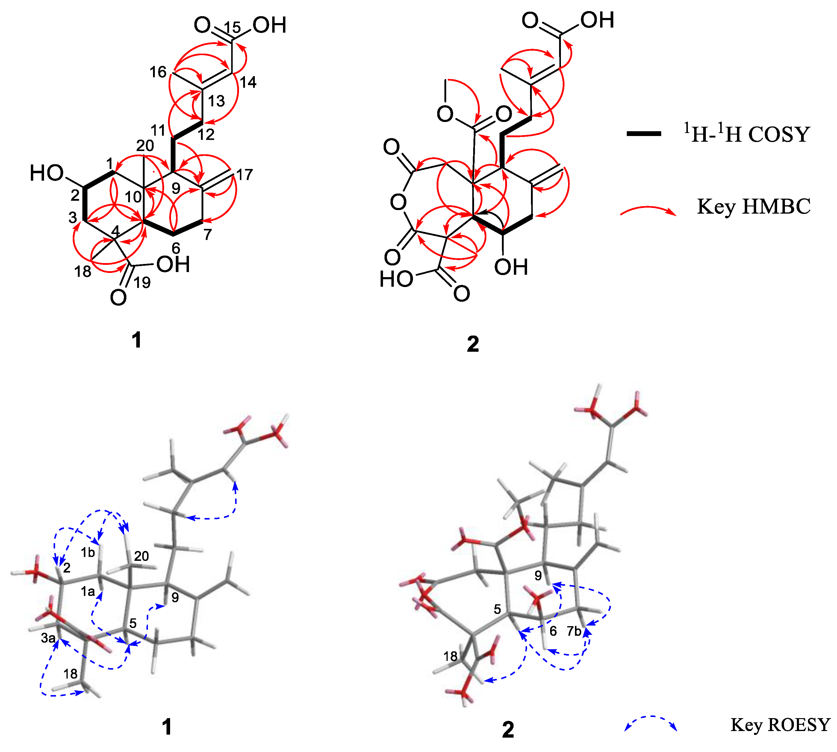
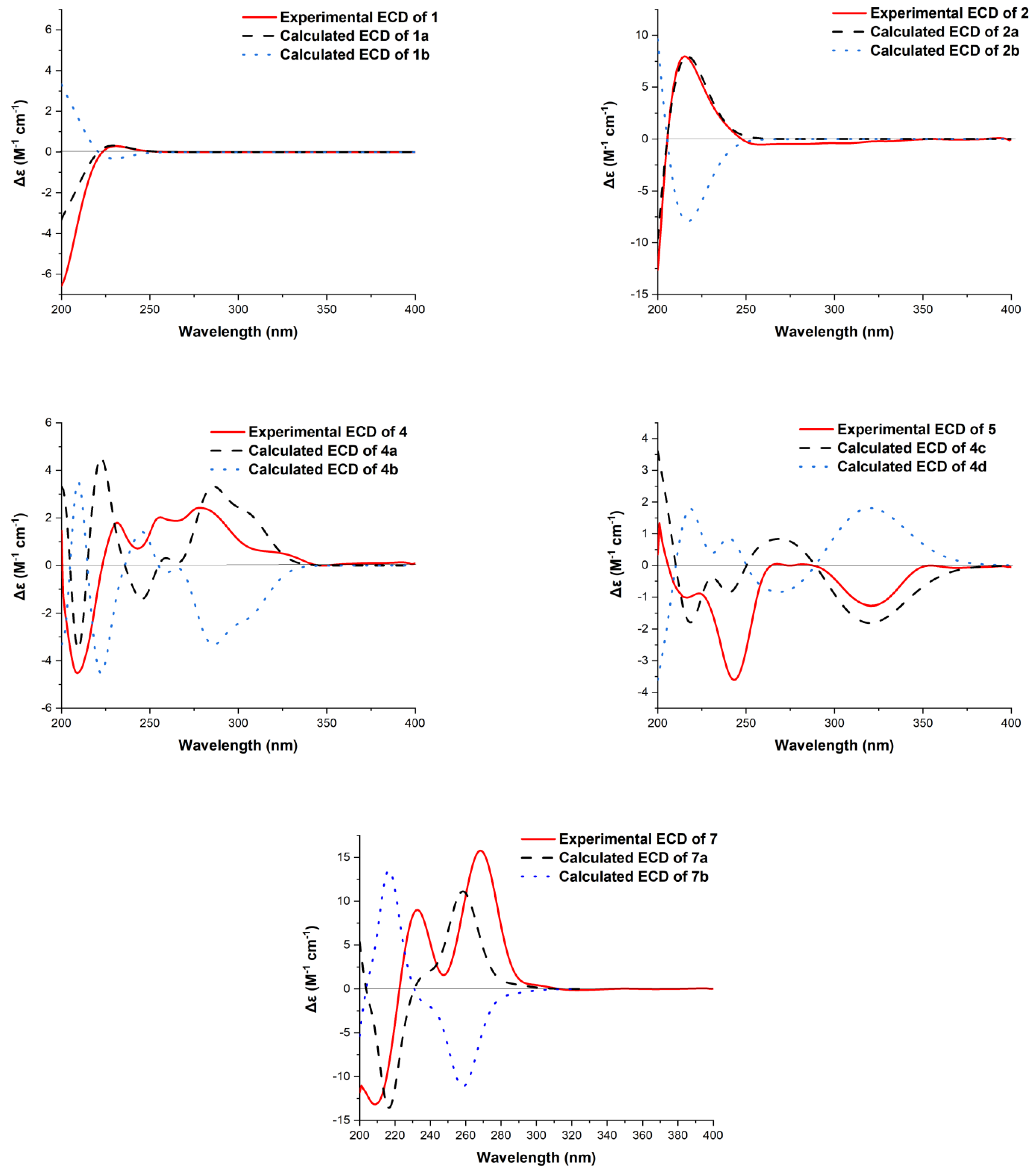
2. Results and Discussion

The fungus Hypoxylon sp. (Hsl2-6) was cultured in rice medium at room temperature for 30 days. The EtOAc extracts were combined to afford an organic residue (30.0 g) that was fractionated and purified by column chromatography (CC) on silica gel, RP-18, and semipreparative HPLC columns to acquire compounds 113.
Hypoxyterpoid A (1) was obtained as a brown oil. It had a molecular formula of C20H30O5 (six degrees of unsaturation) on the basis of its high-resolution electrospray ionization mass spectrometry (HRESIMS) [M + Na]+ at m/z 373.1995 (calcd for C20H30O5Na 373.1991). The infrared (IR) spectrum of 1 at 1704, 1644 cm−1 showed the presence of carbonyl and double bond groups. Analysis of the 1H and 13C nuclear magnetic resonance (NMR, Figures S1 and S2) and heteronuclear single quantum correlations (HSQC) data (Table 1) revealed the presence of two carbonyl carbons (δC 178.3 and 167.6), two sp3 quaternary carbons (δC 44.2 and 40.9), four olefinic carbons (two protonated), three sp3 methine (one oxygenated), six sp3 methylene carbons, and three methyl carbons. These data accounted for all 1H and 13C NMR resonances except for three unobserved exchangeable protons and suggested that 1 was a bicyclic compound. Analysis of 1H-1H correlation spectroscopy (COSY) spectrum revealed three spin system: C-1/C-2/C-3, C-5/C-6/C-7, and C-9/C-11/C-12. The heteronuclear multiple bond correlations (HMBC) correlations from H2-1 to C-3 and C-5, H2-3 to C-5, H-5 to C-4, C-10 and C-18, and from H3-18 to C-3, C-4, C-5 and C-19 completed the ring A with the methyl carbon C-18 and the carboxylic carbon C-19 attached to C-4 directly. HMBC correlations from H3-20 to C-1, C-5 and C-10 established the location of the methyl group C-20 at C-10. The exchangeable proton was located at C-2 supported by the chemical shift value for C-2 (δC 62.9). While HMBC correlations from H2-6 to C-8 and C-10, H-9 to C-8, C-10 and C-20, and from H2-17 to C-7, C-8 and C-9 established the ring B, fused with ring A at C-5 and C-10. Additional HMBC correlations from H3-16 to C-12, C-13, C-14 and C-15, from H-14 to C-12 and C-15, from H2-12 to C-13 and C-14, and from H2-11 to C-13 established the C-13–C-16 subunit, with C-12 attached to C-13 of the olefin C-13/C-14. The hydroxyl groups were located at C-2, C-15, and C-19 by default supported by the chemical shift value for C-2 (δC 62.9), C-15 (δC 167.6), and C-19 (δC 178.3). Thus, The compound 1 and rel-(1R,3S,4aS,5R,8aS)-5-[(3E)-4-carboxy-3-methylbut-3-en-1-yl]decahydro-3-hydroxy-1,4a-dimethyl-6-methylidenenaphthalene-1-carboxylic [25] shared the same planar structure, as depicted in Figure 1.
The relative configuration of 1 was assigned by Rotating Frame Overhauser Effect Spectroscopy (ROESY) experiment (Figure 2) and, by comparison, with the known compound penicichrysogene B (3). The large J value observed for H-3a/H-2 (12.0 Hz) indicated their trans-diaxial orientations. The ROESY correlations of H-5 with H-1a, H-3a, and H-9, and of H-3a with H3-18 suggested the α-orientated of these protons, whereas those of H-2 with H-1b and H3-20 indicated these protons were β-orientated. Additionally, the ROESY correlations of H-14 with H2-12 suggested the E-configuration of the Δ13 double bond. The absolute configuration of 1 was assessed by comparison of the experimental and simulated ECD spectra generated by the time-dependent density functional theory (TDDFT) for two enantiomers (2R,4S,5R,9S,10R)-1 (1a) and (2S,4R,5S,9R,10S)-1 (1b). The experimental ECD spectrum of 1 was nearly identical to the calculated ECD spectrum for 1a (Figure 3), clearly indicating the 2R,4S,5R,9S,10R absolute configuration for 1. Thus, the structure of 1 was elucidated, as depicted in Figure 1.
Hypoxyterpoid B (2) was obtained as a brown oil. Its molecular formula C21H26O10 was established by HRESIMS [M + H]+ at m/z 439.1605 (calcd for C21H27O10 439.1604), indicating the nine degrees of unsaturation. Its 1H and 13C NMR data (Table 1) indicated the presence of five carbonyl carbons (δC 175.9, 173.5, 172.8, 172.6, and 167.7, respectively), two quaternary carbons, four olefinic carbons with two protonated, three sp3 methine carbons with one oxygenated, four sp3 methylene carbons, and three methyl carbons (one oxygenated). These data suggested that 2 was also a bicyclic compound. Detailed comparison of the 1H and 13C NMR data of 2 (Table 1, Figures S7 and S8) with those of 1 revealed the same fragment of C-11–C-16, which was confirmed by interpretation of the 1H-1H COSY and HMBC data (Figure 2, Figures S9 and S11). However, obvious differences in chemical shifts were also observed for some signals for rings A and B. Specifically, the resonances for methylene carbon C-6 (δH/C 1.70; 1.88/25.5) in ring B of 1 were replaced by the oxygenated methine carbon C-6 (δH/C 4.93/77.7), which was confirmed by the 1H-1H COSY correlations H-5/H-6/H-7 and the HMBC correlations from H-5 to C-9 and C-10, from H-6 to C-5 and C-10, from H2-17 to C-7, C-8 and C-9, and from H-9 to C-8 and C-10. Other HMBC correlations from H2-1 to C-2, C-5 and C-10, from H-5 to C-1, C-3, C-4 and C-10 and from H3-18 to C-3, C-4, C-5 and C-19, as well as the bicyclic feature of 2 established a seven-membered ring (ring A) with the methyl carbon C-18 and the carboxylic carbon C-19 attached at C-4. Additionally, HMBC correlations from H2-1 and H-9 to C-10 and C-20 suggested the carbonyl carbon C-20 was attached to C-10 directly. While HMBC correlation from H3-21 to C-20 located the methoxyl group at C-10. On the basis of these data, the gross structure of 2 was established as shown.
The ROESY correlations of H-7b with H-5, H-6 and H-9, and of H-6 with H3-18 revealed the cofacial relationships of these groups (Figure 2). The relative configuration for C-10 was proposed to be the same as 1 and penicichrysogene B (3) from the biosynthetic considerations. The absolute configuration for 2 was also proposed, by a comparison of the experimental and calculated ECD spectra, for the enantiomers (4S,5R,6R,9S,10R)-2 (2a) and (4R,5S,6S,9R,10S)-2 (2b). The result showed that the calculated spectrum of (4S,5R,6R,9S,10R)-2a agreed with the experimental one (Figure 3), indicating the absolute configuration of 2 to be 4S,5R,6R,9S,10R. Compound 2 is a rare type of new diterpenoid derivative with an anhydride moiety. There are few precedents, in the natural products literature, for this type of structures [26,27,28].
Hypoxymarin A (4) was obtained as a yellow oil. Its molecular formula C14H16O5 was established by HRESIMS analysis (m/z 263.0925 [M − H]), indicating seven degrees of unsaturation. The IR spectrum of 4 at 3306, 1697, 1658, 1598 cm−1 showed the presence of hydroxy, ester carbonyl and aromatic groups. Analysis of its NMR data (Table 2, Figures S13 and S14) and revealed the presence of one exchangeable proton (δH 9.63), three methyl groups (including one methoxy), two sp3 methines (one oxygenated), eight aromatic or olefinic carbons (three protonated), and one carbonyl carbon. These data accounted for all 1H and 13C NMR resonances except for one unobserved exchangeable proton. The remaining two degrees of unsaturation suggested that 4 was a bicyclic compound. The 1H-1H correlations (Figure 3) of H-9 with H-10 and H3-11, and of H-10 with H3-12 allowed the assignment of the C-11–C-9–C-10–C-12 butan-2-ol subunit. The HMBC correlations (Figure 3) from H-4 (δH 6.22) to C-3 and C-9, H-9 to C-3 and C-4, and from H-10 and H3-11 to C-3 indicated that C-9 was directly attached to C-3 of the trisubstituted olefin C-3/C-4. Additional HMBC correlations from H-4 to C-1, C-4a, C-8a and C-5, H-5 to C-4, C-6, C-7, and C-8a, and from H-7 to C-1, C-5, C-6 (δC 164.7), C-8 and C-8a, as well as from the methoxy group H3-13 to C-8 established the isochromenone core structure, with the methoxy group carbon C-13 located at C-8. The remaining two exchangeable protons were located at C-6 and C-10 by default. Thus, the planar structure of 4 was established as shown (Figure 1).
The absolute configuration for 4 was also assigned by ECD calculations. The calculated ECD spectra were obtained by the time-dependent density functional theory (TD-DFT) at the B3LYP/6-311G (2d, p) level for four stereoisomers (9S,10S)-4 (4a), (9R,10R)-4 (4b), (9R,10S)-4 (4c) and (9S,10R)-4 (4d). The overall calculated ECD spectrum of 4a4d were generated according to Boltzmann weighting of the conformers. The experimental ECD spectrum of 4 was nearly identical to the calculated ECD spectrum for (9S,10S)-4 (4a) (Figure 3), indicating the absolute configuration to be 9S,10S-4.
Hypoxymarin B (5) was obtained as a yellow oil. The molecular formula was determined as C14H16O5 (seven degrees of unsaturation) by HRESIMS (m/z 263.0924 [M − H]), which is the same with that of 4. The 1H and 13C NMR data (Table 2, Figures S19 and S20) and 2D NMR were very similar, indicating that 5 was a stereoisomer of 4. Subsequent comparison of the experimental ECD spectra of 5, and the calculated ECD spectrum for (9R,10S)-4 (4c), unambiguously assigned its absolute configuration as 9R,10S. Thus, the structure of 5 was elucidated, as depicted in Figure 1.
The molecular formula of hypoxymarin C (6) was determined to be C14H18O5 (six degrees of unsaturation) by HRESIMS (m/z 267.1226 [M + H]+). In the 1H NMR spectrum (Table 3), the proton signals and the coupling constants at δH 7.12 (d, J = 8.8 Hz), 6.71 (d, J = 8.8 Hz) indicated the presence of a 1,2,3,6-tetrasubstituted benzene system. Analysis of its NMR data (Table 3, Figures S25 and S26) the presence of one methyl group, four methylenes, two oxygenated methines, six aromatic carbons (two protonated) and one carbonyl carbon (δC 170.8). The NMR data of 6 resembled those of aspergillumarin B (10) [21] except that the proton H-5 in 10 were replaced by hydroxyl group in 6, which was confirmed by the chemical shift of C-5 (δC 146.3) and the HMBC correlations from H2-4 and H-6 to C-5. On the basis of these results, the planar structure of 6 was elucidated (Figure 1). In order to determine the absolute configuration of C-4′ at the side chain, a [Rh2(OCOCF3)4]-induced ECD experiment was applied (Figure S34). In the induced ECD spectrum, the positive Cotton effect, at 350 nm, indicated a 4′S configuration according to the bulkiness rule [29]. By comparison with the dihydroisocoumarin data described in the literature [20,21], the negative circular dichroism at 259 nm indicated the R configuration at C-3. Thus, the absolute configuration of 6 was established as 3R,4′S.
Hypoxymarin D (7) was isolated as a yellow oil with the molecular formula assigned as C11H12O5 on the basis of its HREIMS (m/z 223.0614 [M − H]), indicating six degrees of unsaturation. Analysis of the 1H and 13C NMR data (Table 3, Figures S28 and S29) revealed the presence of one methyl, one oxygenated methylene, two methines (one oxygenated), six aromatic carbons with two protonated, and one carbonyl carbon (δC 169.1). The 1H and 13C NMR data of 7 were almost consistent with penicimarin A (8). Further analysis of its 1H-1H COSY correlations (Figure 4) of H-9 with H3-10 and H-4, and of H2-3 with H-4, as well as the HMBC correlations (Figure 4) from H3-10 to C-9, from H-9 to C-3, C-4 and C-4a, from H2-3 to C-1, C-4 and C-4a, from H-4 to C-5 and C-8a, and from H-5 and H-6 to C-7 indicated that 7 and 8 share the same planar structure.
By treating 7 with Rh2(OCOCF3)4 in anhydrous CH2Cl2, a negative Cotton effect at 350 nm in the ECD spectrum was observed for the complex (Figure S34). Therefore, successful implementation of the bulkiness rule allowed C-9 to be assigned as R. The absolute configuration of C-4 was further determined by a comparison of the experimental and calculated ECD spectra. The calculated spectrum of (4S,9R)-7a agreed with the experimental one (Figure 3), indicating the absolute configuration of 7 to be 4S,9R.
The known compounds, penicichrysogene B (3) [19] penicimarin A (8) [20], aspergillumarin A (9) [21], aspergillumarin B (10) [21], 5-hydroxysescandelin (11) [22], sescandelin A (12) [23], and sescandelin B (13) [24] (Figure 1) were determined by comparison of their spectroscopic data with those in the literature.
The antibacterial activities of all isolated compounds were evaluated with Bacillus subtilis, Escherichia coli, and Staphylococcus aureus. However, these compounds showed no antibacterial activities against B. subtilis, E. coli, and S. aureus at the concentration of 200 μg/mL. All of the isolated compounds were also evaluated for radical-scavenging activity against 2,2-diphenyl-1-picrylhydrazyl (DPPH) radicals. Compounds 6 and 11 showed free radical scavenging activity with IC50 values of 15.36 ± 0.24 and 3.69 ± 0.07 μM, respectively (ascorbic acid as the positive control with IC50 values of 20.49 ± 0.43 μM). The α-glucosidase inhibitory effects of these compounds were evaluated along with the clinical α-glucosidase inhibitor acarbose. Compound 1 exhibited moderate inhibitory effects against α-glucosidase with IC50 value of 741.50 ± 2.83 μM (acarbose as positive control with IC50 value of 636.80 ± 1.49 μM).
Penicichrysogene B (3) was investigated in the anti-platelets assay and showed no inhibitory activities against AChE and BuChE [19]. Isocoumarin derivatives 813 was evaluated for antibacterial activities and cytotoxic activities in vitro, while only compounds 9 and 10 showed weak antibacterial activity against Staphylococcus aureus and Bacillus subtilis [20,21,22,23,24,30]. It is the first time, for 11, that the antioxidant activity has been evaluated in our study.

3. Experimental Section

3.1. General Experimental Procedure

Optical rotations were measured with an Anton Paar MCP 200 Automatic Polarimeter (Anton Paar GmbH, Graz, Austria). Infrared spectra were obtained on a Nicolet IS5 FT-IR spectrophotometer (Thermo Scientific, Madison, WI, USA). The NMR data were measured on a Bruker Avance-500 MHz spectrometer (Bruker, Rheinstetten, Germany). The UV data were recorded on a Thermo Scientific Genesys 10S spectrophotometer (Thermo Scientific, Madison, WI, USA). ECD spectra were acquired on an Applied Photophysics Chirascan spectropolarimeter (Applied Photophysics Ltd., Leatherhead, UK). Mass data were performed on an Agilent Accurate-Mass-Q-TOF LC/MS 6520 instrument (Agilent Technologies, Santa Clara, CA, USA). Preparative HPLC was conducted with an Agilent 1200 HPLC system using a C18 column (Reprosil-Pur Basic C18 column; 5 μm; 10 × 250 mm) with a flow rate of 2.0 mL/min. Preparative HPLC was performed on the Waters system, using a C18 column (SunFire C18 column; 5 μm; 19 × 250 mm), with a flow rate of 15 mL/min. The absorbance of contents, in the 96-well clear plate, was detected by a SpectraMax Paradigm microplate reader (Molecular Devices, Sunnyvale, CA, USA).

3.2. Strain and Fermentation

Hypoxylon sp. (Hsl2-6) was isolated from the branches of Bruguiera gymnorrhiza collected in Beilun River Mouth, Fangchenggang City, Guangxi Province, P. R. China. The isolated strain was identified by sequence analysis (Genbank Accession Number MN547522) of the rDNA internal transcribed spacer (ITS) region. The strain was first placed on potato-dextrose agar medium and cultured at 25 °C for 7 days. Then the agar was cut into grain size to inoculate in four conical flasks (500 mL), each containing 100 mL autoclave sterilized potato dextrose broth. Seed culture medium was cultured at 25 °C and 180 rpm for 7 days. Finally, Erlenmeyer flasks (500 mL) containing 80 g of rice, 120 mL of distilled H2O, and 10.0 mL seed culture incubated at 25 °C for 30 days.

3.3. Extraction and Isolation

At the end of the fermentation, the rice culture was extracted with EtOAc (three times, each 12 L) and vacuum-dried to afford the crude extract (30.0 g). The extract was fractionated by ODS column chromatography (CC), eluted with 10–100% MeOH to afford twelve fractions (Frs.1–12). The fractions Frs.1–5 (8.7 g) were combined and subjected to silica gel CC and eluted with CHCl3/MeOH (20:1–5:1) to afford five subfractions (Frs.1.1–1.5). The subfraction Fr.1.1 (1.8 g) was subjected to Sephadex LH-20 CC, eluted with CHCl3/MeOH (2:1) to generate five fractions (Frs.1.1.1–1.1.5). Fr.1.1.2 (0.7 g) was loaded on normal pressure silica gel CC with petroleum ether (PE)/acetone (200:1–5:1) to yield Frs.1.1.2.1–1.1.2.6. The Fr.1.1.2.5 was further purified by RP HPLC (50% MeOH in H2O with 0.1% HCOOH; 15.0 mL/min) to afford 9 (9.0 mg, tR 29.3 min). The subfraction Fr.1.2 (2.0 g) was loaded on Sephadex LH-20 CC, eluted with CHCl3/MeOH (2:1), to afford five tertiary fractions (Frs.1.2.1–1.2.5). The precipitation 11 (30.0 mg) was obtained from Fr.1.2.2. The remaining of Fr.1.2.2. was then purified by RP HPLC (40% MeOH in H2O with 0.1% HCOOH; 15.0 mL/min) to afford 13 (2.0 mg, tR 19.0 min) and 12 (3.0 mg, tR 20.8 min). Fr.1.2.4 (0.8 g) was fractionated by normal pressure silica gel CC with PE/acetone (15:1–5:1) to afford three fractions (Frs.1.2.4.1–1.2.4.3). Fr.1.1.4.2 was purified by RP HPLC (36% MeOH in H2O with 0.1% HCOOH; 15.0 mL/min) to afford 7 (5.0 mg, tR 18.0 min) and 8 (8.1 mg, tR 21.0 min). The subfraction Fr.1.3 (100.2 mg) was loaded on Sephadex LH-20 CC eluting with CHCl3/MeOH (2:1) to afford seven fractions (Frs.1.3.1–1.3.7). Fr.1.3.7 was further purified by RP HPLC (42% MeOH in H2O with 0.1% HCOOH; 2.0 mL/min) to afford 6 (5.7 mg, tR 24.0 min). Fr.1.5 (1.6 g) was loaded on Sephadex LH-20 CC eluted with CHCl3/MeOH (2:1) to afford Frs.1.5.1–1.5.3. Fr.1.5.2 was further purified by RP HPLC (45% MeOH in H2O with 0.1% HCOOH; 15.0 mL/min) to afford 4 (5.0 mg, tR 15.0 min) and 5 (2.5 mg, tR 18.0 min). The subfraction Fr.6.2 was purified by RP HPLC (50% MeOH in H2O with 0.1% HCOOH; 2.0 mL/min) to afford 2 (9.2 mg, tR 23.0 min) and 10 (4.3 mg, tR 31.5 min). The fraction Fr.8 was purified by RP HPLC (60% MeOH in H2O with 0.1% HCOOH; 2.0 mL/min) to afford 1 (3.0 mg, tR 31.9 min) and 3 (3.0 mg, tR 45.5 min).
Hypoxyterpoid A (1): brown oil; [α ] D 25 +28.8 (c 0.15, MeOH); UV (MeOH) λmax (log ε): 200 (2.44) nm; IR (neat) νmax 2941, 2851, 1703, 1644, 1446, 1384, 1231, 1155, 1025, 1003, 889, 824, 762 cm−1; CD (MeOH) λmaxε) 202 (−0.56), 222 (+1.96), 287 (−0.33); 1H and 13C NMR data (500 MHz, DMSO-d6) see Table 1; HRESIMS: m/z 373.1995 [M + Na]+ (calcd for C20H30O5Na 373.1991).
Hypoxyterpoid B (2): brown oil; [α ] D 25 +28.2 (c 0.85, MeOH); UV (MeOH) λmax (log ε): 200 (3.02); 210 (2.20) nm; IR (neat) νmax 2952, 1779, 1726, 1644, 1435, 1219, 1165, 1115, 1008, 968, 907 cm−1; CD (MeOH) λmaxε) 200 (−12.61), 215 (+7.96); 1H and 13C NMR data (500 MHz, Acetone-d6) see Table 1; HRESIMS: m/z 439.1605 [M + H]+ (calcd for C21H27O10 439.1604).
Hypoxymarin A (4): Yellow oil; [α ] D 25 −0.6 (c 0.50, MeOH); UV (MeOH) λmax (log ε): 245 (0.69); 324 (0.13) nm; IR (neat) νmax 3306, 2975, 1697, 1658, 1598, 1440, 1369, 1237, 1200, 1174, 1123, 1074, 1026, 947, 907, 855 cm−1; CD (MeOH) λmaxε) 200 (+1.47), 209 (−4.52), 231 (+1.79), 278 (+2.42) nm; 1H and 13C NMR data (500 MHz, Acetone-d6) see Table 2; HRESIMS m/z 263.0925 [M − H] (calcd for C14H15O5, 263.0919).
Hypoxymarin B (5): Yellow oil; [α ] D 25 + 26.4 (c 0.25, MeOH); UV (MeOH) λmax (log ε): 245 (1.27); 324 (0.21) nm; IR (neat) νmax 3420, 2976, 1693, 1656, 1598, 1496, 1441, 1369, 1302, 1238, 1199, 1176, 1121, 1078, 1026, 966, 833, 793cm−1; CD (MeOH) λmaxε) 201 (+1.33), 243 (−3.61), 320 (−1.27) nm; 1H and 13C NMR data (500 MHz, Acetone-d6) see Table 2; HRESIMS m/z 263.0924 [M − H] (calcd for C14H15O5, 263.0919).
Hypoxymarin C (6): White powder; [α ] D 25 –37.2 (c 0.50, MeOH); UV (MeOH) λmax (log ε): 228 (3.06); 247 (1.73) nm; IR (neat) νmax 3218, 2962, 2929, 2860, 1657, 1590, 1471, 1406, 1354, 1286, 1127, 1065, 1013, 944, 820, 701 cm−1; CD (MeOH) λmaxε) 200 (−2.01), 221 (+1.96), 260 (−3.18), 283 (+0.75) nm; 1H and 13C NMR data (500 MHz, Acetone-d6) see Table 3; HRESIMS m/z 267.1226 [M + H]+ (calcd for C14H19O5, 267.1227).
Hypoxymarin D (7): Yellow oil; [α ] D 25 −30.8 (c 0.50, MeOH); UV (MeOH) λmax (log ε): 200 (2.54), 268 (0.36), 302 (0.19) nm; IR (neat) νmax 3140, 2977, 1662, 1628, 1466, 1400, 1323, 1248, 1167, 1130, 1092, 1047, 1024, 1001, 852, 824, 761 cm−1; CD (MeOH) λmaxε) 209 (−13.17), 233 (+9.00), 248 (+1.59), 268 (+15.77) nm; 1H and 13C NMR data (500 MHz, Acetone-d6) see Table 3; HRESIMS m/z 223.0614 [M − H] (calcd for C11H11O5, 223.0606).

3.4. Antimicrobial Assay

All compounds were tested for antibacterial activities against three pathogenic bacteria (S. aureus CGMCC1.2465, B. subtilis ATCC6663, and E. coli CGMCC1.2340) in 96-well microplates, according to the method of Fan et al. [31]. Ampicillin was used as positive controls, and DMSO was used as a negative control. Repeated all experiments three times.

3.5. DPPH Assay

The scavenging activity of DPPH radical was measured as described in previous literature [31]. The scavenging ability was calculated as follows: scavenging ability (%) = (1-A517 of sample/A517 of control) × 100. Ascorbic acid was used as a positive control, and DMSO was used as a negative control. All experiments were repeated three times.

3.6. α-Glucosidase Inhibitory Assay

First, 10.88 U/mL of α-glucosidase from Saccharomyces cerevisiae was diluted with 0.1 M phosphate buffer to 0.2 U/mL. The assay was performed in a 50 µL reaction system, which contains 10 µL of diluted enzyme solution, 20 µL of 0.1 M phosphate buffer, and 10 µL of dimethyl sulfoxide (DMSO) or sample (dissolved in DMSO). After 10 min of incubation in 96-well plates at 37 °C, a 10 μL portion of 4 mM 4-nitrophenyl-α-d-glucopyranoside (PNPG) was added as a substrate to begin the enzymatic reaction (final concentrations of 50, 100, 200, 400, 800 μM). The plate was incubated for an additional 30 min at 37 °C, and the reaction was quenched by adding 50 μL of 0.2 M Na2CO3. The optical density (OD) was measured at an absorbance wavelength of 405 nm using a Spectra Max 190 microplate reader (Molecular Devices, Sunnyvale, CA, USA). Each assay was repeated three times, and acarbose was used as the positive control.

3.7. Rh2(OCOCF3)4-Induced ECD Experiments of 6 and 7

The sample of 6 and 7 (0.5 mg) was dissolved in a dry solution of the stock [Rh2(OCOCF3)4] complex (1.0 mg) in CDCl3 (200 μL) and was received, to CD measurements, at a concentration of 2.5 mg/mL. The first ECD spectrum was recorded immediately after mixing, and its time evolution was monitored until stationary (30 min after mixing). The inherent CD was subtracted. The absolute configurations of the secondary alcohols in 6 and 7 were determined [29].

3.8. ECD Calculation

Conformational analyses for compound 1, 2, 4, and 7 were performed using Maestro 10.2 in the OPLS3 molecular mechanics force-field within an energy window of 3.0 kcal/mol. All of the conformers were then further optimized by the DFT methods at the B3LYP/6-311G(2d,p) level in the Gaussian 09 software package, respectively [32]. The TD-DFT methods at the B3LYP/6-311G(2d,p) were applied to calculate the 60 lowest electronic transitions, which obtained conformers in vacuum, respectively. The Gaussian function was applied to simulate the ECD spectrum of the conformers. The calculated ECD spectra were obtained according to the Boltzmann weighting of each conformer’s ECD spectrum in MeOH solution.

4. Conclusions

In conclusion, two new diterpenoids, hypoxyterpoids A and B (1 and 2), and four new isocoumarin derivatives, hypoxymarins A–D (47), together with seven known metabolites (3 and 813), were isolated from the fermentation broth of the mangrove-derived fungus Hypoxylon sp. The structures of the new compounds were elucidated on the basis of NMR spectroscopic and mass spectrometric analysis. The absolute configurations of compounds 1, 2, 4, 5, and 7 were determined by comparison of experimental and calculated ECD spectra, and the absolute configurations of C-4′ in compound 6 and C-9 in compound 7 were determined by [Rh2(OCOCF3)4]-induced ECD spectra. Compound 1 showed moderate α-glucosidase inhibitory activities with IC50 values of 741.5 ± 2.83 μM. Compounds 6 and 11 exhibited potent DPPH scavenging activities with IC50 values of 15.36 ± 0.24 and 3.69 ± 0.07 μM, respectively. Our findings also suggest that the fungal genus Hypoxylon spp. are a rich source of bioactive secondary metabolites, and thus worthy of in-depth investigations.

Supplementary Materials

The following are available online at https://www.mdpi.com/article/10.3390/md19070362/s1, Figures S1–S33: 1D and 2D NMR for compounds 1, 2 and 47; Figure S34: ECD spectra of the [Rh2(OCOCF3)4] complexes of compounds 6 and 7; Figure S35: ECD conformers of 1, 2, 4 and 7.

Author Contributions

B.H. isolated and identified the fungus. S.L. and R.H. performed fermentation, extraction, isolation, structure elucidation and paper preparation. Y.L., J.R., W.W. and T.W. participated in the structure elucidation. X.J., W.Y. and H.L. evaluated biological data. L.L. and E.L. designed the experiments and revised the manuscript. All authors have read and agreed to the published version of the manuscript.

Funding

This work was supported by the National Key R&D Program of China (2018YFC0311002), the National Natural Science Foundation of China (32022002, 21772228 and 21977113), and the Senior User Project of RV KEXUE (No. KEXUE2019GZ05) of Center for Ocean Mega-Science, Chinese Academy of Sciences.

Institutional Review Board Statement

Not applicable.

Informed Consent Statement

Not applicable.

Data Availability Statement

Data is contained within the article or supplementary material.

Conflicts of Interest

The authors declare no conflict of interest.

References

  1. Zhang, H.W.; Bai, X.L.; Zhang, M.; Chen, J.W.; Wang, H. Bioactive natural products from endophytic microbes. Nat. Prod. J. 2018, 8, 86–108. [Google Scholar] [CrossRef]
  2. Newman, D.J.; Cragg, G.M. Natural products as sources of new drugs over the nearly four decades from 01/1981 to 09/2019. J. Nat. Prod. 2020, 83, 770–803. [Google Scholar] [CrossRef]
  3. Carroll, A.R.; Copp, B.R.; Davis, R.A.; Keyzers, R.A.; Prinsep, M.R. Marine natural products. Nat. Prod. Rep. 2020, 37, 175–223. [Google Scholar] [CrossRef]
  4. Xu, J. Bioactive natural products derived from mangrove-associated microbes. RSC Adv. 2015, 5, 841–892. [Google Scholar] [CrossRef]
  5. Guo, L.F.; Liu, G.R.; Liu, L. Caryophyllene-type sesquiterpenoids and α-furanones from the plant endophytic fungus Pestalotiopsis theae. Chin. J. Nat. Med. 2020, 18, 261–267. [Google Scholar] [CrossRef]
  6. Li, S.J.; Jiao, F.W.; Zhang, X.; Yan, W.; Jiao, R.H. Cytotoxic xanthone derivatives from the mangrove-derived endophytic fungus Peniophora incarnate Z4. J. Nat. Prod. 2020, 83, 2976–2982. [Google Scholar] [CrossRef]
  7. Gan, Q.; Lin, C.Y.; Lu, C.J.; Chang, Y.M.; Che, Q.; Zhang, G.J.; Zhu, T.J.; Gu, Q.Q.; Wu, Z.Q.; Li, M.Y.; et al. New furo [3,2-h] isochroman from the mangrove endophytic fungus Aspergillus sp. 085242. Chin. J. Nat. Med. 2020, 18, 855–859. [Google Scholar]
  8. Yu, X.Q.; Müller, W.E.G.; Meier, D.; Kalscheuer, R.; Guo, Z.; Zou, K.; Umeokoli, B.O.; Liu, Z.; Proksch, P. Polyketide derivatives from mangrove derived endophytic fungus Pseudopestalotiopsis theae. Mar. Drugs 2020, 18, 129. [Google Scholar] [CrossRef] [PubMed] [Green Version]
  9. Liao, H.X.; Shao, T.M.; Mei, R.Q.; Huang, G.L.; Zhou, X.M.; Zheng, C.J.; Wang, C.Y. Bioactive secondary metabolites from the culture of the mangrove-derived fungus Daldinia eschscholtzii HJ004. Mar. Drugs 2019, 17, 710. [Google Scholar] [CrossRef] [Green Version]
  10. Li, W.S.; Hu, H.B.; Huang, Z.H.; Yan, R.J.; Tian, L.W.; Wu, J. Phomopsols A and B from the mangrove endophytic fungus Phomopsis sp. xy21: Structures, neuroprotective effects, and biogenetic relationships. Org. Lett. 2019, 21, 7919–7922. [Google Scholar] [CrossRef] [PubMed]
  11. Liu, G.R.; Niu, S.B.; Liu, L. Alterchromanone A, one new chromanone derivative from the mangrove endophytic fungus Alternaria longipes. J. Antibiot. 2020, 74, 152–155. [Google Scholar] [CrossRef]
  12. Qiu, P.; Cai, R.L.; Li, L.; She, Z.G. Three new isocoumarin derivatives from the mangrove endophytic fungus Penicillium sp. YYSJ-3. Chin. J. Nat. Med. 2020, 18, 256–260. [Google Scholar] [CrossRef]
  13. Quang, D.N.; Hashimoto, T.; Stadler, M.; Asakawa, Y. New azaphilones from the inedible mushroom Hypoxylon rubiginosum. J. Nat. Prod. 2004, 67, 1152–1155. [Google Scholar] [CrossRef]
  14. Quang, D.N.; Stadler, M.; Fournier, J.; Asakawa, Y. Carneic acids A and B, chemotaxonomically significant antimicrobial agents from the xylariaceous ascomycete Hypoxylon carneum. J. Nat. Prod. 2006, 69, 1198–1202. [Google Scholar] [CrossRef]
  15. Leman-Loubière, C.; Le Goff, G.; Retailleau, P.; Debitus, C.; Ouazzani, J. Sporothriolide-related compounds from the fungus Hypoxylon monticulosum CLL-205 isolated from a Sphaerocladina sponge from the Tahiti coast. J. Nat. Prod. 2017, 80, 2850–2854. [Google Scholar] [CrossRef]
  16. Fukai, M.; Tsukada, M.; Miki, K.; Suzuki, T.; Sugita, T.; Kinoshita, K.; Takahashi, K.; Shiro, M.; Koyama, K. Hypoxylonols C−F, benzo[j]fluoranthenes from Hypoxylon truncatum. J. Nat. Prod. 2012, 75, 22–25. [Google Scholar] [CrossRef]
  17. Koyama, K.; Kuramochi, D.; Kinoshita, K.; Takahashi, K. Hypoxylonols A and B, novel reduced benzo[j]fluoranthene derivatives from the mushroom Hypoxylon truncatum. J. Nat. Prod. 2002, 65, 1489–1490. [Google Scholar] [CrossRef] [PubMed]
  18. Surup, F.; Kuhnert, E.; Lehmann, E.; Heitkämper, S.; Hyde, K.D.; Fournier, J.; Stadler, M. Sporothriolide derivatives as chemotaxonomic markers for Hypoxylon monticulosum. Mycology 2014, 5, 110–119. [Google Scholar] [CrossRef] [PubMed] [Green Version]
  19. Qi, B.W.; Jia, F.F.; Luo, Y.; Ding, N.; Li, S.N.; Shi, F.Y.; Hai, Y.; Wang, L.L.; Zhu, Z.X.; Liu, X.; et al. Two new diterpenoids from Penicillium chrysogenum MT-12, an endophytic fungus isolated from Huperzia serrata. Nat. Prod. Res. 2020, 25, 1–8. [Google Scholar] [CrossRef] [PubMed]
  20. Qi, J.; Shao, C.L.; Li, Z.Y.; Gan, L.S.; Fu, X.M.; Bian, W.T.; Zhao, H.Y.; Wang, C.Y. Isocoumarin derivatives and benzofurans from a sponge-derived Penicillium sp. Fungus. J. Nat. Prod. 2013, 76, 571–579. [Google Scholar] [CrossRef] [PubMed]
  21. Li, S.D.; Wei, M.Y.; Chen, G.Y.; Lin, Y.C. Two new dihydroisocoumarins from the endophytic fungus Aspergillus sp. collected from the south China sea. Chem. Nat. Compd. 2012, 48, 371–373. [Google Scholar] [CrossRef]
  22. Findlay, J.A.; Li, G.Q.; Miller, J.D.; Womiloju, T.O. Insect toxins from spruce endophytes. Can. J. Chem. 2003, 81, 284–292. [Google Scholar] [CrossRef]
  23. Kimura, Y.; Nakajima, H.; Hamasaki, T. Sescandelin, a new root promoting substance produced by the fungus, Sesquicillium candelabrum. Agric. Biol. Chem. 1990, 54, 2477–2479. [Google Scholar] [CrossRef]
  24. Kimura, Y.; Nakadoi, M.; Nakajima, H.; Hamasaki, T.; Nagai, T.; Kohmoto, K.; Shimada, A. Structure of sescandelin-B, a new metabolite produced by the fungus, Sesquicillium candelabrum. Agric. Biol. Chem. 2006, 55, 1887–1888. [Google Scholar]
  25. Zhou, K.; Zhao, X.L.; Han, L.P.; Cao, M.M.; Chen, C.; Shi, B.Z.; Luo, D.Q. Paecilomycines A and B, novel diterpenoids, isolated from insect-pathogenic fungi Paecilomyces sp. ACCC 37762. Helv. Chim. Acta 2015, 98, 642–649. [Google Scholar] [CrossRef]
  26. Appendino, G.; Prosperini, S.; Valdivia, C.; Ballero, M.; Colombano, G.; Billington, R.A.; Genazzani, A.A.; Sterner, O. Serca-inhibiting activity of C-19 terpenolides from Thapsia garganica and their possible biogenesis. J. Nat. Prod. 2005, 68, 1213–1217. [Google Scholar] [CrossRef]
  27. Chen, X.; Ding, J.; Ye, Y.M.; Zhang, J.S. Bioactive abietane and seco-abietane diterpenoids from Salvia prionitis. J. Nat. Prod. 2002, 65, 1016–1020. [Google Scholar] [CrossRef] [PubMed]
  28. Kuo, Y.H.; Chen, C.H.; Huang, S.L. New diterpenes from the heartwood of Chamaecyparis obtusa var. formosana. J. Nat. Prod. 1998, 61, 829–831. [Google Scholar] [CrossRef] [PubMed]
  29. Frelek, J.; Szczepek, W.J. [Rh2(OCOCF3)4] as an auxiliary chromophore in chiroptical studies on steroidal alcohols. Tetrahedron: Asymmetry 1999, 10, 1507–1520. [Google Scholar] [CrossRef]
  30. Chen, S.H.; Liu, Y.Y.; Liu, Z.M.; Cai, R.L.; Lu, Y.J.; Huang, X.S.; She, Z.G. Isocoumarins and benzofurans from the mangrove endophytic fungus Talaromyces amestolkiae possess α-glucosidase inhibitory and antibacterial activitie. RSC Adv. 2016, 6, 26412–26420. [Google Scholar] [CrossRef]
  31. Fan, W.W.; Li, E.W.; Ren, J.W.; Wang, W.Z.; Liu, X.Z.; Zhang, Y.J. Cordycepamides A−E and cordyglycoside A, new alkaloidal and glycoside metabolites from the entomopathogenic fungus Cordyceps sp. Fitoterapia 2020, 142, 104525. [Google Scholar] [CrossRef] [PubMed]
  32. Frisch, M.J.; Trucks, G.W.; Schlegel, H.B.; Scuseria, G.E.; Robb, M.A.; Cheeseman, J.R.; Scalmani, G.; Barone, V.; Mennucci, B.; Petersson, G.A.; et al. Gaussian 09; Revision C 01; Gaussian, Inc.: Wallingford, CT, USA, 2009. [Google Scholar]
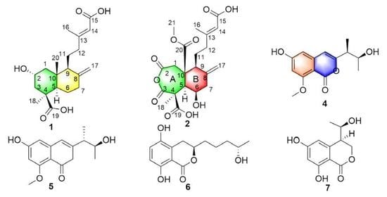
Figure 1. Structures of compounds 113.
Marinedrugs 19 00362 g001
Figure 2. Key 1H-1H COSY, HMBC and ROESY correlations of 1 and 2.
Marinedrugs 19 00362 g002
Figure 3. Calculated and experimental ECD spectra of 1, 2, 4, 5 and 7.
Marinedrugs 19 00362 g003
Figure 4. Key COSY and HMBC correlations of 47.
Marinedrugs 19 00362 g004
Table 1. 1H NMR and 13C NMR data (500 and 125 MHz) for 1 (in DMSO-d6) and 2 (in Acetone-d6).
Position12
δCδH (J in Hz)δCδH (J in Hz)
1a48.0, CH20.87, td (12, 3)35.8, CH22.72, d (18.3)
1b1.96, m3.11, d (18.3)
262.9, CH3.88, tt (4.2, 12)172.8, C
3a46.9, CH20.87, td (12, 3)173.5, C
3b2.19, m
444.2, qC 55.0, C
554.5, CH1.26, m54.7, CH3.50, d (11.3)
6a25.5, CH21.70, m77.7, CH4.93, m
6b1.88, m
7a38.0, CH21.84, m42.4, CH22.38, m
7b2.34, m3.05, dd (4.5, 11.3)
8147.7, qC 142.7, C
954.6, CH1.60, m47.4, CH3.18, m
1040.9, C 51.8, C
11a21.5, CH21.44, m24.1, CH21.47, m
11b1.62, m1.77, m
12a38.9, CH22.21, m40.1, CH22.10, m
12b1.94, m2.34, m
13158.6, C 160.1, C
14116.8, CH5.54, s116.5, CH5.65, s
15167.6, C 167.7, C
1618.3, CH32.07, s18.7, CH32.13, s
17a106.8, CH24.51, s113.7, CH24.91, m
17b4.88, s5.21, s
1828.7, CH31.15, s13.3, CH31.25, s
19178.3, C 175.9, C
2013.5, CH30.54, s172.6, C
21 52.0, CH33.67, s
Table 2. 1H NMR and 13C NMR data (500 and 125 MHz) for 4 and 5 in Acetone-d6.
Position45
δCδH (J in Hz)δCδH (J in Hz)
1158.6, C 158.6, C
3161.6, C 161.3, C
4103.5, CH6.22, s103.8, CH6.20, s
4a143.2, C 143.3, C
5103.6, CH6.43, s103.5, CH6.42, s
6164.6, C 164.6, C
799.4, CH6.51, s99.4, CH6.51, s
8164.7 C 164.7, C
8a102.8, C 102.9, C
946.4, CH2.45, m46.7, CH2.53, m
1069.1, CH3.98, m 69.3, CH3.98, m
1113.7, CH31.26, d (7.0)14.2, CH31.19, d (7.0)
1222.0, CH31.16, d (6.3)20.7, CH31.17, d (6.2)
1356.2, CH33.87, s56.3, CH33.87, s
OH-6 9.63, br s 9.59, br s
Table 3. 1H NMR and 13C NMR data (500 and 125 MHz) for 6 (in Acetone-d6) and 7 (in DMSO-d6).
Position6Position7
δCδH (J in Hz)δCδH (J in Hz)
1170.8, C 1169.1, C
380.5, CH4.63, m368.3, CH24.51, dd (4.1, 11.6)
4.61, dd (1.8, 11.6)
427.4, CH22.68, dd (3.4, 16.8)
3.19, dd (10.6, 16.8)
443.2, CH2.79, m
4a125.6, C 4a143.0, C
5146.3, C 5108.2, CH6.26, d (2.2)
6124.7, CH7.12, d (8.8)6164.5, C
7116.1, CH6.71, d (8.8)7101.2, CH6.19, d (2.2)
8156.2, C 8163.1, C
8a109.3, C 8a100.1, C
1′35.7, CH21.79, m
1.89, m
968.0, CH3.85, m
2′22.0, CH21.62, m1019.9, CH31.04, d (6.2)
3′39.3, CH21.48, m8-OH 11.16, s
4′67.4, CH3.77, m
5′24.1, CH31.14, d (6.2)
8-OH 10.6, s
Publisher’s Note: MDPI stays neutral with regard to jurisdictional claims in published maps and institutional affiliations.

