Article

Agelasine Diterpenoids and Cbl-b Inhibitory Ageliferins from the Coralline Demosponge Astrosclera willeyana

1
Marine Science & Technology Institute, College of Environmental Science & Engineering, Yangzhou University, Yangzhou 225127, China
2
Molecular Targets Program, Center for Cancer Research, National Cancer Institute, National Institutes of Health, Frederick, MD 21702-1201, USA
3
Basic Science Program, Frederick National Laboratory for Cancer Research, National Institutes of Health, Frederick, MD 21702-1201, USA
4
Advanced Biomedical Computational Science, Frederick National Laboratory for Cancer Research, Frederick, MD 21702-1201, USA
5
Women’s Malignancies Branch, Center for Cancer Research, National Cancer Institute, National Institutes of Health, Bethesda, MD 20892-1578, USA
6
Natural Products Branch, Developmental Therapeutics Program, Division of Cancer Treatment and Diagnosis, National Cancer Institute, National Institutes of Health, Frederick, MD 21702-1201, USA
*
Author to whom correspondence should be addressed.
Mar. Drugs 2021, 19(7), 361; https://doi.org/10.3390/md19070361
Submission received: 7 June 2021 / Revised: 17 June 2021 / Accepted: 21 June 2021 / Published: 24 June 2021

Abstract

:
An extract of the coralline demosponge Astrosclera willeyana inhibited the ubiquitin ligase activity of the immunomodulatory protein Cbl-b. The bioassay-guided separation of the extract provided ten active compounds, including three new N-methyladenine-containing diterpenoids, agelasines W–Y (13), a new bromopyrrole alkaloid, N(1)-methylisoageliferin (4), and six known ageliferin derivatives (510). The structures of the new compounds were elucidated from their spectroscopic and spectrometric data, including IR, HRESIMS, and NMR, and by comparison with spectroscopic data in the literature. While all of the isolated compounds showed Cbl-b inhibitory activities, ageliferins (410) were the most potent metabolites, with IC50 values that ranged from 18 to 35 μM.

Graphical Abstract

1. Introduction

The ubiquitin protein ligase (E3), referred to as Casitas B-lineage lymphoma proto-oncogene-b (Cbl-b), negatively regulates the costimulatory pathway in T cells, decreasing the immune response and setting the threshold for anergy in T cells [1]. Cbl-b is essential for the negative regulation of T-cell activation, and thus, it reduces the immune response to cancer cells [2,3]. In line with this function, cells that lack the cblb gene rejected tumors in various models and were resistant to rechallenge with tumors after initial tumor rejection in a variety of tumor models [4,5,6,7]. Thus, targeting Cbl-b may be an effective strategy to enhance antitumor immunity. As part of an ongoing effort to identify small molecule inhibitors of the Cbl-b function from natural products [8], an extract of the sponge Astrosclera willeyana was screened and showed a marked reduction of Cbl-b ligase activity.
The calcareous demosponge Astrosclera willeyana is often referred to as a “living fossil” that is representative of late Paleozoic and Mesozoic reef sponges, and it has provided unique secondary metabolites such as N-methylated ageliferin derivatives and manzacidin D [9,10]. These compounds contain pyrrole-2-carboxylic acid moieties, and their novel structures have been the focus of numerous synthetic efforts [11,12,13,14,15]. To date, little is known about the biological properties of these compounds, except for the reported cytotoxic and antibacterial activities of the nonmethylated form of ageliferin [16,17]. In our current study, bioassay-guided fractionation of the A. willeyana extract provided ten active compounds, including three new N-methyladenine-containing diterpeneoids named agelasines W–Y (13). The agelasines represent a family of diterpene–adenine conjugates that has only been described from sponges in the genus Agelas. In addition, a new bromopyrrole alkaloid, N(1)-methylisoageliferin (4), along with six known ageliferin derivatives (510) were isolated and identified. Herein, we describe the isolation, structure elucidation, and biological activities of the A. willeyana metabolites.

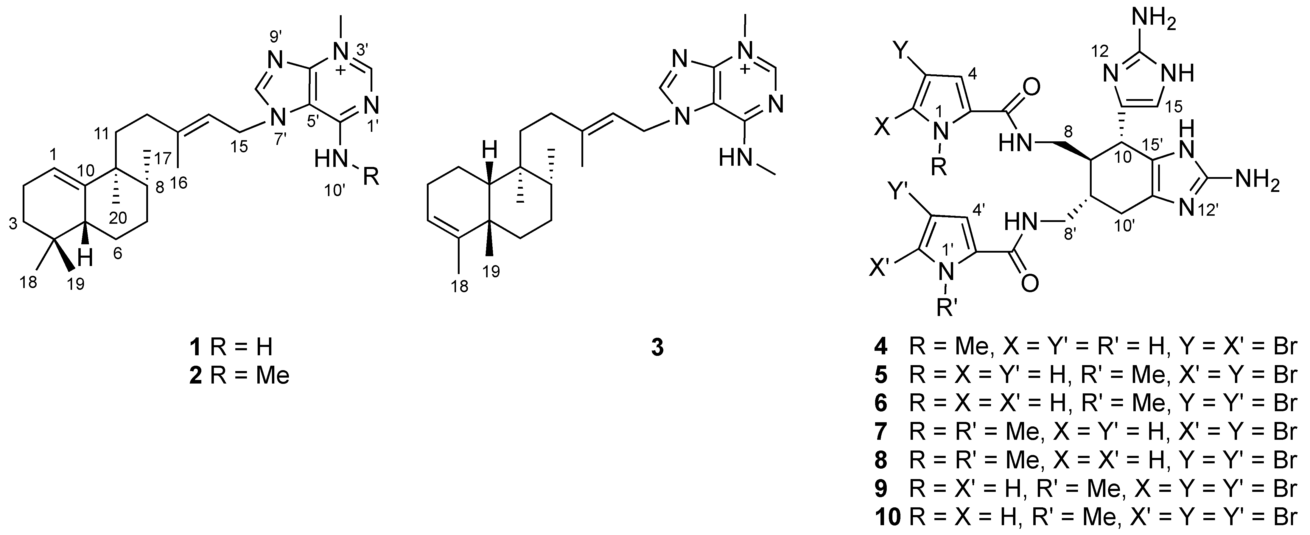
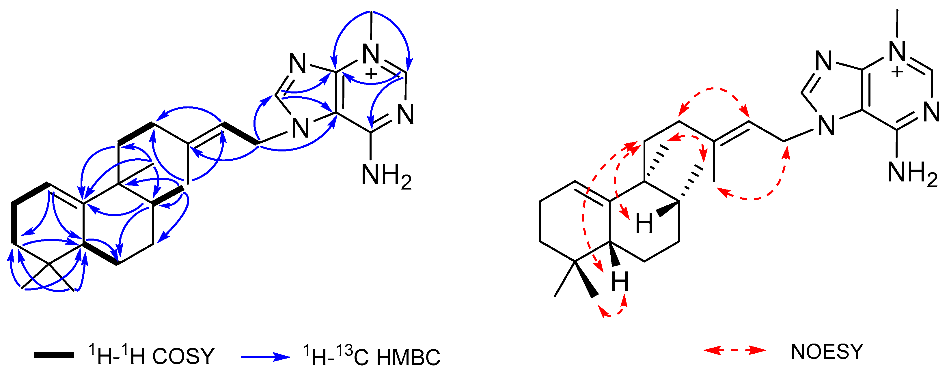
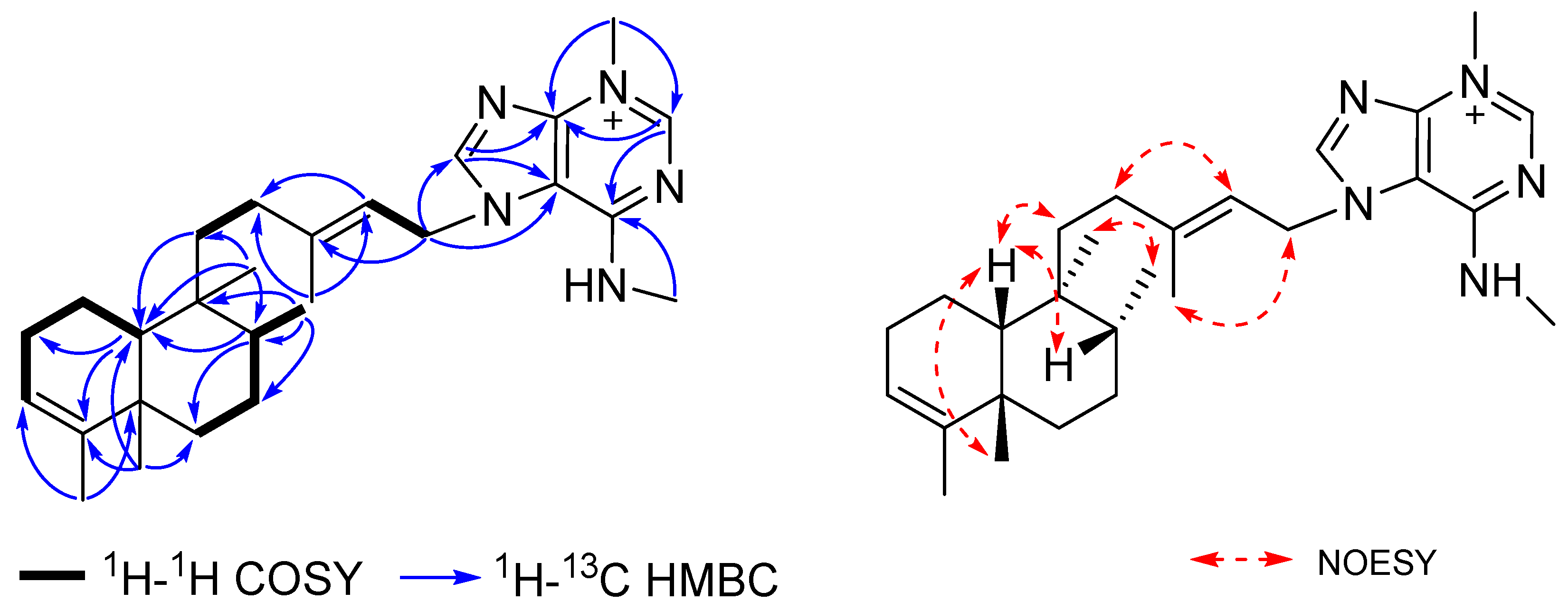
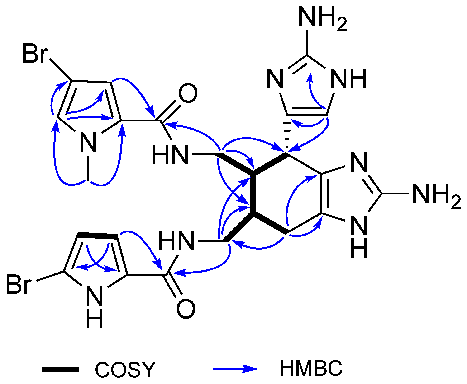
2. Results and Discussion

The organic solvent extract of the sponge Astrosclera willeyana was separated by bioassay-guided diol flash chromatography and C18 HPLC to yield four new metabolites named agelasines W–Y (13) and N(1)-methylisoageliferin (4) (Figure 1), along with six known compounds: N(1′)-methylisoageliferin (5), N(1′)-methylageliferin (6), N(1),N(1′)-dimethylisoageliferin (7), N(1),N(1′)-dimethylageliferin (8), N(1′)-methyl-2-bromoageliferin (9), and N(1′)-methyl-2′-bromoageliferin (10).
Agelasine W (1) was obtained as a colorless oil. The molecular formula C26H40N5+, with 10 degrees of unsaturation, was determined by HRESIMS measurements ([M]+ m/z 422.3285, calcd for C26H40N5+, 422.3278). The 1H and 13C NMR data (Table 1) of compound 1 showed characteristic signals of an adenine moiety at δH/δC 8.44 (1H, s, H-8′)/148.0 (C-8′), 8.57 (1H, s, H-2′)/149.5 (C-2′), δC 112.4 (C-5′), 151.5 (C-4′), and 155.1 (C-6′), and an N-methyl group at δH/δC 4.04 (3H, s)/36.6, revealing the presence of an N-methyladeninium unit in (1). The remaining C20H33 portion was defined as a halimane diterpenoid moiety by comparison of its NMR spectroscopic data with those of related diterpenes [18,19,20]. The four methyl singlets at δH 0.84 (H3-18), 0.88 (H3-19), 0.94 (H3-20), and 1.84 (H3-16), and a doublet at δH 0.83 (3H, d, J = 6.4 Hz, H-17), were compatible with a bicyclic halimane ring system, while the 1H NMR signals of δH 2.10 (1H, m, H-11a), 1.26 (1H, m, H-11b), 2.00 (1H, m, H-12a), 1.81 (1H, m, H-12b), 5.45 (1H, t, J = 6.9 Hz, H-14), 5.12 (2H, br d, J = 6.9 Hz, H2-15), and 1.84 (3H, s, H3-16) were assigned to a 3-methyl-2-pentenyl chain, according to their COSY and HMBC correlations (Figure 2). The HMBC correlations from H-1 to C-3 and C-5, and from H-8 and H2-11 to C-10, established the location of a trisubstituted olefin at Δ1−10, while the HMBC correlations from H2-15 to C-5′ and C-8′ defined the attachment of C-15 to N-7′ of the adenine subunit. Moreover, the 1H and 13C data of (1) were highly similar to those of agelasine C [21]. However, the N-methyl group in (1) showed HMBC correlations to C-2′ and C-4′, which revealed its location on N-3′, while agelasine C had a methyl group substituted at N-9′. The E configuration of the C-13/C-14 olefin was assigned from NOESY correlations between H-14/H-12b and H2-15/H3-16. Additional NOESY correlations of H-5/H-11a and H-8/H-11b suggested that H-5, H-8, and H2-11 were on the same face of the molecule, while a correlation between H3-17 and H3-20 supported this assignment. The chemical shift of C-20 appeared at δH 0.94 ppm, which was consistent with the C-17 and C-20 methyl groups being cis, since it was reported that C-20 is more shielded in a cis than a trans orientation of these methyls [21]. The absolute configuration of agelasine W (1) is suggested as 5R, 8R, 9,S according to the positive optical rotation of (1) ([α] D 25 + 17) compared to those for (+)-and (−)-agelasine C, +36.7 and −55.1, respectively [20,21].
Agelasine X (2) was obtained as a colorless oil, and the HRESIMS spectrum displayed a [M]+ ion at m/z 436.3452, corresponding to the molecular formula of C27H42N5+ with 10 degrees of unsaturation. The 1H and 13C NMR data of compound 2 were nearly identical to those of (1), except for the presence of an additional N-methyl group at δH/δC 3.27 (3H, s)/29.3. The location of the N-methyl group was assigned at N-10′ based on an HMBC correlation to C-6′ (δC 153.8). The relative and absolute configurations of agelasine X (2) were assigned the same as (1) based on their close spectroscopic similarities and its positive optical rotation ([α] D 25 + 20).
Agelasine Y (3) was also isolated as a colorless oil, and HRESIMS ([M]+ m/z 436.3439, calcd for C27H42N5+, 436.3435) established a molecular formula of C27H42N5+ that was isomeric with compound 2. Agelasine Y (3) shared many similar NMR features with those of (2), except for differences in select signals in the bicyclic diterpene ring system (Table 1). The diterpene portion of (3) was assigned as a clerodane skeleton by NMR analysis and a comparison of its spectroscopic data with those of previously reported clerodane diterpenes [20,22,23,24]. The location of the trisubstituted C-3/C-4 olefin was defined by a COSY correlation between the H-2/H-3 and HMBC correlations from H2-1 to C-3, H-10 to C-4, H3-18 to C-3 and C-5, and from H3-19 to C-4 (Figure 3). The relative configuration of the bicyclic ring system of (3) was deduced as a cis-clerodane from the characteristic deshielded carbon signals at δC 25.0 (CH2, C-2) and 33.6 (CH3, C-19) [22], in addition to a NOESY correlation between H-10 and H3-19. Additional NOESY correlations of H-8/H-10, H-10/H-11a, and H3-17/H3-20 established the relative configurations at C-8 and C-9. Furthermore, a comparison of the 13C NMR data in CDCl3 with four closely related clerodane diastereomers (cis-cis, cis-trans, trans-trans, and trans-cis) showed that the chemical shifts of (3) (Supplementary Materials) were in good agreement with those of neo-cis-cis-kolavenol [25], supporting the assigned configuration of the bicyclic scaffold. The E configuration of the side chain olefin was assigned from NOESY correlations of H-14/H-12b and H2-15/H3-16. The structure of (3) resembled that of agelasine Except for the position of the N-methyl groups, the rotation for (3) ([α] D 25 + 15) and agelasine A ([α] D 25 − 31.3) have opposite signs [20]. Therefore, the absolute configuration of agelasine Y (3) is suggested as shown.
N(1)-methylisoageliferin (4) was obtained as a pale-yellow glass. The molecular formula C23H26Br2N10O2 with 15 degrees of unsaturation was determined by an HRESIMS of the doubly charged ion ([M + 2H]2+ m/z 317.0374, calcd for C23H28N10O279Br22+, 317.0376). The 1H and 13C NMR data of (4) (Table 2) closely resembled those of the known compound N(1′)-methylisoageliferin (5) [10], and the molecular formula of (4) was isomeric with (5). NMR signals characteristic of the 3-bromo-N-methylpyrrole 5-carbozamide and 2-bromopyrrole 5-carboxamide ring systems were apparent, as well as signals for a highly substituted cyclohexene and two amino imidazole rings. The HMBC correlations from H-4 and H2-8 to C-6 (Figure 4) revealed that the 3-bromo-N-methylpyrrole 5-carboxamide ring was linked to C-8, and the 2-bromopyrrole 5-carboxamide ring was linked to C-8′ via the HMBC correlations from H-4′ and H2-8′ to C-6′. Thus, the constitution of (4) only differed from (5) by the location of N-methyl substitution on the pyrrole ring. The configuration of N(1)-methylisoageliferin (4) was identical to that of (5), since its NMR data for the cyclohexene ring, including proton coupling constants and NOE enhancements, and its optical rotation, as well as its ECD data, were fully consistent with those of (5) [10].
The known compounds 510 were identified as N(1′)-methylisoageliferin (5), N(1′)-methylageliferin (6), N(1),N(1′)-dimethylisoageliferin (7), N(1),N(1′)-dimethylageliferin (8), N(1′)-methyl-2-bromoageliferin (9), and N(1′)-methyl-2′-bromoageliferin (10) by comparison of their spectroscopic data with the appropriate literature values [10].
Compounds 110 were tested for their ability to inhibit the in vitro enzymatic activity of the Cbl-b ubiquitin ligase (Table 3). The adenine–diterpenoid metabolites agelasines W–Y (13) showed weak inhibitory activities against Cbl-b (IC50 > 50 µM), while the ageliferin derivatives (410) had more pronounced inhibitory effects (IC50 = 18~35 µM). Compounds 79 were the most potent metabolites among the tested compounds, but definitive SAR conclusions were difficult to make. There was no clear pattern of N-methylation or bromine substitution of the two pyrrole rings that directly correlated with the observed Cbl-b inhibitory activity. While the ageliferins are rather modest inhibitors of Cbl-b, the ageliferin structural scaffold could serve as a starting point for the development of more potent analogs with enhanced inhibitory properties.

3. Materials and Methods

3.1. General Experimental Procedures

Flash chromatography was performed using a CombiFlash system (Teledyne Isco, Lincoln, NE, USA). High-performance liquid chromatography (HPLC) was performed using a Varian ProStar 215 solvent delivery module equipped with a Varian ProStar 340 UV-Vis detector, operating under Star 6.41 chromatography workstation software (Agilent Technologies, Santa Clara, CA, USA). HPLC fractions were subsequently dried on Explorer-220 (Thermo Fisher Scientific, Waltham, MA, USA). NMR spectra were acquired with a Bruker Avance III NMR spectrometer equipped with a 3-mm cryogenic probe and operated at 600 MHz for 1H and 150 MHz for 13C (Bruker, Billerica, MA, USA). Spectra were calibrated to their residual solvent signals at δH 3.31 and δC 49.0 for CD3OD. LRESIMS studies were measured on an Agilent 6130 Quadrapole LC/MS system (Agilent Technologies, Santa Clara, CA, USA). HRESIMS data were carried out on an Agilent Technology 6530 Accurate-mass Q-TOF LC/MS (Agilent Technologies, Santa Clara, CA, USA). UV spectra were measured with a PerkinElmer Lambda 465 UV/Vis photodiode array spectrophotometer (PerkinElmer, Waltham, MA, USA). ECD spectra were obtained with a Jasco-1500 circular dichroism spectrophotometer (JASCO, Tokyo, Japan). Optical rotations were recorded on a Rudolph research analytical AUTOPOL IV spectropolarimeter (Rudolph Research Analytical, Hackettstown, NJ, USA). IR spectra were measured with a Bruker ALPHA II FT-IR spectrometer (Bruker, Billerica, MA, USA).

3.2. Animal Material

Specimens of the sponge Astrosclera willeyana were collected in Tonga in November 1997 and kept frozen until extraction. The collection was carried out by the Coral Reef Research Foundation under contract with the Natural Products Branch, U.S. National Cancer Institute. A voucher specimen (voucher ID # 0CDN5435) was deposited at the Smithsonian Institution, Washington, DC, USA.

3.3. Extraction and Isolation

Following the standard NCI protocol for marine samples [26], the frozen sponge sample (2127 g, wet weight) was ground and processed to provide 4.50 g of organic solvent (CH2Cl2-MeOH 1:1 and 100% MeOH) extract (NSC #C017821). A 3.10-g aliquot of the extract was loaded onto a cotton plug and then applied to a diol flash chromatography column (150 g) through a CombiFlash system, sequentially eluting with hexane, CH2Cl2, EtOAc/CH2Cl2 1:1 (v/v), EtOAc, MeOH/EtOAc 1:4 (v/v), MeOH/EtOAc 1:1 (v/v), MeOH/EtOAc 4:1 (v/v), and MeOH to obtain nine fractions (Frac. A–I). The active Frac. E (eluted by MeOH/EtOAc 1:4, (v/v) 735 mg) was fractionated by semipreparative HPLC (Phenomenex Luna C18(2), 5 µM, 100 Å, 250 × 21.2 mm), using a linear gradient of CH3CN/H2O 3:7–1:0 with 0.1% trifluoroacetic acid (TFA) as the mobile phase to afford agelasine W (1, 7.0 mg), agelasine X (2, 18.9 mg), and agelasine Y (3, 4.7 mg). Another more active Frac. F (eluted by EtOAc/MeOH 1:1, 1787 mg) was separated by semipreparative HPLC in the same way as Frac. E to afford N(1)-methylisoageliferin (4, 10.7 mg), N(1′)-methylisoageliferin (5, 20.0 mg), N(1′)-methylageliferin (6, 26.3 mg), N(1),N(1′)-dimethylisoageliferin (7, 53.1 mg), N(1), N(1′)-dimethylageliferin (8, 6.9 mg), N(1′)-methyl-2-bromoageliferin (9, 26.9 mg), and N(1′)-methyl-2′-bromoageliferin (10, 14.7 mg).
Agelasine W (1): colorless oil; [α] D 25 + 17 (c 0.06, MeOH); UV (MeOH) λmax (log ε) 280 (3.70) nm; IR (neat) νmax 2951, 2357, 1659, 1434, 1196, 1133 cm−1; 1H and 13C NMR, Table 1; HRESIMS m/z 422.3285 [M]+ (calcd for C26H40N5+, 422.3278).
Agelasine X (2): colorless oil; [α] D 25 + 20 (c 0.06, MeOH); UV (MeOH) λmax (log ε) 285 (3.89); IR (neat) νmax 2952, 2357, 1643, 1395, 1196, 1132 cm−1; 1H and 13C NMR, Table 1; HRESIMS m/z 436.3452 [M]+ (calcd for C27H42N5+, 436.3435).
Agelasine Y (3): colorless oil; [α] D 25 + 15 (c 0.06, MeOH); UV (MeOH) λmax (log ε) 285 (3.95) nm; IR (neat) νmax 2944, 2357, 1643, 1394, 1196, 1132 cm−1; 1H and 13C NMR, Table 1; HRESIMS m/z 436.3439 [M]+ (calcd for C27H42N5+, 436.3435).
N(1)-Methylisoageliferin (4): pale-yellow glass; [α] D 25 + 50 (c 0.06, MeOH); UV (MeOH) λmax (log ε) 218 (4.30), 228 (4.25) and 270 (4.32) nm; ECD (c 7.12 × 10⁻³ M, MeOH) λmaxε) 284 (−0.75), 250 (+0.96), 229 (−4.56), and 212 (+1.33) nm; IR (neat) νmax 3172, 1678, 1200, 1139 cm−1; 1H and 13C NMR, Table 2; HRESIMS m/z 317.0374 [M + 2H]2+ (calcd for C23H28N10O279Br22+, 317.0376).

3.4. Cbl-b Biochemical Assay

An extract of Astrosclera willeyana was identified as an active source in a screening campaign of prefractionated natural product samples for the inhibition of Cbl-b ubiquitin ligase activity [8]. The bulk extract was fractionated as described above, and the resulting fractions were evaluated for activity in the Cbl-b bioassay, the details of which have already been reported [8]. In brief, dose response experiments with chromatography fractions and purified compounds were carried out in a Tris-HCl-based enzymatic assay buffer with the following final composition: 75-nM Cbl-b (N1/2 construct) [27], 50-nM Ube2d2 protein (E2) [28], 10-nM UBE1protein (E1), 50-nM biotinylated ubiquitin, and 0.5-µM unlabeled recombinant ubiquitin. The assay buffer also contained the following additives: 0.1-mM dithiothreitol, 0.5-mg/mL bovine gelatin (type B), 0.5-mM magnesium chloride, and 0.01% Triton X-100. Reactions were initiated by the addition of ATP to a final concentration of 100 µM. Following initiation, reactions were transferred to plates previously precoated overnight with 10 µg/mL of the polyubiquitin-binding domain of Cbl-b (UBA) [29]. The UBA domain of Cbl-b binds ubiquitin chains, which results in the capture and enrichment of autopolyubiquitinated Cbl-b. Following a reaction interval of 60 min, the reactions were quenched by the addition of zinc to a final concentration of 2.4 mM. Quenched reactions were incubated at room temperature overnight. Following this incubation period reaction, plates were processed as standard ELISA plates: washed thrice in Tris-buffered saline (with 0.1% Tween-20, 1X TBST), probed with avidin-conjugated horse radish peroxidase, which binds captured biotinylated ubiquitin, washed again in 1X TBST (3X), and then, an avidin-HRP-dependent fluorescent signal (indicating avidin-HRP/biotin-polyubiquitin complexes captured by the UBA coating on the plate) was detected (excitation 325 nm, emission 420 nm) using an Infinite M1000 (Tecan US, Inc., Raleigh, NC, USA) plate reader.

4. Conclusions

Two different classes of Cbl-b inhibitory metabolites, including three new adenine-diterpenoid conjugates (13) and seven ageliferin derivatives (410), were obtained from the marine sponge Astrosclera willeyana. Agelasines W–Y (13) have bicyclic terpenoid skeletons with a prenyl side chain that terminates with an N-methyladenine subunit. These alkaloids arise from a mixed biosynthetic process that incorporates both terpene and purine components, and their new structures enrich the known chemo-diversity of Astrosclera sponges. Diterpene alkaloids with an N-methyladenine moiety are generally methylated at the N-9′ position [30,31,32,33,34]; however, agelasines W–Y (13), along with the previously reported agelasimines [35,36], have methyl substituents at both the N-3′ and N-10′ positions. The new compound N(1)-methylisoageliferin (4) and six known analogs of ageliferin (510) exhibited significant Cbl-b inhibitory properties, and they could provide a structural framework for lead compound development.

Supplementary Materials

The following are available online at https://www.mdpi.com/article/10.3390/md19070361/s1, Figure S1: 1H NMR spectrum (600 MHz) of Agelasine W (1) in CD3OD, Figure S2: 13C NMR spectrum (150 MHz) of Agelasine W (1) in CD3OD, Figure S3: HSQC spectrum of Agelasine W (1) in CD3OD, Figure S4: HMBC spectrum of Agelasine W (1) in CD3OD, Figure S5: COSY spectrum of Agelasine W (1) in CD3OD, Figure S6: NOESY spectrum of Agelasine W (1) in CD3OD, Figure S7: HRESIMS of Agelasine W (1), Figure S8: IR spectrum of Agelasine W (1), Figure S9: UV spectrum of Agelasine W (1), Figure S10: 1H NMR spectrum (600 MHz) of Agelasine X (2) in CD3OD, Figure S11: 13C NMR spectrum (150 MHz) of Agelasine X (2) in CD3OD, Figure S12: HSQC spectrum of Agelasine X (2) in CD3OD, Figure S13: HMBC spectrum of Agelasine X (2) in CD3OD, Figure S14: COSY spectrum of Agelasine X (2) in CD3OD, Figure S15: HRESIMS of Agelasine X (2), Figure S16: IR spectrum of Agelasine X (2), Figure S17: UV spectrum of Agelasine X (2), Figure S18: 1H NMR spectrum (600 MHz) of Agelasine Y (3) in CD3OD, Figure S19: 13C NMR spectrum (150 MHz) of Agelasine Y (3) in CD3OD, Figure S20: HSQC spectrum of Agelasine Y (3) in CD3OD, Figure S21: HMBC spectrum of Agelasine Y (3) in CD3OD, Figure S22: COSY spectrum of Agelasine Y (3) in CD3OD, Figure S23: NOESY spectrum of Agelasine Y (3) in CD3OD, Figure S24: HRESIMS of Agelasine Y (3), Figure S25: IR spectrum of Agelasine Y (3), Figure S26: UV spectrum of Agelasine Y (3), Figure S27: 1H NMR spectrum (600 MHz) of N(1)-methylisoageliferin (4) in CD3OD, Figure S28: 13C NMR spectrum (150 MHz) of N(1)-methylisoageliferin (4) in CD3OD, Figure S29: HSQC spectrum of N(1)-methylisoageliferin (4) in CD3OD, Figure S30. HMBC spectrum of N(1)-methylisoageliferin (4) in CD3OD, Figure S31: COSY spectrum of N(1)-methylisoageliferin (4) in CD3OD, Figure S32: ECD spectrum of N(1)-methylisoageliferin (4), Figure S33: HRESIMS of N(1)-methylisoageliferin (4), Figure S34: IR spectrum of N(1)-methylisoageliferin (4), Figure S35: UV spectrum of N(1)-methylisoageliferin (4), Table S1: 1H NMR (600 MHz) data for compounds 510 in CD3OD, Table S2: 13C NMR (150 MHz) data for compound 510 in CD3OD, Table S3: 13C NMR (150 MHz) data for compound 3 and closely related clerodane diastereomers in CDCl3.

Author Contributions

Conceptualization, S.L., B.R.O. and K.R.G.; methodology, W.J., D.W., B.A.P.W., E.A.S. and D.V.; software, A.W. and E.I.G.; investigation, W.J., D.W. and B.A.P.W.; resources, S.L. and B.R.O.; data curation, E.I.G., U.K. and H.R.B.; writing—original draft preparation, W.J.; writing—review and editing, D.W., S.L. and K.R.G.; supervision, K.R.G.; project administration, S.L., B.R.O. and K.R.G.; funding acquisition, S.L. and B.R.O. All authors have read and agreed to the published version of the manuscript.

Funding

This research was funded in part by the National Natural Science Foundation of China 81903772. It was also supported in part by the Intramural Research Program of the NIH, National Cancer Institute, Center for Cancer Research and with federal funds from the National Cancer Institute, National Institutes of Health under contract HHSN261200800001E.

Institutional Review Board Statement

Not applicable.

Data Availability Statement

The data presented in this study are available in the Supplementary Materials.

Acknowledgments

We thank the Natural Products Support Group (NCI at Frederick) for the extract preparation and Lauren Procter, Morgan Pagonis, and Jane Jones of the FNLCR Protein Expression Laboratory for their recombinant protein production efforts. This research was funded in part by the National Natural Science Foundation of China 81903772. It was also supported in part by the Intramural Research Program of the NIH, National Cancer Institute, Center for Cancer Research and with federal funds from the National Cancer Institute, National Institutes of Health under contract HHSN261200800001E. The content of this publication does not necessarily reflect the views or policies of the Department of Health and Human Services, nor does the mention of trade names, commercial products, or organizations imply endorsement by the U.S. Government.

Conflicts of Interest

The authors declare no conflict of interest.

References

  1. Lutz-Nicoladoni, C.; Wolf, D.; Sopper, S. Modulation of immune cell functions by the E3 ligase Cbl-b. Front. Oncol. 2015, 5, 58. [Google Scholar] [CrossRef] [Green Version]
  2. Paolino, M.; Penninger, J.M. Cbl-b in T-cell activation. Semin. Immunopathol. 2010, 32, 137–148. [Google Scholar] [CrossRef] [PubMed]
  3. Wallner, S.; Gruber, T.; Baier, G.; Wolf, D. Releasing the brake: Targeting Cbl-b to enhance lymphocyte effector functions. Clin. Dev. Immunol. 2012, 692639. [Google Scholar] [CrossRef] [PubMed]
  4. Bachmaier, K.; Krawczyk, C.; Kozieradzki, I.; Kong, Y.-Y.; Sasaki, T.; Oliveira-Dos-Santos, A.; Mariathasan, S.; Bouchard, D.; Wakeham, A.; Itie, A.; et al. Negative regulation of lymphocyte activation and autoimmunity by the molecular adaptor Cbl-b. Nature 2000, 403, 211–216. [Google Scholar] [CrossRef] [PubMed]
  5. Chiang, Y.J.; Kole, H.K.; Brown, K.; Naramura, M.; Fukuhara, S.; Hu, R.-J.; Jang, I.K.; Gutkind, J.S.; Shevach, E.; Gu, H. Cbl-b regulates the CD28 dependence of T-cell activation. Nature 2000, 403, 216–220. [Google Scholar] [CrossRef]
  6. Chiang, J.Y.; Jang, I.K.; Hodes, R.; Gu, H. Ablation of Cbl-b provides protection against transplanted and spontaneous tumors. J. Clin. Investig. 2007, 117, 1029–1036. [Google Scholar] [CrossRef] [Green Version]
  7. Liyasova, M.S.; Ma, K.; Lipkowitz, S. Molecular pathways: Cbl proteins in tumorigenesis and antitumor immunity-opportunities for cancer treatment. Clin. Cancer Res. 2015, 21, 1789–1794. [Google Scholar] [CrossRef] [Green Version]
  8. Wilson, B.A.P.; Voeller, D.; Smith, E.A.; Wamiru, A.; Goncharova, E.I.; Liu, G.; Lipkowitz, S.; O’Keefe, B.R. In vitro ubiquitination platform identifies methyl ellipticiniums as ubiquitin ligase inhibitors. SLAS Discov. 2021. [Google Scholar] [CrossRef]
  9. Jahn, T.; Konig, G.M.; Wright, A.D.; Worheide, G.; Reitner, J. Manzacidin D: An unprecedented secondary metabolite from the “living fossil” sponge Astrosclera willeyana. Tetrahedron Lett. 1997, 38, 3883–3884. [Google Scholar] [CrossRef]
  10. Williams, D.H.; Faulkner, D.J. N-methylated ageliferins from the sponge Astrosclera willeyana from Pohnpei. Tetrahedron 1996, 52, 5381–5390. [Google Scholar] [CrossRef]
  11. Hashimoto, T.; Maruoka, K. Synthesis of manzacidins: A stage for the demonstration of synthetic methodologies. Org. Biomol. Chem. 2008, 6, 829–835. [Google Scholar] [CrossRef]
  12. Ma, Z.; Wang, X.; Wang, X.; Rodriguez, R.A.; Moore, C.E.; Gao, S.; Tan, X.; Ma, Y.; Rheingold, A.L.; Baran, P.S.; et al. Asymmetric synthesis of sceptrin and massadine and evidence for biosynthetic enantiodivergence. Science 2014, 346, 219–224. [Google Scholar] [CrossRef] [PubMed] [Green Version]
  13. Ohfune, Y.; Oe, K.; Namba, K.; Shinada, T. Total synthesis of manzacidins. An overview and perspective. Heterocycles 2012, 85, 2617–2649. [Google Scholar] [CrossRef]
  14. Wang, X.; Ma, Z.; Lu, J.; Tan, X.; Chen, C. Asymmetric synthesis of ageliferin. J. Am. Chem. Soc. 2011, 133, 15350–15353. [Google Scholar] [CrossRef] [PubMed] [Green Version]
  15. Wang, X.; Wang, X.; Tan, X.; Lu, J.; Cormier, K.W.; Ma, Z.; Chen, C. Correction to a biomimetic route for construction of the [4 + 2] and [3 + 2] core skeletons of dimeric pyrrole-imidazole alkaloids and asymmetric synthesis of ageliferins. J. Am. Chem. Soc. 2013, 135, 1163. [Google Scholar] [CrossRef] [Green Version]
  16. Eder, C.; Proksch, P.; Wray, V.; van Soest, R.W.M.; Ferdinandus, E.; Pattisina, L.A.; Sudarsono, S. New bromopyrrole alkaloids from the Indopacific sponge Agelas nakamurai. J. Nat. Prod. 1999, 62, 1295–1297. [Google Scholar] [CrossRef]
  17. Hamed, A.N.E.; Schmitz, R.; Bergermann, A.; Totzke, F.; Kubbutat, M.; Mueller, W.E.G.; Youssef, D.T.A.; Bishr, M.M.; Kamel, M.S.; Edrada-Ebel, R.; et al. Bioactive pyrrole alkaloids isolated from the Red Sea: Marine sponge Stylissa carteri. Z. Naturforsch. C J. Biosci. 2018, 73, 199–210. [Google Scholar] [CrossRef] [PubMed] [Green Version]
  18. Chu, M.-J.; Tang, X.-L.; Qin, G.-F.; Sun, Y.-T.; Li, L.; de Voogd, N.J.; Li, P.-L.; Li, G.-Q. Pyrrole derivatives and diterpene alkaloids from the South China Sea sponge Agelas nakamurai. Chem. Biodivers. 2017, 14, e1600446. [Google Scholar] [CrossRef]
  19. Hattori, T.; Adachi, K.; Shizuri, Y. New agelasine compound from the marine sponge Agelas mauritiana as an antifouling substance against macroalgae. J. Nat. Prod. 1997, 60, 411–413. [Google Scholar] [CrossRef]
  20. Nakamura, H.; Wu, H.; Ohizumi, Y.; Hirata, Y. Agelasine-A, -B, -C and -D, novel bicyclic diterpenoids with a 9-methladeninium unit possessing inhibitory effects on Na,K-ATPase from the Okinawa sea sponge Agelas sp. Tetrahedron Lett. 1984, 25, 2989–2992. [Google Scholar] [CrossRef]
  21. Marcos, I.S.; Garcia, N.; Sexmero, M.J.; Basabe, P.; Diez, D.; Urones, J.G. Synthesis of (+)-agelasine C. A structural revision. Tetrahedron 2005, 61, 11672–11678. [Google Scholar] [CrossRef]
  22. Pettit, G.R.; Tang, Y.; Zhang, Q.; Bourne, G.T.; Arm, C.A.; Leet, J.E.; Knight, J.C.; Pettit, R.K.; Chapuis, J.-C.; Doubek, D.L.; et al. Isolation and structures of axistatins 1–3 from the Republic of Palau marine sponge Agelas axifera Hentschel. J. Nat. Prod. 2013, 76, 420–424. [Google Scholar] [CrossRef] [Green Version]
  23. Capon, R.J.; Faulkner, D.J. Antimicrobial metabolites from a Pacific sponge, Agelas sp. J. Am. Chem. Soc. 1984, 106, 1819–1822. [Google Scholar] [CrossRef]
  24. Du, K.; De Mieri, M.; Neuburger, M.; Zietsman, P.C.; Marston, A.; van Vuuren, S.F.; Ferreira, D.; Hamburger, M.; van der Westhuizen, J.H. Labdane and clerodane diterpenoids from Colophospermum mopane. J. Nat. Prod. 2015, 78, 2494–2504. [Google Scholar] [CrossRef]
  25. Pelot, K.A.; Hagelthorn, D.M.; Hong, Y.J.; Tantillo, D.J.; Zerbe, P. Diterpene synthase-catalyzed biosynthesis of distinct clerodane stereoisomers. ChemBioChem 2019, 20, 111–117. [Google Scholar] [CrossRef] [PubMed] [Green Version]
  26. McCloud, T.G. High throughput extraction of plant, marine and fungal specimens for preservation of biologically active molecules. Molecules 2010, 15, 4526–4563. [Google Scholar] [CrossRef] [PubMed] [Green Version]
  27. Ettenberg, S.A.; Magnifico, A.; Cuello, M.; Nau, M.M.; Rubinstein, Y.R.; Yarden, Y.; Weissman, A.M.; Lipkowitz, S. Cbl-b dependent coordinated degradation of the epidermal growth factor receptor signaling complex. J. Biol. Chem. 2001, 276, 27677–27684. [Google Scholar] [CrossRef] [Green Version]
  28. Lorick, K.L.; Jensen, J.P.; Fang, S.; Ong, A.M.; Hatakeyama, S.; Weissman, A.M. RING fingers mediate ubiquitin-conjugating enzyme (E2)-dependent ubiquitination. Proc. Natl. Acad. Sci. USA 1999, 96, 11364–11369. [Google Scholar] [CrossRef] [PubMed] [Green Version]
  29. Davies, G.C.; Ettenberg, S.A.; Coats, A.O.; Mussante, M.; Ravichandran, S.; Collins, J.; Nau, M.M.; Lipkowitz, S. Cbl-b interacts with ubiquitinated proteins; differential functions of the UBA domains of c-Cbl and Cbl-b. Oncogene 2004, 23, 7104–7115. [Google Scholar] [CrossRef] [Green Version]
  30. Calcul, L.; Tenney, K.; Ratnam, J.; McKerrow, J.H.; Crews, P. Structural variations to the 9-N-methyladeninium diterpenoid hybrid commonly isolated from Agelas sponges. Aust. J. Chem. 2010, 63, 915–921. [Google Scholar] [CrossRef]
  31. Gordaliza, M. Terpenyl-purines from the sea. Mar. Drugs 2009, 7, 833–849. [Google Scholar] [CrossRef] [PubMed] [Green Version]
  32. Kubota, T.; Iwai, T.; Takahashi-Nakaguchi, A.; Fromont, J.; Gonoi, T.; Kobayashi, J. Agelasines O-U, new diterpene alkaloids with a 9-N-methyladenine unit from a marine sponge Agelas sp. Tetrahedron 2012, 68, 9738–9744. [Google Scholar] [CrossRef]
  33. Stout, E.P.; Yu, L.C.; Molinski, T.F. Antifungal diterpene alkaloids from the Caribbean sponge Agelas citrina: Unified configurational assignments of agelasidines and agelasines. Eur. J. Org. Chem. 2012, 2012, 5131–5135. [Google Scholar] [CrossRef] [Green Version]
  34. Yang, F.; Hamann, M.T.; Zou, Y.; Zhang, M.-Y.; Gong, X.-B.; Xiao, J.-R.; Chen, W.-S.; Lin, H.-W. Antimicrobial metabolites from the Paracel Islands sponge Agelas mauritiana. J. Nat. Prod. 2012, 75, 774–778. [Google Scholar] [CrossRef] [PubMed] [Green Version]
  35. Fathi-Afshar, R.; Allen, T.M. Biologically active metabolites from Agelas mauritiana. Can. J. Chem. 1988, 66, 45–50. [Google Scholar] [CrossRef]
  36. Ohba, M.; Iizuka, K.; Ishibashi, H.; Fujii, T. Synthesis and absolute configurations of the marine sponge purines (+)-agelasimine-A and (+)-agelasimine-B. Tetrahedron 1997, 53, 16977–16986. [Google Scholar] [CrossRef]
Figure 1. Structures of compounds 110.
Marinedrugs 19 00361 g001
Figure 2. Key 2D correlations for agelasine W (1).
Marinedrugs 19 00361 g002
Figure 3. Key 2D NMR correlations for agelasine Y (3).
Marinedrugs 19 00361 g003
Figure 4. Key 2D NMR correlations for N(1)-methylisoageliferin (4).
Marinedrugs 19 00361 g004
Table 1. 1H NMR (600 MHz) and 13C NMR (150 MHz) data for agelasines W–Y (13) in CD3OD.
Position123
δH (J in Hz)δC, TypeδH (J in Hz)δC, TypeδH (J in Hz)δC, Type
15.36, t (4.0)121.4, CH5.36, t (4.0)121.4, CH2.01, m 1.83, m18.8, CH2
22.04, m24.1, CH22.04, m24.1, CH22.15, m 2.01, m25.0, CH2
31.37, m 1.13, m34.2, CH21.37, m 1.13, m34.2, CH25.28, br s124.4, CH
4 32.4, C 32.4, C 141.0, C
51.69, m44.8, CH1.69, m44.9, CH 38.0, C
61.59, m 1.30, m24.8, CH21.59, m 1.30, m24.8, CH22.03, m 1.09, m38.8, CH2
72.02, m 1.37, m30.2, CH22.02, m 1.37, m30.2, CH21.25, m29.9, CH2
81.55, m40.6, CH1.55, m40.6, CH1.48, m38.6, CH
9 44.1, C 44.1, C 41.3, C
10 142.7, C 142.6, C1.40, m45.9, CH
112.10, m 1.26, m38.6, CH22.10, m 1.26, m38.7, CH21.65, m 1.37, m37.6, CH2
122.00, m 1.81, m35.5, CH22.00, m 1.81, m35.5, CH22.03, m33.9, CH2
13 147.4, C 147.7, C 147.7, C
145.45, t (6.9)117.5, CH5.45, t (6.9)117.2, CH5.50, t (7.0)117.3, CH
155.12, br d (6.9)46.6, CH25.13, br d (6.9)46.7, CH25.15, br d (7.0)46.7, CH2
161.84, s17.0, CH31.84, s17.0, CH31.86, s17.0, CH3
170.83, d (6.4)16.0, CH30.83, d (6.4)16.0, CH30.80, d (6.4)16.3, CH3
180.84, s26.6, CH30.84, s26.6, CH31.69, s20.0, CH3
190.88, s28.7, CH30.88, s28.7, CH31.04, s33.6, CH3
200.94, s22.8, CH30.94, s22.8, CH30.85, s17.9, CH3
2′8.57, s149.5, CH8.67, s149.5, CH8.67, s149.5, CH
3′-NMe4.04, s36.6, CH34.06, s36.6, CH34.05, s36.6, CH3
4′ 151.5, C 150.0, C 150.4, C
5′ 112.4, C 113.1, C 113.1, C
6′ 155.1, C 153.8, C 153.8, C
8′8.44, s148.0, CH8.38, s147.1, CH8.39, s147.1, CH
10′-NMe 3.27, s29.3, CH33.27, s29.3, CH3
Table 2. 1H NMR (600 MHz) and 13C (150 MHz) NMR data for N(1)-methylisoageliferin (4) in CD3OD.
PositionδH (J in Hz)δC, Type
26.91, d (1.5)129.1, CH
2′ 104.7, C
3 95.6, C
3′6.14, d (4.0)112.5, CH
46.84, d (1.5)116.1, CH
4′6.81, d (4.0)113.5, CH
5 127.0, C
5′ 128.4, C
6 163.9, C
6′ 163.0, C
83.72, dd (14.8, 3.2); 3.43, dd (14.8, 4.3)40.4, CH2
8′3.63, dd (14.0, 2.7); 3.37, dd (14.0, 2.7)42.5, CH2
92.17, m43.8, CH
9′2.25, m37.2, CH
103.82, br d (8.5)33.6, CH
10′2.72, dd (16.3, 5.3); 2.47, ddd (16.3, 9.0, 2.9)23.5, CH2
11 127.6, C
11′ 122.8, C
13 149.3, C
13′ 149.2, C
156.77, s112.9, CH
15′ 119.1, C
NMe3.90, s37.2, CH3
Table 3. Cbl-b inhibitory activities of compounds 110 (IC50 values in μM).
CompoundIC50CompoundIC50
157630
272718
366819
433919
5251035
Publisher’s Note: MDPI stays neutral with regard to jurisdictional claims in published maps and institutional affiliations.

