Abstract
Chitosans represent a group of multifunctional drug excipients. Here, we aimed to estimate the impact of high-molecular weight chitosan on the physicochemical properties of clotrimazole–chitosan solid mixtures (CL–CH), prepared by grinding and kneading methods. We characterised these formulas by infrared spectroscopy, differential scanning calorimetry, and powder X-ray diffractometry, and performed in vitro clotrimazole dissolution tests. Additionally, we examined the antifungal activity of clotrimazole–chitosan mixtures against clinical Candida isolates under neutral and acid conditions. The synergistic effect of clotrimazole and chitosan S combinations was observed in tests carried out at pH 4 on Candida glabrata strains. The inhibition of C. glabrata growth reached at least 90%, regardless of the drug/excipient weight ratio, and even at half of the minimal inhibitory concentrations of clotrimazole. Our results demonstrate that clotrimazole and high-molecular weight chitosan could be an effective combination in a topical antifungal formulation, as chitosan acts synergistically with clotrimazole against non-albicans candida strains.
1. Introduction
Excipients are inactive ingredients that perform defined functions in pharmaceutical preparations. Examples include bulking, solubilising, hydrophilising, gelling, and viscosity-increasing agents. Advances in drug formulations and delivery systems have led to the development of multifunctional excipients, agents which possess additional functions aside from their primary purpose. These multifunctional excipients can favourably influence the properties of drugs [1,2]. Macromolecular compounds, such as chitosans, belong to this group of excipients. Chitosans can be used as disintegrants, film-forming agents, coating agents, tablet binders, viscosity enhancers, and mucoadhesive agents [3,4]. Chitosans also have a broad spectrum of biological properties that have been well described in the scientific literature, including antibacterial, antioxidant, haemostatic, and antifungal activity [5,6,7]. Many of chitosan’s physicochemical and biological properties, including its antifungal activity, are related to its molecular weight (MW) and degree of deacetylation [8,9,10,11]. Low-molecular weight chitosan (LMWC), i.e., less than 150 kDa, is more successful in inhibiting mycelial growth than high-molecular weight chitosan (HMWC). This difference is a consequence of its enhanced interactions with cell membranes, allowing the compound to successfully disturb membrane function and inhibit major metabolic processes after penetrating the fungal cell [12]. Calamari et al. evaluated the effect of HMWC and sodium alginate on Candida albicans proteinase activity. Their results indicated that HMWC (300 kDa) chitosan significantly inhibits the proteinase secretion of Candida albicans [13].
Candida species are the predominant cause of fungal infections, and Candida albicans is the most commonly identified opportunistic pathogen of the Candida genus in clinical contexts. In recent years, Candida glabrata has become the second most common cause of mucosal and invasive candidiasis, responsible for 15–20% of all known Candida infections [14]. Notably, the incidence of Candida infections increases every year [15].
The emergence of antifungal resistance has created the need for new formulations that can effectively treat candidiasis [12]. There are several classes of drugs used as fungal therapies, and azole derivatives are most frequently employed to treat Candida infections [16]. One example is clotrimazole, a synthetic imidazole derivative with broad-spectrum antifungal activity [17]. The low aqueous solubility (0.49 mg/L) of this active pharmaceutical ingredient (API) is an obstacle to achieving sufficient concentrations for topical application of the drug [18,19]. To prepare clotrimazole suspensions with potent antimycotic activity, excipients that improve solubility and the dissolution rate must be incorporated [20,21,22,23,24,25].
The wide spectrum of chitosan biopharmaceutical properties, including the ability for fungal growth inhibition, prompted us to examine chitosan’s potential as a multifunctional carrier in a solid mixture with the most employed antifungal API. For the first time, we investigated the possibility of combining chitosan and clotrimazole in simple powder solid mixtures. In this study, we posited that a formula containing clotrimazole and HMWC as an excipient could act as an effective antifungal agent. Our work aimed to determine the physicochemical properties of solid powder formulations. We carried out in vitro dissolution studies and determined the antifungal activity of solid clotrimazole mixtures (prepared by grinding or kneading) with three samples of high-molecular weight chitosan: chitosan A, chitosan B, and chitosan S.
2. Results and Discussion
2.1. Clotrimazole Content
We first determined the clotrimazole content in mixtures obtained by grinding (GM) and kneading (KM) methods (Table 1). We determined that the clotrimazole content ranged between 96.52% and 101.83%.

Table 1.
The compositions of clotrimazole–chitosan mixtures prepared by grinding (GM) and kneading (KM) methods.
2.2. Spectroscopic Studies of Clotrimazole–Chitosan Mixtures
To determine possible interactions in the obtained solid clotrimazole and chitosan mixtures, we obtained Fourier-transform infrared spectroscopy (FTIR) spectra and powder X-ray diffraction (PXRD) patterns. FTIR spectra for clotrimazole (CL), chitosan, and their corresponding mixtures are shown for CHA, CHB, and CHS in Figure 1, Figure 2 and Figure 3, respectively. The characteristic absorption bands observed on clotrimazole FTIR spectra, corresponding to aromatic C-H stretching (3063 cm−1), aromatic C=C stretching (1585 cm−1), C=N stretching (1566 cm−1), C-N stretching (1081 cm−1, 1040 cm−1), and aromatic C-H bending (764 cm−1), are consistent with the literature data [26]. The FTIR spectra obtained for different chitosan types (CHA, CHB, and CHS) showed wide absorption bands in the 3280–3360 cm−1 region, related to the superposition of O-H and N-H stretching, which is characteristic for polysaccharide compounds. Absorption bands were also observed on all registered chitosan spectra in the region of 2870–2930 cm−1, corresponding to the presence of C-H aliphatic stretching vibrations, at 1024 cm−1, attributed to the C-O stretching, and at 1150 cm−1, attributed to the asymmetric stretching of the C-O-C bridge (typical for glycoside linkages) [27]. The FTIR spectra registered for all prepared CL–CH mixtures showed a characteristic intensity reduction compared to the bands corresponding to the pure drug. No additional bands were observed in the registered spectra that could indicate chemical interactions between the drug and chitosan in the prepared mixtures, as well as no shifts or broadening of bands that could indicate drug–polymer hydrogen bonds.
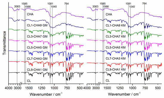
Figure 1.
FTIR spectra of clotrimazole (CL), chitosan A (CHA), and CL–CHA mixtures prepared by grinding (GM) and kneading (KM) methods.
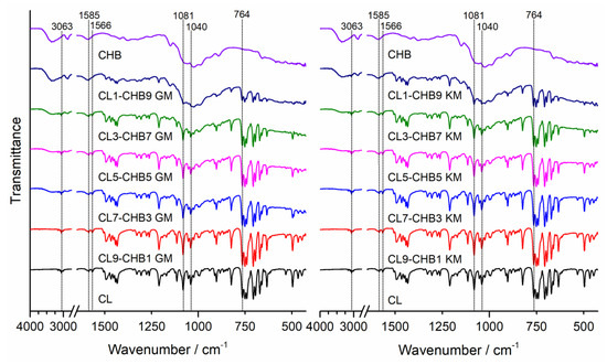
Figure 2.
FTIR spectra of clotrimazole (CL), chitosan B (CHB), and CL–CHB mixtures prepared by grinding (GM) and kneading (KM) methods.
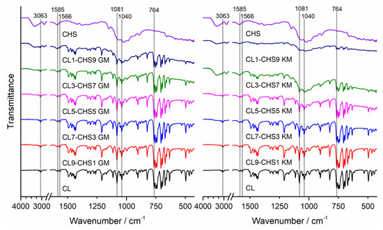
Figure 3.
FTIR spectra of clotrimazole (CL), chitosan S (CHS), and CL–CHS mixtures prepared by grinding (GM) and kneading (KM) methods.
The same conclusion can be drawn from analysing PXRD observations, shown in Figure 4, Figure 5 and Figure 6 for CL–CHA, CL–CHB, and CL–CHS mixtures, respectively. According to published data [26], the CL diffractogram shows distinct peaks successively at 2θ: 9.3°, 10.0°, 12.5°, 16.8°, 18.7°, 20.8°, 22.6°, 24.3°, 27.7°, and 28.2°. These PXRD data are compatible with the structure of clotrimazole presented in the Cambridge Structural Database (CSD ref. PUTRIH) [28]. Consistent with Yen at al. [29], the broad peaks at an angle 2θ of 10° and 20° occurred for all analysed chitosans and examined mixtures; these peaks correspond to the crystalline phase dispersed in the more hydrated and amorphous phase [30]. The comparative analysis of diffraction patterns obtained for the CL–CH mixtures shows a decrease in CL diffraction intensity for each of the mixtures obtained by grinding and kneading methods, as a consequence of the reduction in the CL content in the mixture, crystallinity decrease, and an effective dispersibility in the polymer matrix [31].
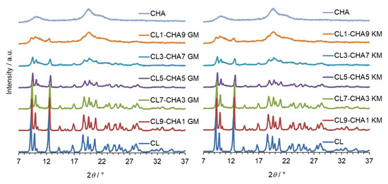
Figure 4.
PXRD patterns of clotrimazole (CL), chitosan A (CHA), and CL–CHA mixtures prepared by grinding (GM) and kneading (KM) methods.
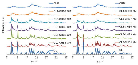
Figure 5.
PXRD diffraction patterns of clotrimazole (CL), chitosan B (CHB), and CL–CHB mixtures prepared by grinding (GM) and kneading (KM) methods.
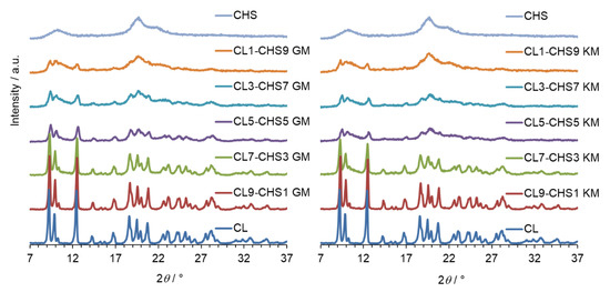
Figure 6.
PXRD patterns of clotrimazole (CL), chitosan S (CHS), and CL–CHS mixtures prepared by grinding (GM) and kneading (KM) methods.
2.3. In Vitro Dissolution Studies
The method of preparing the solid-state formulation influences on its physicochemical properties, such as the dissolution rate of API from a solid drug–polymer mixture. In the study, two different methods of preparing clotrimazole–chitosan mixtures: the solvent-free method of mechanochemical grinding and the method with the use of a solvent, were selected. The grinding method appears as a fast, highly efficient, convenient, and eco-friendly solvent-free solid mixtures formulation method, and the second method used, the kneading method, can be used as an industrially prepared solid binary system. Numerous reports indicate that chitosan can improve the dissolution rate and bioavailability of drugs with poor solubility [31,32,33,34,35,36,37,38]. Therefore, we compared the dissolution profile of pure CL in 1.0% sodium lauryl sulphate (SLS) to the dissolution profile of CL released from CL–CHA, CL–CHB, and CL–CHS mixtures obtained from different drug/polymer weight ratios. Figure 7, Figure 8 and Figure 9 present CL dissolution profiles following release from CL–CHA, CL–CHB, CL–CHS mixtures, respectively. The addition of chitosan causes the CL dissolution rate to increase, independent of the chitosan molecular weight, even in the first 5 min of the dissolution test. The CL dissolution rate also increases with enhanced chitosan content in the appropriate CL–CH mixture. All CL–CH mixtures containing 90 wt.% polymer had the best CL dissolution profile, an observation also made for naproxen–chitosan [31] and telmisartan–chitosan [32] solid systems. We found that the grinding method was more effective than the kneading method, which is also more difficult to scale-up for industrial applications, as it requires the addition of an organic solvent and using high temperatures [33]. The dissolution highest rate was observed for mixtures obtained by grinding with chitosan S, which has the highest molecular weight. An almost 60-fold increase in CL cumulative release was obtained for the CL1–CHS9 GM mixture compared to the pure drug dissolution profile after 60 min (Table 2). The effectiveness of chitosan on antifungal drug release from various dosage forms was confirmed by Giunchedi et al. [39] in in vitro and in vivo studies. Additionally, combination therapy can result in a synergistic effect that leads to drug dose reduction, limiting the development of antifungal resistance [40].
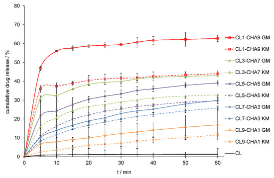
Figure 7.
Dissolution profiles of CL and CL–CHA mixtures obtained by grinding (GM) and kneading (KM) methods in 1.0% SLS at 37 °C. Results are expressed as mean ± S.D. (n = 3).
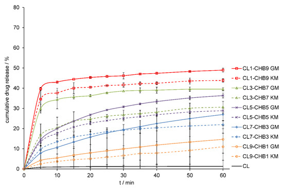
Figure 8.
Dissolution profiles of CL and CL–CHB mixtures obtained by grinding (GM) and kneading (KM) methods in 1.0% SLS at 37 °C. Results are expressed as mean ± S.D. (n = 3).
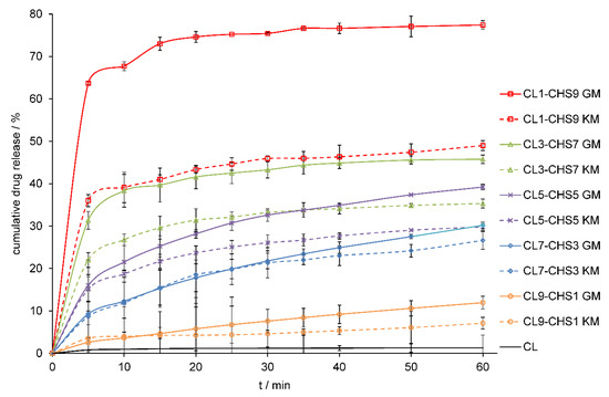
Figure 9.
Dissolution profiles of CL and CL–CHS mixtures obtained by grinding (GM) and kneading (KM) methods in 1.0% SLS at 37 °C. Results are expressed as mean ± S.D. (n = 3).
Table 2.
Cumulative percentage of drug released from CL–CH mixtures prepared by grinding (GM) and kneading methods (KM) 60 min after the dissolution test.
2.4. Differential Scanning Calorimetry (DSC) Analysis
Differential scanning calorimetry (DSC) thermograms registered for CL–CHS mixtures confirm the thermal stability of the drug. The heating curve of pure clotrimazole (Figure 10) shows a sharp, single endothermic peak at 145.6 (∆H = 103.5 J g−1) related to its melting, whereas the thermogram for chitosan S shows a broad endothermic event in the range of 40–110°, indicating an evaporation of residual moisture present in the polymer sample [41]. Table 3 contains the temperatures and enthalpy values of endothermic events related to the melting of CL, visible on DSC curves registered for CL–CHS mixtures. An increase in chitosan concentration causes a reduction in CL melting enthalpy. The sharp shape of the clotrimazole melting peak, visible on DSC curves of all examined mixtures, indicates that the substance remains in a crystalline form. Only slight shifts in the clotrimazole melting peak towards lower temperatures were observed on the DSC curves. The same trends were observed for mixtures obtained by kneading. No decomposition was observed in any tested mixtures after heating to 160 °C. Finally, there were no new thermal effects, which could indicate the occurrence of drug–polymer interactions.
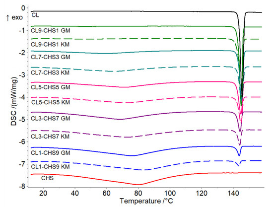
Figure 10.
DSC thermograms of clotrimazole (CL), chitosan S (CHS), and CL–CHS mixtures prepared by grinding (GM) and kneading methods (KM).
Table 3.
The temperature and enthalpy values for clotrimazole melting observed on DSC thermograms during heating of CL–CHS examined formulations.
2.5. Analysis of Antifungal Activity
Based on their superior dissolution properties, we examined the anticandidal activity of clotrimazole–chitosan S combinations, under neutral and acid conditions. The minimal inhibitory concentrations of chitosan S (MICCH), determined in neutral pH 7 (Table 4), indicated that it had no activity against tested representatives of Candida glabrata and Candida albicans (M.I.C. > 250 mg/L). The statistical analysis was performed by Student’s t-test, and p values of <0.05 were considered as significant.
Table 4.
The susceptibility of Candida glabrata and Candida albicans strains to clotrimazole and chitosan S, determined in standard (pH 7) and modified conditions (pH 4).
Following the addition of 0.1% acetic acid to the growth medium, the MICCH decreased for all tested strains as well as for four clinical isolates (three strains of C. glabrata and one strain of C. albicans) to 0.8–3.9 mg/L. In the case of 13 isolates (9 strains of C. albicans and 4 strains of C. glabrata), the values of MICCH did not change at pH 4.
The MIC of clotrimazole (MICCL), determined in standard conditions (pH 7), ranged from 0.125 to 4 mg/L for C. glabrata and from <0.0156 to 2 mg/L for C. albicans. In acidic conditions (pH 4), the MIC of clotrimazole increased from 8- to 30-fold; the only exceptions were two isolates of C. albicans (No 1050 and 1342), which showed higher MICCL at a standard dose (2 mg/L), and decreased to 0.03 and 0.0156 mg/L, respectively, at pH 4. The antifungal activity of clotrimazole in combination with chitosan S was independently tested in four strains—C. glabrata 769, C. glabrata 773, C. glabrata 1941, and C. albicans 488—in standard and modified conditions. The chequerboard assay indicated that chitosan and clotrimazole were synergistic at an acidic pH against C. glabrata isolates (FICI of 0.072, 0.0796, and 0.2644). In the case of Candida albicans 488, indifference was observed, independent of pH (Table 5 and Table 6).
Table 5.
Antifungal activity of the combination of clotrimazole and chitosan S tested in standard conditions (pH 7).
Table 6.
Antifungal activity of the combination of clotrimazole and chitosan S tested in modified conditions (pH 4).
These findings were supported by tests of antifungal activity at different weight ratios of CL–CH mixtures. The antifungal activity of clotrimazole and chitosan mixtures at pH 4 is presented as the percentage of fungal growth compared to the control (Figure 11). In all examined Candida glabrata strains, the inhibition of fungal growth reached at least 80%, regardless of CL–CH weight ratio. On the other hand, the addition of chitosan showed no substantial effect on the survival of Candida albicans 488.
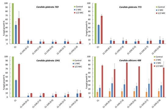
Figure 11.
Fungal growth in the presence of clotrimazole (CL) and chitosan S (CHS) at pH 4. Minimal inhibitory concentration (MIC) = minimal inhibitory concentration of clotrimazole for tested strain, ½ (MIC) = clotrimazole concentration equal to half of the value of MIC, 2 MIC = clotrimazole concentration equal to twice the value of MIC, CL–CHS = mixtures of clotrimazole with chitosan S at the weight ratios of 1:9, 3:7, 5:5, 7:3, and 9:1.
3. Materials and Methods
3.1. Materials
Clotrimazole (CL) used in the study (Figure 12a) was obtained from Hasco Lek S.A. (Wrocław, Poland). Sodium lauryl sulphate (SLS) was purchased from P.P.H. Stanlab (Lublin, Poland). Ethanol was obtained from Chempur (Piekary Śląskie, Poland). High-molecular weight chitosan (CHS) (Figure 12b), with 92% deacetylation degree and a viscosity-average molecular weight of Mv =1087 kDa, was purchased from France-Chitine (Orange, France). Samples of chitosan A (CHA) and chitosan B (CHB) were prepared in the Institute of Applied Radiation Chemistry Technical University of Łódź (Łódź, Poland) by irradiation of dry CHS in an air atmosphere by greys from a 60Co source (average energy of quantum 1.25 MeV) at 5 and 30 kGy, respectively. The average viscosimetric molecular weight of chitosan samples, presented in Table 7, was determined by the Ubbelohde capillary viscometry technique.
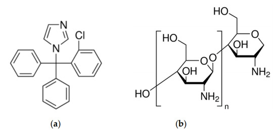
Figure 12.
Chemical structure of: (a) clotrimazole, and (b) chitosan repeated monomer unit.
Table 7.
Physicochemical parameters of investigated chitosan samples.
3.2. Mixture Preparation
Mixtures of clotrimazole–chitosan were prepared by grinding (GM) and kneading (KM) methods after mixing clotrimazole and the selected polymer in different ratios.
3.2.1. Grinding Method (GM)
Mixtures of CL–CHS, CL–CHA, and CL–CHB were prepared by grinding mixtures of each component in an agate mortar for 10 min. The weight ratios of components in mixtures of clotrimazole–polymer were 1:9, 3:7, 5:5, 7:3, and 9:1. The obtained mixtures were sieved through a 315 µm mesh and stored in a desiccator at room temperature until use.
3.2.2. Kneading Method (KM)
Clotrimazole and chitosan were weighed and mixed in an agate mortar with the addition of a sufficient volume of ethanol to achieve a slurry-like consistency. The solvent used to prepare mixtures was then completely evaporated at 30–35 °C with continuous stirring to obtain a dry mass. The dry mass was then triturated in an agate mortar and sieved through a 315 μm mesh. The pulverised solid dispersions were stored in a desiccator at room temperature until use. The weight ratios of CL–CHS, CL–CHA, and CL–CHB in the obtained mixtures were 1:9, 3:7, 5:5, 7:3, and 9:1.
3.3. Determination of Drug Content
Samples of obtained mixtures containing the equivalent of 10 mg CL were dissolved in 10 mL of methanol. The solutions were filtered and diluted by methanol in a 1:10 ratio, and the clotrimazole concentration in solutions was determined spectrophotometrically at λ = 260 nm using a UV–Vis spectrophotometer (Jasco V-650, Tokyo, Japan). Clotrimazole concentrations in the mixtures were calculated against a predetermined calibration curve (R2 = 0.999) over the concentration range of 50–500 μg/mL.
3.4. Physicochemical Characterisation of Clotrimazole–Chitosan Mixtures
3.4.1. Fourier Transform Infrared Spectroscopy (FTIR)
FTIR spectra of clotrimazole, chitosan S, A, and B, and CL–CH mixtures were obtained using a Perkin-Elmer Spectrum Two FTIR spectrometer (Perkin Elmer, Waltham, MA, U.S.A.). The spectra were collected in the range of 450 to 4000 cm−1, using the attenuated total reflection (ATR) sampling mode. An ATR device with a diamond crystal was used. Each FTIR spectrum was registered at room temperature in transmittance mode, by an accumulation of 32 scans with a resolution of 4 cm−1. After scanning of each sample, an air spectrum recording for the clean and dry ATR crystal was taken and subtracted automatically as background.
3.4.2. Powder X-ray Diffraction Analysis (XRPD)
Powder X-ray diffraction patterns for mixtures containing clotrimazole and chitosan S, A, and B were measured on a powder diffractometer (D2 Phaser, Bruker, Germany) equipped with CuKα radiation and 1D Lynxeye detector. The diffraction patterns were measured in the range of 7°–50° (2θ) with a 0.02° step (2θ) and an exposure time of 0.5 s per step.
3.4.3. Differential Scanning Calorimetry (DSC)
DSC curves of pure components and CL–CH mixtures were recorded using a 214 Polyma (Netzsch, Selb, Germany) heat flux-type calorimeter. The measurements and data analysis were carried out using Proteus 7.0 software (Netzsch, Selb, Germany). Calibration of the DSC instrument was performed using the melting points of indium (156.6 °C), tin (231.9 °C), bismuth (271.4 °C), and zinc (419.5 °C) as standards [42]. DSC samples were prepared by weighing approximately 4–5 mg of CL, CH, or the appropriate CL–CH mixture in a standard 40 µL aluminium crucible. The crucible was next sealed with a pierced lid. An empty standard crucible, with a pierced lid, was used as reference. DSC measurements were run in triplicate, in the temperature range of 10–160 °C and a heating rate of 5 °C min−1. Dry nitrogen (99.999% purity), with a flow rate of 50 cm3 min−1, was used as a purge gas.
3.5. In Vitro Dissolution Release Profile of Clotrimazole
To perform dissolution studies, samples of 100 mg clotrimazole and CL–CH mixtures were pressed under a pressure of 1 tonne using a hydraulic press (Specac, Orpington, UK). In vitro drug release studies were carried out in triplicate, using the U.S.P. type 1 apparatus (Vankel VK7025, Varian Inc., Cary, NC, USA) with 500 mL of water with 1.0% SLS as dissolution medium at a paddle speed of 50 rpm and temperature of 37 ± 0.5 °C. At predetermined intervals (5, 10, 15, 20, 25, 35, 40, 50, and 60 min), 3 mL aliquots were withdrawn, and clotrimazole concentrations were determined using a UV–Vis spectrophotometer (Jasco V-650, Tokyo, Japan) at λ = 260 nm. Clotrimazole concentrations in mixtures were calculated against a predetermined calibration curve (R2 = 0.997) over the concentration range of 2–20 μg/mL.
3.6. Antifungal Activity
3.6.1. Determination of Minimal Inhibitory Concentration (MIC) of Clotrimazole and Chitosan S
The minimal inhibitory concentrations of clotrimazole (MICCL) and chitosan S (MICCH) were tested in acid and neutral conditions following standard and modified EUCAST procedures [43]. The susceptibility tests were performed on 10 clinical isolates of Candida albicans and 7 clinical isolates of Candida glabrata, as well as Candida albicans ATCC 90028, Candida albicans ATCC 10231, and Candida glabrata ATCC 90030. The strains were stored at −80 °C and were cultured on Sabouraud Dextrose Agar and incubated at 37 °C before testing. After 24 h, the cultures were resuspended at a density of 1–5 × 10⁵ CFU/mL and used to inoculate the 96-well microplates loaded with different dilutions of tested compounds.
Standard Procedure (pH 7)
In accordance with the EUCAST (European Committee on Antimicrobial Susceptibility Testing) document, the microdilution method and liquid medium RPMI 1640 (supplemented with 2% glucose and buffered with MOPS (4-Morpholinepropanesulfonic acid)) of pH 7 were used. The stock ethanol (96%) solutions of clotrimazole (1600 mg/L) were used to prepare serial ethanol dilutions ranging from 3.1 to 800 mg/L, which were subsequently diluted 100 times in double-concentrated RPMI 1640 (RPMI 1640 2x). After the addition of equal volume (50 µL) of the water solution of tested microorganisms, the final concentrations of clotrimazole were 0.0078–8 mg/L. The stock solution of chitosan (10,000 mg/L in 1% acetic acid) was used to prepare serial dilutions in 1% acetic acid to obtain concentrations of 19.5–2500 mg/L. Finally, 5 µL of each solution was transferred to 45 µL of RPMI 1640 2x dispersed in appropriate wells of the microplates. After adding 50 µL of microbial suspensions, the range of obtained chitosan concentrations was 250–0.8 mg/L.
Modified Procedure (pH 4)
Experiments were performed as in the standard procedure, except we used acid-RPMI 1640 (RPMI 1640 supplemented with 2% glucose, 0.1% acetic acid and MOPS) at pH 4 and prepared yeast suspensions in 0.1% water solution of acetic acid. Chitosan S was tested in the same concentrations as in the standard method, but clotrimazole concentrations were higher. The stock ethanol solution of clotrimazole was 25,000 mg/L, working solutions ranged from 50 to 12,500 mg/L, and the final tested concentrations were 0.25–125 mg/L. The plates, prepared according to standard or modified procedures, were incubated at 37 °C for 24 h and then measured spectrophotometrically (530 nm). MIC was calculated as the lowest concentration of the tested drug that produced a 50% reduction in the strain absorbance compared to the absorbance of the negative (untreated) control.
3.6.2. Determination of Minimal Inhibitory Concentrations (MIC) of Clotrimazole–Chitosan S Combinations
The MIC of clotrimazole–chitosan S combinations were assessed on three strains of Candida glabrata and one Candida albicans. In the chequerboard assay, the wells of the 96-well microplate were inoculated simultaneously with both tested compounds and the investigated microorganisms. The combinations of clotrimazole and chitosan were also tested in acid and neutral conditions using RPMI 1640 media. The range of concentrations of chitosan tested in acidic conditions was 0.25–500 mg/L, whereas clotrimazole was 0.25–125 mg/L for isolates of Candida glabrata and 0.0156–8 mg/L for the isolate of Candida albicans. In neutral conditions, the range of tested clotrimazole concentrations was 0.0156–8 mg/L, and chitosan S was 7.8–500 mg/L. After a 24-h incubation, the absorbance at 530 nm was measured. The fractional inhibitory concentration of clotrimazole (FICCL) was calculated as MICCL-CH of clotrimazole obtained in the presence of chitosan, divided by MICCL of clotrimazole alone. The fractional inhibitory concentration of chitosan (FICCH) was a quotient of MICCH-CL of chitosan S acting in combination with clotrimazole and MICCH of chitosan acting alone. The sum of FICCL and FICCH represents the fractional inhibitory concentration index (FICI), which is indicative for synergism (SYN) if its value is less than 0.5, indifference (IND) if ranged from 0.5 to 4, and antagonism when it is higher than 4 [44,45].
3.6.3. Antifungal Activity of Clotrimazole in the Presence of Chitosan
Our experiment tested the antifungal activity of the mixtures prepared with a defined ratio of clotrimazole–chitosan, namely 1:9, 3:7, 5:5, 7:3, and 9:1. For each of the tested strains, two concentrations of clotrimazole were prepared (one equal to MIC, and a second representing ½ of MIC) and pipetted into five wells of the microplate. Next, the appropriate amounts of chitosan were added, keeping the defined ratio. After a 24-h incubation at 37 °C, the viability of the tested samples was measured at 530 nm. Results were expressed as a percentage of fungal growth inhibition compared to control wells, where the fungal growth was 100%.
4. Conclusions
Previous studies have investigated the impact of different types of chitosan on yeast species, including Candida. The results demonstrated the anti-yeast activity of high-molecular weight and low-molecular weight chitosans [46]. In this work, we analysed the physicochemical properties and activity of high-molecular weight chitosan (M.W. > 400 kDa) and clotrimazole against clinical isolates of Candida species. The dissolution results obtained in our work led to the conclusion that chitosan can be used as an excipient which improves the dissolution rate of poorly soluble clotrimazole (II class in the Biopharmaceutical Classification System). The dispersion of drug molecules between the polymer chains increases the effect of wetting the particles of medication and prevents their agglomeration [47,48]. We observed the fastest dissolution of the drug in mixtures containing chitosan S (which has the highest molecular weight) and proceeded to analyse their activity against both albicans and non-albicans strains. We observed significant differences related to the susceptibility of tested strains to CL–CHS. The examined CL–CHS combinations demonstrated significant activity against Candida, especially C. glabrata strains, which have an innate resistance to azole antimycotic therapy [49,50,51]. Our studies indicate that high-molecular weight chitosan and clotrimazole could have a synergistic effect on C. glabrata isolates at acidic pHs. The development of pharmaceutical formulations based on clotrimazole–chitosan solid-state mixtures is a promising step towards developing alternative effective topical antifungal formulations.
Author Contributions
Conceptualisation, B.G.; methodology, B.G., U.N. and K.W.; writing—original draft preparation, B.K., B.G. and A.G.; writing—review and editing, B.K., A.G., and U.N.; supervision, B.K., A.G.; project administration. All authors have read and agreed to the published version of the manuscript.
Funding
This study was financed with national funds for scientific research by the Ministry of Science and Higher Education, Poland (Grant No. SUB.D190.20.004).
Conflicts of Interest
The authors declare no conflict of interest.
References
- Karolewicz, B. A review of polymers as multifunctional excipients in drug dosage form technology. Saudi Pharm. J. 2016, 24, 525–536. [Google Scholar] [CrossRef] [PubMed]
- Jøraholmen, M.W.; Bhargava, A.; Julin, K.; Johannessen, M.; Skalko-Basnet, N. The Antimicrobial Properties of Chitosan Can Be Tailored by Formulation. Mar. Drugs 2020, 18, 96. [Google Scholar] [CrossRef] [PubMed]
- Rowe, R.C.; Sheskey, P.J.; Owen, S.C. Handbook of Pharmaceutical Excipients, 5th ed.; The Pharmaceutical Press: London, UK, 2006; pp. 132–135. [Google Scholar]
- Badwan, A.A.; Rashid, I.; Al Omari, M.M.; Darras, F.H. Chitin and Chitosan as Direct Compression Excipients in Pharmaceutical Applications. Mar. Drugs 2015, 13, 1519–1547. [Google Scholar] [CrossRef] [PubMed]
- Cheung, R.C.F.; Ng, T.; Wong, J.H.; Chan, W.-Y. Chitosan: An Update on Potential Biomedical and Pharmaceutical Applications. Mar. Drugs 2015, 13, 5156–5186. [Google Scholar] [CrossRef] [PubMed]
- Younes, I.; Rinaudo, M. Chitin and Chitosan Preparation from Marine Sources. Structure, Properties and Applications. Mar. Drugs 2015, 13, 1133–1174. [Google Scholar] [CrossRef]
- Bano, I.; Arshad, M.; Yasin, T.; Ghauri, M.A.; Younus, M. Chitosan: A potential biopolymer for wound management. Int. J. Biol. Macromol. 2017, 102, 380–383. [Google Scholar] [CrossRef]
- Szymańska, E.; Winnicka, K. Stability of Chitosan—A Challenge for Pharmaceutical and Biomedical Applications. Mar. Drugs 2015, 13, 1819–1846. [Google Scholar] [CrossRef]
- Alburquenque, C.; Bucarey, S.A.; Neira-Carrillo, A.; Urzúa, B.; Hermosilla, G.; Tapia, C.V. Antifungal activity of low molecular weight chitosan against clinical isolates of Candida spp. Med. Mycol. 2010, 48, 1018–1023. [Google Scholar] [CrossRef]
- Minh, N.C.; Van Hoa, N.; Trung, T.S. Preparation, properties, and application of low-molecular-weight chitosan. In Handbook of Chitin and Chitosan, Preparation and Properties, 1st ed.; Thomas, S., Pius, A., Gopi, S., Eds.; Elsevier Inc.: Amsterdam, The Netherlands, 2020; Volume 1, pp. 453–471. [Google Scholar]
- Delgado, C.L.D.; Rodríguez-Cruz, I.M.; Fuentes-Prado, E.; Escobar-Chávez, J.J.; Vidal-Romero, G.; García-González, L.; Puente-Lee, R.I. Drug Carrier Systems Using Chitosan for Non Parenteral Routes. In Pharmacology and Therapeutics; IntechOpen: London, UK, 2014; pp. 273–325. [Google Scholar]
- Shih, P.-Y.; Liao, Y.-T.; Tseng, Y.-K.; Deng, F.-S.; Lin, C.-H. A Potential Antifungal Effect of Chitosan Against Candida albicans Is Mediated via the Inhibition of SAGA Complex Component Expression and the Subsequent Alteration of Cell Surface Integrity. Front. Microbiol. 2019, 10. [Google Scholar] [CrossRef]
- Calamari, S.; Bojanich, A.; Barembaum, S.; Azcurra, A.; Virga, C.; Dorronsoro, S. High molecular weight chitosan and sodium alginate effect on secretory acid proteinase of Candida albicans. Rev. Iberoam. Micol. 2004, 21, 206–208. [Google Scholar]
- Roetzer, A.; Gabaldón, T.; Schüller, C. From Saccharomyces cerevisiae to Candida glabrata in a few easy steps: Important adaptations for an opportunistic pathogen. FEMS Microbiol. Lett. 2011, 314, 1–9. [Google Scholar] [CrossRef] [PubMed]
- Kabir, M.A.; Ahmad, Z. CandidaInfections and Their Prevention. ISRN Prev. Med. 2013, 2013, 1–13. [Google Scholar] [CrossRef] [PubMed]
- Whaley, S.G.; Berkow, E.L.; Rybak, J.M.; Nishimoto, A.T.; Barker, K.S.; Rogers, P.D. Azole Antifungal Resistance in Candida albicans and Emerging Non-albicans Candida Species. Front. Microbiol. 2017, 7, 2173. [Google Scholar] [CrossRef]
- Heel, R.C.; Brogden, R.N.; Speight, T.M.; Avery, G.S. Econazole: A review of its antifungal activity and therapeutic efficacy. Drugs 1978, 16, 424–447. [Google Scholar] [CrossRef] [PubMed]
- Karolewicz, B.; Gajda, M.; Owczarek, A.; Pluta, J.; Górniak, A. Physicochemical Characterization and Dissolution Studies of Solid Dispersions of Clotrimazole with Pluronic F127. Trop. J. Pharm. Res. 2014, 13, 1225–1232. [Google Scholar] [CrossRef]
- Dhake, A.S.; Shinkar, D.M.; Shayle, S.; Patil, S.B.; Setty, C.M. Development and evaluation of mucoadhesive tablets of clotrimazole and its β-cyclodextrin complex for treatment of candidiasis. Int. J. Pharm. Pharm. Sci. 2011, 3, 159–164. [Google Scholar]
- Balata, G.; Mahdi, M.; Abu Bakera, R. Improvement of Solubility and Dissolution Properties of Clotrimazole by Solid Dispersions and Inclusion Complexes. Indian J. Pharm. Sci. 2011, 73, 517–526. [Google Scholar] [CrossRef] [PubMed]
- Santos, S.S.; Lorenzoni, A.; Ferreira, L.M.; Mattiazzi, J.; Adams, A.I.; Denardi, L.B.; Alves, S.H.; Schaffazick, S.R.; Cruz, L. Clotrimazole-loaded Eudragit® RS100 nanocapsules: Preparation, characterization and in vitro evaluation of antifungal activity against Candida species. Mater. Sci. Eng. C 2013, 33, 1389–1394. [Google Scholar] [CrossRef]
- El-Garhy, O.H. Investigating the potential of polyethylene glycols in solubilization of imidazole drugs of interest. Int. J. Pharm. Pharm. Sci. 2013, 5, 266–272. [Google Scholar]
- Yuen, C.-W.M.; Yip, J.; Liu, L.; Cheuk, K.; Kan, C.-W.; Cheung, H.-C.; Cheng, S.-Y. Chitosan microcapsules loaded with either miconazole nitrate or clotrimazole, prepared via emulsion technique. Carbohydr. Polym. 2012, 89, 795–801. [Google Scholar] [CrossRef]
- Szymańska, E.; Winnicka, K. Preparation and in vitro evaluation of chitosan microgranules with clotrimazole. Acta Pol. Pharm. Drug Res. 2012, 69, 509–513. [Google Scholar]
- Queiroz, M.F.; Melo, K.R.T.; Sabry, D.A.; Sassaki, G.L.; Rocha, H.A.O. Does the Use of Chitosan Contribute to Oxalate Kidney Stone Formation? Mar. Drugs 2014, 13, 141–158. [Google Scholar] [CrossRef] [PubMed]
- Hani, U.; Krishna, G.; Shivakumar, H.G. Design and optimization of clotrimazole–hydroxypropyl-β-cyclodextrin bioadhesive vaginal tablets using Anacardium occidentale gum by 32 factorial design. RSC Adv. 2015, 5, 35391–35404. [Google Scholar] [CrossRef]
- Kweon, H.; Um, I.C.; Park, Y.H. Structural and thermal characteristics of Antheraea pernyi silk fibroin/chitosan blend film. Polym. 2001, 42, 6651–6656. [Google Scholar] [CrossRef]
- Song, H.; Shin, H.-S. The Antifungal Drug Clotrimazole. Acta Cryst. C 1998, C54, 1675–1677. [Google Scholar] [CrossRef]
- Yen, M.-T.; Yang, J.-H.; Mau, J.-L. Physicochemical characterization of chitin and chitosan from crab shells. Carbohydr. Polym. 2009, 75, 15–21. [Google Scholar] [CrossRef]
- Kasai, D.; Chougale, R.B.; Masti, S.; Chalannavar, R.; Malabadi, R.B.; Gani, R. Influence of Syzygium cumini leaves extract on morphological, thermal, mechanical, and antimicrobial properties of PVA and PVA/chitosan blend films. J. Appl. Polym. Sci. 2018, 135, 46188. [Google Scholar] [CrossRef]
- Mura, P.; Zerrouk, N.; Mennini, N.; Maestrelli, F.; Chemtob, C. Development and characterization of naproxen-chitosan solid systems with improved drug dissolution properties. Eur. J. Pharm. Sci. 2003, 19, 67–75. [Google Scholar] [CrossRef]
- Zhong, L.; Zhu, X.; Luo, X.; Su, W. Dissolution Properties and Physical Characterization of Telmisartan–Chitosan Solid Dispersions Prepared by Mechanochemical Activation. AAPS PharmSciTech 2013, 14, 541–550. [Google Scholar] [CrossRef]
- Shete, A.S.; Yadav, A.V.; Murthy, S.M. Chitosan and chitosan chlorhydrate based various approaches for enhancement of dissolution rate of carvedilol. DARU J. Pharm. Sci. 2012, 20, 93–101. [Google Scholar] [CrossRef]
- Kumar, S.G.V.; Mishra, D.N. Preparation, characterization and in vitro dissolution studies of solid systems of valdecoxib with chitosan. Chem. Pharm. Bull. 2006, 54, 1102–1106. [Google Scholar] [CrossRef] [PubMed][Green Version]
- Portero, A.; Remuñán-López, C.; Vila-Jato, J. Effect of chitosan and chitosan glutamate enhancing the dissolution properties of the poorly water soluble drug nifedipine. Int. J. Pharm. 1998, 175, 75–84. [Google Scholar] [CrossRef]
- Corti, G.; Maestrelli, F.; Cirri, M.; Mura, P.; Zerrouk, N. Dissolution and Permeation Properties of Naproxen From Solid-State Systems With Chitosan. Drug Deliv. 2008, 15, 303–312. [Google Scholar] [CrossRef] [PubMed][Green Version]
- Maestrelli, F.; Zerrouk, N.; Chemtob, C.; Mura, P. Influence of chitosan and its glutamate and hydrochloride salts on naproxen dissolution rate and permeation across Caco-2 cells. Int. J. Pharm. 2004, 271, 257–267. [Google Scholar] [CrossRef] [PubMed]
- Sawayanagi, Y.; Nambu, N.; Nagai, T. Dissolution properties and bioavailability of phenytoin from ground mixtures with chitin or chitosan. Chem. Pharm. Bull. 1983, 31, 2064–2068. [Google Scholar] [CrossRef]
- Giunchedi, P.; Juliano, C.; Gavini, E.; Cossu, M.; Sorrenti, M. Formulation and in vivo evaluation of chlorhexidine buccal tablets prepared using drug-loaded chitosan microspheres. Eur. J. Pharm. Biopharm. 2002, 53, 233–239. [Google Scholar] [CrossRef]
- Cui, J.; Ren, B.; Tong, Y.; Dai, H.; Zhang, L. Synergistic combinations of antifungals and anti-virulence agents to fight against Candida albicans. Virulence 2015, 6, 362–371. [Google Scholar] [CrossRef]
- Chollakup, R.; Uttayarat, P.; Chworos, A.; Smitthipong, W. Noncovalent Sericin-Chitosan Scaffold: Physical Properties and Low Cytotoxicity Effect. Int. J. Mol. Sci. 2020, 21, 775. [Google Scholar] [CrossRef]
- Karolewicz, B.; Gajda, M.; Górniak, A.; Owczarek, A.; Mucha, I. Pluronic F127 as a suitable carrier for preparing the imatinib base solid dispersions and its potential in development of a modified release dosage forms. J. Therm. Anal. Calorim. 2017, 130, 383–390. [Google Scholar] [CrossRef]
- The European Committee on Antimicrobial Susceptibility Testing (EUCAST). Method for the Determination of Broth Dilution Minimum Inhibitory Concentrations of Antifungal Agents for Yeasts. In EUCAST Definitive Document E.DEF 7.3.1; European Society of Clinical Microbiology and Infectious Diseases: Basel, Switzerland, 2017. [Google Scholar]
- Hindler, J. Antimicrobial Susceptibility Testing. In Clinical Microbiology Procedures Handbook, 2nd ed.; Isenberg, H.D., Ed.; A.S.M. Press: Washington, DC, USA, 1995; pp. 5.18.11–15.18.20. [Google Scholar]
- Odds, F.C. Synergy, antagonism, and what the chequerboard puts between them. J. Antimicrob. Chemother. 2003, 52, 1. [Google Scholar] [CrossRef]
- Kulikov, S.N.; Lisovskaya, S.A.; Zelenikhin, P.V.; Bezrodnykh, E.A.; Shakirova, D.R.; Blagodatskikh, I.V.; Tikhonov, V.E. Antifungal activity of oligochitosans (short chain chitosans) against some Candida species and clinical isolates of Candida albicans: Molecular weight - activity relationship. Eur. J. Med. Chem. 2014, 74, 169–178. [Google Scholar] [CrossRef] [PubMed]
- Vasconcelos, T.; Sarmento, B.; Costa, P.G. Solid dispersions as strategy to improve oral bioavailability of poor water soluble drugs. Drug Discov. Today 2007, 12, 1068–1075. [Google Scholar] [CrossRef] [PubMed]
- Teja, S.B.; Patil, S.P.; Shete, G.; Patel, S.; Bansal, A.K. Drug-excipient behaviour in polymeric amorphous solid dispersions. J. Excip. Food Chem. 2013, 4, 70–94. [Google Scholar]
- Fidel, P.L., Jr.; Vazquez, J.A.; Sobel, J.D. Candida glabrata: Review of epidemiology, pathogenesis, and clinical disease with comparison to C. albicans. Clin. Microbiol. Rev. 1999, 2, 80–96. [Google Scholar] [CrossRef]
- Berila, N.; Borecká, S.; Dzugasova, V.; Bojnansky, J.; Šubík, J. Mutations in the CgPDR1 and CgERG11 genes in azole-resistant Candida glabrata clinical isolates from Slovakia. Int. J. Antimicrob. Agents 2009, 33, 574–578. [Google Scholar] [CrossRef] [PubMed]
- Seyfarth, F.; Schliemann, S.; Elsner, P.; Hipler, U. Antifungal effect of high- and low-molecular-weight chitosan hydrochloride, carboxymethyl chitosan, chitosan oligosaccharide and N-acetyl-d-glucosamine against Candida albicans, Candida krusei and Candida glabrata. Int. J. Pharm. 2007, 353, 139–148. [Google Scholar] [CrossRef]


Publisher’s Note: MDPI stays neutral with regard to jurisdictional claims in published maps and institutional affiliations. |
© 2020 by the authors. Licensee MDPI, Basel, Switzerland. This article is an open access article distributed under the terms and conditions of the Creative Commons Attribution (CC BY) license (http://creativecommons.org/licenses/by/4.0/).