Article

New Peptides from The Marine-Derived Fungi Aspergillus allahabadii and Aspergillus ochraceopetaliformis

1
Natural Products Research Institute, College of Pharmacy, Seoul National University, San 56-1, Sillim, Gwanak, Seoul 151-742, Korea
2
Department of Agricultural Biotechnology, College of Agriculture and Life Science, Seoul National University, San 56-1, Sillim, Gwanak, Seoul 151-921, Korea
*
Authors to whom correspondence should be addressed.
Mar. Drugs 2019, 17(9), 488; https://doi.org/10.3390/md17090488
Submission received: 23 July 2019 / Revised: 19 August 2019 / Accepted: 19 August 2019 / Published: 21 August 2019

Abstract

:
Four new peptides were isolated from the culture broths of the marine-derived fungi Aspergillus allahabadii and A. ochraceopetaliformis. Based on the results of chemical and spectroscopic analyses, two compounds (1 and 2) from A. allahabadii were determined to be cyclopentapeptides, while those from A. ochraceopetaliformis were a structurally-related cyclodepsihexapeptide (3) and its linear analog (4). In addition to the presence of a D-amino acid residue, the almost reversed sequence of peptides in 3 and 4, relative to those of the 1 and 2, is notable. These new compounds exhibited moderate inhibition against the enzyme sortase A as well as a weak inhibition against isocitrate lyase (2).

Graphical Abstract

1. Introduction

Recently, fungal peptides have attracted significant interest due to their beneficial effects in promoting health and reducing disease [1]. Indeed, bioactive peptides and peptide-bearing compounds have been used as lead compounds in drug development. One representative example is the lipopeptide pneumocandin B0 from Glarea lozoyensis, which was approved as the antifungal drug caspofungin acetate (CANCIDAS®) [2,3]. Another notable example is the diketopiperazine halimide from a marine-derived Aspergillus sp., as its synthetic analog plinabulin is currently in clinical trials as a new anticancer agent [4].
As found from their counterparts from terrestrial environments, peptides are among the major groups of marine fungal natural products [5,6,7]. Considering the explosive progress in chemical investigation of marine fungi, demonstrated by the isolation of more than 3500 novel compounds, the potential of these organisms as the prolific sources of bioactive peptides is expected to continually increase [8,9,10,11,12,13]. Among the marine fungi, peptides and structurally-related compounds have mainly been isolated from the genera Acremonium, Aspergillus, Fusarium, and Penicillium [3]. In particular, the ascomycete genus Aspergillus is well known producer of small to medium-sized cyclic tri- to penta-peptides [3].
During the course of a search for new compounds from marine-derived fungi, we isolated strains of Aspergillus allahabadii and A. ochraceopetaliformis from underwater sediment obtained off the coast of Jaeju-do (island), Korea. The LC-UV and LC-ESI-MS analyses of the culture broths were conducted and compared with our in-house spectrum library of fungal extracts. This approach indicated the presence of four putatively unknown compounds, prompting an extensive chemical investigation. A large-scale cultivation followed by solvent-partitioning and chromatographic separation afforded four peptides. Here, we report the structures of two cyclopentapeptides (1 and 2), a related cyclohexadepsipeptide (3) and its linear analog (4). These compounds moderately inhibited microbe-derived enzymes, sortase A (SrtA) and isocitrate lyase (ICL, only 2).

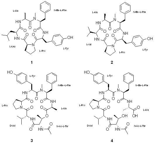
2. Results and Discussion

The molecular formula of compound 1 was deduced to be C33H43N5O6 with 15 degrees of unsaturation by HRFABMS analysis ([M + H]+ m/z 606.3296, calcd for C33H44N5O6, 606.3292). The 13C NMR data of this compound showed signals at δC 171.2, 170.6, 170.4, 168.9, and 168.2 for carbonyl carbons (Table 1). In conjunction with the strong absorption band at 1649 cm−1 in the IR spectrum, these carbons were thought to be amide carbons, indicating a peptidic compound. Signals for the corresponding exchangeable peptide NH protons were observed at δH 8.29, 7.27, and 6.96 in the 1H NMR spectra. Other conspicuous features in the NMR data were several de-shielded carbons (δC 155.7–14.9) and protons (δH 7.28–6.66), accounting for two aromatic moieties. All the remaining carbon and proton signals were in the shielded region, suggesting the presence of aliphatic amino acids. Thus, compound 1 must possess two rings from the 15 degrees of unsaturation inherent in its formula.
Given this information, the structure of compound 1 was determined by a stepwise interpretation of the 2-D NMR data. First, based on the results of HSQC analysis, all the protons and their attached carbons were precisely matched. Then, the 1H-1H COSY data defined several proton spin systems, including two involving exchangeable NH protons: NH-CH-CH3 and NH-CH-CH2-CH(CH3)2. In this way, two NH-bearing aliphatic moieties were readily identified as alanine (Ala) and leucine (Leu) (Figure 1). This interpretation was confirmed by the HMBC data, which showed diagnostic intra-residual three-bond correlations with the β-carbonyl-β-NH protons, allowing the assignment of the carbonyl carbons for each amino acid residue. Thus, the carbonyl carbons at δC 171.2 and 170.4 were assigned as the amide carbonyls of Ala and Leu, respectively.
The COSY data identified the two aromatic moieties as p-hydroxyphenyl and phenyl groups (Tables S2–S5). Starting with the HMBC correlations between the ring protons (δH 7.04) and a benzylic carbon (δC 37.3), the tracing of proton–proton and carbon–proton correlations identified a carbonyl (δC 168.9), an α-methine (δC 52.6, δH 4.78) and an NH (δH 7.27) of a tyrosine (Tyr) residue. Similar 2-D NMR analyses indicated carbonyl (δC 168.1), α-methine (δC 61.7, δH 4.17), and β-methylene/benzylic (δC 33.9, δH 3.24, and 2.70) groups. However, instead of NH, the presence of an NCH3 group (δC 30.5, δH 2.61) was confirmed by crucial HMBC correlation of H3-NCH3/C-α, indicating the presence of an N-methylphenylalanine (N-MePhe) residue.
For the remaining residue, the HSQC and COSY data identified the linear connectivity of a methine (δC 60.9, δH 4.10) and three methylenes (δC 31.5, δH 1.95, 1.88; δC 21.4, δH 1.77, δH 1.58; δC 46.4, δH 3.47, 3.37). The characteristic chemical shifts of a terminal methine and methylene groups as well as the lack of spin-coupled NH protons were indicative of a proline (Pro) residue, accounting for one of the two rings in the structure. The presence of this residue was confirmed by the HMBC data. The ring moiety of Pro was assigned based on the key correlations of H-α/C-δ and CO, H2-β/C-δ and CO, and H2-δ/C-γ (Figure 2).
The assembly of these amino acid residues into a cyclopeptide was accomplished using sequential inter-residue HMBC correlations between the protons and carbons belonging to neighboring amino acid residues (Figure 2). The peptide linkage between Tyr and N-MePhe was revealed by the two-bond correlation of NH (Tyr)–CO (N-MePhe). The latter amino acid was linked to Ala by the correlations of H3-NCH3 (N-MePhe)–CO (Ala) and H-α (N-MePhe)–CO (Ala). Similarly, the correlation of NH (Ala)–CO (Leu) linked these amino acids. Finally, this Leu was bound via a peptide bond to Pro based on the correlation of NH (Leu)–CO (Pro). Although it was not directly proven by the HMBC data, the molecular formula requires the presence of an additional ring, confirming a peptide linkage between N (Pro) and CO (Tyr), since the structure of 1 has 15 degrees of unsaturation. Five are equivalent to five carbonyl groups from the peptide linkage, eight are equivalent to two aromatic groups of Tyr and N-MePhe, one is equivalent to a ring from Pro. As no more double bond carbon is left, the only way to satisfy the 15 degrees of unsaturation is by connecting a peptide linkage between N (Pro) and CO (Tyr).
The structure of 1 derived from the NMR data was confirmed by high-resolution LC-MS/MS analysis, in which all of the peptide bonds between neighboring amino acid residues were sequentially cleaved (Figure 3). Finally, the absolute configurations of all the amino acids were assigned as L by advanced Marfey’s analysis (Figure S32, Table S1). Thus, the structure of compound 1, designated to be JG002CPA, was determined to be a new cyclopentapeptide.
The molecular formula of compound 2 was established to be C32H41N5O6 by HRFABMS analysis ([M + H]+ m/z 592.3133, calcd for C32H42N5O6, 592.3135). The NMR data of this compound were very similar to those of 1, suggesting the same pentacyclopeptide nature. The most noticeable difference in the 13C and 1H NMR data was the lack of an aliphatic methylene in 2. A detailed examination of the NMR data revealed that the signals of the carbons and protons of the Leu in 1 were replaced with those of a valine (Val), while the signals from other four residues were the same between of 1 (Table 1). This interpretation was unambiguously confirmed by combined 2-D NMR data, as all the key carbon–proton and proton–proton correlations were found (Figure 2). Subsequently, 2 was shown to have the same amino acid sequence as 1 by LC-MS/MS analysis (Figure 3). The absolute configurations of all amino acids were also assigned as L, identical to 1, by advanced Marfey’s analysis (Figure S32, Table S1). Thus, the structure of compound 2, designated to be JG002CPB, was determined to be a cyclopentapeptide structurally closely related to JG002CPA (1). Literature studies showed that the most related cyclopeptides to 1 and 2 are cotteslosin A and B, which were previously isolated from the marine-derived fungus Aspergillus versicolor [14].
Compound 3 was isolated as an amorphous solid from the culture broth of A. ochraceopetaliformis and determined to have a formula of C38H50N6O9 by HRFABMS analysis ([M + H]+ m/z 735.3721, calcd for C38H51N6O9, 735.3718). The 13C NMR data of this compound showed signals at δC 173.3, 172.4, 172.0, 171.8, 170.9, 169.7, and 168.1 for seven carbonyl carbons, indicating that 3 is a larger peptide than 1 and 2. The most noticeable difference in the NMR data was the presence of an isolated methyl group suggesting an acetyl group (δC 23.1, δH 1.83) (Table 2).
As for the previous compounds, the structure of 3 was determined by combined 2-D NMR data. Consequently, the same five amino acid residues (Ala, N-MePhe, Pro, Tyr, and Val) that could be found in 2 were readily identified by the COSY and intra-residual HMBC correlations (Figure 2). Unlike other carbonyl carbons, the carbonyl carbon of Pro failed to show HMBC correlations with neighboring protons and was unassigned at this stage. For the remaining residue, the COSY data revealed a proton spin system linearly consisting of four resonances at δH 6.29, 4.69, 5.53, and 1.18 for NH-CH-CH-CH3, respectively. The proton–proton coupling pattern and remarkably de-shielded methine at δH 5.53 suggested the presence of a threonine (Thr) residue. This was confirmed by the HMBC data in which the long-range couplings of the methine and methyl protons with neighboring carbons provided not only the linear structure but also the assignment of its carbonyl carbon at δC 169.7. The previously described acetyl group was also confirmed by a two-bond correlation between the methyl proton and carbonyl carbon at δC 171.8. Subsequently, the unassigned amide connection of Pro in the cyclopeptide was also confirmed indirectly but logically by constructing an N-AcThr residue with the HMBC correlations of NH and its α-methine protons with the carbonyl carbon (Figure 2). Thus, 3 was found to possess this unit in addition to the same five amino acid residues found in 2. A literature study showed that aspergillicins A–E, cyclohexapeptides from an Australian marine-derived Aspergillus carneus, also contained this modified amino-acid residue as a building block [15].
The assembly of the amino acid residues into the full structure was accomplished by the interpretation of HMBC data for the intra-residual HMBC correlations. Consequently, as shown in Figure 2, two partial structures, Ala-N-MePhe-Tyr and N-AcThr-Val, were readily deduced. An ester linkage connected these fragments according to a crucial HMBC correlation between the β-CH proton of Thr and the CO carbon of Ala. Although it was not directly indicated by the HMBC data, it was interpreted that the only remaining carbon at δC 173.3 must be the carbonyl carbon of Pro, which form a peptide bond with the NH of Tyr.
The structure of 3 derived from its HMBC data was confirmed by high-resolution LC/MS-MS analysis. It was observed that the N-AcThr moiety was readily converted to Thr during the analysis. Despite this, as shown in Figure 3, the linear sequence of this compound was independently found to be N-MePhe-Ala-N-AcThr-Val-Pro-Tyr, securing the placement of Pro between Val and Tyr. Thus, the structure of 3 was defined to be a new cyclohexadepsipeptide. Interestingly, the amino acid sequence of 3 was opposite to those of 2 (also 1) with reference to the five commonly-presented units.
The absolute configurations of the amino acids in 3 were assigned by advanced Marfey’s analysis. Unexpectedly, the Val in this compound was found to have a D-configuration by repeated analyses, while all other units had L-configurations (Figure S32, Table S1). In the LC-MS/MS analysis, N-AcThr was hydrolyzed to Thr. Additionally, the Marfey’s analysis for the β-stereogenic center of Thr assigned this to a common L-Thr. Thus, the structure of 3, designated to be FJ120DPA, was determined to be a new cyclohexadepsipeptide containing unusual D-Val and L-N-AcThr units.
A minor constituent congener of 3, compound 4, was isolated as an amorphous solid that was found to have the formula C38H52N6O10 by HRFABMS data ([M + Na]+ m/z 775.3646, calcd for C38H52N6NaO10, 775.3643). Compound 4 was analyzed in MeOH-d4 because the compound possesses a better solubility in methanol. Furthermore, the NMR data of compound 4 showed a better peak separation as well as minimal conformer formation. Despite compound 4 being analyzed in another solvent, the 13C and 1H NMR data of this compound were very similar to those of 3 obtained in CDCl3 (Table 2). A detailed examination of these data revealed that all the amino acid residues of 3 were exactly the same as those of 4. These NMR results, in conjunction with the presence of an additional H2O unit in its molecular formula, suggested 4 to be a hydrolyzed derivative of 3, possibly at the ester linkage between the Ala and the N-AcThr residue. This interpretation was confirmed by combined 2-D NMR experiments in which the carbon–proton and proton–proton correlations found for these compounds were virtually identical. Thus, compound 4, designated to be FJ120DPB, was structurally defined to be a hydrolyzed derivative of FJ120DPA (3). The structural similarity between 3 and 4 may bring the possibility of abiotic origin of the latter. However, the presence of compound 4 could already be detected in the unprocessed crude extract by LC-ESI-MS analysis. Furthermore, no acid or water was used for the extraction from the semi-solid cultivation medium. Consequently, it was presumed that compound 4 is not an artifact but rather a natural product.
To investigate the bioactivity of isolated compounds, antimicrobial activities against pathogenic microorganisms and enzyme inhibitory activities toward anti-virulence drug targets SrtA and ICL were first examined in vitro. In bioassays, these compounds were inactive (MIC (minimum inhibitory concentration) > 128 μg/mL) against the Gram-positive and Gram-negative bacterial strains Enterococcus faecalis (ATCC19433), Enterococcus faecium (ATCC19434), Staphylococcus aureus (ATCC25923), Escherichia coli (ATCC25922), Klebsiella pneumoniae (ATCC10031), and Salmonella enterica (ATCC14028). These compounds were also inactive (MIC > 128 μg/mL) against the fungal strains Aspergillus fumigatus (HIC6094), Candida albicans (ATCC10231), Trichophyton mentagrophytes (IFM40996), and Trichophyton rubrum (NBRC9185). There have been several investigations for antimicrobial cyclopeptides from natural product screening. A cyclic pentapeptide asperpeptide A isolated from Aspergillus sp. XS-20090B15 showed antibacterial activity against Bacillus cereus and Staphylococcus epidermidis with the same MIC value of 12.5 μM [16]. A cyclic hexapeptide (ASP2397) produced by Acremonium persicinum MF-347833 exhibited potent antifungal activity against A. fumigatus (MIC = 0.78 μg/mL) [17].
SrtA and ICL are excellent targets for the design and development of new anti-virulence drugs against pathogenic microorganisms. SrtA, a type of transpeptidase, plays a critical role in the pathogenesis of Gram-positive bacteria, including S. aureus, by modulating the ability of the bacterium to adhere to host tissue via the covalent anchoring of adhesins and other virulence-associated proteins to cell wall peptidoglycans [18]. The glyoxylate cycle is a sequence of anaplerotic reactions catalyzed by the key enzymes ICL and malate synthase [19]. The expression of ICL is upregulated during infection of macrophages by pathogenic microorganisms such as the pulmonary bacterium Mycobacterium tuberculosis [20] and the human pathogenic fungus C. albicans [21]. The mutant strains lacking srtA or icl are markedly less virulent in mice than the wild-type without affecting microbial viability [18,20,21]. In further bioassays, these compounds were moderately active against S. aureus SrtA and exhibited no antibacterial activity against S. aureus growth (MIC > 128 μg/mL). Compound 2 exhibited the strongest inhibitory activity (IC50 = 53.1 μM) of the test compounds with the inhibition comparable to the positive controls berberine chloride (IC50 = 104.3 μM) and curcumin (IC50 = 47.8 μM) (Table 3). In a similar assay, compound 2 showed weak inhibition (IC50 = 104.3 μM) of isocitrate lyase (ICL) derived from C. albicans. The IC50 value of compound 2 is comparable to the previously reported diketopiperazine cyclo(l-Phe-l-Val) (IC50 = 109.5 μM) isolated from marine-derived Streptomyces puniceus [22].

3. Materials and Methods

3.1. General Experimental Procedures

Optical rotations were measured on a JASCO P1020 polarimeter (Jasco, Tokyo, Japan) using a 1 cm cell. UV spectra were acquired with a Hitachi U-3010 spectrophotometer (Hitachi High-Technologies, Tokyo, Japan). IR spectra were recorded on a JASCO 4200 FT-IR spectrometer (Jasco, Tokyo, Japan) using a ZnSe cell. 1H and 13C NMR spectra were measured in DMSO-d6, CDCl3, or CD3OD solutions on Bruker Avance –400, –500, –600, or –800 instruments (Billerica, MA, USA). High resolution FAB mass spectrometric data were obtained at the Korea Basic Science Institute (Daegu, Korea) and were acquired using a JEOL JMS 700 mass spectrometer (Jeol, Tokyo, Japan) with meta-nitrobenzyl alcohol (NBA) as the matrix. High-resolution LC-MS/MS data were obtained at the National Instrumentation Center for Environmental Management (Seoul, Korea) on a Q-TOF 5600 instrument equipped with a Dionex U-3000 HPLC system. Semi-preparative HPLC separations were performed on a Spectrasystem p2000 equipped with a Spectrasystem RI-150 refractive index detector. All solvents used were spectroscopic grade or distilled from glass prior to use.

3.2. Fungal Material

3.2.1. Aspergillus allahabadii (Strain Number JG002)

The fungal strain A. allahabadii (strain number JG002) was isolated from underwater sediments collected at –20 m off the coast of Jeju-do, Korea, in April 2018. The sample was diluted using sterile seawater. One milliliter of diluted sample was processed utilizing the spread plate method in YPG medium (5 g of yeast extract, 5 g of peptone, 10 g of glucose, 0.15 g of penicillin G, 0.15 g of streptomycin sulfate, 24.8 g of Instant Ocean, and 16 g of agar in 1 L of distilled water) plates. The plates were incubated at 28 °C for 5 days. The strain was identified using standard molecular biology protocols by DNA amplification and sequencing of the ITS region. Genomic DNA extraction was performed using Intron’s i-genomic BYF DNA Extraction Mini Kit according to the manufacturer’s protocol. The strain was identified using standard molecular biology protocols by DNA amplification and sequencing of the ITS region [23]. The 18S rDNA sequence of this strain exhibited 100% identity (564/564) with that of A. candidus, A. allahabadii, and A. niveus. Due to the limitations of the ITS regions to identify intrasection species, sequence comparison of the β-tubulin was further analyzed [24]. This strain exhibited 100% identity with that of A. allahabadii strain CGMCC_3.02584 (GenBank accession number MH292842.1) in β-tubulin region (477/477). The nucleotide sequence of JG002 was deposited in the GenBank database under accession number MK424488.

3.2.2. Aspergillus ochraceopetaliformis (Strain Number FJ120)

The fungal strain A. ochraceopetaliformis (strain number FJ120) was isolated from underwater sediments collected off the coast of Jeju-do, Korea, in July 2007. The sample was diluted using sterile seawater. The isolation was performed in the same conditions as for the strain JG002. The identification was also processed using the same protocols. The nucleotide sequence of FJ120 was deposited in the GenBank database under accession number KF384187. The 18S rDNA sequence of this strain exhibited 100% identity (588/588) with that of A. ochraceopetaliformis strain RKI08-134 (GenBank accession number FJ797698).

3.3. Fermentation

3.3.1. JG002

The fungal strain was cultured on solid YPG media (5 g of yeast extract, 5 g of peptone, 10 g of glucose, 24.8 g of Instant Ocean, and 16 g of agar in 1 L of distilled water) for 7 days. An agar plug (1 × 1 cm) was inoculated in a 250 mL flask containing 100 mL of YPG media. After 7 days of growth, 10 mL of each culture was transferred to 2.8 L Fernbach flasks containing rice media (200 g of rice, 0.5 g of yeast extract, 0.5 g of peptone, and 12.4 g of Instant Ocean in 200 mL of distilled water). In total, 800 g of rice media was prepared and cultivated for 4 weeks at 28 °C, with agitating once every week.

3.3.2. FJ120

The seed preparation and inoculation were performed under the same culturing conditions as for the strain JG002. Then, 10 mL of each culture was transferred to 2.8 L Fernbach flasks containing YMM media (5g of yeast extract, 5g of malt extract, 10g of mannitol and 24.8 g of Instant Ocean in 1000 mL of distilled water). In total, 20 L of YMM media was prepared and cultivated under static conditions for 8 weeks at 28 °C.

3.4. Extraction and Isolation

3.4.1. JG002

The entire culture was macerated and extracted with MeOH (1 L × 3). The solvent was evaporated in vacuo to afford a brown organic gum (4.2 g). The extract was separated by C18 reversed-phase vacuum flash chromatography using sequential mixtures of H2O and MeOH (six fractions of H2O-MeOH, gradient from 50:50 to 0:100), acetone, and finally EtOAc as the eluents. Based on the results of 1H NMR analysis, the fractions eluted with H2O-MeOH 40:60 (380 mg) and 30:70 (760 mg) were chosen for further separation. The fraction that eluted with H2O-MeOH (40:60) was separated by semi-preparative reversed-phase HPLC (YMC-ODS-A column, 250 × 10 mm, 5 μm; H2O-MeCN, 70:30, 2.0 mL/min), affording compound 1. The H2O-MeOH (30:70) fraction from vacuum flash chromatography was separated by semi-preparative reversed-phase HPLC (H2O-MeCN, 58:42, 2.0 mL/min), and afforded compound 2. The overall isolated amounts were 10.3 and 5.8 mg for 1 and 2, respectively.

3.4.2. FJ120

The entire culture was filtered and extracted with EtOAc (20 L × 3). The solvent was evaporated in vacuo to afford a brown organic gum (5.2 g). The extract was separated by C18 reversed-phase vacuum flash chromatography using sequential mixtures of H2O and MeOH (five fractions of H2O-MeOH, gradient from 80:20 to 0:100), finally MeOH:MC (1:1) as the eluents. Based on the results of 1H NMR analysis, the fractions eluted with H2O-MeOH 80:20 (1370 mg) and 40:60 (1380 mg) were chosen for further separation. The fraction that was eluted with H2O-MeOH (80:20) was separated by semi-preparative reversed-phase HPLC (YMC-ODS-A column, 250 x 10 mm, 5 μm; H2O-MeOH, 50:50, 2.0 mL/min), affording compound 4. This compound was further purified by reversed-phase HPLC (YMC-ODS-A column, 4.6 × 250 nm, 5 μm; H2O-MeCN, 85:15, 0.7 mL/min). The H2O-MeOH (40:60) fraction from vacuum flash chromatography was separated by semi-preparative reversed-phase HPLC (H2O-MeOH, 50:50, 2.0 mL/min) and afforded pure compound 3. The overall isolated amounts were 24.6 and 3.8 mg for 3 and 4, respectively.
JG002CPA (1): Pale yellow, amorphous solid, [α] D 25 +61 (c 0.20, MeOH); UV (MeOH) λmax (log ε) 218 (3.48), 277 (2.68) nm; 1H and 13C NMR: see Table 1; IR (ZnSe) νmax 3326, 2956, 1649, 1517, 1425 cm−1; HRFABMS, m/z 606.3296 [M + H]+ (calcd for C33H44N5O6, 606.3292).
JG002CPB (2): Pale yellow, amorphous solid, [α] D 25 +69 (c 0.20, MeOH); UV (MeOH) λmax (log ε) 221 (3.47), 281 (2.67) nm; 1H and 13C NMR: see Table 1; IR (ZnSe) νmax 3300, 2971, 2920, 1649, 1512, 1452 cm−1; HRFABMS, m/z 592.3133 [M + H]+ (calcd for C32H42N5O6, 592.3135).
FJ120DPA (3): Pale yellow, amorphous solid, [α] D 25 +66 (c 0.20, MeOH); UV (MeOH) λmax (log ε) 212 (3.56), 280 (2.16) nm; 1H and 13C NMR: see Table 2; IR (ZnSe) νmax 3304, 2964, 2922, 1732, 1644, 1540, 1514, 1453 cm−1; HRFABMS, m/z 735.3721 [M + H]+ (calcd for C38H51N6O9, 735.3718).
FJ120DPB (4): Pale yellow, amorphous solid, [α] D 25 +73 (c 0.20, MeOH); UV (MeOH) λmax (log ε) 220 (3.57), 280 (2.65) nm; 1H and 13C NMR: see Table 2; IR (ZnSe) νmax 3315, 2953, 2912, 1685, 1649, 1546, 1453 cm−1; HRFABMS, m/z 775.3646 [M + Na]+ (calcd for C38H52N6NaO10, 775.3643).

3.5. Stereochemical Analysis of the Amino Acid Residues

Compounds 13 (1 mg each) were hydrolyzed in 0.5 mL of 6 N HCl at 120 °C for 8 h. After removing the HCl in vacuo, 0.5 mg portions of the hydrolysate were transferred into two vials and dissolved in 100 μL of 1 N NaHCO3. L-FDAA (100 μL of 10 mg/mL in acetone) was added to one reaction vial, and D-FDAA was added to the other vial. The reactions were maintained at 80 °C for 3 min. Then, the reaction mixtures were neutralized by adding 50 μL of 2 N HCl and diluted with 300 μL of 50% aqueous CH3CN−H2O. Twenty microliters of each reaction mixture were analyzed by LC/MS using a Phenomenex C18(2) column (Luna, 100 × 4.6 mm, 5 μm) with gradient solvent conditions (flow rate 0.7 mL/min; UV 340 nm detection; 10% to 60% CH3CN−H2O with 0.1% formic acid over 50 min). L-FDAA derivatives were eluted before D-FDAA derivatives for all amino acid residues in the hydrolysate of 1 and 2. Thus, the absolute configurations of all the amino acid residues in these compounds were determined to be L. D-FDAA derivative eluted before L-FDAA derivative for Val residue in the hydrolysate of 3, while other amino acid residues eluted the L-FDAA derivatives faster than the D-FDAA derivatives (Supporting Information). In order to assign between L-Thr and L-allo-Thr in 3, a Marfey’s analysis using additional HPLC analysis with a YMC-ODS-A column (250 × 4.6 mm, 5 μm) and gradient solvent conditions (flow rate 0.7 mL/min; UV 360 nm detection; 20% to 90% MeOH−H2O with 0.1% TFA over 60 min) was applied, and the residue was determined to be L-Thr rather than L-allo-Thr.

3.6. Biological Assays

3.6.1. Antibacterial Activity Assay

The antibacterial activity assay was performed according to the Clinical and Laboratory Standards Institute (CLSI) method [25]. Gram-positive bacteria (S. aureus ATCC25923, E. faecalis ATCC19433, E. faecium ATCC19434) and Gram-negative bacteria (K. pneumoniae ATCC10031, S. enterica ATCC14028, E. coli ATCC25922) were cultured in MHB (Mueller Hinton broth) at 37 °C. Each test compound was dissolved in dimethyl sulfoxide (DMSO) and diluted with MBH to prepare serial twofold dilutions in the range of 0.06–128 μg/mL (final DMSO concentration: 1%). An aliquot of 190 μL of MBH containing the test compound was mixed with 10 μL of the broth containing approximately 106 colony-forming units (cfu)/mL of test bacterium (final concentration: 5 × 10⁴ cfu/mL) in each well of a 96-well plate. The plates were incubated for 24 h at 37 °C. The MIC was defined as the lowest concentration of test compound that prevented cell growth. Ampicillin and tetracycline were used as reference compounds.

3.6.2. Antifungal Activity Assay

The antifungal activity assay was performed in accordance with the guidelines in CLSI document M38 [26]. C. albicans ATCC10231, A. fumigatus HIC6094, T. rubrum NBRC9185, and T. mentagrophytes IFM40996 were used as test organisms. Each stock solution of the compound was diluted in RPMI 1640 broth with the concentration range of 0.06–128 μg/mL. The final inoculum concentration was 104 cells/mL in each well of a 96-well plate. The MIC was defined after incubation for 24 h (for C. albicans), 48 h (for A. fumigatus), and 96 h (for T. rubrum and T. mentagrophytes) at 37 °C. Amphotericin B was used as a positive control.

3.6.3. ICL Inhibition Assay

The recombinant ICL from C. albicans was prepared and the inhibitory activity of the test compounds against ICL was determined by previously documented procedures [23]. The reaction mixture consisted of 20 mM sodium phosphate buffer (pH 7.0), 4.1 mM phenylhydrazine, 3.75 mM MgCl2, 1.27 mM threo-dl(+)isocitrate, and 2.5 μg/mL ICL, and was incubated with the test compounds at a concentration range of 1 to 128 μg/mL at 37 °C for 30 min. The increase in intensity of absorbance resulting from the formation of glyoxylate phenylhydrazone was observed using an UVmini-1240 spectrophotometer (SHIMADZU, Tokyo, Japan) at a wavelength of 324 nm. A known ICL inhibitor, 3-nitropropionic acid, was used as a reference control.

3.6.4. SrtA Inhibition Assay

S. aureus-derived recombinant SrtA was purified from transformed E. coli by nickel-based affinity chromatography, and enzyme activity was determined by quantifying the intensity of augmented fluorescence upon cleavage of a synthetic peptide containing LPETG motifs [27]. For the reactions, 300 μL of reaction mixture (50 mM Tris–HCl, pH 7.5, 150 mM NaCl, 5 mM CaCl2, 55 μg SrtA, and 0.75 μg synthetic peptide substrate, Dabcyl-QALPETGEE-Edans) was added to a 96-well microtiter plate. After incubation at 37 °C for 1 h, the increase in the fluorescence intensity was recorded by an UVmini-1240 spectrophotometer (SHIMADZU, Tokyo, Japan) using excitation and emission wavelengths of 350 and 495 nm, respectively. Curcumin and berberine chloride were used as positive controls.

4. Conclusions

Four new peptides were isolated and structurally elucidated from the culture broths of marine-derived fungi A. allahabadii and A. ochraceopetaliformis. Based upon the results of combined chemical and spectroscopic analyses, the amino acid sequences of two cyclopentapeptides (1 and 2) from A. allahabadii were elucidated. Furthermore, one cyclodepsihexapeptide (3) and its linear analog (4) from A. ochraceopetaliformis could be elucidated. In addition, the presence of a D-Val residue in the two hexapeptides was also found by Marfey’s analysis. The new compounds moderately inhibited the microbial enzyme sortase A and (2) weakly inhibited isocitrate lyase.

Supplementary Materials

The following are available online at https://www.mdpi.com/1660-3397/17/9/488/s1. Figures S1–S28: HRFABMS, 1-D and 2-D NMR spectra of 14, Figures S29–S31: HRLC/MS-MS fragmentation analysis of 13, Figure S32: The advanced Marfey’s analysis of 14, Table S1: The advanced Marfey’s analysis of 14, Figure S33: The LC analysis of L-FDAA derivatives of Thr and allo-Thr for 3, Figure S34: The LC-MS analysis of FJ120 extract, Tables S2-S5: The NMR table of compound 14.

Author Contributions

J.-Y.H., J.-H.L. and S.C.P. carried out the isolation and structural elucidation; J.L. and K.-B.O. performed antimicrobial and enzyme inhibition bioassays; J.S. and D.-C.O. reviewed and evaluated all data; J.S. and K.-B.O. supervised the research work and prepared the paper.

Funding

This study was supported by the National Research Foundation (NRF, grant No. 2018R1A4A1021703) funded by the Ministry of Science, ICT, and Future Planning, Korea. This study was also supported by the project titled “A study of the massive production and the development of novel therapeutic agents from marine microbial bioactive natural products by biotechnological methods (grant No. 20170158)” funded by the Ministry of Oceans and Fisheries, Korea.

Acknowledgments

We thank the BK21 plus program (2019) for supporting the publication.

Conflicts of Interest

The authors declare no conflicts of interest.

References

  1. Phyo, Y.Z.; Ribeiro, J.; Fernandes, C.; Kijjoa, A.; Pinto, M.M.M. Marine natural peptides: Determination of absolute configuration using liquid chromatography methods and evaluation of bioactivities. Molecules 2018, 23, 306. [Google Scholar] [CrossRef] [PubMed]
  2. Balkovec, J.M.; Hughes, D.L.; Masurekar, P.S.; Sable, C.A.; Schwartz, R.E.; Singh, S.B. Discovery and development of first in class antifungal caspofungin (CANCIDAS®)—A case study. Nat. Prod. Rep. 2014, 31, 15–34. [Google Scholar] [CrossRef] [PubMed]
  3. Wang, X.; Lin, M.; Xu, D.; Lai, D.; Zhou, L. Structural diversity and biological activities of fungal cyclic peptides, excluding cyclodipeptides. Molecules 2017, 22, 2069. [Google Scholar] [CrossRef] [PubMed]
  4. Mayer, A.M.S.; Glaser, K.B.; Cuevas, C.; Jacobs, R.S.; Kem, W.; Little, R.D.; McIntosh, J.M.; Newman, D.J.; Potts, B.C.; Shuster, D.E. The odyssey of marine pharmaceuticals: A current pipeline perspective. Trends Pharmacol. Sci. 2010, 31, 255–265. [Google Scholar] [CrossRef] [PubMed]
  5. Rateb, M.E.; Ebel, R. Secondary metabolites of fungi from marine habitats. Nat. Prod. Rep. 2011, 28, 290–344. [Google Scholar] [CrossRef] [PubMed]
  6. Hasan, S.; Ansari, M.I.; Ahmad, A.; Mishra, M. Major bioactive metabolites from marine fungi: A review. Bioinformation 2015, 11, 176–181. [Google Scholar] [CrossRef] [PubMed]
  7. Sun, W.; Wu, W.; Liu, X.; Zaleta-Pinet, D.A.; Clark, B.R. Bioactive compounds isolated from marine-derived microbes in China: 2009–2018. Mar. Drugs 2019, 17, 339. [Google Scholar] [CrossRef] [PubMed]
  8. Kang, H.K.; Seo, C.H.; Park, Y. Marine peptides and their anti-infective activities. Mar. Drugs 2015, 13, 618–654. [Google Scholar] [CrossRef]
  9. Deshmukh, S.K.; Prakash, V.; Ranjan, N. Marine fungi: A source of potential anticancer compounds. Front. Microbiol. 2018, 8, 2536. [Google Scholar] [CrossRef]
  10. Srivastava, A.; Mishra, V. Marine peptides act as novel chemotherapeutic agent. J. Microbiol. Exp. 2018, 6, 267–270. [Google Scholar] [CrossRef]
  11. Carroll, A.R.; Copp, B.R.; Davis, R.A.; Keyzers, R.A.; Prinsep, M.R. Marine natural products. Nat. Prod. Rep. 2019, 36, 122–173. [Google Scholar] [CrossRef] [Green Version]
  12. Liao, L.; Bae, S.Y.; Won, T.H.; You, M.; Kim, S.H.; Oh, D.C.; Lee, S.K.; Oh, K.B.; Shin, J. Asperphenin A and B, lipopeptidyl benzophenones from a marine-derived Aspergillus sp. fungus. Org. Lett. 2017, 19, 2066–2069. [Google Scholar] [CrossRef]
  13. Hu, G.P.; Yuan, J.; Sun, L.; She, Z.G.; Wu, J.H.; Lan, X.J.; Zhu, X.; Lin, Y.C.; Chen, S.P. Statistical research on marine natural products based on data obtained between 1985 and 2008. Mar. Drugs 2011, 9, 514–525. [Google Scholar] [CrossRef]
  14. Fremlin, L.J.; Piggott, A.M.; Lacey, E.; Capon, R.J. Cottoquinazoline A and Cotteslosins A and B, Metabolites from an Australian Marine-Derived Strain of Aspergillus versicolor. J. Nat. Prod. 2009, 72, 666–670. [Google Scholar] [CrossRef]
  15. Capon, R.J.; Skene, C.; Stewart, M.; Ford, J.; O’Hair, R.A.J.; Williams, L.; Lacey, E.; Gill, J.H.; Heiland, K.; Friedel, T. Aspergillicins A–E: Five novel depsipeptides from the marine-derived fungus Aspergillus carneus. Org. Biomol. Chem. 2003, 1, 1856–1862. [Google Scholar] [CrossRef]
  16. Chen, M.; Shao, C.L.; Fu, X.M.; Kong, C.J.; She, Z.G.; Wang, C.Y. Lumazine peptides penilumamides B–D and the cyclic pentapeptide asperpeptide A from a gorgonian-derived Aspergillus sp. fungus. J. Nat. Prod. 2014, 77, 1601–1606. [Google Scholar] [CrossRef]
  17. Nakamura, I.; Yoshimura, S.; Masaki, T.; Takase, S.; Ohsumi, K.; Hashimoto, M.; Furukawa, S.; Fujie, A. ASP2397: A novel antifungal agent produced by Acremonium persicinum MF-347833. J. Antibiot. 2017, 70, 45–51. [Google Scholar] [CrossRef]
  18. Mazmanian, S.K.; Liu, G.; Jensen, E.R.; Lenoy, E.; Schneewind, O. Staphylococcus aureus sortase mutants defective in the display of surface proteins and in the pathogenesis of animal infections. Proc. Natl. Acad. Sci. USA 2000, 97, 5510–5515. [Google Scholar] [CrossRef]
  19. Dunn, M.F.; Ramirez-Trujillo, J.A.; Hernández-Lucas, I. Major roles of isocitrate lyase and malate synthase in bacterial and fungal pathogenesis. Microbiology 2009, 155, 3166–3175. [Google Scholar] [CrossRef] [Green Version]
  20. McKinney, J.D.; Höner Zu Bentrup, K.; Muñoz-Elías, E.J.; Miczak, A.; Chen, B.; Chan, W.T.; Swenson, D.; Sacchettini, J.C.; Jacobs, W.R., Jr.; Russell, D.G. Persistence of Mycobacterium tuberculosis in macrophages and mice requires the glyoxylate shunt enzyme isocitrate lyase. Nature 2000, 406, 735–738. [Google Scholar] [CrossRef]
  21. Lorenz, M.C.; Fink, G.R. The glyoxylate cycle is required for fungal virulence. Nature 2001, 412, 83–86. [Google Scholar] [CrossRef]
  22. Kim, H.; Hwang, J.Y.; Shin, J.; Oh, K.B. Inhibitory effects of diketopiperazines from marine-derived Streptomyces puniceus on the isocitrate lyase of Candida albicans. Molecules 2019, 24, 2111. [Google Scholar] [CrossRef]
  23. Schoch, C.L.; Seifert, K.A.; Huhndorf, S.; Robert, V.; Spouge, J.L.; Levesque, C.A.; Chen, W. Fungal Barcoding Consortium. Nuclear ribosomal internal transcribed spacer (ITS) region as a universal DNA barcode marker for Fungi. Proc. Natl. Acad. Sci. USA 2012, 109, 6241–6246. [Google Scholar] [CrossRef]
  24. Samson, R.A.; Peterson, S.W.; Frisvad, J.C.; Varga, J. New species in Aspergillus section Terrei. Stud. Mycol. 2011, 69, 39–55. [Google Scholar] [CrossRef]
  25. Clinical and Laboratory Standards Institute. Methods for Dilution Antimicrobial Susceptibility Tests for Bacteria That Grow Aerobically, 11th ed.; M07; CLSI: Wayne, PA, USA, 2018. [Google Scholar]
  26. Clinical and Laboratory Standards Institute. Reference Methods for Broth Dilution Antifungal Susceptibility Testing of Filamentous Fungi, 3rd ed.; M38; CLSI: Wayne, PA, USA, 2017. [Google Scholar]
  27. Oh, K.B.; Kim, S.H.; Lee, J.; Cho, W.J.; Lee, T.; Kim, S. Discovery of diarylacrylonitriles as a novel series of small molecule sortase A inhibitors. J. Med. Chem. 2004, 47, 2418–2421. [Google Scholar] [CrossRef]
Figure 1. The structures of 14.
Marinedrugs 17 00488 g001
Figure 2. The COSY (bold) and HMBC (arrows) key correlations of compounds 1 and 3.
Marinedrugs 17 00488 g002
Figure 3. The high-resolution LC/MS-MS fragmentation analysis of compounds 13 (ac).
Marinedrugs 17 00488 g003
Table 1. NMR data of compounds 1 and 2 in DMSO-d6.
12
Unit PositionδC, TypeδH, Mult (J in Hz)δC, TypeδH, Mult (J in Hz)
AlaCO171,2, C 171.2, C
α44.2, CH3.54, dd (7.9, 6.8) 43.9, CH3.60, dd (8.0, 6.8)
β17.1, CH30.70, d (6.4)17.1 CH30.71, d (6.4)
NH 8.29, d (8.4) 8.32, d (8.3)
N-Me-PheCO168.1, C 168.2, C
α61.7, CH4.17, dd (11.6, 3.6) 61.6, CH4.16, dd (11.4, 3.4)
β33.9, CH23.24, dd (14.3, 3.3) 33.9, CH23.25, dd (14.3, 3.3)
2.70, dd (14.4, 11.8) 2.72, dd (14.4, 11.6)
γ137.5, C 137.5, C
ortho129.0, CH7.09, d (7.2)129.0, CH7.10, d (7.2)
meta128.5, CH7.28, d (7.6)128.5, CH7.28, d (7.6)
para126.7, CH7.22, t (7.4)126.7, CH7.22, t (7.4)
N-CH330.5, CH32.61, s30.5, CH32.63, s
TyrCO168.9, C 168.9, C
α52.6, CH4.78, td (8.5, 5.2)52.7, CH4.76, td (8.5, 5.2)
β37.3, CH23.04, dd (13.4, 8.8)37.3, CH23.02, dd (13.4, 9.0)
2.73, dd (13.5, 5.7) 2.74, dd (13.5, 4.7)
γ127.4, C 127.4, C
ortho130.2, CH7.04, d (8.4) 130.2, CH7.04, d (8.4)
meta114.9, CH6.66, d (8.4)114.9, CH6.66, d (8.4)
para155.7, C 155.8, C
OH 9.19, s 9.21, s
NH 7.27, m 7.26, m
ProCO170.6, C 170.6, C
α60.9, CH4.10, dd (7.9, 1.7)60.9, CH4.10, dd (7.3, 2.6)
β31.5, CH21.95, m31.5, CH21.93, m
1.88, m
γ21.4, CH21.77, m21.5, CH21.78, m
1.58, m 1.61, dq (12.3, 9.1)
δ46.4, CH23.47, m46.2, CH23.49, m
3.37, m 3.35, m
LeuCO170.4, C
α53.3, CH4.19, m
β41.3, CH21.37, m
1.34, m
γ24.5, CH1.37, m
δ22.4, CH30.88, d (6.1)
21.7, CH30.80, d (6.2)
NH 6.96, d (9.0)
ValCO 169.2, C
α 61.0, CH3.82, t (9.6)
β 30.7, CH1.69, m
γ 19.3, CH30.80, d (6.6)
18.9, CH30.78, d (6.7)
NH 7.00, d (8.7)
Table 2. NMR data of compounds 3 and 4.
3 a4 b
Unit PositionδC, TypeδH, Mult (J in Hz)δC, TypeδH, Mult (J in Hz)
ValCO172.0, C 172.8, C
α57.1, CH4.43, t (9.8)59.4, CH4.10, d (9.0)
β30.3, CH1.89, m 30.8, CH1.97, m
γ19.2, CH30.86, d (6.5)19.4, CH31.02, d (6.6)
18.8, CH30.92, d (6.5)19.2, CH30.91, d (6.8)
NH 6.43, d (9.1)
ProCO173.3, C 173.7, C
α59.5, CH4.31, d (5.8)61.7, CH4.35, dd (8.3, 2.4)
β29.7, CH22.13, m30.5, CH21.88, m
1.74, m
γ24.7, CH21.88, m24.2, CH21.63, m
1.04, m
δ48.1, CH23.93, m48.6, CH23.72, m
3.52, m 3.46, m
TyrCO172.4, C 173.9, C
α50.6, CH4.54, dd (13.3, 6.6)51.2, CH4.62, dd (11.2, 4.0)
β35.9, CH22.42, dd (13.6, 9.2)36.0, CH22.63, dd (13.1, 11.5)
1.87, dd (13.2, 6.2) 1.43, dd (13.2, 3.5)
γ126.6, C 129.2, C
ortho130.2, CH6.77, d (7.8)131.5, CH6.92, d (8.3)
meta115.9, CH6.66, d (8.0)115.9, CH6.64, d (8.4)
para155.9, C 157.3, C
NH 6.44, d (9.1)
N-Me-PheCO168.1, C 170.5, C
α63.2, CH4.82, t (6.8) 63.7, CH5.28, dd (10.9, 3.4)
β34.5, CH23.34, dd (14.0, 5.7)35.4, CH23.16, dd (14.3, 3.5)
2.52, dd (13.9, 7.8) 2.97, dd (14.1, 9.3)
γ137.9, C 139.4, C
ortho129.4, CH7.13, d (7.4)130.7, CH7.28, d (7.2)
meta129.1, CH7.24, d (7.5)130.1, CH7.30, d (7.3)
para127.3, CH7.23, d (7.3)128.1, CH7.17, d (7.5)
N-CH329.5, CH32.87, s30.2, CH32.90, s
AlaCO170.9, C 180.8, C
α48.5, CH4.67, d (7.9) 52.1, CH4.19, m
β18.6, CH31.32, d (7.3)18.5, CH31.34, d (6.5)
NH 8.16, d (8.3)
N-Ac-ThrCO169.7, C 173.0, C
α55.4, CH4.69, m59.7, CH4.47, d (5.2)
β70.1, CH5.53, q (5.9)68.6, CH4.17, q (5.9)
γ17.0, CH31.18, d (6.5)19.7, CH31.21, d (6.4)
NH 6.29, d (9.0)
CO171.8, C 174.1, C
α23.1, CH31.83, s22.8, CH32.09, s
a,b Measured in CDCl3 and CD3OD, respectively.
Table 3. Results of bioactivity tests.
IC50 (μM)
CompoundSrtA InhibitionICL Inhibition
170.0>128
253.1116.8
3131.9>128
477.0>128
Berberine chloride a104.3
Curcumin a47.8
3-Nitropropionic acid a 18.5
a Positive control.

